Introduction
Pancreatic cancer is an aggressive disease,
frequently diagnosed (in ~80% of cases) at an advanced stage
(1) when the tumor is unresectable
due to infiltration of local arteries or distant metastasis
(2). Unfortunately, pancreatic
resection is possible in few (5-10%) patients (3). Pancreatic adenocarcinoma remains one
of the deadliest cancers, with a five-year survival rate of 6–8%
(4,5) and a median duration of survival of
less than two years even in surgically-treated patients (5).
Pancreatic cancer is the fourth-leading cause of
cancer-related deaths in the USA (4) and is predicted to become the
second-leading cause among individuals older than 65 years by 2030
(6). Globally, this trend has
remained unchanged, with more than 200000 deaths attributed to
pancreatic cancer annually (7).
The overall survival (OS) of pancreatic cancer
patients has not improved significantly in the past 30 years
(1). The high mortality rate is
probably due to diagnosis at an advanced stage when the currently
available therapies have limited effects. The addition of
nab-paclitaxel (nab-PTX) to gemcitabine (GEM) increased the OS from
6.6 to 8.7 months (8,9). For patients in good physical
condition, treatment with FOLFIRINOX (5-fluorouracil, oxaliplatin,
and irinotecan) increased the survival to 11.1 months compared to
the OS for GEM alone, but had potentially severe side effects
(10).
Chemoresistance is a reason for high mortality in
pancreatic cancer (7,11). Gemcitabine and nab-PTX are the
standard of care for treating advanced pancreatic cancer (8,9),
exhibiting benefits over GEM. However, systemic chemotherapy has a
limited effect on OS due to low response rates and chemoresistance,
ascribed to the poorly understood mechanism of action. Early
diagnosis using novel biomarkers is an important goal for
pancreatic cancer researchers.
The polymeric immunoglobulin receptor (pIgR)
responsible for transcytosis of polymeric Igs (dimeric IgA and
pentameric IgM) across mucosal surfaces (12) facilitates the secretion of IgA and
IgM, the first-line antibodies against infection (13,14).
The extracellular portion of pIgR is then cleaved off as a
secretory component (SC) bound to polymeric IgA, protecting it from
proteolytic degradation (14) and
ensuring effective mucosal secretion (15). pIgR is expressed on epithelial cells
and is upregulated by proinflammatory cytokines in response to
viral and bacterial infections, thus linking innate and adaptive
immunity (13,14,16,17).
The extracellular component of pIgR can be cleaved to produce the
SC that is not bound to IgA, which then acts as a scavenger on the
mucosal lining (14).
pIgR is highly expressed in several cancers; its
upregulation was detected in colon (18), breast (19,20),
endometrial (21), bladder
(22), hepatocellular (23,24),
epithelial ovarian (25), and
esophageal and gastric cancers (26). High levels of the SC were also
detected in the sera of patients with lung (27,28),
pancreatic (29), and colon cancer
with liver metastasis (30).
However, the clinical significance and prognostic value of pIgR
remain unclear.
Herein, pIgR, as a novel protein associated with
chemoresistance, was investigated using pancreatic cancer
patient-derived xenograft (PDX) lines. To demonstrate the
association between pIgR expression and clinicopathological
features, the expression and prognostic ability of pIgR were
evaluated using immunohistochemistry (IHC) of 77 human pancreatic
cancer tissues following surgical resection.
Materials and methods
Establishment of pancreatic cancer PDX
lines
NSG mice, obtained from The Jackson Laboratory, were
housed aseptically in plastic cages at 22±1°C under 45±10% relative
humidity and a 12-h light/12-h dark cycle. All mice were fed a
standard diet and were allowed free access to food and water. All
experiments involving animals were performed in accordance with the
care and use guidelines of the Kanagawa Cancer Center Research
Institute, Japan. The study was approved by the Research Ethics
Committee of Kanagawa Cancer Center Research Institute (approval
no. 176).
Surgically resected tumor tissues from ten
pancreatic cancer patients (age range, 51–79 years; mean age, 67.2
years; 4 males and 6 females; Table
SI) were subcutaneously transplanted into 6–12-week-old mice
using transplantation needles (Fig.
S1A) as previously described (31,32).
These patients provided informed consent and tumor tissues were
resected at the Kanagawa Cancer Center Hospital (Kanagawa, Japan)
from June, 2008 to March, 2015. Briefly, after each patient
underwent surgery, fresh tumor tissues were cut into small pieces
of approximately 0.8–5.0 mm3 using scissors or minced
under sterile conditions. A small incision was made on the lower
back of the mouse near the base on the hindlimb, and a transplant
needle was inserted until the tip reached the dorsal subcutaneous
area of the upper part of the back, followed by closure. This
method was used to prevent outflow of the engraftment. These PDX
lines were designated as Generation 1 (G1). When the tumor volumes
in G1 mice reached 1000 mm3, the tumor tissues were
removed and re-implanted into other NSG mice. Ten PDX lines up to
G7 were generated after repeated passaging (Fig. S1B).
Antitumor effects of chemotherapy in
the pancreatic cancer PDX model
Anticancer drugs [GEM or GEM + nab-PTX] or saline
(used as a control) were administered intraperitoneally to PDX mice
with 200–400 mm3 tumors. Tumor volumes (mm3)
were determined weekly using the formula [length (mm)2 ×
width (mm)]/2. The chemotherapy was administered via
intra-abdominal injection on days 1, 3, and 6.
Identification of
chemoresistance-related molecules
Tumor tissues from treated and control PDX lines
were harvested when the tumor volume in the control group exceeded
1500 mm3. After the tumor-bearing mice were sacrificed,
the tumors were cut into 4-µm thick pieces that were immediately
frozen in liquid nitrogen. The frozen tissue was crushed using a
Cryo-Press (Microtec Co., Ltd.). Total RNA was isolated and
purified with ZR-Duet DNA/RNA MiniPrep (Zymo Research Corp.). RNA
quality was assessed using the Agilent 2100 Bioanalyzer (Agilent
Technologies, Inc.), followed by next-generation sequencing (NGS)
on an Illumina HiSeq 4000 system at BGI with paired-end sequencing.
The read lengths were 2×100 bp for GEM monotherapy and 2×150 bp for
GEM + nab-PTX combination therapy.
Transcriptome analysis
Paired-end reads were mapped to all the RefSeq human
(hg38 coordinates) and mouse (mm10 coordinates) transcripts using
Bowtie 1.1.2 (33), allowing up to
one mismatch, and reads mapped to both the species or to multiple
genes were discarded. To avoid bias due to read length differences,
only the first 100 bp of each read for samples with read lengths of
150 bp were used for mapping. Reads mapping to noncoding
transcripts were removed, and the remaining were used for gene
expression profiling of human cancer and mouse stromal cells
(34). The expression values were
normalized for cancer and stromal cells independently; the sum of
expression values below the 95th percentile being 300000.
Bioinformatics analysis using The
Cancer Genome Atlas (TCGA; http://www.cancer.gov/about-nci/organization/ccg/research/structural-genomics/tcga)
database
The Human Protein Atlas (http://www.proteinatlas.org) was used as a tool for
antibody-based biomarker discovery (35). TCGA RNASeq data for pancreatic
cancer were used to generate Kaplan-Meier curves on the basis of
pIgR expression. Patients were classified into two groups, and the
association between survival and gene expression was examined. The
expression cut-off based on the fragments per kilobase of exon
model per million reads mapped (FPKM) value that yielded the
maximal difference between the two groups with regard to survival
at the lowest log-rank P-value was selected.
Immunohistological analysis of samples
from pancreatic cancer patients
A consecutive cohort of 77 histopathologically
confirmed pancreatic cancer patients who underwent surgical
resection at Showa University Hospital in Tokyo, Japan, from
January 1, 2008 to December 31, 2017 was included (Table I). Patients with no history of
chemotherapy (GEM or GEM + nab-PTX) were included. Archived
formalin-fixed, paraffin-embedded (FFPE) tumor tissues from
resected specimens from the cohort of patients were used.
Histological classifications were based on the World Health
Organization system. Tumor staging was performed per the criteria
described in the UICC TNM classification (7th edition, 2009). The
included patients had stage IIA or stage IIB disease, per the 7th
edition of UICC. The study protocol was approved by the Ethics
Committee of the Showa University School of Medicine (Tokyo, Japan;
Approval no. 2611) and adhered to the principles of the Declaration
of Helsinki.
 | Table I.Correlation between pIgR expression
and clinicopathological features in 77 cases of pancreatic
cancer. |
Table I.
Correlation between pIgR expression
and clinicopathological features in 77 cases of pancreatic
cancer.
|
| pIgR
expression |
|
|---|
|
|
|
|
|---|
|
Characteristics | Low n=47 | High n=30 | P-value |
|---|
| Age (years) (mean ±
SD) | 72.4 | 69.8 | 0.339a |
| Sex |
|
| 0.0608b |
|
Male | 23 | 14 |
|
|
Female | 24 | 16 |
|
| Tumor location |
|
| 0.6588b |
|
Pancreatic head | 29 | 20 |
|
|
Pancreatic body/tail | 18 | 10 |
|
| Histological
type |
|
| 0.1008b |
|
Adenocarcinoma | 43 | 30 |
|
|
Others | 4 | 0 |
|
| TNM (UICC 7th) |
|
| 0.3395b |
|
IIA | 14 | 6 |
|
|
IIB | 33 | 24 |
|
| Histological
differentiation |
|
| 0.1638b |
| G1 | 16 | 15 |
|
|
G2-4 | 31 | 15 |
|
| Lymphatic
invasion |
|
| 0.8813b |
| ly0,
ly1 | 18 | 12 |
|
| ly2,
ly3 | 29 | 18 |
|
| Venous
invasion |
|
| 0.0697b |
| v0,
v1 | 3 | 6 |
|
| v2,
v3 | 44 | 24 |
|
| Perineural
invasion |
|
| 0.4773b |
|
ne01 | 11 | 5 |
|
|
ne23 | 36 | 25 |
|
| Resection
margin |
|
| 0.0618b |
| R0 | 32 | 19 |
|
|
R1-2 | 13 | 10 |
|
| Adjuvant
chemotherapy |
|
| 0.8645b |
|
Absent | 21 | 14 |
|
|
Present | 26 | 16 |
|
Immunohistological analysis of pIgR expression in
human pancreatic cancer tissues was performed using standard
protocols employing a Leica Bond system (Leica Microsystems GmbH).
Briefly, heat-mediated antigen retrieval in FFPE tissue sections
(3-µm thick) was performed in sodium citrate buffer (pH 9.0) for 20
min at 100°ç. The sections were incubated with an anti-pIgR
antibody (1:500 dilution; product code ab96196; Abcam) for 15 min
at room temperature, and a signal was detected using a horseradish
peroxidase-conjugated compact polymer system [HRP-polymer secondary
antibody; Goat Anti-Rabbit IgG H&L (HRP polymer); product code
ab214880; Abcam]. DAB was applied as the chromogen and incubated
for 5 min at room temperature. The sections were counterstained
with hematoxylin for 5 min at room temperature and viewed under a
bright-field microscope at an ×100 magnification.
The immunostained sections were reviewed and scored
separately by two pancreas-specialist pathologists who were blinded
to the clinical parameters. pIgR expression in the tumor tissue was
assessed by comparing the staining intensity with the percentage of
immunoreactive cells. The staining intensity was graded based on
the following criteria: 0, negative; 1, weak; 2, moderate; 3,
strong (uncolored, light yellow, yellowish brown, and brown,
respectively). The staining percentages were graded based on the
proportion of positively stained tumor cells as 0, 1, 2, and 3 for
0%, 1–25%, 26–50%, and ≥51% positive tumor cells. The pIgR staining
was scored using the following formula: Overall score=positive
percentage score × staining intensity score. The pIgR expression
was evaluated based on the staining index (scored as 0, 1, 2, 3, 4,
6 or 9). A score of 0 was designated as 0, >0 and ≤2 as 1, >2
and ≤6 as 2, and >6 and ≤9 as 3. Tumor samples graded as level 0
or 1 had low pIgR expression, whereas those graded as level 2 or 3
had high pIgR expression. In addition, the percentage of
pIgR-stained area was quantified using the Hybrid Cell Count
BZ-X800 software (Keyence) (36).
For analysis of clinicopathological factors, an
invasive factor was evaluated based on the pancreatic cancer
classification for further stratification, as recommended by the
Japan Pancreas Society. Lymphatic invasion was graded depending on
the degree of invasion: ly0, negative; ly1, weak; ly2, moderate;
ly3, strong. Similarly, venous invasion was graded as v0, v1, v2 or
v3, and perineural invasion was graded as ne0, ne1, ne2 or ne3.
Evaluation of histological differentiation and resection margin was
performed according to the 7th edition of UICC. Histological
differentiation was graded per the following criteria: G1, well
differentiated; G2, moderately differentiated; G3, poorly
differentiated; G4, undifferentiated. For the resection margin, the
grading was performed as follows: R0, negative resection margins;
R1, microscopic tumor infiltration; R2, macroscopic residual
tumor.
Statistical analysis
Statistical significance was analyzed using JMP Pro
14.0 (SAS Institute Inc.). Associations between the IHC status of
pIgR expression and various clinicopathological characteristics
were evaluated using Student's t-tests or Pearson chi-square
tests. Classification and regression tree analysis were used to
assess the optimal prognostic cut-off for pIgR expression in OS
studies. OS was defined as the interval, in months, between the
initial pancreatic resection surgery and either death or the last
observation. Kaplan-Meier analysis and the log-rank test were
applied for estimating the differences in OS depending on high or
low pIgR expression. Univariate and multivariate analyses were
based on the Cox proportional hazards regression model. All the
tests were two-sided, and results with P-values <0.05 were
considered to indicate a statistically significant difference.
Results
Identification of a
chemoresistance-related molecule
Ten pancreatic cancer PDX lines were established
(Table SI). The tumor growth curve
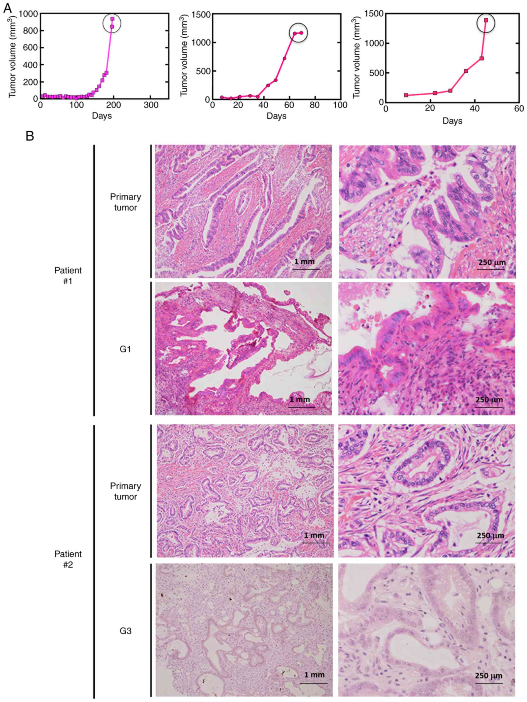
for one representative line is presented in Fig. 1A. The tumor tissues of the PDX lines
retained the pathological (Fig. 1B)
and genetic (data not shown) features of the original pancreatic
cancer tissues, even after repeated passages.
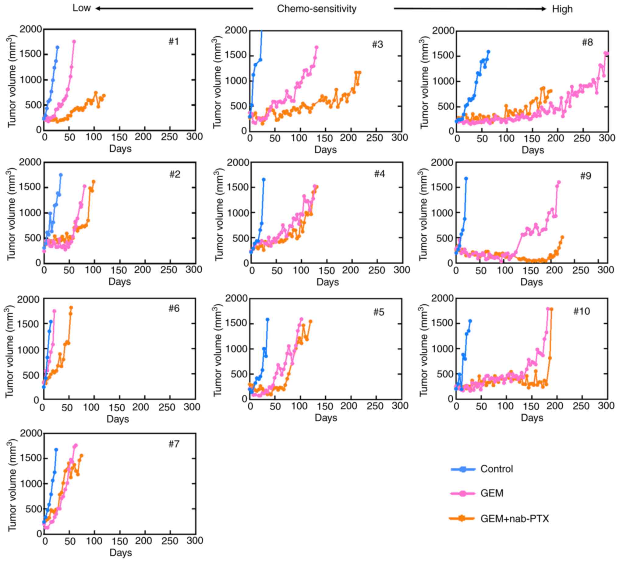
To identify the chemoresistance-related molecules,
the antitumor effects of the mono- or combination therapy in the
PDX models were characterized in terms of tumor growth after the
treatments (Fig. 2). Although
antitumor effects were transiently observed in some cases in the
treatment group, tumor growth was finally observed in all the
lines.
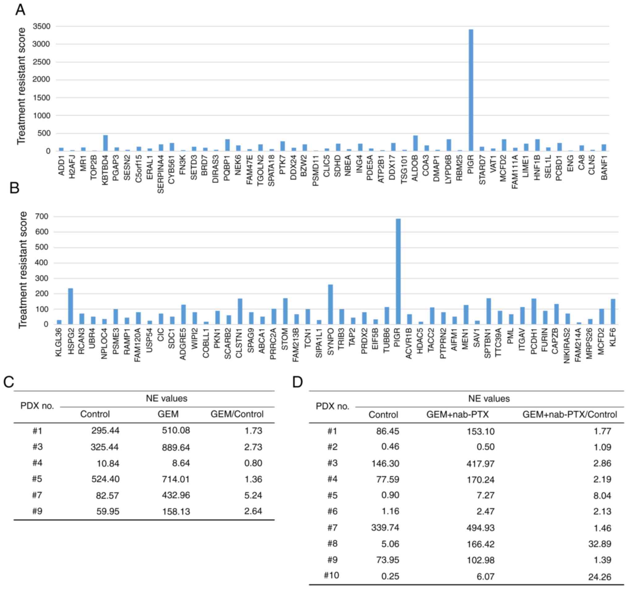
The mRNA expression in tumor tissues was analyzed
using NGS. We performed NGS of tumor tissues from PDXs before and
after treatment with standard chemotherapy. The number of genes
analyzed by RNA sequencing using NGS was 26732. Among them, there
were 193 genes with FPKM values of 10 or more and expression
fold-change ratios between the treatment group and control group of
3 or more (Table SII). Using the
normalized expression (NE) values, the ratio of the control to
treated PDX lines was calculated. For the mono- and combination
therapies, genes with NE values greater than 10 and NE ratios
(treated group to control group) greater than 2 were selected
(Fig. 3A and B). The ratio of NE
values for pIgR mRNA expression (Fig. 3C and D) between the treated and
control groups was greater than 1 for most of the lines treated
with GEM or GEM + nab-PTX. This analysis revealed that the
expression of pIgR mRNA was more increased in the PDX group
that received chemotherapy than in untreated PDXs. These findings
indicated that pIgR mRNA was expressed before the
administration of anticancer drugs and may have been induced by
chemotherapy. Moreover, the expression level of pIgR mRNA
was higher in the treated group than in the control group, which
may indicate that increased expression of pIgR mRNA was
involved in chemoresistance.
Prognostic analysis from TCGA RNA
database for pancreatic cancer
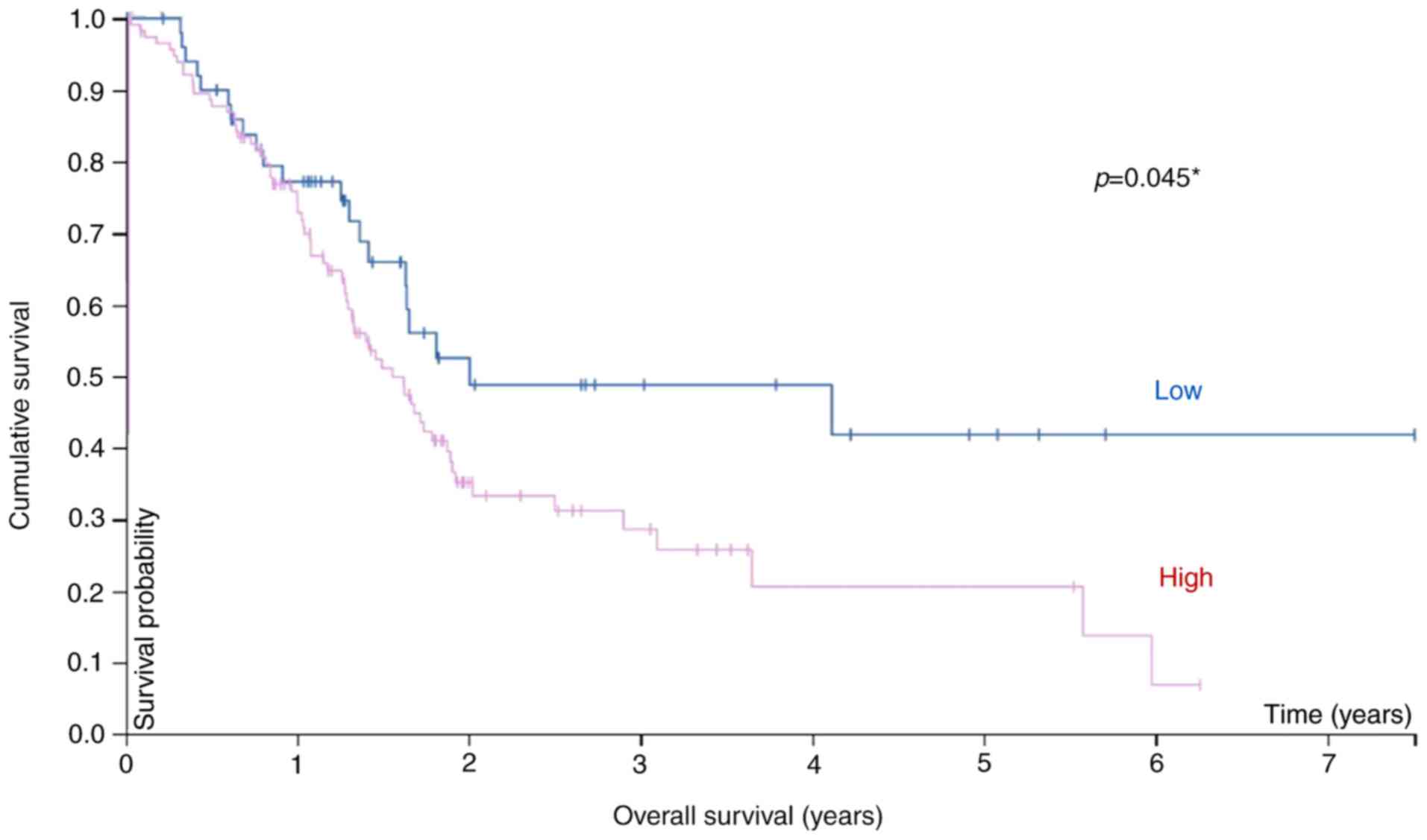
Additional analysis of pIgR expression and OS
in 171 pancreatic cancer samples from TCGA database revealed an
association between survival and the mRNA level (Fig. 4). Pancreatic cancer patients (stages
I–IV) were divided into high and low pIgR mRNA groups using
41 FPKM as the cut-off value, and the prognosis of each group was
examined. Patients in the high-pIgR mRNA group exhibited
significantly poorer survival rates than those in the low
pIgR mRNA group (P=0.045). The five-year survival rate was
42 and 21% for the low- and high-pIgR mRNA groups. Thus, it
was inferred that pIgR may be a putative prognostic
biomarker of pancreatic cancer.
Immunohistological and prognostic
analysis in pancreatic cancer patients
We corroborated our findings in the PDX model with
the expression of pIgR in clinical specimens, the
clinicopathological features of which are summarized in Table I. Immunohistochemistry revealed that
pIgR was strongly expressed on the tumor cells (Fig. 5A). The samples were divided into the
high [39.0% (30/77)] and low [61.0% (47/77)] pIgR groups (Table I), as described in the Materials and
methods section (Fig. 5B).
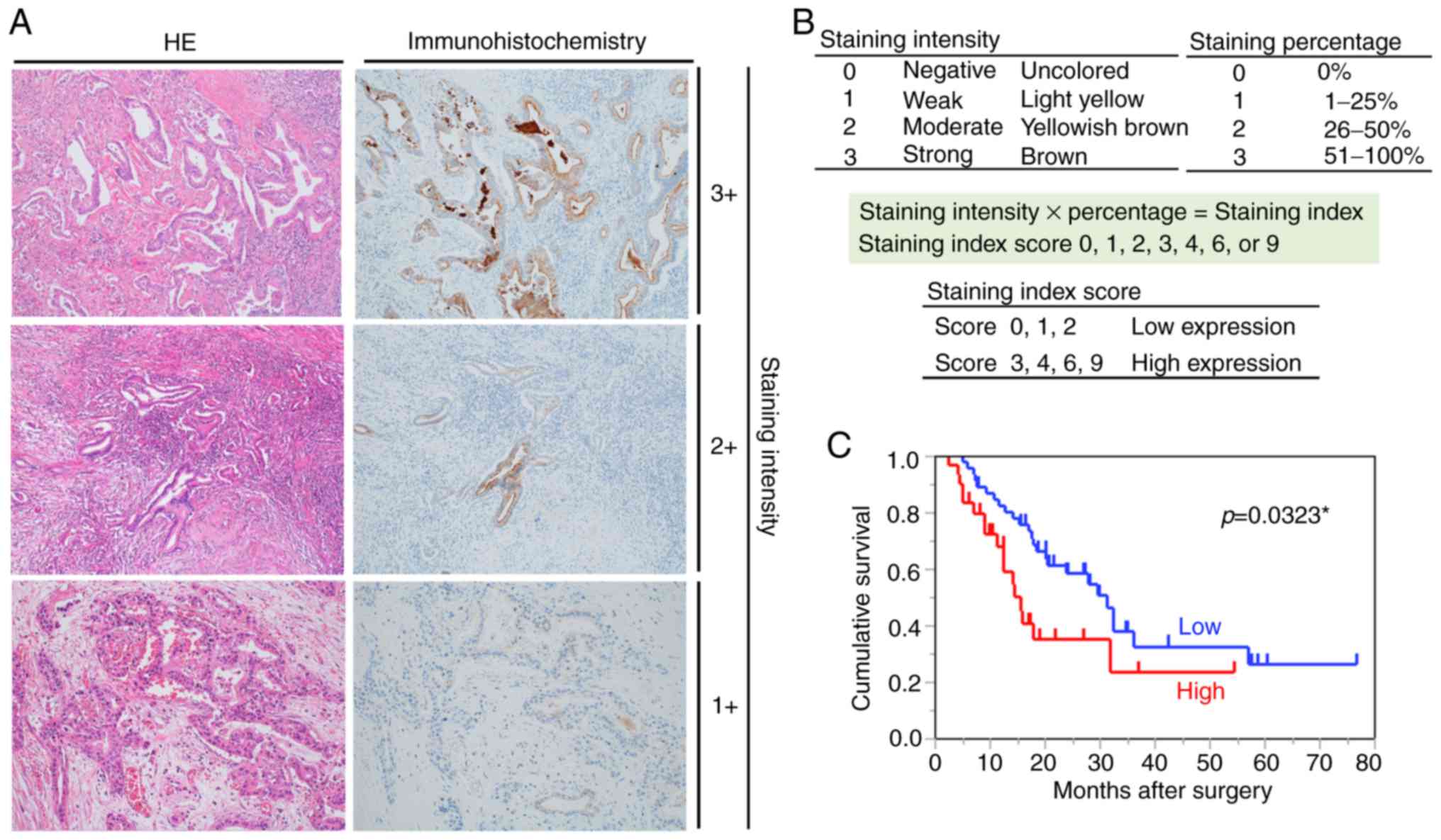 | Figure 5.Relationship between pIgR expression
and prognosis. (A) Immunohistochemical staining for pIgR in
pancreatic cancer tissues (magnification, ×100). The left and right
images show the same sample tissue blocks and correspond to the
staining intensity. Left, H&E staining; right,
immunohistochemical staining for pIgR. (B) Criteria for
determination of pIgR expression levels. The pIgR expression levels
in immunostaining were determined based on the intensity of
staining and percentage of stained cells. The staining intensity
and staining percentage criteria are presented. (C) Kaplan-Meier
survival analysis in patients with pancreatic cancer (n=77,
revealing OS based on the expression of pIgR protein. Red line,
high-expression group (n=47), blue line, low-expression group
(n=30). pIgR, polymeric immunoglobulin receptor; H&E,
hematoxylin and eosin; OS, overall survival. *Statistically
significant. |
Furthermore, whether pIgR was useful for prognostic
evaluation of pancreatic cancer was analyzed. Notably, there was no
statistical difference in patient or pathological features,
including age (P=0.34), sex (P=0.06), tumor location (head or
body/tail, P=0.66), histological type (adenocarcinoma or others,
P=0.10), TNM stage (IIA or IIB, P=0.34), histological
differentiation (P=0.16), lymphatic invasion (P=0.88), venous
invasion (P=0.07), perineural invasion (P=0.48), resection margin
(P=0.06), and adjuvant chemotherapy (P=0.86), due to the separation
of patients into the two groups of high and low pIgR expression
(Table I). Next, the association
between the pIgR expression levels and patient survival was
assessed using Kaplan-Meier analysis with log-rank tests. Among 77
pancreatic cancer patients, those with high pIgR expression had
poorer survival rates than those with low pIgR expression
(P=0.0323) (Fig. 5C). Furthermore,
the percentage of pIgR-stained area was analyzed using the Hybrid
Cell Count software with respect to survival time and the
expression of pIgR determined by two pathologists. The percentage
of pIgR-stained area was significantly associated with the
expression of pIgR as determined by the pathologists (P<0.001)
(Fig. S2A). In addition, higher
percentages of pIgR-stained area determined by the image analysis
software were associated with shorter OS (r=−0.3801,
P=0.00065) (Fig. S2B).
Univariate analysis of OS revealed four prognostic
parameters: Histological differentiation (P=0.0012), resection
margin (P=0.0004), adjuvant chemotherapy (P<0.0001), and pIgR
expression (P=0.0404). Multivariate analysis using Cox proportional
hazards revealed that histological differentiation (P=0.0004),
resection margin (P=0.0004), adjuvant chemotherapy (P=0.0013), and
pIgR expression (P=0.0045) were independent prognostic factors for
poor outcome (Table II).
| Table II.Univariate and multivariate analyses
of prognostic factor for overall survival in 77 pancreatic cancer
patients. |
Table II.
Univariate and multivariate analyses
of prognostic factor for overall survival in 77 pancreatic cancer
patients.
|
| Univariate
analysis | Multivariate
analysis |
|---|
|
|
|
|
|---|
| Clinicopathological
factors | HR | 95% CI |
P-valuea | HR | 95% CI |
P-valuea |
|---|
| Age(years) |
| (≦71
vs. >71) | 1.03 | 0.54–1.92 | 0.9214 | – | – | – |
| Sex |
| (male
vs. female) | 0.57 | 0.30–1.06 | 0.076 | – | – | – |
| TNM stage UICC
7th |
| (IIA
vs. IIB) | 0.59 | 0.26–1.21 | 0.1575 | – | – | – |
| Tumor location |
|
(body/tail vs. head) | 0.65 | 0.33–1.22 | 0.1861 | – | – | – |
| Histological
differentiation |
| (G1 vs.
G2, G3, G4) | 0.34 | 0.16–0.66 | 0.0012b | 0.26 | 0.11–0.56 | 0.0004b |
| Lymphatic
invasion |
| (ly0,
ly1 vs. ly2, ly3) | 0.71 | 0.37–1.31 | 0.2754 | – | – | – |
| Venous
invasion |
| (v0, v1
vs. v2, v3) | 0.94 | 0.35–2.09 | 0.8849 | – | – | – |
| Perineural
invasion |
| (ne0,
ne1 vs. ne2, ne3) | 0.62 | 0.27–1.29 | 0.2108 | – | – | – |
| Resection
margin |
| (R0 vs.
R1, R2) | 0.36 | 0.22–0.63 | 0.0004b | 0.28 | 0.14–0.56 | 0.0004b |
| Adjuvant
chemotherapy |
|
(present vs. absent) | 0.26 | 0.13–0.50 |
<0.0001b | 0.34 | 0.17–0.71 | 0.0013b |
| pIgR |
| (low
vs. high) | 0.56 | 0.27–0.97 | 0.0404b | 0.35 | 0.17–0.71 | 0.0045b |
Discussion
Pancreatic cancer is characterized by late
diagnosis, early metastasis, limited response to chemotherapy, and
extremely poor prognosis (7,37,38),
highlighting the need for novel prognostic biomarkers and
therapeutic targets. Using PDX lines, it was determined that
pIgR mRNA was upregulated after treatment with anticancer
drugs, indicating that pIgR may be associated with chemoresistance.
It was also revealed that high pIgR expression was an independent
prognostic factor for poor survival in pancreatic cancer patients.
Analysis of TCGA data supported our findings at the RNA level in
pancreatic cancer patients.
Various mouse tumor models and tumor cell lines have
been used for predicting the efficacy and possible toxicities of
anticancer drugs in cancer patients (31,39).
However, the results of these studies do not necessarily reflect
human clinical data (40),
primarily due to the lack of a tumor microenvironment in such cell
and tumor models. Moreover, studies in mice are also not always
translatable to human patients (41,42).
Therefore, better models, reflecting human clinical pathology, are
required.
PDX lines of immunodeficient mice have emerged as
relevant in vivo models for human tumors (43); they not only recapitulate the
interactions with the host microenvironment but also reflect tumor
heterogeneity, including cancer stem cells. In the present study
the PDX lines were confirmed to retain most of the histological and
genetic characteristics of their donor tumors and remained stable
after repeated passaging. In pancreatic cancer, it is often
challenging to perform surgical resection or biopsy using
endoscopic ultrasound/fine needle aspiration at diagnosis and to
collect sufficient pancreatic tissue samples (44,45).
Therefore, PDX models are optimal for research in pancreatic cancer
pathology (46).
It was determined that an increased pIgR mRNA
level was associated with chemoresistance. However, variations in
mRNA expression do not always correspond to changes in protein
expression owing to various post-transcriptional protein
modifications (47–49). Thus, mRNA abundance can be a poor
predictor of the protein levels. Analysis of colon and rectal tumor
proteomics data in TCGA database showed that mRNA abundance did not
reliably predict the differences in protein abundance between
tumors (50). To confirm the
correlation between the mRNA and protein levels of pIgR and the
relationship between pIgR protein and clinicopathological factors,
pIgR protein expression was evaluated in tissues from 77 pancreatic
cancer patients. It was revealed that high pIgR expression was an
independent prognostic factor for survival in pancreatic cancer
patients.
Few studies have investigated the role of pIgR in
cancers, especially in pancreatic cancer. Improved survival of
patients with low pIgR expression and low in vitro and ex
vivo stromal activity was observed using data from 88
pancreatic ductal adenocarcinoma patient samples; however, pIgR
expression alone had no statistically significant effect on the
survival (51). The present data
revealed statistical significance for pIgR expression. Low pIgR
expression in pancreatic adenocarcinoma was significantly
associated with progressive disease, a shorter time to recurrence,
and death within five years (52);
however, our results stand in contrast with these, possibly because
the target patient population was different, with the patient
cohort in the previous study having intestinal as well as
pancreatobiliary type adenocarcinoma. Moreover, differences in
surgical procedures and different types of anticancer drugs may
have led to contrasting results. Further investigations are
required to determine the relationship between pIgR expression and
its role in pancreatic cancer patients.
A limitation of the present study was the subjective
assessment of IHC staining. Because a single analytical method for
IHC has not been established, expression analysis of pIgR in the
tumor tissues was performed using previously reported methods
(53–55). To decrease subjectivity, the use of
image analysis software is an option (56). However, the specificity and
sensitivity of automated software remain unclear (57), and therefore, a semiquantitative
assessment strategy was deemed more appropriate. In addition, none
of the patients had received neoadjuvant chemotherapy before
surgical resection. The inclusion of specimens obtained after
neoadjuvant chemotherapy would have been ideal to compare tissues
with and without chemotherapy. However, only chemo-naïve patients
were included in this study because neoadjuvant chemotherapy is not
yet permitted in Japan. Third, for survival analysis using TCGA RNA
database, the optimal expression cut-off that yielded the maximal
difference between the two groups with regard to survival at the
lowest log-rank P-value was selected; nonetheless, this cut-off may
not be suitable for other patient groups.
In summary, in the present study it was revealed
that the pIgR mRNA level was associated with
chemoresistance, and that high expression of pIgR protein was
significantly associated with poor prognosis of pancreatic cancer
patients. Thus, pIgR may be a novel predictor of poor prognosis of
pancreatic cancer patients after surgical resection and a promising
candidate for targeted therapy of pancreatic cancer.
Supplementary Material
Supporting Data
Acknowledgements
Not applicable.
Funding
No funding was received.
Availability of data and materials
The datasets used during the present study are
available from the corresponding author upon reasonable request.
All data generated or analyzed during this study are included in
this published article. The results published in the present study
are in part based upon data generated by the TCGA Research Network:
https://www.cancer.gov/tcga.
Authors' contributions
RO, TT, SK and SW conceived and designed this study.
RO, YK, KH, AH, TI, YH, HA, JT, KY, KA, MW, MS, RO, TA, MM, TT and
SW collected samples and recorded the general data and the
observation indicators of the patients. EY, TS and SW performed all
the experiments using PDX models. SI and DK analyzed the data for
NGS analysis. MT, NO and TN performed the pathological diagnosis
and analyzed the immunostained samples. RO analyzed all the data
and wrote the first draft of the manuscript. JT, KY, TT, SK and SW
critically reviewed and corrected the manuscript. All authors
reviewed and approved the final version of the manuscript.
Ethics approval and consent to
participate
The study protocol was approved by the Ethics
Committee of the Showa University School of Medicine (Tokyo, Japan;
Approval no. 2611) and adhered to the principles of the Declaration
of Helsinki. All experiments involving animals were performed in
accordance with the care and use guidelines of the Kanagawa Cancer
Center Research Institute, Japan. This study was approved by the
Research Ethics Committee of Kanagawa Cancer Center Research
Institute (approval no. 176).
Patient consent for publication
Not applicable.
Competing interests
The authors declare that they have no competing
interests.
References
|
1
|
Hidalgo M, Cascinu S, Kleeff J, Labianca
R, Löhr JM, Neoptolemos J, Real FX, Van Laethem JL and Heinemann V:
Addressing the challenges of pancreatic cancer: Future directions
for improving outcomes. Pancreatology. 15:8–18. 2015. View Article : Google Scholar : PubMed/NCBI
|
|
2
|
Lefebvre AC, Maurel J, Boutreux S, Bouvier
V, Reimund JM, Launoy G and Arsene D: Pancreatic cancer: Incidence,
treatment and survival trends-1175 cases in Calvados (France) from
1978 to 2002. Gastroenterol Clin Biol. 33:1045–1051. 2009.
View Article : Google Scholar : PubMed/NCBI
|
|
3
|
Kocher HM and Alrawashdeh W: Pancreatic
cancer. BMJ Clin Evid. 2010(pii): 04092010.PubMed/NCBI
|
|
4
|
Siegel RL, Miller KD and Jemal A: Cancer
statistics in USA, 2018. CA Cancer J Clin. 68:7–30. 2018.
View Article : Google Scholar : PubMed/NCBI
|
|
5
|
Li HB, Zhou J and Zhao FQ: A prognostic
nomogram for disease-specific survival in patients with pancreatic
ductal adenocarcinoma of the head of the pancreas following
pancreaticoduodenectomy. Med Sci Monit. 24:6313–6321. 2018.
View Article : Google Scholar : PubMed/NCBI
|
|
6
|
Rahib L, Smith BD, Aizenberg R, Rosenzweig
AB, Fleshman JM and Matrisian LM: Projecting cancer incidence and
deaths to 2030: The unexpected burden of thyroid, liver, and
pancreas cancers in the United States. Cancer Res. 74:2913–2921.
2014. View Article : Google Scholar : PubMed/NCBI
|
|
7
|
Kamisawa T, Wood LD, Itoi T and Takaori K:
Pancreatic cancer. Lancet. 388:73–85. 2016. View Article : Google Scholar : PubMed/NCBI
|
|
8
|
Von Hoff DD, Ervin T, Arena FP, Chiorean
EG, Infante J, Moore M, Seay T, Tjulandin SA, Ma WW, Saleh MN, et
al: Increased survival in pancreatic cancer with nab-paclitaxel
plus gemcitabine. N Engl J Med. 369:1691–1703. 2013. View Article : Google Scholar : PubMed/NCBI
|
|
9
|
Goldstein D, El-Maraghi RH, Hammel P,
Heinemann V, Kunzmann V, Sastre J, Scheithauer W, Siena S,
Tabernero J, Teixeira L, et al: Nab-Paclitaxel plus gemcitabine for
metastatic pancreatic cancer: Long-term survival from a phase III
trial. J Natl Cancer Inst. 107:1–10. 2015. View Article : Google Scholar
|
|
10
|
Conroy T, Desseigne F, Ychou M, Bouché O,
Guimbaud R, Bécouarn Y, Adenis A, Raoul JL, Gourgou-Bourgade S, de
la Fouchardière C, et al: FOLFIRINOX versus gemcitabine for
metastatic pancreatic cancer. N Engl J Med. 364:1817–1825. 2011.
View Article : Google Scholar : PubMed/NCBI
|
|
11
|
Cecconi D, Palmieri M and Donadelli M:
Proteomics in pancreatic cancer research. Proteomics. 11:816–828.
2011. View Article : Google Scholar : PubMed/NCBI
|
|
12
|
Mostov KE, Kraehenbuhl JP and Blobel G:
Receptor-mediated transcellular transport of immunoglobulin:
Synthesis of secretory component as multiple and larger
transmembrane forms. Proc Natl Acad Sci USA. 77:7257–7261. 1980.
View Article : Google Scholar : PubMed/NCBI
|
|
13
|
Rojas R and Apodaca G: Immunoglobulin
transport across polarized epithelial cells. Nat Rev Mol Cell Biol.
3:944–955. 2002. View
Article : Google Scholar : PubMed/NCBI
|
|
14
|
Kaetzel CS: The polymeric immunoglobulin
receptor: Bridging innate and adaptive immune responses at mucosal
surfaces. Immunol Rev. 206:83–99. 2005. View Article : Google Scholar : PubMed/NCBI
|
|
15
|
Phalipon A and Corthésy B: Novel functions
of the polymeric Ig receptor: Well beyond transport of
immunoglobulins. Trends Immunol. 24:55–58. 2003. View Article : Google Scholar : PubMed/NCBI
|
|
16
|
Denning GM: IL-4 and IFN-gamma
synergistically increase total polymeric IgA receptor levels in
human intestinal epithelial cells. Role of protein tyrosine
kinases. J Immunol. 156:4807–4814. 1996.PubMed/NCBI
|
|
17
|
Kvale D, Løvhaug D, Sollid LM and
Brandtzaeg P: Tumor necrosis factor-alpha up-regulates expression
of secretory component, the epithelial receptor for polymeric Ig. J
Immunol. 140:3086–3089. 1988.PubMed/NCBI
|
|
18
|
Poger ME, Hirsch BR and Lamm ME: Synthesis
of secretory component by colonic neoplasms. Am J Pathol.
82:327–338. 1976.PubMed/NCBI
|
|
19
|
Harris JP, Caleb MH and South MA:
Secretory component in human mammary carcinoma. Cancer Res.
35:1861–1864. 1975.PubMed/NCBI
|
|
20
|
Harris JP and South MA: Secretory
component: A glandular epithelial cell marker. Am J Pathol.
105:47–53. 1981.PubMed/NCBI
|
|
21
|
DeSouza LV, Krakovska O, Darfler MM,
Krizman DB, Romaschin AD, Colgan TJ and Siu KW: mTRAQ-based
quantification of potential endometrial carcinoma biomarkers from
archived formalin-fixed paraffin-embedded tissues. Proteomics.
10:3108–3116. 2010. View Article : Google Scholar : PubMed/NCBI
|
|
22
|
Rossel M, Billerey C, Bittard H, Ksiazek
P, Alber D, Revillard JP and Vuitton DA: Alterations in polymeric
immunoglobulin receptor expression and secretory component levels
in bladder carcinoma. Urol Res. 19:361–366. 1991. View Article : Google Scholar : PubMed/NCBI
|
|
23
|
Rossel M, Seilles E, Voigt JJ, Vuitton D,
Legait N and Revillard JP: Polymeric Ig receptor expression in
hepatocellular carcinoma. Eur J Cancer. 28:1120–1124. 1992.
View Article : Google Scholar
|
|
24
|
Ai J, Tang Q, Wu Y, Xu Y, Feng T, Zhou R,
Chen Y, Gao X, Zhu Q, Yue X, et al: The role of polymeric
immunoglobulin receptor in inflammation-induced tumor metastasis of
human hepatocellular carcinoma. J Natl Cancer Inst. 103:1696–1712.
2011. View Article : Google Scholar : PubMed/NCBI
|
|
25
|
Berntsson J, Lundgren S, Nodin B, Uhlén M,
Gaber A and Jirström K: Expression and prognostic significance of
the polymeric immunoglobulin receptor in epithelial ovarian cancer.
J Ovarian Res. 7:262014. View Article : Google Scholar : PubMed/NCBI
|
|
26
|
Fristedt R, Gaber A, Hedner C, Nodin B,
Uhlén M, Eberhard J and Jirström K: Expression and prognostic
significance of the polymeric immunoglobulin receptor in esophageal
and gastric adenocarcinoma. J Transl Med. 12:832014. View Article : Google Scholar : PubMed/NCBI
|
|
27
|
Xiao T, Ying W, Li L, Hu Z, Ma Y, Jiao L,
Ma J, Cai Y, Lin D, Guo S, et al: An approach to studying lung
cancer-related proteins in human blood. Mol Cell Proteomics.
4:1480–1486. 2005. View Article : Google Scholar : PubMed/NCBI
|
|
28
|
Rossel M, Brambilla E, Billaud M, Vuitton
DA, Blanc-Jouvan F, Biichle S and Revillard JP: Nonspecific
increased serum levels of secretory component in lung tumors:
Relationship to the gene expression of the transmembrane receptor
form. Am J Respir Cell Mol Biol. 9:341–346. 1993. View Article : Google Scholar : PubMed/NCBI
|
|
29
|
Makawita S, Smith C, Batruch I, Zheng Y,
Rückert F, Grützmann R, Pilarsky C, Gallinger S and Diamandis EP:
Integrated proteomic profiling of cell line conditioned media and
pancreatic juice for the identification of pancreatic cancer
biomarkers. Mol Cell Proteomics. 10:M111.008599. 2011. View Article : Google Scholar : PubMed/NCBI
|
|
30
|
Kvale D, Norstein J, Meling GI, Børmer OP,
Brandtzaeg P, Langmark F and Rognum TO: Circulating secretory
component in relation to early diagnosis and treatment of liver
metastasis from colorectal carcinomas. J Clin Pathol. 45:568–571.
1992. View Article : Google Scholar : PubMed/NCBI
|
|
31
|
Morton CL and Houghton PJ: Establishment
of human tumor xenografts in immunodeficient mice. Nat Protoc.
2:247–250. 2007. View Article : Google Scholar : PubMed/NCBI
|
|
32
|
Chijiwa T, Kawai K, Noguchi A, Sato H,
Hayashi A, Cho H, Shiozawa M, Kishida T, Morinaga S, Yokose T, et
al: Establishment of patient-derived cancer xenografts in
immunodeficient NOG mice. Int J Oncol. 47:61–70. 2015. View Article : Google Scholar : PubMed/NCBI
|
|
33
|
Langmead B, Trapnell C, Pop M and Salzberg
SL: Ultrafast and memory-efficient alignment of short DNA sequences
to the human genome. Genome Biol. 10:R252009. View Article : Google Scholar : PubMed/NCBI
|
|
34
|
Komura D, Isagawa T, Kishi K, Suzuki R,
Sato R, Tanaka M, Katoh H, Yamamoto S, Tatsuno K, Fukayama M, et
al: CASTIN: A system for comprehensive analysis of cancer-stromal
interactome. BMC Genomics. 17:8992016. View Article : Google Scholar : PubMed/NCBI
|
|
35
|
Pontén F, Jirström K and Uhlen M: The
human protein atlas-A tool for pathology. J Pathol. 216:387–393.
2008. View Article : Google Scholar : PubMed/NCBI
|
|
36
|
Iida Y, Tanaka H, Sano H, Suzuki Y,
Shimizu H and Urano T: Ectopic expression of PCSK9 by smooth muscle
cells contributes to aortic dissection. Ann Vasc Surg. 48:195–203.
2018. View Article : Google Scholar : PubMed/NCBI
|
|
37
|
Zijlstra M, Bernards N, de Hingh IH, van
de Wouw AJ, Goey SH, Jacobs EM, Lemmens VE and Creemers GJ: Does
long-term survival exist in pancreatic adenocarcinoma? Acta Oncol.
55:259–264. 2016. View Article : Google Scholar : PubMed/NCBI
|
|
38
|
Ying H, Dey P, Yao W, Kimmelman AC,
Draetta GF, Maitra A and DePinho RA: Genetics and biology of
pancreatic ductal adenocarcinoma. Genes Dev. 30:355–385. 2016.
View Article : Google Scholar : PubMed/NCBI
|
|
39
|
Jin K, Li G, Cui B, Zhang J, Lan H, Han N,
Xie B, Cao F, He K, Wang H, et al: Assessment of a novel VEGF
targeted agent using patient-derived tumor tissue xenograft models
of colon carcinoma with lymphatic and hepatic metastases. PLoS One.
6:e283842011. View Article : Google Scholar : PubMed/NCBI
|
|
40
|
Yada E, Wada S, Yoshida S and Sasada T:
Use of patient-derived xenograft mouse models in cancer research
and treatment. Futur Sci OA. 4:FSO2712018. View Article : Google Scholar
|
|
41
|
Hidalgo M, Amant F, Biankin AV, Budinská
E, Byrne AT, Caldas C, Clarke RB, de Jong S, Jonkers J, Mælandsmo
GM, et al: Patient-derived xenograft models: An emerging platform
for translational cancer research. Cancer Discov. 4:998–1013. 2014.
View Article : Google Scholar : PubMed/NCBI
|
|
42
|
Cho SY, Kang W, Han JY, Min S, Kang J, Lee
A, Kwon JY, Lee C and Park H: An integrative approach to precision
cancer medicine using patient-derived xenografts. Mol Cells.
39:77–86. 2016. View Article : Google Scholar : PubMed/NCBI
|
|
43
|
Fujii E, Suzuki M, Matsubara K, Watanabe
M, Chen YJ, Adachi K, Ohnishi Y, Tanigawa M, Tsuchiya M and Tamaoki
N: Establishment and characterization of in vivo human tumor models
in the NOD/SCID/γcnull mouse. Pathol Int. 58:559–567. 2008.
View Article : Google Scholar : PubMed/NCBI
|
|
44
|
Erickson RA, Sayage-Rabie L and Beissner
RS: Factors predicting the number of EUS-guided fineneedle passes
for diagnosis of pancreatic malignancies. Gastrointest Endosc.
51:184–190. 2000. View Article : Google Scholar : PubMed/NCBI
|
|
45
|
LeBlanc JK, Ciaccia D, Al-Assi MT, McGrath
K, Imperiale T, Tao LC, Vallery S, DeWitt J, Sherman S and Collins
E: Optimal number of EUS-guided fineneedle passes needed to obtain
acorrect diagnosis. Gastrointest Endosc. 59:475–481. 2004.
View Article : Google Scholar : PubMed/NCBI
|
|
46
|
Ohkuma R, Yada E, Ishikawa S, Komura D,
Ishizaki H, Tamada K, Kubota Y, Hamada K, Ishida H, Hirasawa Y, et
al: High expression of olfactomedin-4 is correlated with
chemoresistance and poor prognosis in pancreatic cancer. PLoS One.
15:e02267072020. View Article : Google Scholar : PubMed/NCBI
|
|
47
|
De Sousa Abreu R, Penalva LO, Marcotte EM
and Vogel C: Global signatures of protein and mRNA expression
levels. Mol Biosyst. 5:1512–1526. 2009.PubMed/NCBI
|
|
48
|
Schwanhüusser B, Busse D, Li N, Dittmar G,
Schuchhardt J, Wolf J, Chen W and Selbach M: Global quantification
of mammalian gene expression control. Nature. 473:337–342. 2011.
View Article : Google Scholar : PubMed/NCBI
|
|
49
|
Wu L, Candille SI, Choi Y, Xie D, Jiang L,
Li-Pook-Than J, Tang H and Snyder M: Variation and genetic control
of protein abundance in humans. Nature. 499:79–82. 2013. View Article : Google Scholar : PubMed/NCBI
|
|
50
|
Zhang B, Wang J, Wang X, Zhu J, Liu Q, Shi
Z, Chambers MC, Zimmerman LJ, Shaddox KF, Kim S, et al:
Proteogenomic characterization of human colon and rectal cancer.
Nature. 513:382–387. 2014. View Article : Google Scholar : PubMed/NCBI
|
|
51
|
Arumugam P, Bhattacharya S, Chin-Aleong J,
Capaso M and Kocher HM: Expression of polymeric immunoglobulin
receptor and stromal activity in pancreatic ductal adenocarcinoma.
Pancreatology. 17:295–302. 2017. View Article : Google Scholar : PubMed/NCBI
|
|
52
|
Fristedt R, Elebro J, Gaber A, Jonsson L,
Heby M, Yudina Y, Nodin B, Uhlén M, Eberhard J and Jirström K:
Reduced expression of the polymeric immunoglobulin receptor in
pancreatic and periampullary adenocarcinoma signifies tumour
progression and poor prognosis. PLoS One. 9:e1127282014. View Article : Google Scholar : PubMed/NCBI
|
|
53
|
Liu F, Ye P, Bi T, Teng L, Xiang C, Wang
H, Li Y, Jin K and Mou X: COLORECTAL polymeric immunoglobulin
receptor expression is correlated with hepatic metastasis and poor
prognosis in colon carcinoma patients with hepatic metastasis.
Hepatogastroenterology. 61:652–659. 2014.PubMed/NCBI
|
|
54
|
Wang X, Du J, Gu P, Jin R and Lin X:
Polymeric immunoglobulin receptor expression is correlated with
poor prognosis in patients with osteosarcoma. Mol Med Rep.
9:2105–2110. 2014. View Article : Google Scholar : PubMed/NCBI
|
|
55
|
Niu H, Wang K and Wang Y: Polymeric
immunoglobulin receptor expression is predictive of poor prognosis
in glioma patients. Int J Clin Exp Med. 7:21852014.PubMed/NCBI
|
|
56
|
Aguilar-Mahecha A, Hassan S, Ferrario C
and Basik M: Microarrays as validation strategies in clinical
samples: Tissue and protein microarrays. OMICS. 10:311–326. 2006.
View Article : Google Scholar : PubMed/NCBI
|
|
57
|
Simon R and Sauter G: Tissue microarrays
for miniaturized high-throughput molecular profiling of tumors. Exp
Hematol. 30:1365–1372. 2002. View Article : Google Scholar : PubMed/NCBI
|