Introduction
Endocrine disruptor chemicals (EDCs) are substances
that can alter the regular functioning of the endocrine system of
living organisms (1,2) by interfering with the effect,
biosynthesis and metabolism of hormones (3). Consequently, the disruption of hormone
homeostasis can affect different physiological processes leading to
various diseases in humans (4).
Although these compounds can occur naturally, a large number is
mainly human-made (5). They can
easily reach the environment since they are released from several
daily products, such as food cans, metals or pesticides (6), industrial chemicals, organochloride
pesticides and other chemicals (3).
Due to the massive use of these chemical compounds, they are widely
scattered in the environment (7) and
are then bio-accumulated by living organisms (8). The principal source of exposure is oral
intake of contaminated food (3) and
direct contact or inhalation from polluted air (1). Moreover, the possibility of vertical
transfer of these compounds through the placenta and breast milk
has been reported (6).
It has been observed that EDCs are still present in
the environment regardless of their prohibition in some countries;
levels of these compounds have been detected in non-pregnant women,
pregnant women, men and newborns (9)
as a consequence affecting the entire population. Thus, in terms of
human health impact (10), these
substances have become a priority for international policymakers
and the scientific community (11).
Unfortunately, once these compounds reach the organism, they can
disrupt the endocrine function by mimicking or blocking naturally
occurring hormones due to their similar chemical structure
(11), therefore affecting the
development later in adult life (7).
Regarding the mechanism of action, Fig. 1 shows that EDCs can act through
various signaling pathways modulating the action of androgenic
thyroid and retinoid receptors, as well as interacting with
estrogen receptors (ERs) (3,7) and other non-nuclear receptors, such as
membrane ERs, non-steroid receptors and orphan receptors (3). The carcinogenicity attributable to the
EDCs is given by genotoxicity, epigenetic modifications or immune
system alterations, especially for hormone-associated types of
cancer where endocrine disruption is the most relevant cause
(12). Therefore, the study and
association of EDCs with carcinogenesis are of high priority.
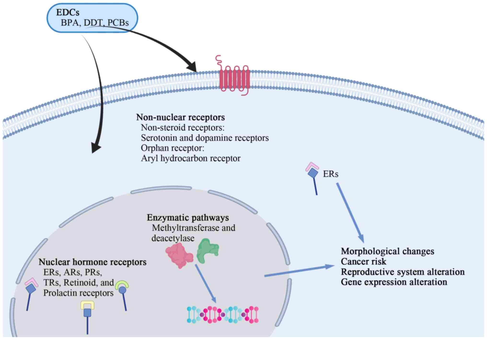 | Figure 1.Summary of the mechanism of action of
EDCs. BPA, DDT and PCBs can exert their action by modulating
different signaling pathways, through the interaction with nuclear
hormone receptors, such as ERs, ARs, PRs, TRs, retinoid receptors
and prolactin receptors. EDCs can also interact with non-nuclear
receptors, such as non-steroid receptors, orphan receptors,
membrane ERs and enzymes. EDCs, endocrine disruptor chemicals; BPA,
bisphenol-A; DDT, dichlorodiphenyltrichloroethane; PCBs,
polychlorinated biphenyls; ERs, estrogen receptors; ARs, androgen
receptors; PRs, progesterone receptors; TRs, thyroid receptors. |
The present review aimed to describe the impact of
environmental compounds such as bisphenol A (BPA),
dichlorodiphenyltrichloroethane (DDT) and polychlorinated biphenyls
(PCBs) on breast and prostate glands.
Data collection method
In the present review, a search on MEDLINE (through
PubMed: http://pubmed.ncbi.nlm.nih.gov/), Web of Science
(https://www.webofknowledge.com) and
SCOPUS (https://www.scopus.com/) was conducted
between November 2019 and March 2020 to identify studies published
from 1982 to the present addressing the association between BPA,
DDT, PCBs and carcinogenesis of the breast and prostate glands. The
search terms used were ‘mammary gland’, ‘breast cancer’, ‘prostate
gland’, ‘prostate cancer’, ‘bisphenol A’, ‘BPA’,
‘dichlorodiphenyltrichloroethane’, ‘DDT’, ‘polychlorinated
biphenyls’, ‘PCBs’, ‘exposure’, ‘epidemiology’, ‘monotonic
response’, ‘hormone sensitivity’ and ‘epigenetic’, and the data
collection approach is shown in Fig.
2.
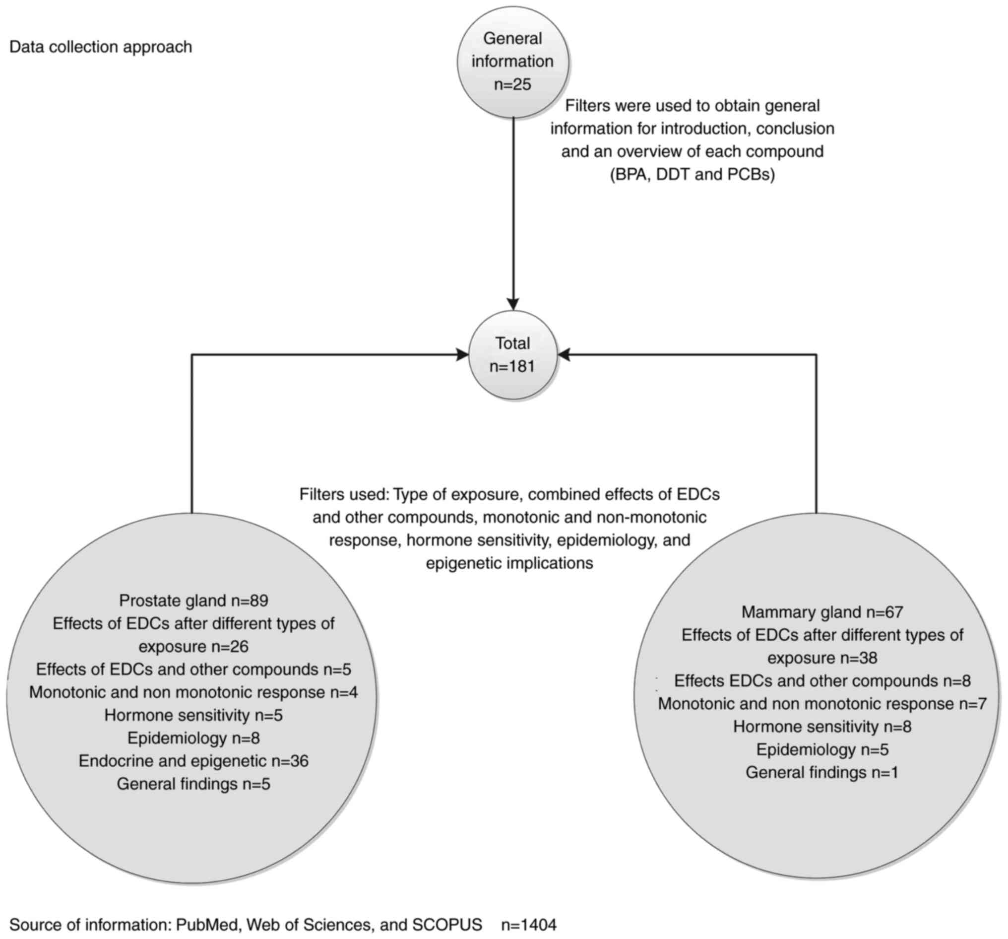 | Figure 2.Search on MEDLINE (through PubMed),
Web of Science and SCOPUS was conducted between November 2019 and
March 2020 to identify studies addressing the association between
BPA, DDT, PCBs and carcinogenesis of the breast and prostate
glands. The search terms used were ‘mammary gland’, ‘breast
cancer’, ‘prostate gland’, ‘prostate cancer’, ‘bisphenol A’, ‘BPA’,
‘dichlorodiphenyltrichloroethane’, ‘DDT’, ‘polychlorinated
biphenyls’, ‘PCBs’, ‘exposure’, ‘epidemiology’, ‘monotonic
response’, ‘hormone sensitivity’ and ‘epigenetic’. EDCs, endocrine
disruptor chemicals; BPA, bisphenol-A; DDT,
dichlorodiphenyltrichloroethane; PCBs, polychlorinated
biphenyls. |
EDCs: BPA, DDT and PCBs
First, it has been reported that BPA can interact
with ERs due to its chemical structure, thus modulating different
signaling pathways (13). Similarly,
DDT has been considered as a xenoestrogen compound with estrogen
and androgen receptor influence (12). At the same time, PCBs have shown
similar action mimicking natural hormones (14) and affecting reproductive development
(15). A brief description of each
compound is presented below.
BPA is known as a xenoestrogen and can be found in
filters, polymers, cosmetics, plasticizers, safety equipment, food
cans, thermal paper and medical devices (5,16,17). BPA is popular due to its advantages
over other materials such as strength, stability and durability
(5). More than 5 million tons of BPA
are produced every year (16). BPA
can reach living organisms through contaminated food (such as
damaged food cans) or water, inhalation or direct contact (5,10).
Notably, BPA has been considered a selective ER modulator (10) acting as an estrogen agonist or
antagonist depending on the tissue. Consequently, BPA has been
implicated in various endocrine disorders, such as infertility,
early puberty, metabolic disarrangement and cancer (13).
DDT is a chlorinated hydrocarbon with insecticidal
activity; moreover, it was extensively used to control malaria,
typhus and other agricultural diseases (18). Due to its physicochemical properties
(8), DDT and its metabolites are
extremely stable (18). These
properties facilitate their bioaccumulation in fatty tissue
(8) and their continuous circulation
in the environment (19). The main
route of exposure to DDT is direct contact or consumption of
contaminated food (8). Regardless of
the prohibition and restriction of its use in many industrialized
countries, DDT and its main metabolite,
p,p′-dichlorodiphenyldichloroethylene (DDE), are still present in
animals and humans (19). Similarly
to BPA, DDT has demonstrated estrogenic properties, either
mimicking the estrogen action (20)
or blocking the ER activity (19).
Additionally, it has been suggested that DDT and its metabolites
antagonize the activity of androgens by competitively binding to
androgen receptors (ARs) (20).
Finally, PCBs are organochlorine compounds that were
extensively used, mainly for industrial purposes (21), until its control and prohibition in
the 1970s (1). PCBs consist of a
mixture of 209 chlorinated aromatic compounds, known as congeners
(15), each with individual
properties and action upon the endocrine system. Additionally, it
has been reported that these compounds have estrogenic activity,
and the interaction with other organochlorines have a synergistic
effect (22). According to the
toxicological mechanisms of these compounds, they can be divided
into two groups: The dioxin-like coplanar PCBs and the
non-dioxin-like PCBs. The former are characterized by activating
the aryl hydrocarbon receptor (AhR), while the latter interact with
nuclear receptors (some of them hormone receptors) and modulate the
estrogen and androgen signaling pathways and activate some
carcinogenic processes (23).
Therefore, considering that hormones regulate numerous types of
cancer, it is urgent to investigate the effects of environmental
endocrine disruptors and their long-term exposure on
hormone-dependent cancers (24).
Hence, the present study focuses on two important
hormone-associated types of cancer: Breast and prostate cancer
(17).
Effects of EDCs on mammary gland tissue
Exposure at early-life
Since breast tissue is not fully developed at birth,
hormones are crucial for its proper and unique development
throughout life (25). During this
period, cells are under rapid growth and differentiation.
Therefore, any exposure to endocrine disruptors at this stage can
be critical for breast cancer incidence (26) and denote an important issue that must
be addressed by legislators and researchers. Studying different
environmental substances with known endocrine-disrupting effects on
morphology and human health gives valuable information regarding
the consequences. Unfortunately, some of these toxic compounds are
still constantly released into the environment despite current
regulations. An example of this is the release of DDT from melted
glaciers every day; similarly, PCBs accumulated in adipose tissues
are released into the body under specific physiologic conditions
exerting their effects on the endocrine system (27).
How BPA, DDT and PCBs are associated with
carcinogenesis and how they interfere with the normal
hormone-tissue response are valid questions in this context. From
that perspective, a previous study (28) has suggested that endocrine disruptors
directly affect breast cancer incidence; nevertheless, the complete
knowledge regarding the immediate association between these
environmental toxicants and initiation and progression of cancer
remains under investigation. Another study has suggested that in
prenatal exposures, BPA interacts with ER inducing the expression
of certain genes that cause cellular proliferation, particularly
branching ductal growth (26). It is
well known that ER is crucial in the carcinogenesis process
(29); therefore, any interaction
will alter such a process.
Due to its long half-life, DDT accumulates in human
tissues, and it has been demonstrated that its metabolites are
transferred from breast milk to newborns, thus increasing the serum
level in children exposed to such compounds (30). Although a previous study (31) used DDE serum levels as a marker of DDT
exposure, design limitations paused the progress regarding
conclusive evidence to determine exactly the grade of influence of
DDT exposure in early life and breast cancer development.
Regarding the association between early-life PCBs
exposure and an increase in breast cancer development, studies are
scarce. However, a previous study (32) evaluated whether PCBs concentrations
were demonstrative of early-life exposure using a
physiologically-based pharmacokinetic (PBPK) model. This evaluation
took into consideration some information obtained from the French
population-based case-control study (32), and the half-life of PCB153 congener
was detected with high frequency in most of the samples used; even
though this tool can serve as a predictor of early-life exposure,
some hindrances must be considered, for instance, the window of
breast cancer susceptibility is not clear for PCB compounds
(32). Therefore, it is difficult to
establish a direct and close association between PCB compounds and
breast cancer when exposed early in life.
Exposure in adults
Paradoxically, as endocrine disruptors disrupt the
endocrine function, one of their effects is the derangement of the
normal cell interactions (tissue organization). Some interactions,
such as the epithelial-epithelial and stromal-epithelial
interactions, are fundamental for the definition of any tissue
organization, and any change at this level can affect the proper
communication with consequences in cell proliferation, motility and
cell adhesion (33); therefore, the
analysis of these interactions during mammogenesis and
carcinogenesis is necessary. It is inevitable not to mention a
tissue-based theory, the so-called tissue organization field theory
(TOFT) of carcinogenesis; this proposes that morphogenetic fields
coordinate histogenesis and organogenesis (33). Then, during the perinatal phase, it
leads to tissue arrangement and remodeling processes, affecting
morphogenesis; however, this phenomenon can be reversible (26,34,35).
Similarly, it has been reported that BPA affects
tissue organization and intensifies the estrogen-tissue-target
response, along with the expression of mediators of progesterone,
altering the progesterone hormone response (36). Notably, major morphological changes in
the mammary gland development were observed when BPA exposure
occurred during the embryogenesis phase (37). Moreover, studies have revealed that
fetal exposures to BPA cause a more sensitive response to estradiol
on certain tissues, such as the uterus, vagina and mammary glands
(25,38). Consequently, this leads to mammary
gland changes and increases the susceptibility to develop diseases
later in life (26).
On the other hand, although DDT has
endocrine-disrupting features, its direct effects on cancer remain
unknown (39). A previous study that
evaluated breast cancer supported the TOFT theory and DDT effects
on mammary tissue (34). Concerning
DDT and PCBs, the aforementioned study assessed a mixture of
different pesticides, including DDT, PCBs and polybrominated
biphenyls (PBBs), in normal human breast epithelial cells (34). This evaluation was particularly
conducted to determine their effects upon gap junctional
intercellular communication in mammary tissue (34). Major effects on intercellular
communication integrity in normal epithelial cells were observed
after the treatment with the mixture of DDT, halogenated compounds
and dieldrin (40). Similar effects
were observed in epithelial cells from other tissues under the
effects of PCBs (41,42).
Overall, it was suggested that epithelial cell
communication was affected by different environmental compounds
such as BPA, DDT and PCBs, and the collaboration of individual
compounds contributed to this phenomenon (40). In other words, each toxic compound may
grant a certain grade of risk to the breast cancer process and
other types of cancer. Additionally, to tackle this issue from a
broader perspective, other factors such as compound interactions,
chemical structure and physiological conditions should be
considered.
Exposure during prenatal and postnatal
life
In vivo studies that evaluated the long-term
effects of BPA based their concentration on the safe reference dose
(SRD) provided by the Environmental Protection Agency (EPA) and the
United States (US) Food and Drug Administration (USFDA) (29,37). The
SRD established for humans is 50 µg/kg/day based on a 1,000-fold
reduction of the dose used in the US National Toxicology Program
analysis (29,37). Several studies have suggested that
early exposure to BPA using doses below the SRD causes different
changes at a morphological level, including proliferation,
over-growth of structures like terminal end buds (TEBs) and
hyperplasia (37,43).
Additionally, it was reported that fetal exposure to
low BPA doses affected epithelia-epithelia interactions; for
instance, a relevant morphological alteration occurred in fetal
mammary tissue after 0.25 µg BPA per kg−1 BW per
day−1 exposure (38).
Different modifications were observed, such as enlargement of
ductal area, change in the matrix organization, suppression of
lumen formation, abnormal collagen distribution and alteration of
the fat pad in the extracellular matrix (44). Moreover, under the influence of BPA, a
similar gene expression in stromal and epithelial compartments was
observed compared with ethinylestradiol treatment (44). Therefore, this supported the idea of
BPA as an endocrine disruptor with xenoestrogen action.
A study was designed to evaluate the effects of BPA
in utero exposure in pregnant Sprague Dawley rats, treated
with 25 and 250 µg BPA/kg (45). The
measurement of the mammary gland architecture was followed at
different ages of the female offspring, and it was observed that
morphological components such as TEBs and terminal ducts (TDs),
which are epithelial structures, increased at the highest doses of
BPA (250 µg), and this was time-dependent (45), demonstrating the effects of early
exposure to BPA upon the susceptibility to mammary tumor
formation.
A previous study (43)
also studied the effects of BPA on the female mammary gland at
prenatal exposure, reporting an increase in neoplastic lesions,
such as ductal hyperplasia, comprising epithelial and stromal
alterations near the affected ducts. Notably, the study also
observed that most of these effects were not present before
puberty, indicating that BPA exerts its influence on estrogen
sensitivity, and that there was a notorious presence of mast cells
associated with the hyperplastic ducts, suggesting a reinforcement
of the angiogenesis process (43).
After perinatal BPA exposure of the mouse mammary
gland, some morphological changes were also observed. For instance,
mice exposed to BPA from day 8 of pregnancy until day 16 of
lactation, with doses 0.25–25 µg BPA/kg BW/d showed a direct
correlation between time and increment of alveolar buds (ABs) and
intraductal hyperplasia (46).
Similarly, in a study where BPA exposure was extended from prenatal
to postnatal, an increase of ABs was observed at 3 and 9 months of
age (26). Notably, the same study
also reported that perinatal exposure to estrogens promoted
intraductal hyperplasia (26).
Another study used ovariectomized female mice
exposed to BPA via osmotic pumps in a concentration of 25–250 ng
BPA/kg BW/d, from day 9 of pregnancy to postnatal day 4, reporting
an increase in lateral branching, TEBs per ductal area,
mammographic density and ABs (47).
Other cellular changes, such as an increased number of cells
expressing ER and progesterone receptor (PR) in the epithelia, were
observed, increasing the sensitivity to these hormones (47).
Another morphological characteristic observed in
rodents exposed to BPA was a more fibroblastic and denser stroma
near the ducts (48), possibly due to
a desmoplastic reaction. Moreover, it was observed that perinatal
exposure affects the hormone-tissue response translated into
sensitivity to estradiol and overexpression of PR, increasing
breast cancer risk (26,43,47).
Furthermore, it was shown that BPA exposure led to
paraneoplastic and neoplastic lesions, even in the absence of other
carcinogenic compounds (37,45). For instance, Sprague Dawley rats were
exposed to different BPA concentrations using osmotic pumps during
gestation and postnatal stages, resulting in rats with mini tumors
>1 cm2 after gestational day 9 until postnatal day
21, even when no other carcinogen compound was present (49).
Another study (50)
that evaluated the effects of BPA on the mammary gland was
performed using non-human primate mammary gland in a model called a
‘high-estrogen level model’, with some advantages over other rodent
models. This model allowed to observe important morphological
alterations in the mammary tissue after the in utero BPA
exposure, such as epithelial density in monkeys, which is key in
breast carcinogenesis (50).
Similarly, DDT has also been investigated in
vivo regarding breast cancer (51). However, the results remain
inconclusive. In a case-control study performed between 1994 and
1997, it was reported that there was no association between DDE and
DDT accumulation and breast cancer risk (51). However, the aforementioned study
presented some drawbacks and therefore no definitive conclusions
could be stated at that point. Later, a further study involving
postnatal and in uterus exposure contributed to investigate
the effects of DDT exposure (27).
The susceptibility to develop breast cancer when exposure occurred
in the early stages of life in prospective human studies was
evaluated, revealing an association between DDT early-life exposure
and breast cancer risk; moreover, this risk was increased (5-fold)
when the exposure occurred early in life compared with when it
occurred later in life (27,34).
A case-control study in patients with breast cancer
in India during 2015–2016 analyzed the serum levels of
organochlorine compounds, such as hexachlorocyclohexane (HCH)-α, β,
γ, endosulfan, DDT and its metabolites, among other compounds
(52). This study revealed that the
most frequent stage of breast cancer detected was Stage II (36%),
and invasive ductal carcinoma was the most common type (78.6%) in
those patients (52). Additionally,
it was found that DDE levels were higher in patients with breast
cancer compared with in healthy individuals (52).
Mammographic breast density is considered a marker
of breast cancer risk (28,53,54). It is
the result of a combination of hormonal activity, and it is
important during mammary tissue development, for example during
puberty and pregnancy (53). A study
conducted to determine whether in utero environmental
exposure affected the mammography density provided valuable
evidence associating DDT with different morphological changes, and
results indicated that p,p′-DDT and DDE were associated with a more
sensitive and dense area, while o,p′-DDT with a non-dense area;
notably, o,p′-DDT was associated with a dense area in women with a
maternal breast cancer history (53,55).
Therefore, it is necessary to consider the type of exposure, the
timing and the grade of biotransformation suffered from the toxic
compound.
PCBs comprise highly halogenated congeners,
providing lipophilicity, which allows bioaccumulation in adipose
tissue (24,56). Therefore, it is suggested that this
bioaccumulation has a causal association with breast cancer risk.
To check this possible association, a previous study collected
samples from 51 women (ranging from disease-free to metastatic
breast cancer), and PCB concentrations were assayed directly from
the breast tissue; additionally, the clinical and pathological
features were evaluated (24).
Notably, the bioaccumulation of PCBs did not show any direct
association with breast cancer; however, this bioaccumulation was
associated with age, where the more the exposure, the more the
quantity of PCBs accumulated in breast tissue (24).
A hospital-based case-control study conducted in
Canada evaluated samples from biopsies of 217 breast cancer cases
and 213 benign controls (57). A
total of 14 PCBs congeners were considered along with 10 other
organochlorine deposits in breast adipose tissue (57). The study detected that some congeners,
such as PCB105 and PCB118, had a certain connection with high
cancer risks, with a high risk even for premenopausal women, while
others, such as PCB170 and PCB180, were also higher but with no
clear consistency (57).
Furthermore, a case-control study conducted in
Connecticut studied the number of 9 PCB congeners in the adipose
breast tissue of 490 women (304 cases and 186 controls), suggesting
that individual congeners contributed in a particular manner; for
instance, PCB156 showed protective properties, while PCB180 and PCB
183 increased the risk of breast cancer (58). According to Wolff et al
(59), congeners can be divided into
three groups: i) Potentially estrogenic; ii) potentially
antiestrogenic and immunotoxic, dioxin-like; and iii)
phenobarbital, CYP inducers, biologically persistent. Therefore,
PCB156 can be classified in group 2, according to the
aforementioned description and another study (60).
Regarding the possible association between PCBs and
the risk of breast cancer, a case-control study was conducted in a
high-exposed population in eastern Slovakia, analyzing the serum
levels of PCB of 24 patients with breast cancer and 88 healthy
individuals (60). From this
investigation, a poor association between PCB levels and the risk
of cancer was observed; by contrast, a positive association was
found between DDE and DDT in the breast cancer samples (60). Nevertheless, the authors of the
aforementioned study discussed some limitations, such as the
collection of insufficient samples gathered and insufficient sample
collection time (60).
Accordingly, another study indicated that PCBs are
structure-dependent and supported that each congener contributed in
a specific manner and at a certain grade to develop the disease
according to its chemical structure (61). Therefore, individual congener
evaluation may provide further insight and more understanding
regarding its specific biological effects on breast tissue; such
evaluation may serve as a preliminary approach to determine breast
cancer risk (58). However, it could
not be taken as definitive evidence of causality (57) due to some discrepancies (62) in the magnitude of the effect. Besides,
other substances that were not evaluated should be considered since
they could also exert a certain effect along with PCBs.
Effects of EDCs and other
compounds
As aforementioned, the embryo is highly susceptible
to any stimulus, especially to chemicals and hormones, and results
indicated that BPA prenatal exposure increased the risk of mammary
neoplasia under N-nitroso-N-methylurea (NMU) influence;
morphologically, a high number of mast cells was observed in the
mammary gland stroma (43). Although
the uterus is an important organ that protects the fetus from
external factors, total isolation is impossible; different routes
allow exogenic compounds to reach the organism during developmental
phases and consequently different substances can act concomitantly
(63). Therefore, the analysis of
their effect can give insight regarding these effects on the
embryo. A study has been done on mammary gland tumor formation and
the influence of another carcinogenic compound, NMU, alongside BPA;
NMU is a chemical compound that has been classified by the
International Agency for Research on Cancer (IARC) as possibly
carcinogenic to humans (63). Another
study reported that in utero BPA-exposed animals had a
higher number of neoplastic lesions when they were subsequently
treated with low doses of NMU (37),
reinforcing proliferation and abnormal development, and increasing
the risk of developing mammary gland tumors. Furthermore, a
reduction in latency time was observed when pregnant mice were
exposed to BPA with subsequent female offspring treatment with 7,
12-dimethylbenz (a) anthracene (DMBA), and similar results were
observed in rats, in which the number and size of tumors increased
after BPA and DMBA treatment (64,65).
Some studies (66,67) have
suggested that flavonoids are protective molecules. Furthermore,
some infant supplements contain soy to counteract the effects of
contaminants in infants (66,67). Nevertheless, in vivo studies
evaluated the effect of flavonoids and a mixture of common
contaminants on the mammary gland, such as p,pY-DDT, p,p′-DDE,
endosulfan, PCBs and Aldrin (66,68). The
quantities administered were in the range of safety, according to
the Agency for Toxic Substances and Disease Registry or the US EPA.
Sprague-Dawley rats, 9–16 days of gestation and following postnatal
feeding, were administered genistein at 10 µg/g BW/d. After 200
days of age, the rats exposed perinatally to the mixture and the
naturally occurring estrogenic compounds acquired ductal
hyperplasia, lactational changes and fibrosis (66,68). It is
important to consider that it was suggested that, inversely,
prepubertal genistein exposure was associated with a protective
action against mammary tumors (66).
Another study in rats revealed that acute pubertal
exposure to diethylstilbestrol, genistein and o,p′-DDT can increase
mammary cell proliferation and enhanced mammary gland
differentiation, especially in TEBs (69). The same study demonstrated that
Aroclor 1221, Aroclor 1254 and
2,3,7,8-tetrachlorodibenzo-p-dioxin (TCDD) did not show any
significant difference compared with the control (69). This may be due to inadequate dose,
weak anti-estrogenic properties or wrong exposure timing.
Other examples showed the effects of a mixture of
compounds compared with a single compound. For instance, a group of
neonate rats fed with a mixture of DDT, DDE and 19 PCB congeners
was compared with a group exposed to TCDD alone, and both groups
were injected with a cancer-initiator, NMU 30 mg/kg BW/d (70). Mammary tumors were extracted when they
reached the size of ~1 cm or at 308 days of age, and observations
indicated that TCDD and high doses of the mixture induced mammary
tumors (70).
Monotonic and non-monotonic
response
Hormones, exogenous estrogens and oral
contraceptives are known factors that increase breast cancer risk
(56). It may be hypothesized that
high doses of these factors may increase the risk of breast or
other types of cancer, but this is not always the case for EDCs.
Similar to some hormones, in vitro and in vivo data
demonstrated that EDCs responded to a non-monotonic dose-response
(28,37,45,71). Some
notable results about non-monotonic and ER expression were obtained
in a study conducted on Chinese women (28). Exposure to PCBs was measured in breast
adipose tissues of 230 women undergoing a biopsy, lumpectomy or
mastectomy: First, PCB153 was detected with the highest molar
concentrations compared with the other six congeners that were
evaluated; second, postmenopausal women presented the highest
levels of PCBs compared with premenopausal women; and third, PCB
levels were different from ER expression, showing a non-linear
change, presumably due to its non-monotonic response curve, the
same as E2 behavior, affecting the ER transcription (56). Similarly, a population-based study
suggested that DDE increased breast cancer risk at lower doses
(25).
After measuring PCBs and other lipophilic-compound
concentrations in breast and abdominal adipose tissue, it was found
that the amount of compounds in Europe (72) and the Americas was as follows:
Organochlorine pesticides>polychlorinated biphenyls>spider
mite control>polybrominated
diphenylethers>2,2′,4,4′,5,5′-hexabromobiphenyl
(OCPs>PCBs>SMCs>PBDEs>PBB153, respectively). PCBs
(73) and DDTs were ~70% of the
concentration of the total analyzed (74), whereas in Asia, DDT and HCH were found
in high concentrations in adipose tissue (75–77).
These studies established an association between
abdominal adipose tissue and breast fat tissue regarding PCBs and
other lipophilic contaminants with some specific exceptions
(73). This approach may be useful
for further analysis considering pharmacokinetic tools as the PBPK
model, enabling researchers to predict or evaluate breast cancer
risk in a retrospective manner. Therefore, time, period of exposure
and dosage should be considered in terms of breast cancer
susceptibility, in addition to the characteristics of the compound.
Since some of them as PCBs are lipid-soluble compounds, their
distribution and bioaccumulation serve a crucial role in
pharmacokinetics and toxicological analysis.
Hormone sensitivity and receptor
expression
Hormones are important for the development of tissue
architecture (37). Therefore, any
change in this normal structure can have serious consequences later
in life. Upon hormone influence, the mammary gland undergoes
several morphological changes in the stromal and epithelial
sections (37). During this period,
the mammary tissue is highly responsive to any hormone impairment.
On the other hand, hormones such as prolactin, estradiol and
progesterone are recognized as important factors involved in breast
cancer (28,78). Although estrogen is associated with
cancer etiology, it cannot be considered the only cause of this
disease; other hormones and factors are necessary to contribute to
that process (28).
A previous study in rodents confirmed that BPA
exposure during their prenatal phase had a high impact on the
proliferative process and ERα and prolactin expression (48). The same observations were made in both
mice and rats, in which BPA increased the sensitivity to estrogen
and overexpression of PR (26,46,47,79).
Similarly, in utero DDT exposure was evaluated concerning
the estrogen sensitivity. Findings revealed that most breast cancer
cases were ER- and PR-positive and erbB-2 (HER2)-negative, and that
in utero DDT exposure was associated with HER2-positive
breast cancer (27,34,80,81). These
results were confirmed using MCF-7 cell lines, where HER2, among
other oncogenes, was activated by low DDT doses (82,83).
Regarding PCBs and their estrogenic activity, it was
demonstrated that some congeners do possess this activity (84,85);
presumably, this activity may increase the risk of
hormone-responsive cancers. Conversely, the congeners that
exhibited anti-estrogenic characteristics may restrain the harmful
effects of estrogens (58). In
general, PCB congeners with a high content of chlorinated groups
exhibited mainly anti-estrogenic effects (84), in contrast to the poorly halogenated
ones. However, there are some discrepancies concerning the hormone
receptor expression and its sensitivity. For instance, another
epidemiological study indicated that DDE increased breast cancer
risk with no association with ER and PR status (25). Similarly, PCB congeners showed no
positive association with breast cancer and its relation to ER and
PR status (78).
Epidemiology
BPA is present in containers and canned food and is
thus present in daily activities (86). It has been shown to influence the
mortality rate and reduction of fetal survival (86). Therefore, epidemiological studies and
animal studies are necessary to establish a positive and reliable
association beyond the molecular aspects of the effects of EDCs.
This may encourage epidemiologists and scientists to improve their
methods, work together and exert a serious movement to harden
public policies regarding environmental toxicants. Several
epidemiological studies (86,87) have been performed in the Americas,
Asia and Europe, but no direct association has been found between
DDT and DDE and breast cancer. However, the influence on the risk
of breast cancer at early age exposure cannot be excluded (87).
To evaluate the influence of PCBs in breast cancer
survival, a population-based cohort study was performed by
collecting blood samples from women diagnosed with first primary
invasive or in situ breast cancer between 1996 and 1997
(88). Notably, the analysis was
separated for each congener, knowing that each compound had
different estrogenic activity. Therefore, anti-estrogenic and
persistent congeners such as PCB118 and those part of Group2A
(59) were inversely associated with
all-cause mortality, in contrast to congeners that clearly showed
estrogenic activity, such as PCB174 and PCB177, and were
potentially estrogenic and persistent (88). Another study aimed to investigate
mortality incidence after a breast cancer diagnosis, providing
information regarding the inverse association between PCBs and
mortality, particularly in those cases with adverse prognostic
factors (89). The study was
conducted prospectively and evaluated the adipose concentration of
PCBs in Danish women. A slow metabolism could explain this inverse
association, where high lipid concentration meant slow
metabolization by CYP450, which showed anti-apoptotic effects in
vitro (89). Additionally,
lipophilic properties may also partly explain the quantities found
in adipose tissue, higher than circulating PCBs (89). Similarly, in Canada, Woolcott et
al (90) observed that
organochlorines were regularly associated with tumors with poor
prognosis (ER- and PR-negative, large size and advanced grade),
while there was no association with ER status. Therefore, higher
levels of organochlorides may be associated with a higher level of
aggressiveness of the tumor. An evaluation performed by the IARC
concluded that despite the bias and discrepancies, the possible
association between PCBs and breast cancer risk cannot be discarded
(84).
General findings
Regardless of the route, type or time of exposure,
BPA has been reported to alter mammary gland morphology and gene
expression (26), and has been
associated with breast cancer risk upon early exposure (5). In general, PCBs do not show a positive
correlation with breast cancer risk; therefore, larger studies are
required to identify a consistent association, even though the
concentration of these compounds has decreased due to the
prohibited or regulated policies use (74).
To assume that exposure to endocrine disruptors is
not a social or economic issue does not comprehensively approach
the problem. For instance, the association between DDT exposure and
the risk of breast cancer is associated with emerging countries,
while developed countries have stricter policies regarding
pesticides and hence their population is better protected (20,52,91).
Effects of EDCs on prostate
Exposure at early-life
Considering that embryo development is highly
vulnerable to environmental toxicants (10), any exposure to substances such as BPA,
DDT or PCBs may affect the correct morphological development of the
prostate. It was reported that neonatal exposure to EDCs was
associated with obstruction of male reproductive organs (10) and even dysregulation of some prostate
cancer (PCa)-associated genes (4).
The prostate is part of the reproductive male
system (92), and like the mammary
gland, it is sensitive to external factors such as environmental
contaminants, especially during critical windows of development
(7). Prostate malfunction affects
primarily elderly males compared with younger cases (93,94). PCa
is one of the primary causes of cancer-associated deaths among
American men (95) and in western
countries (5). Additionally, benign
prostatic hyperplasia is the most common benign neoplasia among
older men (95).
PCa is associated with different risk factors,
including genetics, infection, diet and hormone impairment
(5). Age is fundamental in this
context, considering that changes in the biological endocrine
system are more common at an advanced age (17). Moreover, older men present a higher
estrogen/testosterone intra-prostatic ratio and higher ER
overexpression compared with young men, and an autopsy study
revealed that the prevalence of pathological benign lesions such as
hyperplasia increased markedly in 90% of men older than 80 years
old (5).
Studies reported that BPA exerted prostate toxicity
(10,17), causing physiological changes at the
fetal, pubertal and adult stages (96,97),
affecting male fertility (10) and
causing hormonal disruption inducing benign hyperplasia and
prostate cancer in adults and elderly men (5). Additionally, it was reported that low
doses of BPA induced cellular proliferation of the ventral prostate
and promoted the synthesis of prostaglandin D2 in adult rats
(5,98)
via its estrogenic activity. Furthermore, other EDCs are involved
in PCa. For example, PCB-153 congener and DDE were observed in the
plasma of patients with PCa (5) and
were associated with PCa risk (8,21).
A previous study (9)
provided an in vitro human-prostate model from embryonic
stem cells in an attempt to simulate in utero conditions;
prostate organoids were carefully grown and differentiated with
growth factors, steroids and testosterone. Organoids developed
branches creating a complex network of epithelial-like ducts
confined by a membrane and stromal cells similar to the human
prostate (9). Using this model, and
during differentiation under BPA exposure (1 or 10 nM), a
disruption of prostate morphogenesis and cell homeostasis was
observed in prostate structures in a dose-dependent manner
(99), providing evidence in
vitro that BPA caused an impaired development in the human
fetal prostate that affected maturing prostate structures. In
another study (100), cells obtained
from the prostate gland of healthy young men, expressing ERα and β,
were transplanted for eventual tissue formation into a
kidney-capsule mouse model. When mice were treated with
testosterone and estradiol, the in-develop tissue showed improper
development, followed by prostatic intraepithelial neoplasia
(100,101). On the other hand, a de novo
generation study provided evidence to hypothesize that prostate
cells may behave like breast tissue and that early embryonic BPA
exposure may affect prostate development by increasing hormone
susceptibility (9).
Tyl et al (102), in a two-generation study, identified
BPA as a non-reproductive selective compound at any dose in mice.
On the other hand, the abnormal prostate stem cell self-renewal
caused by BPA in early-life exposure, particularly during
development, could be considered as an important risk factor for
cancer in adulthood (5). Gestational,
postnatal and epidemiological studies suggested that during
decisive windows of development, exposure to EDCs affected the
normal function and increased the risk of PCa in adult life
(95,103,104).
Some of the points of these studies are presented in the current
review.
Exposure in adults
Benign prostatic hyperplasia (BPH) is a
non-malignant growth of the prostate gland, affecting older men
(17,31). A study based on epithelial-mesenchymal
transition (EMT) induction by BPA determined that this substance
promoted BPH in aged rats via EMT (17). Sprague-Dawley SD rats (male, 5–7 weeks
old) were administered BPA in smaller doses than those indicated by
the USFDA (105). Results indicated
that BPA promoted the growth of the dorsolateral prostate,
increasing the incidence of prostate epithelial tumors (17). Additionally, the ventral prostate lobe
became more sensitive to low doses with long-term exposure, and an
increase of the estrogen to androgen ratio was observed (17). Even though protein expression
suggested an association between BPH and EMT, the specific
signaling pathway requires to be studied in detail for further
confirmation.
BPA exposure has been associated with obesity and
endocrine/metabolic disease (106).
A histopathological study confirmed that long-term BPA exposure and
a high-fat diet induced lesions typical of proliferative and
inflammatory processes; additionally, some metabolic changes
altered the normal prostatic function (107). The dorsolateral lobe was the most
affected, showing a high number of lesions in rats administered
with BPA and a diet rich in fatty acids, suggesting an additive
effect (107). In general, BPA
increased the number of epithelial alterations and inflammatory
foci, and adding a hypercaloric diet affected similar signaling
pathways (107). No data were found
regarding PCBs and DDT concerning specific histological or
morphological prostate tissue changes under these compounds.
Exposure during prenatal and postnatal
life
A previous study has investigated BPA exposure
in utero to evaluate and determine its specific prostate
development effects and a possible association with PCa in animals
(108). Similarly to the
aforementioned flavonoids, melatonin
(N-acetyl-5-methoxy-tryptamine) has some prophylactic features. It
has been demonstrated that melatonin protected from cellular damage
caused by reactive oxygen species (109). Thus, Olukole et al (110) studied the effects of melatonin
against the toxicity caused by BPA in adult rodents using
prostate-specific antigen (PSA) measurement, revealing that the
probability of developing PCa increased after 14 days of low-dose
exposure. Furthermore, histopathological alterations of the
prostate were similar to those found when in-utero exposure
took place (108). Notably,
melatonin decreased lesions in the epithelium compartments, tubular
atrophy and vascular congestion (110), suggesting a protective function.
In humans, the evaluation of PCa demonstrated that
BPA concentration in urine was higher in patients suffering from
PCa than that in healthy individuals, suggesting an association
between amounts of BPA circulating in urine and PCa prognosis
(5).
Regarding DDT, it was reported that chlorinated
hydrocarbons exposure induced mutagenesis in rodents; this provided
the first highlights of DDT and its effects on other organs
inducing metastatic liver tumors, hepatocarcinogenesis, lymphomas
and lung tumors in animals (18). In
a feeding study of 25 years, 24 cynomolgus and rhesus monkeys,
which are phylogenetically close to humans, were given DDT (20
mg/kg) in the diet for 130 months and held for observation until
the age of 18–24 years (18). Results
indicated that there was evidence of liver and central nervous
system toxicity; two neoplastic developments were observed in the
dosed cynomolgus monkey group, one case of metastatic
hepatocellular carcinoma and one of prostate adenocarcinoma
(18). Additionally, it was observed
that the rhesus monkeys were the most susceptible to suffer from
neurotoxicity, suggesting a difference in the metabolism within
these two species of monkeys (18).
Therefore, differences such as compound biotransformation, diet,
body fat and serum levels should be considered in each
analysis.
Regarding DDT and its effects on ERs in
vivo, a study conducted in transgenic mice to express a
reporter of ER activity (ERE-tkLUC mouse) investigated the
transcriptional activity of ERs measured via luciferase induction
(19). Engineered male mice (2 months
old) were injected intraperitoneally with 100 µl DDT and its two
isomers p,p′-DDT and o,p′-DDT to reach ~50 µg/kg, a dose high
enough to interfere with normal fertility in animals; after
measuring the luciferase signal at different periods, it was
observed that DDT isomers could modulate ER activity of the
reproductive tissue (prostate) and other tissues, such as the
liver, brain and thymus (19).
Additionally, DDT effects were observed at 16 h, almost 10 h after
the estradiol injection response, presumably due to the kinetic
distribution of DDT and derivative compounds (19). These observations show that DDT exerts
an effect even at postnatal exposure and that other organs may be
affected in time, similarly to the effects observed in mammary
tissue after long-term exposure to DDT (18).
Another perspective is the analysis of the
epigenetic transgenerational inheritance of epimutations caused by
DDT. DNA methylation of sperm is one of these epimutations
(111). It was reported that
maternal and paternal outcross of the fourth generation caused
pathologies in the male prostate, kidney and other abnormalities
such as obesity; in particular, DDT lineage animals in the third
generation presented prostate disease inherited through
mother-of-origin; similarly, kidney disease was transmitted via
parent-of-origin (111). However,
the aforementioned effects were exhibited by DDT as well as by its
main metabolite, DDE. A study conducted in Sprague-Dawley rats
demonstrated that in utero, lactation and direct exposure to
DDT, DDE and a mixture composed of DDT, deltamethrin, p-nonylphenol
and phytoestrogens harmed the male reproductive system, as well as
affected the male offspring (7). At
concentrations typically found in malarial areas of South Africa,
an increment of prostate mass was observed in DDT exposed rats;
other abnormalities were observed in other treated groups, such as
increased steroid hormone in serum (7), indicating that long-term exposure to
pesticides, such as DDT that has a long half-life, is detrimental
for living organisms.
On the other hand, it was observed that high levels
of DDE in serum induced alterations of the reproductive process in
humans (112–114). Exposure to pesticides was
demonstrated to be a hindrance to male fertility. This was
evaluated in the semen of fertile and infertile men in India. After
chemical analysis, an association was found between DDT
metabolites, DDE and 1,1-dichloro-2,2 bis(p-chlorophenyl) ethane
(DDD) and infertility in Indian men (115). Several mechanisms have been
postulated, including alteration of quality and quantity of sex
gland secretion, chemical infiltration to seminal plasma affecting
the sperm or alteration of the ova at the time of conceptus
(116).
Another typical mechanism to evaluate PCa evolution
is the measurement of biochemical recurrence after prostatectomy.
Blood levels of 326 men were evaluated for chlordecone, DDE and
PCB-153 concentrations at the time of diagnosis; notably, DDE and
PCB-153 outcomes were not conclusive concerning the association
with elevated PSA levels (117).
These results support the fact that determining the net effect of
EDCs on PCa is difficult due to the estrogenic and androgenic
modulation by DDE on the prostate and the multiple effects of each
congener in the case of PCBs, being troublesome to predict or
establish causality of PCa initiation and progression.
Effects of EDCs and other
compounds
BPA was banned in some developed countries, and it
was replaced by other analogs such as bisphenol-F, bisphenol-B and
bisphenol-S (BPS) (118). BPS is a
more stable compound, thus more resistant to degradation than BPA
(119). It was reported that the
prostate in gerbils presented some morphological particularities
(120–122). Silva et al (119) assessed the effects of the BPA
analogs on the prostate of gerbils. A total of 30 male gerbils (90
days old) were divided into three groups of 10 animals each and
were orally administered with either the dilution vehicle alone
(control), 40 µg/kg BPA or 40 µg/kg BPS for 28 days (119). Changes in prostatic tissue,
glandular hyperplasia, AR and ERα immunostaining and augmented cell
proliferation were observed in the prostate, and neither BPA nor
BPS caused changes in testosterone and estradiol serum levels
(119).
Another notable combination is DDT and its
metabolite DDE; as in breast cancer, this combination also affected
the normal functioning and structure of the prostate (8). Moreover, it partly affected the clinical
diagnosis and eventual course of the disease due to late treatment
(8). Additionally, low quantities of
DDT or DDE were able to repress PSA at the mRNA and protein levels;
consequently, the PSA test came out altered (8), giving a false negative result for
patients with PCa. The specific mechanism was described as blocking
the AR binding to the PSA promoter via conformational changes to
the AR-ligand complex; therefore, detection of PSA alone should not
be regarded as a unique screening detection procedure.
Additionally, it was observed that these compounds altered the
response of androgen-sensitive cells in traditional therapies
(8).
On the other hand, to determine the effects of
different environmental pollutants, researchers from Singapore
studied the association of several organohalogen compounds and
their association with PCa risk (21). The levels of OCPs, PCBs and
halogenated flame retardants were measured in a hospital-based
case-control study in patients with PCa between 50 and 83 years
old; among the pollutants, the combination of DDT and PCBs was more
associated with PCa risk, especially for p,p′-DDE, p,p′-DDD,
p,p′-DDT, PCB-153 and PCB-138 (21).
Inversely, other researchers estimated that proper PCa risk
evaluation should be performed by analyzing specific pesticides,
thus establishing their potential association (20). Therefore, a systematic meta-analysis
review of different case-control and cohort studies published up to
March 2015 was performed, and no association was found after
pooling the results; nevertheless, the authors did not discard the
possibility of an association between different OCPs, basing this
assumption on factors such as the small number of studies analyzed,
heterogeneity between the studies, experiment design and
methodology used to evaluate the exposure (20).
Monotonic and non-monotonic
response
It was proposed that EDCs possessed a
non-traditional dose-response dynamic (20), giving u-shaped curves, exerting higher
effect at low doses (3). The biphasic
dose-response of BPA was demonstrated in rodent prostate (123), and the combination with ER led to an
increase of ER expression, promoting cell proliferation, as in
breast cells. Moreover, low-dose and chronic exposure greatly
altered the homeostasis of stem cells in rats (124). Controversially, other studies
suggested that the effects on adult prostate were dose-dependent
(102,124). Therefore, it is difficult to
establish a conclusion.
Regarding DDT behavior, an epidemiological study
revealed that an analysis performed by separating different
contaminants allowed to detect specific associations of
compound-effect; however, any possible effect related to the
mixture could be unrecognized (20).
In a case-cohort study, PCBs, along with OCPs, were evaluated,
revealing that PCBs were associated with PCa risk (125) and that long-term exposure to
low-doses of the contaminant increased the risk in the normal
population (126).
Hormone sensitivity and receptor
expression
Similarly as in breast cancer, the influence of
hormones should not be underestimated in PCa. It was observed that
BPA alone was not enough to induce a prostate pathology, but it
increased the prostate susceptibility to estrogen, thus
contributing to carcinogenesis later in life; however, rats exposed
to BPA in developmental phases with subsequent testosterone and
estradiol treatment showed more relevant results (16), suggesting a possible synergy to induce
prostate carcinogenesis (92).
In a previous study, after rats were treated with
BPA, it was observed that the estrogen to androgen ratio increased,
promoting the proliferation of dorsolateral prostate, upregulating
ERα and AR with an eventual EMT occurrence (17). Moreover, it was reported that low
doses of estrogen could stimulate prostate hyperplasia in rats
(127), suggesting its influence
upon BPH (17), which was aggravated
by higher BPA doses. Thus, this supported the association between
BPA influence on sensitive-hormone tissue and its effects on
prostate pathology.
Another study evaluated the effects of BPA on adult
rats, despite the knowledge that aged rats were prone to develop
prostate hyperplasia, and revealed an upregulation of ERα in adult
rats (92). A molecular study
observed that BPA treatment upregulated the pituitary tumor
transforming gene 1, epidermal growth factor, Sh3kbp I and PCNA
(92) involved in cell growth and
proliferation. Moreover, BPA exposure modified several other
molecular elements including enzymes involved in histone
modification, such as methyltransferase and deacetylase, in
addition to other epigenetic alterations (5). BPA alone was not enough to initiate PCa,
but it was suggested that early-life exposure to BPA might serve as
an initiator in estrogen-sensitive tissue through epigenetic
programming of a set of genes that act upon tumorigenesis later in
life (124,128).
On the other hand, it was observed that DDT
affected predominantly reproductive tissues in mice, such as testis
and prostate, over lung or liver tissues; additionally, it was
observed that the DDT mobilization process from fat deposits during
fasting or certain diseases or caloric restriction was sufficient
to cause ERα modulation at different physiological levels (129). Moreover, modulation of ER and AR by
DDT and its metabolite DDE has been suggested; however, final
effects upon prostate and eventual causality of PCa remain hard to
determine (20,130,131).
Epidemiology
Although PCa is the most common type of cancer in
men in North America, its etiology remains unclear, with age,
ethnicity, hormone status, diet and lifestyle being some of the
known risk factors (12,132). The evaluation of current treatments
is crucial for patients with extremely invasive cancer, since
conventional treatments are not fully effective due to resistance
to these cytotoxic therapies (133,134).
One of the standard treatments is androgen deprivation therapy,
which is based on the androgen-dependence of the tumor; this
deprivation is reversed by BPA, which activates ARs highly
expressed in PCa (135).
Consequently, the resistance to certain prostate therapy is
aggravated by the exposure to substances that revert the
therapeutic action, affecting the survival of patients.
Based on the high number of farmers affected by
PCa, a case-control study to determine the cumulative exposure to
pesticides was conducted in British Columbia (12). Through a questionnaire (Job Exposure
Matrix) and histological sample evaluation, it was determined that
DDT was among the pesticides with a high significance and PCa risk
(12).
A retrospective analysis in two Caribbean islands
determined that environmental factors were highly associated with
the risk of developing PCa, confirmed by the detection of p,p′-DDE
serum concentration. It was observed that evaluating over one
generation with similar ethnicity and geographical area, the
outcomes were different (132). This
difference was based on genetic and environmental factors. Another
observation was that countries that normally presented low PCa
incidence rates, such as Asia, presented a rise later; this
phenomenon might be due to lifestyle (132) where the diet was probably the major
external factor that changed. Therefore, preventive-protective
medical advice became fundamental in the population, especially for
pregnant women and children (136).
In contrast to the studies that have reported a
significant correlation between the plasma levels of PCBs and PCa
(137,138), a nested case-control study with a
total of 14,203 Japanese men (40–69 years old) was conducted,
revealing no correlation in the incidence of PCa for any of the
PCBs congeners (137). Similar
results were obtained from another case-control study conducted in
Canada between 1997 and 1999, in which PSA and digital rectal
examinations were performed in men aged 50–80 years old.
Participants who took hormone-related medication were excluded, and
it was revealed that long-term low-dose exposure to PCBs did not
contribute to PCa risk (138).
Factors such as differences in exposure, lifestyle or exposure to
other substances could contribute to this discrepancy.
Additionally, it was reported that plasma measurements did not
represent real biological effects on the organism and were less
significant for those effects that occurred during important
periods of development (137,138).
Finally, it is necessary to consider that not all PCBs have the
same metabolism, meaning that some can circulate longer in the body
compared with other environmental compounds (139).
Endocrine disruptor and epigenetic
changes
Epigenetic changes involve inheritable alterations
of gene expression that do not involve the DNA sequence
modification (140). The gene
expression is then altered by changes in the chromatin structure
due to histone modifications or DNA methylation (141). These changes are catalyzed by
epigenetic regulatory enzymes, including DNA methyltransferases,
histone methyltransferases and histone deacetylases (141). In general, epigenetics is the
connection between genetics and environment in modulating the
physiological functions (142,143).
The association between epigenetic alterations and tumor
development and progression has recently gained more support, and
it has been suggested that these epigenetic alterations are given
by the mutation of the epigenetic regulatory enzymes (144,145).
Endocrine disruptors disrupt the normal endocrine function, as well
as the epigenome in a transgenerational manner, especially at early
life exposure (146).
On the other hand, it has been reported that the
endocrine system interacts with the same modifying enzymes,
particularly via nuclear steroid receptors at a certain level
(147). Regarding prostate diseases,
PCa or BPH incidence has increased presumably due to exposure to
environmental compounds causing epigenetic transgenerational
changes (148). EDCs, such as BPA,
produce epigenetic changes in the prostate at early exposure
(5). BPA affects directly prostate
stem and progenitor cells, causing the epigenetic alterations and
thus promoting carcinogenetic occurrence (99,149–151).
A previous study (124) has suggested that BPA influences the
predisposition of prostate cells to hormonal sensitivity in adult
life through these epigenetic changes. Perinatal prostate exposure
to low doses of BPA generates epigenetic alteration as hypo- or
hyper-methylation of DNA (4,152,153),
histone methylation (154) and
changes in the expression levels of non-coding RNAs (149). These changes under estrogen
influence can intensify the estrogenic response (124). Another consideration is that
estrogen and BPA exposure cause epigenetic modifications on the
same genes, possibly altering similar signaling pathways associated
with carcinogenesis at early gland development (5,152). Both
are involved in the alteration of protein expression, such as that
of the histone deacetylase SIRT1 and the histone methyltransferase
SET8, which are associated with altered gene expression in PCa
cells (155). Therefore, these
epigenetic changes are mediated by BPA, and this influence has been
reported to occur in a dose-responsive manner (124), possibly through changes in the
activity of DNA methyltransferases, methyl-CpG binding domain
proteins and histone methyltransferases (153,154).
Additionally, BPA or estradiol alters the
expression of enzymes, such as the histone deacetylase SIRT1 and
the histone methyltransferase SET8, both associated with PCa gene
alterations (155). High levels of
estradiol together with BPA can intensify prostate carcinogenesis
and progression (4,37,149,152–154,156).
Thus, BPA can be considered as a compound that initiates epigenetic
alteration in prostate cells, increasing the risk of cancer, which
is then intensified in the presence of estrogen (128). The germline-mediated epigenetic
transgenerational inheritance has been reported as a consequence of
environmental substances (157,158).
These changes are described as the transference of epigenetic
information through generations when the exposition to
environmental toxicants is not present (159). In particular, DDT was shown to
induce the epigenetic transgenerational inheritance of diseases
such as obesity, testis disease, ovary disease, kidney disease and
prostate disease (160). An analysis
of the signaling pathways affected by DDT exposure during fetal
gonadal development and the epimutation-gene alteration (DNA
methylation, non-coding RNA and histone retention) revealed that
pathways associated with cancer and endocytosis were present at the
transgenerational F3 generation sperm (161). Moreover, it has been reported that
long non-coding RNAs (lncRNAs) are part of the memory regulation
via chromatin remodeling, DNA methylations or histone modifications
(162). Consequently, DDT ancestral
exposure has been shown to result in a differentiation of lncRNAs
expression in sperm of males through the germline-mediated
epigenetic transgenerational inheritance (161). Therefore, DDT can be considered as a
compound with long-term effects via epigenetic modifications,
causing changes that can eventually be involved in carcinogenesis
in organs such as the prostate. However, future investigations
should further explore the role of DDT and epigenetic changes in
primordially inducing prostate diseases.
On the other hand, early exposure to PCBs induces
epigenetic modulations closely associated with steroid receptors,
which in turn serve as cofactors of histone remodeling enzymes
(163). Although it is known that
most PCBs (particularly the dioxin-like compounds) activate the
AhR, these compounds can also cause epigenetic effects by changing
the activity of DNA methyltransferases in the liver of the
offspring of rats (164). Casati
et al (163) have reported
that steroid receptors serve as cofactors of histone remodeling
enzyme, via PCBs-AR-Jarid1b (a demethylase enzyme) interaction.
PCBs interact with specific sites of the ligand-binding domain of
the AR (165). Presumably, Jarid1b
modulates the AR ligand interaction, and it has been reported that
in PCa Jarid1b interacts with AR particularly in the AR
transcriptional activity (166). One
element that allows the binding of AR/Jarid1b on the target gene is
the presence of the androgen-responsive elements and a specific
binding site for Jarid1b (PLU1) on the DNA (163). However, the exact mechanism of these
interactions remains unclear.
In general, it can be postulated that exposure to
EDCs can interact directly in the endocrine system through steroid
receptors, which in turn will affect carcinogenic processes via
epigenetic mechanisms, altogether with the direct activity of EDCs
upon the epigenetic regulatory enzyme, producing epimutations that
will be translated into long term transgenerational effects
(167).
General findings
The absence of familial history in some
epidemiological studies affected the proper association between
prostate disease incidence and carcinogenetic effects of compounds.
As aforementioned, other external factors, such as diet, can affect
prostate lesions (107). Thus,
further in vivo studies are required to establish the
underlying molecular mechanisms (107). Therefore, it is highly recommended
to consider diet and resting time as risk factors. Besides time and
route of exposure (168), genetics
and even ethnicity should be considered (95), since they have been associated with
the incidence of developing PCa. Although the mechanism by which
EDCs affect prostate carcinogenesis remains unclear, it is clear
that one single pathway is not responsible for all outcomes
obtained from in vitro and in vivo studies. In this
context, another consideration is that some EDCs exhibited
synergistic action through the same signaling pathways, while
others could activate different pathways (169).
During the present review, some drawbacks were
detected. For example, some animal models can be resistant to
endocrine disruptors (37). The type
of organs to be analyzed can provide biased information depending
on when the sample was taken, too early in some cases, therefore
limiting tumor initiation (37). Time
of exposure is critical in early-life stages when development is
crucial (24).
Since the influence of other environmental
compounds was inevitable (169), an
important limitation in epidemiological investigations is to
associate a particular compound with a specific morphological
change, apart from associating a specific compound with a single
and particular cancer. Similarly, in vivo studies may serve
as an approximation, but not as definitive conclusions. However,
these studies may provide valuable information regarding structural
changes due to exposure to EDCs.
Some of the disadvantages present in most
epidemiological studies involve the single sample consideration,
which does not reflect real-life long-term exposure (20), time of exposure to EDCs (170,171).
In most epidemiological studies, late exposure is analyzed,
ignoring the effects of early-stage exposure. Other differences,
such as statistical methodology and duration of sample collection,
represent an obstacle when analyzing and comparing epidemiological
studies (20).
In general, the analysis of different environmental
pollutants is not easy. Its complexity lays in the difference
between the compounds and the organ-specific features. At a glance,
it was possible to mention that exposure to any contaminant was a
risk factor. When exposed to a mixture of substances, it is also
necessary to understand and consider the biological point of when
the exposure occurred, particularly if the exposure took place in a
window of susceptibility, since that is when organs such as the
prostate and mammary glands are under development, thus increasing
their vulnerability (53,172).
On the other hand, it is important to determine the
susceptibility of each organ to different environmental EDCs. For
instance, some compounds, such as DDT, affected reproductive organs
as well as other organs, including the liver, brain and thymus
(19). Thus, there was an urgency to
study prostate and breast cancer as a whole, with more integrative
models (172) to understand the real
effects of these compounds on specific organs fully.
Although one study supported a time-dependent
response, indicating that the longer the exposure, the higher the
risk (46), some compounds exhibited
non-monotonic dose behavior. In living organisms, being exposed to
a mixture of environmental toxicants does not facilitate the
prediction of changes and effects with precision. Additionally,
some in vitro and in vivo studies showed additive,
synergistic or inhibitory effects (7,12,66). Due to the high number of chemicals
involved in the exposure during their evaluation in epidemiological
studies, it became difficult to determine the association of one
specific environmental compound to a specific tissue (12). Moreover, another study indicated that
exposure to a single compound did not reflect the effects that the
exposure of a mixture of EDCs could cause, having a significant
physiological effect (7). Thus,
studies with multiple EDCs combined and different dosages should
understand and observe significant physiological effects. This is
even more urgent for those compounds with long half-lives, which
can be present and exert their long-term effects on humans.
It is highly recommended to consider various
elements, including time of exposure, dose-effect (66), tumor susceptibility, target tissue,
type of exposure (53), the
association with factors such as hormones and exposure to other
compounds at different concentrations, to evaluate morphological
changes in human mammary and prostate glands. Particularly, BPA
showed a certain limit of biotransformation in the liver (37). Therefore, it is crucial to consider
whether the exposure occurred during liver development in any
specific species.
Concerning PCBs, individual evaluation is required,
considering the total universe of 209 congeners to determine the
type of sample and type of measurement that may affect results, For
instance, it was reported that PCB measurements in adipose tissue,
blood level and serum were different, affecting the net outcomes
(58). Thus, to state that PCBs are
carcinogenic or to determine their effects on any particular
tissue, it is necessary to have more data; despite a large number
of epidemiological studies, there is still a lack of correspondence
among them. This may be due to the contribution of each congener
and their different quantities in a mixture (56,84), and
to difficulties in identifying the critical window of exposure
(89). Therefore, this could affect a
particular population differently after exposure. Thus, the type of
cancer would depend on a specific feature, such as the molecular
structure, metabolites and estrogenic activity.
Concerning DDT, animal studies may be used to
confirm the effects of EDCs on humans due to the high correlation
among them. Therefore, results from animal and epidemiological
studies should be considered by policymakers (34). Species variation is another factor
that should be considered when DDT or any compound is evaluated.
For instance, similar effects after DDT exposure were detected in
monkeys and rats, including tremors and neurotoxicity;
nevertheless, hamsters did not show the same effects, and within
the same monkey family, differences in metabolisms were present
across species (18).
Conclusions and future perspectives
Despite a large amount of evidence, the
consequences of EDCs on human health remain under debate (169). The resultant evidence of animal
studies may not be enough for some researchers or legislators.
Therefore, additional epidemiological studies are required to
address and establish a direct effect of these contaminants on
cancer initiation (95). These
studies should also involve multiple-dose analysis and other
factors, such as body weight, diets, time of exposure, age, hormone
levels and lifestyle.
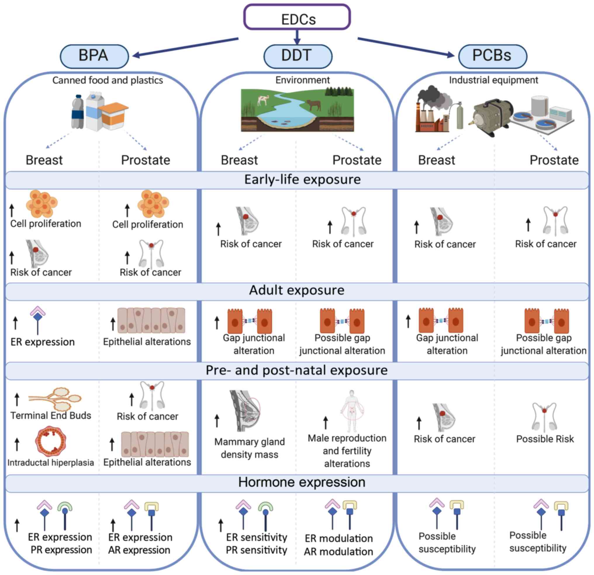
It can be concluded that contaminants classified as
EDCs are still circulating in water, sediments and soil affecting
the human mammary and prostate glands (Fig. 3). BPA exposure can induce cell
proliferation, morphological alterations and alter carcinogenesis
in both glands. Furthermore, BPA, DDT and PCBs contribute to the
risk of cancer particularly in mammary and prostate glands, in
which postnatal exposure to DDT induces male infertility.
Since hormone-associated cancers are presented, it
is imperative to mention that both BPA and DDT affect the
expression and sensitivity of the ER and prolactin receptor in the
mammary gland, and of AR in the prostate gland.
This phenomenon is caused by the indiscriminate use
of these compounds and their lipophilic nature (21). Even though some manufacturers claim
their products to be BPA-free (119), BPA analogs are not part of the
regulatory frame. Consequently, the overuse of ‘healthy-safe
products’ has been overlooked and should be addressed by public
health agencies.
Considering that most of the referred compounds
modulate estrogenic activity, a possible substance may be proposed
to treat the effects of EDCs such as BPA, DDT and PCBs. Compounds
of the selective estrogen receptor modulators category, such as
tamoxifen, toremifene and clomifene, should be studied to assess
whether their implication in the estrogen signaling pathway is
affected by these EDCs. However, it must be considered that EDCs,
such as BPA and PCB congeners, can have estrogen and antiestrogen
effects depending on the tissue.
Finally, the use of pesticides can be approached
from a socio-economical perspective since some epidemiological
studies have demonstrated high concentration levels of PCPs in
developing countries with no or partial regulations, such as
countries in Africa, Asia and Latin America (20,173).
These studies suggest that regulation and control in the use of
these compounds may contribute to decreasing either the exposure or
the development of cancer or of other associated diseases in
humans.
Acknowledgements
The authors would like to thank Mr. Leodán A.
Crispin (Instituto de Alta Investigación, Universidad de Tarapacá,
Arica, Chile) for providing technical support.
Funding
The current study was supported by grants from
Convenio de Desempeño (grant no. UTA1117) and from Universidad de
Tarapacá and Fondo Nacional de Ciencias (grant no. 1200656).
Availability of data and materials
Not applicable.
Authors' contributions
TCB and GMC assessed the authenticity of all the
raw data, wrote, edited and reviewed the manuscript and agreed to
be accountable for all aspects of the revision in ensuring that the
accuracy or integrity of any part of the work was appropriately
conducted. TCB and GMC read and approved the final manuscript.
Ethics approval and consent to
participate
Not applicable.
Patient consent for publication
Not applicable.
Competing interests
The authors declare that they have no competing
interests.
References
|
1
|
Rattan S, Zhou C, Chiang C, Mahalingam S,
Brehm E and Flaws JA: Exposure to endocrine disruptors during
adulthood: Consequences for female fertility. J Endocrinol.
233:R109–R129. 2017. View Article : Google Scholar : PubMed/NCBI
|
|
2
|
Sifakis S, Androutsopoulos VP, Tsatsakis
AM and Spandidos DA: Human exposure to endocrine disrupting
chemicals: Effects on the male and female reproductive systems.
Environ Toxicol Pharmacol. 51:56–70. 2017. View Article : Google Scholar : PubMed/NCBI
|
|
3
|
Diamanti-Kandarakis E, Bourguignon JP,
Giudice LC, Hauser R, Prins GS, Soto AM, Zoeller RT and Gore AC:
Endocrine-disrupting chemicals: An Endocrine Society scientific
statement. Endocr Rev. 30:293–342. 2009. View Article : Google Scholar : PubMed/NCBI
|
|
4
|
Cheong A, Zhang X, Cheung YY, Tang WY,
Chen J, Ye SH, Medvedovic M, Leung YK, Prins GS and Ho SM: DNA
methylome changes by estradiol benzoate and bisphenol A links
early-life environmental exposures to prostate cancer risk.
Epigenetics. 11:674–689. 2016. View Article : Google Scholar : PubMed/NCBI
|
|
5
|
Di Donato M, Cernera G, Giovannelli P,
Galasso G, Bilancio A, Migliaccio A and Castoria G: Recent advances
on bisphenol-A and endocrine disruptor effects on human prostate
cancer. Mol Cell Endocrinol. 457:35–42. 2017. View Article : Google Scholar : PubMed/NCBI
|
|
6
|
WHO/UNEP: State of the science of
endocrine disrupting chemicals-2012: An assessment of the state of
the science of endocrine disruptors prepared by a group of experts
for the United Nations Environment Programme (UNEP) and WHO. 2012,
https://www.who.int/ceh/risks/cehemerging2/en/January.
2020
|
|
7
|
Patrick SM, Bornman MS, Joubert AM, Pitts
N, Naidoo V and de Jager C: Effects of environmental endocrine
disruptors, including insecticides used for malaria vector control
on reproductive parameters of male rats. Reprod Toxicol. 61:19–27.
2016. View Article : Google Scholar : PubMed/NCBI
|
|
8
|
Wong LI, Labrecque MP, Ibuki N, Cox ME,
Elliott JE and Beischlag TV: p,p′-Dichlorodiphenyltrichloroethane
(p,p′-DDT) and p,p′-dichlorodiphenyldichloroethylene (p,p′-DDE)
repress prostate specific antigen levels in human prostate cancer
cell lines. Chem Biol Interact. 230:40–49. 2015. View Article : Google Scholar : PubMed/NCBI
|
|
9
|
Ho SM and Tam NN: Organoid model shows
effect of BPA on prostate development. Nat Rev Urol. 12:658–659.
2015. View Article : Google Scholar : PubMed/NCBI
|
|
10
|
Cariati F, D'Uonno N, Borrillo F,
Iervolino S, Galdiero G and Tomaiuolo R: ‘Bisphenol a: An emerging
threat to male fertility’. Reprod Biol Endocrinol. 17:62019.
View Article : Google Scholar : PubMed/NCBI
|
|
11
|
EPRS: Endocrine disruptors: An overview of
the latest developments at the European level in the context of
plant protection products. European Parliamentary Research Service:
Brussels, European Union. 2019, https://www.europarl.europa.eu/thinktank/en/document.html?reference=EPRS_STU(2019)631743February.
2020
|
|
12
|
Band PR, Abanto Z, Bert J, Lang B, Fang R,
Gallagher RP and Le ND: Prostate cancer risk and exposure to
pesticides in British Columbia farmers. Prostate. 71:168–183. 2011.
View Article : Google Scholar : PubMed/NCBI
|
|
13
|
Konieczna A, Rutkowska A and Rachon D:
Health risk of exposure to Bisphenol A (BPA). Rocz Panstw Zakl Hig.
66:5–11. 2015.PubMed/NCBI
|
|
14
|
Gregoraszczuk EL, Rak A, Ludewig G and
Gasinska A: Effects of estradiol, PCB3, and their hydroxylated
metabolites on proliferation, cell cycle, and apoptosis of human
breast cancer cells. Environ Toxicol Pharmacol. 25:227–233. 2008.
View Article : Google Scholar : PubMed/NCBI
|
|
15
|
McGovern V: PCBs are endocrine disruptors:
Mixture affects reproductive development in female mice. Environ
Health Perspect. 114:A368–A369. 2006. View Article : Google Scholar
|
|
16
|
Prins GS, Hu WY, Xie L, Shi GB, Hu DP,
Birch L and Bosland MC: Evaluation of Bisphenol A (BPA) exposures
on prostate stem cell homeostasis and prostate cancer risk in the
NCTR-sprague-dawley rat: An NIEHS/FDA CLARITY-BPA consortium study.
Environ Health Perspect. 126:1170012018. View Article : Google Scholar : PubMed/NCBI
|
|
17
|
Huang DY, Zheng CC, Pan Q, Wu SS, Su X, Li
L, Wu JH and Sun ZY: Oral exposure of low-dose bisphenol A promotes
proliferation of dorsolateral prostate and induces
epithelial-mesenchymal transition in aged rats. Sci Rep. 8:4902018.
View Article : Google Scholar : PubMed/NCBI
|
|
18
|
Takayama S, Sieber SM, Dalgard DW,
Thorgeirsson UP and Adamson RH: Effects of long-term oral
administration of DDT on nonhuman primates. J Cancer Res Clin
Oncol. 125:219–225. 1999. View Article : Google Scholar : PubMed/NCBI
|
|
19
|
Di Lorenzo D, Villa R, Biasiotto G,
Belloli S, Ruggeri G, Albertini A, Apostoli P, Raviscioni M, Ciana
P and Maggi A: Isomer-specific activity of
dichlorodyphenyltrichloroethane with estrogen receptor in adult and
suckling estrogen reporter mice. Endocrinology. 143:4544–4551.
2002. View Article : Google Scholar : PubMed/NCBI
|
|
20
|
Lewis-Mikhael AM, Olmedo-Requena R,
Martinez-Ruiz V, Bueno-Cavanillas A and Jimenez-Moleon JJ:
Organochlorine pesticides and prostate cancer, is there an
association? A meta-analysis of epidemiological evidence. Cancer
Causes Control. 26:1375–1392. 2015. View Article : Google Scholar : PubMed/NCBI
|
|
21
|
Pi N, Chia SE, Ong CN and Kelly BC:
Associations of serum organohalogen levels and prostate cancer
risk: Results from a case-control study in Singapore. Chemosphere.
144:1505–1512. 2016. View Article : Google Scholar : PubMed/NCBI
|
|
22
|
Safe S: Hydroxylated polychlorinated
Biphenyls (PCBs) and Organochlorine Pesticides as Potential
Endocrine Disruptors, in The Handbook of Environmental Chemistry:
Endocrine Disruptors-Part I. Vol. 3 Series: Anthropogenic
Compounds, M. Metzler. Springer; Heidelberg: pp. 155–167. 2001
|
|
23
|
Pěnčíková K, Svržková L, Strapáčová S,
Neča J, Bartoňková I, Dvořák Z, Hýžďalová M, Pivnička J, Pálková L,
Lehmler HJ, et al: In vitro profiling of toxic effects of prominent
environmental lower-chlorinated PCB congeners linked with endocrine
disruption and tumor promotion. Environ Pollut. 237:473–486. 2018.
View Article : Google Scholar : PubMed/NCBI
|
|
24
|
Ellsworth RE, Mamula KA, Costantino NS,
Deyarmin B, Kostyniak PJ, Chi LH, Shriver CD and Ellsworth DL:
Abundance and distribution of polychlorinated biphenyls (PCBs) in
breast tissue. Environ Res. 138:291–297. 2015. View Article : Google Scholar : PubMed/NCBI
|
|
25
|
Brody JG and Rudel RA: Environmental
pollutants and breast cancer. Environ Health Perspect.
111:1007–1019. 2003. View Article : Google Scholar : PubMed/NCBI
|
|
26
|
Soto AM, Brisken C, Schaeberle C and
Sonnenschein C: Does cancer start in the womb? altered mammary
gland development and predisposition to breast cancer due to in
utero exposure to endocrine disruptors. J Mammary Gland Biol
Neoplasia. 18:199–208. 2013. View Article : Google Scholar : PubMed/NCBI
|
|
27
|
Cohn BA, La Merrill M, Krigbaum NY, Yeh G,
Park JS, Zimmermann L and Cirillo PM: DDT exposure in utero and
breast cancer. J Clin Endocrinol Metab. 100:2865–2872. 2015.
View Article : Google Scholar : PubMed/NCBI
|
|
28
|
Wang Z, Liu H and Liu S: Low-dose
bisphenol a exposure: A seemingly instigating carcinogenic effect
on breast cancer. Adv Sci (Weinh). 4:16002482016. View Article : Google Scholar : PubMed/NCBI
|
|
29
|
Marchese S and Silva E: Disruption of 3D
MCF-12A breast cell cultures by estrogens-an in vitro model for
ER-mediated changes indicative of hormonal carcinogenesis. PLoS
One. 7:e457672012. View Article : Google Scholar : PubMed/NCBI
|
|
30
|
Verner MA, Chevrier J, Ngueta G, Rauch S,
Bornman R and Eskenazi B: Early-life exposure to p,p′-DDT and
p,p′-DDE in South African children participating in the VHEMBE
study: An assessment using repeated serum measurements and
pharmacokinetic modeling. Environ Int. 119:478–484. 2018.
View Article : Google Scholar : PubMed/NCBI
|
|
31
|
Cohn BA: Developmental and environmental
origins of breast cancer: DDT as a case study. Reprod Toxicol.
31:302–311. 2011. View Article : Google Scholar : PubMed/NCBI
|
|
32
|
Verner MA, Bachelet D, McDougall R,
Charbonneau M, Guenel P and Haddad S: A case study addressing the
reliability of polychlorinated biphenyl levels measured at the time
of breast cancer diagnosis in representing early-life exposure.
Cancer Epidemiol Biomarkers Prev. 20:281–286. 2011. View Article : Google Scholar : PubMed/NCBI
|
|
33
|
Paulose T, Speroni L, Sonnenschein C and
Soto AM: Estrogens in the wrong place at the wrong time: Fetal BPA
exposure and mammary cancer. Reprod Toxicol. 54:58–65. 2015.
View Article : Google Scholar : PubMed/NCBI
|
|
34
|
Soto AM and Sonnenschein C: Endocrine
disruptors: DDT, endocrine disruption and breast cancer. Nat Rev
Endocrinol. 11:507–508. 2015. View Article : Google Scholar : PubMed/NCBI
|
|
35
|
Soto AM and Sonnenschein C: The tissue
organization field theory of cancer: A testable replacement for the
somatic mutation theory. Bioessays. 33:332–340. 2011. View Article : Google Scholar : PubMed/NCBI
|
|
36
|
Ayyanan A, Laribi O, Schuepbach-Mallepell
S, Schrick C, Gutierrez M, Tanos T, Lefebvre G, Rougemont J,
Yalcin-Ozuysal O and Brisken C: Perinatal exposure to bisphenol a
increases adult mammary gland progesterone response and cell
number. Mol Endocrinol. 25:1915–1923. 2011. View Article : Google Scholar : PubMed/NCBI
|
|
37
|
Seachrist DD, Bonk KW, Ho SM, Prins GS,
Soto AM and Keri RA: A review of the carcinogenic potential of
bisphenol A. Reprod Toxicol. 59:167–182. 2016. View Article : Google Scholar : PubMed/NCBI
|
|
38
|
Wadia PR, Cabaton NJ, Borrero MD, Rubin
BS, Sonnenschein C, Shioda T and Soto AM: Low-dose BPA exposure
alters the mesenchymal and epithelial transcriptomes of the mouse
fetal mammary gland. PLoS One. 8:e639022013. View Article : Google Scholar : PubMed/NCBI
|
|
39
|
Harada T, Takeda M, Kojima S and Tomiyama
N: Toxicity and carcinogenicity of dichlorodiphenyltrichloroethane
(DDT). Toxicol Res. 32:21–33. 2016. View Article : Google Scholar : PubMed/NCBI
|
|
40
|
Kang KS, Wilson MR, Hayashi T, Chang CC
and Trosko JE: Inhibition of gap junctional intercellular
communication in normal human breast epithelial cells after
treatment with pesticides, PCBs, and PBBs, alone or in mixtures.
Environ Health Perspect. 104:192–200. 1996. View Article : Google Scholar : PubMed/NCBI
|
|
41
|
Machala M, Blaha L, Vondracek J, Trosko
JE, Scott J and Upham BL: Inhibition of gap junctional
intercellular communication by noncoplanar polychlorinated
biphenyls: Inhibitory potencies and screening for potential mode(s)
of action. Toxicol Sci. 76:102–111. 2003. View Article : Google Scholar : PubMed/NCBI
|
|
42
|
Roehrborn CG: Benign prostatic
hyperplasia: An overview. Rev Urol. 7 (Suppl 9):S3–S14.
2005.PubMed/NCBI
|
|
43
|
Durando M, Kass L, Piva J, Sonnenschein C,
Soto AM, Luque EH and Munoz-de-Toro M: Prenatal bisphenol A
exposure induces preneoplastic lesions in the mammary gland in
Wistar rats. Environ Health Perspect. 115:80–86. 2007. View Article : Google Scholar : PubMed/NCBI
|
|
44
|
Vandenberg LN, Maffini MV, Wadia PR,
Sonnenschein C, Rubin BS and Soto AM: Exposure to environmentally
relevant doses of the xenoestrogen bisphenol-A alters development
of the fetal mouse mammary gland. Endocrinology. 148:116–127. 2007.
View Article : Google Scholar : PubMed/NCBI
|
|
45
|
Moral R, Wang R, Russo IH, Lamartiniere
CA, Pereira J and Russo J: Effect of prenatal exposure to the
endocrine disruptor bisphenol A on mammary gland morphology and
gene expression signature. J Endocrinol. 196:101–112. 2008.
View Article : Google Scholar : PubMed/NCBI
|
|
46
|
Vandenberg LN, Maffini MV, Schaeberle CM,
Ucci AA, Sonnenschein C, Rubin BS and Soto AM: Perinatal exposure
to the xenoestrogen bisphenol-A induces mammary intraductal
hyperplasias in adult CD-1 mice. Reprod Toxicol. 26:210–219. 2008.
View Article : Google Scholar : PubMed/NCBI
|
|
47
|
Munoz-de-Toro M, Markey CM, Wadia PR,
Luque EH, Rubin BS, Sonnenschein C and Soto AM: Perinatal exposure
to bisphenol-A alters peripubertal mammary gland development in
mice. Endocrinology. 146:4138–4147. 2005. View Article : Google Scholar : PubMed/NCBI
|
|
48
|
Murray TJ, Maffini MV, Ucci AA,
Sonnenschein C and Soto AM: Induction of mammary gland ductal
hyperplasias and carcinoma in situ following fetal bisphenol A
exposure. Reprod Toxicol. 23:383–390. 2007. View Article : Google Scholar : PubMed/NCBI
|
|
49
|
Acevedo N, Davis B, Schaeberle CM,
Sonnenschein C and Soto AM: Perinatally administered bisphenol a as
a potential mammary gland carcinogen in rats. Environ Health
Perspect. 121:1040–1046. 2013. View Article : Google Scholar : PubMed/NCBI
|
|
50
|
Tharp AP, Maffini MV, Hunt PA, VandeVoort
CA, Sonnenschein C and Soto AM: Bisphenol A alters the development
of the rhesus monkey mammary gland. Proc Natl Acad Sci USA.
109:8190–8195. 2012. View Article : Google Scholar : PubMed/NCBI
|
|
51
|
Zheng T, Holford TR, Mayne ST, Ward B,
Carter D, Owens PH, Dubrow R, Zahm SH, Boyle P, Archibeque S and
Tessari J: DDE and DDT in breast adipose tissue and risk of female
breast cancer. Am J Epidemiol. 150:453–458. 1999. View Article : Google Scholar : PubMed/NCBI
|
|
52
|
Kaur N, Swain SK, Banerjee BD, Sharma T
and Krishnalata T: Organochlorine pesticide exposure as a risk
factor for breast cancer in young Indian women: A case-control
study. South Asian J Cancer. 8:212–214. 2019. View Article : Google Scholar : PubMed/NCBI
|
|
53
|
Krigbaum NY, Cirillo PM, Flom JD, McDonald
JA, Terry MB and Cohn BA: In utero DDT exposure and breast density
before age 50. Reprod Toxicol. 92:85–90. 2020. View Article : Google Scholar : PubMed/NCBI
|
|
54
|
Boyd NF, Jensen HM, Cooke G and Han HL:
Relationship between mammographic and histological risk factors for
breast cancer. J Natl Cancer Inst. 84:1170–1179. 1992. View Article : Google Scholar : PubMed/NCBI
|
|
55
|
McDonald JA, Cirillo PM, Tehranifar P,
Krigbaum NY, Engmann NJ, Cohn BA and Terry MB: In utero DDT
exposure and breast density in early menopause by maternal history
of breast cancer. Reprod Toxicol. 92:78–84, 20120. View Article : Google Scholar : PubMed/NCBI
|
|
56
|
He Y, Peng L, Huang Y, Peng X, Zheng S,
Liu C and Wu K: Association of breast adipose tissue levels of
polychlorinated biphenyls and breast cancer development in women
from Chaoshan, China. Environ Sci Pollut Res Int. 24:4778–4790.
2017. View Article : Google Scholar : PubMed/NCBI
|
|
57
|
Aronson KJ, Miller AB, Woolcott CG, Sterns
EE, McCready DR, Lickley LA, Fish EB, Hiraki GY, Holloway C, Ross
T, et al: Breast adipose tissue concentrations of polychlorinated
biphenyls and other organochlorines and breast cancer risk. Cancer
Epidemiol Biomarkers Prev. 9:55–63. 2000.PubMed/NCBI
|
|
58
|
Holford TR, Zheng T, Mayne ST, Zahm SH,
Tessari JD and Boyle P: Joint effects of nine polychlorinated
biphenyl (PCB) congeners on breast cancer risk. Int J Epidemiol.
29:975–982. 2000. View Article : Google Scholar : PubMed/NCBI
|
|
59
|
Wolff MS, Camann D, Gammon M and Stellman
SD: Proposed PCB congener groupings for epidemiological studies.
Environ Health Perspect. 105:13–14. 1997. View Article : Google Scholar : PubMed/NCBI
|
|
60
|
Pavuk M, Cerhan JR, Lynch CF, Kocan A,
Petrik J and Chovancova J: Case-control study of PCBs, other
organochlorines and breast cancer in Eastern Slovakia. J Expo Anal
Environ Epidemiol. 13:267–275. 2003. View Article : Google Scholar : PubMed/NCBI
|
|
61
|
Zheng T, Holford TR, Tessari J, Mayne ST,
Owens PH, Ward B, Carter D, Boyle P, Dubrow R, Archibeque-Engle S
and Zahm SH: Breast cancer risk associated with congeners of
polychlorinated biphenyls. Am J Epidemiol. 152:50–58. 2000.
View Article : Google Scholar : PubMed/NCBI
|
|
62
|
Guttes S, Failing K, Neumann K, Kleinstein
J, Georgii S and Brunn H: Chlororganic pesticides and
polychlorinated biphenyls in breast tissue of women with benign and
malignant breast disease. Arch Environ Contam Toxicol. 35:140–147.
1998. View Article : Google Scholar : PubMed/NCBI
|
|
63
|
IARC: Some inorganic substances,
chlorinated hydrocarbons, aromatic amines, N-nitroso compounds, and
natural products. IARC Working Group on the Evaluation of
Carcinogenic Risks to Humans. Monograph 1. International Agency for
Research on Cancer; Lyon: 1972, https://publications.iarc.fr/19January. 2020
|
|
64
|
Jenkins S, Raghuraman N, Eltoum I,
Carpenter M, Russo J and Lamartiniere CA: Oral exposure to
bisphenol a increases dimethylbenzanthracene-induced mammary cancer
in rats. Environ Health Perspect. 117:910–915. 2009. View Article : Google Scholar : PubMed/NCBI
|
|
65
|
Jenkins S, Betancourt AM, Wang J and
Lamartiniere CA: Endocrine-active chemicals in mammary cancer
causation and prevention. J Steroid Biochem Mol Biol. 129:191–200.
2012. View Article : Google Scholar : PubMed/NCBI
|
|
66
|
Foster WG, Younglai EV, Boutross-Tadross
O, Hughes CL and Wade MG: Mammary gland morphology in
Sprague-dawley rats following treatment with an organochlorine
mixture in utero and neonatal genistein. Toxicol Sci. 77:91–100.
2004. View Article : Google Scholar : PubMed/NCBI
|
|
67
|
American Academy of Pediatrics. Committee
on Nutrition, . American academy of pediatrics. Committee on
nutrition. Soy protein-based formulas: Recommendations for use in
infant feeding. Pediatrics. 101:148–153. 1998. View Article : Google Scholar : PubMed/NCBI
|
|
68
|
Badger TM, Gilchrist JM, Pivik RT, Andres
A, Shankar K, Chen JR and Ronis MJ: The health implications of soy
infant formula. Am J Clin Nutr. 89 (Suppl 1):1668S–1672S. 2009.
View Article : Google Scholar : PubMed/NCBI
|
|
69
|
Brown NM and Lamartiniere CA:
Xenoestrogens alter mammary gland differentiation and cell
proliferation in the rat. Environ Health Perspect. 103:708–713.
1995. View Article : Google Scholar : PubMed/NCBI
|
|
70
|
Desaulniers D, Leingartner K, Russo J,
Perkins G, Chittim BG, Archer MC, Wade M and Yang J: Modulatory
effects of neonatal exposure to TCDD, or a mixture of PCBs,
p,p′-DDT, and p-p′-DDE, on methylnitrosourea-induced mammary tumor
development in the rat. Environ Health Perspect. 109:739–747. 2001.
View Article : Google Scholar : PubMed/NCBI
|
|
71
|
Welshons WV, Thayer KA, Judy BM, Taylor
JA, Curran EM and vom Saal FS: Large effects from small exposures.
I. Mechanisms for endocrine-disrupting chemicals with estrogenic
activity. Environ Health Perspect. 111:994–1006. 2003. View Article : Google Scholar : PubMed/NCBI
|
|
72
|
Petreas M, Nelson D, Brown FR, Goldberg D,
Hurley S and Reynolds P: High concentrations of polybrominated
diphenylethers (PBDEs) in breast adipose tissue of California
women. Environ Int. 37:190–197. 2011. View Article : Google Scholar : PubMed/NCBI
|
|
73
|
Petreas M, Smith D, Hurley S, Jeffrey SS,
Gilliss D and Reynolds P: Distribution of persistent, lipid-soluble
chemicals in breast and abdominal adipose tissues: Lessons learned
from a breast cancer study. Cancer Epidemiol Biomarkers Prev.
13:416–424. 2004.PubMed/NCBI
|
|
74
|
Li AJ, Feldman SM, McNally RK and Kannan
K: Distribution of organohalogen and synthetic musk compounds in
breast adipose tissue of breast cancer patients in ulster County,
New York, USA. Arch Environ Contam Toxicol. 77:68–78. 2019.
View Article : Google Scholar : PubMed/NCBI
|
|
75
|
Kunisue T, Takayanagi N, Isobe T,
Takahashi S, Nose M, Yamada T, Komori H, Arita N, Ueda N and Tanabe
S: Polybrominated diphenyl ethers and persistent organochlorines in
Japanese human adipose tissues. Environ Int. 33:1048–1056. 2007.
View Article : Google Scholar : PubMed/NCBI
|
|
76
|
Nakata H, Kawazoe M, Arizono K, Abe S,
Kitano T, Shimada H, Li W and Ding X: Organochlorine pesticides and
polychlorinated biphenyl residues in foodstuffs and human tissues
from china: Status of contamination, historical trend, and human
dietary exposure. Arch Environ Contam Toxicol. 43:473–480. 2002.
View Article : Google Scholar : PubMed/NCBI
|
|
77
|
Tan J, Li QQ, Loganath A, Chong YS, Xiao M
and Obbard JP: Multivariate data analyses of persistent organic
pollutants in maternal adipose tissue in Singapore. Environ Sci
Technol. 42:2681–2687. 2008. View Article : Google Scholar : PubMed/NCBI
|
|
78
|
Rusiecki JA, Holford TR, Zahm SH and Zheng
T: Polychlorinated biphenyls and breast cancer risk by combined
estrogen and progesterone receptor status. Eur J Epidemiol.
19:793–801. 2004. View Article : Google Scholar : PubMed/NCBI
|
|
79
|
Jusko TA, Koepsell TD, Baker RJ,
Greenfield TA, Willman EJ, Charles MJ, Teplin SW, Checkoway H and
Hertz-Picciotto I: Maternal DDT exposures in relation to fetal and
5-year growth. Epidemiology. 17:692–700. 2006. View Article : Google Scholar : PubMed/NCBI
|
|
80
|
Cohn BA, Wolff MS, Cirillo PM and Sholtz
RI: DDT and breast cancer in young women: New data on the
significance of age at exposure. Environ Health Perspect.
115:1406–1414. 2007. View Article : Google Scholar : PubMed/NCBI
|
|
81
|
IBCERCC: Breast Cancer and the
Environment: Prioritizing Prevention. National Institute of
Environmental Health Sciences & Interagency Breast Cancer &
Environmental Research Coordinating Committee, USA. 2013,
https://www.niehs.nih.gov/about/boards/ibcercc/index.cfmJanuary.
2020
|
|
82
|
Enan E and Matsumura F: Activation of
c-Neu tyrosine kinase by o,p′-DDT and beta-HCH in cell-free and
intact cell preparations from MCF-7 human breast cancer cells. J
Biochem Mol Toxicol. 12:83–92. 1998. View Article : Google Scholar : PubMed/NCBI
|
|
83
|
Hatakeyama M and Matsumura F: Correlation
between the activation of Neu tyrosine kinase and promotion of foci
formation induced by selected organochlorine compounds in the MCF-7
model system. J Biochem Mol Toxicol. 13:296–302. 1999. View Article : Google Scholar : PubMed/NCBI
|
|
84
|
IARC: Polychlorinated Biphenyls and
Polybrominated Biphenyls. Working Group on the Evaluation of
Carcinogenic Risk to Humans. Monograph 107. International Agency
for Research on Cancer; Lyon: 2016, https://www.ncbi.nlm.nih.gov/books/NBK361696/November.
2018
|
|
85
|
Plísková M, Vondrácek J, Canton RF, Nera
J, Kocan A, Petrik J, Trnovec T, Sanderson T, van den Berg M and
Machala M: Impact of polychlorinated biphenyls contamination on
estrogenic activity in human male serum. Environ Health Perspect.
113:1277–1284. 2005. View Article : Google Scholar : PubMed/NCBI
|
|
86
|
Ranjit N, Siefert K and Padmanabhan V:
Bisphenol-A and disparities in birth outcomes: A review and
directions for future research. J Perinatol. 30:2–9. 2010.
View Article : Google Scholar : PubMed/NCBI
|
|
87
|
IARC: DDT, Lindane, and 2,4-D. Working
Group on the Evaluation of Carcinogenic Risk to Humans. Monograph.
113. International Agency for Research on Cancer; Lyon: 2018,
https://www.ncbi.nlm.nih.gov/books/NBK507424/November.
2018
|
|
88
|
Parada H Jr, Wolff MS, Engel LS, Eng SM,
Khankari NK, Neugut AI, Teitelbaum SL and Gammon MD:
Polychlorinated biphenyls and their association with survival
following breast cancer. Eur J Cancer. 56:21–30. 2016. View Article : Google Scholar : PubMed/NCBI
|
|
89
|
Roswall N, Sorensen M, Tjonneland A and
Raaschou-Nielsen O: Organochlorine concentrations in adipose tissue
and survival in postmenopausal, Danish breast cancer patients.
Environ Res. 163:237–248. 2018. View Article : Google Scholar : PubMed/NCBI
|
|
90
|
Woolcott CG, Aronson KJ, Hanna WM,
SenGupta SK, McCready DR, Sterns EE and Miller AB: Organochlorines
and breast cancer risk by receptor status, tumor size, and grade
(Canada). Cancer Causes Control. 12:395–404. 2001. View Article : Google Scholar : PubMed/NCBI
|
|
91
|
Gray JM, Rasanayagam S, Engel C and Rizzo
J: State of the evidence 2017: An update on the connection between
breast cancer and the environment. Environ Health. 16:942017.
View Article : Google Scholar : PubMed/NCBI
|
|
92
|
Wu S, Huang D, Su X, Yan H, Wu J and Sun
Z: Oral exposure to low-dose bisphenol A induces hyperplasia of
dorsolateral prostate and upregulates EGFR expression in adult
Sprague-Dawley rats. Toxicol Ind Health. 35:647–659. 2019.
View Article : Google Scholar : PubMed/NCBI
|
|
93
|
Liao LM and Schaefer W: Cross-sectional
and longitudinal studies on interaction between bladder compliance
and outflow obstruction in men with benign prostatic hyperplasia.
Asian J Androl. 9:51–56. 2007. View Article : Google Scholar : PubMed/NCBI
|
|
94
|
Xia SJ, Xu XX, Teng JB, Xu CX and Tang XD:
Characteristic pattern of human prostatic growth with age. Asian J
Androl. 4:269–271. 2002.PubMed/NCBI
|
|
95
|
Prins GS: Endocrine disruptors and
prostate cancer risk. Endocr Relat Cancer. 15:649–656. 2008.
View Article : Google Scholar : PubMed/NCBI
|
|
96
|
Vom Saal FS: Triennial Reproduction
Symposium: Environmental programming of reproduction during fetal
life: Effects of intrauterine position and the endocrine disrupting
chemical bisphenol A. J Anim Sci. 94:2722–2736. 2016. View Article : Google Scholar : PubMed/NCBI
|
|
97
|
Herath CB, Jin W, Watanabe G, Arai K,
Suzuki AK and Taya K: Adverse effects of environmental toxicants,
octylphenol and bisphenol A, on male reproductive functions in
pubertal rats. Endocrine. 25:163–1672. 2004. View Article : Google Scholar : PubMed/NCBI
|
|
98
|
Wu J, Huang D, Su X, Yan H and Sun Z: Oral
administration of low-dose bisphenol A promotes proliferation of
ventral prostate and upregulates prostaglandin D2 synthase
expression in adult rats. Toxicol Ind Health. 32:1848–1858. 2016.
View Article : Google Scholar : PubMed/NCBI
|
|
99
|
Calderon-Gierszal EL and Prins GS:
Directed differentiation of human embryonic stem cells into
prostate organoids in vitro and its perturbation by low-dose
bisphenol A exposure. PLoS One. 10:e01332382015. View Article : Google Scholar : PubMed/NCBI
|
|
100
|
Cantone I and Fisher AG: Epigenetic
programming and reprogramming during development. Nat Struct Mol
Biol. 20:282–289. 2013. View Article : Google Scholar : PubMed/NCBI
|
|
101
|
Prusinski L, Al-Hendy A and Yang Q:
Developmental exposure to endocrine disrupting chemicals alters the
epigenome: Identification of reprogrammed targets. Gynecol Obstet
Res. 3:1–6. 2016.PubMed/NCBI
|
|
102
|
Tyl RW, Myers CB, Marr MC, Sloan CS,
Castillo NP, Veselica MM, Seely JC, Dimond SS, Van Miller JP,
Shiotsuka RN, et al: Two-generation reproductive toxicity study of
dietary bisphenol A in CD-1 (Swiss) mice. Toxicol Sci. 104:362–84.
2008. View Article : Google Scholar : PubMed/NCBI
|
|
103
|
Kilian E, Delport R, Bornman MS and de
Jager C: Simultaneous exposure to low concentrations of
dichlorodiphenyltrichloroethane, deltamethrin, nonylphenol and
phytoestrogens has negative effects on the reproductive parameters
in male Spraque-Dawley rats. Andrologia. 39:128–135. 2007.
View Article : Google Scholar : PubMed/NCBI
|
|
104
|
Skakkebaek NE: Endocrine disrupters and
testicular dysgenesis syndrome. Horm Res. 57 (Suppl 2):S432002.
|
|
105
|
USFDA: Bisphenol A (BPA). U.S. Food &
Drug Administration, USA, . 2018, https://www.fda.gov/food/food-additives-petitions/bisphenol-bpaJanuary.
2020
|
|
106
|
Porreca I, Ulloa-Severino L, Almeida P,
Cuomo D, Nardone A, Falco G, Mallardo M and Ambrosino C: Molecular
targets of developmental exposure to bisphenol A in diabesity: A
focus on endoderm-derived organs. Obes Rev. 18:99–108. 2017.
View Article : Google Scholar : PubMed/NCBI
|
|
107
|
Facina CH, Campos SG, Goncalves BF, Goes
RM, Vilamaior PS and Taboga SR: Long-term oral exposure to safe
dose of bisphenol A in association with high-fat diet stimulate the
prostatic lesions in a rodent model for prostate cancer. Prostate.
78:152–163. 2018. View Article : Google Scholar : PubMed/NCBI
|
|
108
|
Amaral FG, Turati AO, Barone M, Scialfa
JH, do Carmo Buonfiglio D, Peres R, Peliciari-Garcia RA, Afeche SC,
Lima L, Scavone C, et al: Melatonin synthesis impairment as a new
deleterious outcome of diabetes-derived hyperglycemia. J Pineal
Res. 57:67–79. 2014. View Article : Google Scholar : PubMed/NCBI
|
|
109
|
Andrabi SS, Parvez S and Tabassum H:
Melatonin and ischemic stroke: Mechanistic roles and action. Adv
Pharmacol Sci. 2015:3847502015.PubMed/NCBI
|
|
110
|
Olukole SG, Ajani SO, Ola-Davies EO,
Lanipekun DO, Aina OO, Oyeyemi MO and Oke BO: Melatonin ameliorates
bisphenol A-induced perturbations of the prostate gland of adult
Wistar rats. Biomed Pharmacother. 105:73–82. 2018. View Article : Google Scholar : PubMed/NCBI
|
|
111
|
Ben Maamar M, King SE, Nilsson E, Beck D
and Skinner MK: Epigenetic transgenerational inheritance of
parent-of-origin allelic transmission of outcross pathology and
sperm epimutations. Dev Biol. 458:106–119. 2020. View Article : Google Scholar : PubMed/NCBI
|
|
112
|
Cohn BA, Cirillo PM, Wolff MS, Schwingl
PJ, Cohen RD, Sholtz RI, Ferrara A, Christianson RE, van den Berg
BJ and Siiteri PK: DDT and DDE exposure in mothers and time to
pregnancy in daughters. Lancet. 361:2205–2206. 2003. View Article : Google Scholar : PubMed/NCBI
|
|
113
|
Hruska KS, Furth PA, Seifer DB, Sharara FI
and Flaws JA: Environmental factors in infertility. Clin Obstet
Gynecol. 43:821–829. 2000. View Article : Google Scholar : PubMed/NCBI
|
|
114
|
Korrick SA, Chen C, Damokosh AI, Ni J, Liu
X, Cho SI, Altshul L, Ryan L and Xu X: Association of DDT with
spontaneous abortion: A case-control study. Ann Epidemiol.
11:491–496. 2001. View Article : Google Scholar : PubMed/NCBI
|
|
115
|
Pant N, Mathur N, Banerjee AK, Srivastava
SP and Saxena DK: Correlation of chlorinated pesticides
concentration in semen with seminal vesicle and prostatic markers.
Reprod Toxicol. 19:209–214. 2004. View Article : Google Scholar : PubMed/NCBI
|
|
116
|
Mann T and Lutwak-Mann C: Passage of
chemicals into human and animal semen: Mechanisms and significance.
Crit Rev Toxicol. 11:1–14. 1982. View Article : Google Scholar : PubMed/NCBI
|
|
117
|
Brureau L, Emeville E, Helissey C, Thome
JP, Multigner L and Blanchet P: Endocrine disrupting-chemicals and
biochemical recurrence of prostate cancer after prostatectomy: A
cohort study in Guadeloupe (French West Indies). Int J Cancer.
146:657–663. 2020. View Article : Google Scholar : PubMed/NCBI
|
|
118
|
Rosenmai AK, Dybdahl M, Pedersen M, Alice
van Vugt-Lussenburg BM, Wedebye EB, Taxvig C and Vinggaard AM: Are
structural analogues to bisphenol a safe alternatives? Toxicol Sci.
139:35–47. 2014. View Article : Google Scholar : PubMed/NCBI
|
|
119
|
Silva JPA, Ramos JG, Campos MS, da Silva
Lima D, de Azevedo Brito PV, Mendes EP, Taboga SR, Biancardi MF,
Ghedini PC and Santos FCA: Bisphenol-S promotes
endocrine-disrupting effects similar to those promoted by
bisphenol-A in the prostate of adult gerbils. Reprod Toxicol.
85:83–92. 2019. View Article : Google Scholar : PubMed/NCBI
|
|
120
|
Zaviacic M and Ablin RJ: The female
prostate and prostate-specific antigen. Immunohistochemical
localization, implications of this prostate marker in women and
reasons for using the term ‘prostate’ in the human female. Histol
Histopathol. 15:131–142. 2000.PubMed/NCBI
|
|
121
|
Biancardi MF, Dos Santos FC, de Carvalho
HF, Sanches BD and Taboga SR: Female prostate: Historical,
developmental, and morphological perspectives. Cell Biol Int.
41:1174–1183. 2017. View Article : Google Scholar : PubMed/NCBI
|
|
122
|
Muto M, Inamura K, Ozawa N, Endo T, Masuda
H, Yonese J and Ishikawa Y: Skene's gland adenocarcinoma with
intestinal differentiation: A case report and literature review.
Pathol Int. 67:575–579. 2017. View Article : Google Scholar : PubMed/NCBI
|
|
123
|
Huang D, Wu J, Su X, Yan H and Sun Z:
Effects of low dose of bisphenol A on the proliferation and
mechanism of primary cultured prostate epithelial cells in rodents.
Oncol Lett. 14:2635–2642. 2017. View Article : Google Scholar : PubMed/NCBI
|
|
124
|
Prins GS, Ye SH, Birch L, Zhang X, Cheong
A, Lin H, Calderon-Gierszal E, Groen J, Hu WY, Ho SM and van
Breemen RB: Prostate cancer risk and DNA methylation signatures in
aging rats following developmental BPA exposure: A dose-response
analysis. Environ Health Perspect. 125:0770072017. View Article : Google Scholar : PubMed/NCBI
|
|
125
|
Lim JE, Nam C, Yang J, Rha KH, Lim KM and
Jee SH: Serum persistent organic pollutants (POPs) and prostate
cancer risk: A case-cohort study. Int J Hyg Environ Health.
220:849–856. 2017. View Article : Google Scholar : PubMed/NCBI
|
|
126
|
Ritchie JM, Vial SL, Fuortes LJ, Guo H,
Reedy VE and Smith EM: Organochlorines and risk of prostate cancer.
J Occup Environ Med. 45:692–702. 2003. View Article : Google Scholar : PubMed/NCBI
|
|
127
|
Wu JH, Jiang XR, Liu GM, Liu XY, He GL and
Sun ZY: Oral exposure to low-dose bisphenol A aggravates
testosterone-induced benign hyperplasia prostate in rats. Toxicol
Ind Health. 27:810–819. 2011. View Article : Google Scholar : PubMed/NCBI
|
|
128
|
Sharma S, Kelly TK and Jones PA:
Epigenetics in cancer. Carcinogenesis. 31:27–36. 2010. View Article : Google Scholar : PubMed/NCBI
|
|
129
|
Villa R, Bonetti E, Penza ML, Iacobello C,
Bugari G, Bailo M, Parolini O, Apostoli P, Caimi L, Ciana P, et al:
Target-specific action of organochlorine compounds in reproductive
and nonreproductive tissues of estrogen-reporter male mice. Toxicol
Appl Pharmacol. 201:137–148. 2004. View Article : Google Scholar : PubMed/NCBI
|
|
130
|
Carruba G: Estrogen and prostate cancer:
An eclipsed truth in an androgen-dominated scenario. J Cell
Biochem. 102:899–911. 2007. View Article : Google Scholar : PubMed/NCBI
|
|
131
|
Leavens TL, Sparrow BR and Devito MJ: Lack
of antiandrogenic effects in adult male rats following acute
exposure to 2,2-bis(4-chlorophenyl)-1,1-dichloroethylene
(p,p′-DDE). Toxicology. 174:69–78. 2002. View Article : Google Scholar : PubMed/NCBI
|
|
132
|
Belpomme D, Irigaray P, Ossondo M, Vacque
D and Martin M: Prostate cancer as an environmental disease: An
ecological study in the French Caribbean islands, Martinique and
Guadeloupe. Int J Oncol. 34:1037–1044. 2009. View Article : Google Scholar : PubMed/NCBI
|
|
133
|
Zeliadt SB, Ramsey SD, Penson DF, Hall IJ,
Ekwueme DU, Stroud L and Lee JW: Why do men choose one treatment
over another?: A review of patient decision making for localized
prostate cancer. Cancer. 106:1865–1874. 2006. View Article : Google Scholar : PubMed/NCBI
|
|
134
|
Kirkpatrick JP and Anscher MS:
Radiotherapy for locally recurrent prostate cancer. Clin Adv
Hematol Oncol. 3:933–942. 2005.PubMed/NCBI
|
|
135
|
Hess-Wilson JK: Bisphenol A may reduce the
efficacy of androgen deprivation therapy in prostate cancer. Cancer
Causes Control. 20:1029–1037. 2009. View Article : Google Scholar : PubMed/NCBI
|
|
136
|
Landau-Ossondo M, Rabia N, Jos-Pelage J,
Marquet LM, Isidore Y, Saint-Aimé C, Martin M, Irigaray P and
Belpomme D; ARTAC international research group on pesticides, : Why
pesticides could be a common cause of prostate and breast cancers
in the French Caribbean Island, Martinique. An overview on key
mechanisms of pesticide-induced cancer. Biomed Pharmacother.
63:383–395. 2009. View Article : Google Scholar : PubMed/NCBI
|
|
137
|
Sawada N, Iwasaki M, Inoue M, Itoh H,
Sasazuki S, Yamaji T, Shimazu T and Tsugane S; Japan Public Health
Center Based Prospective (JPHC) Study Group, : Plasma
organochlorines and subsequent risk of prostate cancer in Japanese
men: A nested case-control study. Environ Health Perspect.
118:659–665. 2010. View Article : Google Scholar : PubMed/NCBI
|
|
138
|
Aronson KJ, Wilson JW, Hamel M, Diarsvitri
W, Fan W, Woolcott C, Heaton JP, Nickel JC, Macneily A and Morales
A: Plasma organochlorine levels and prostate cancer risk. J Expo
Sci Environ Epidemiol. 20:434–445. 2010. View Article : Google Scholar : PubMed/NCBI
|
|
139
|
Wolff MS, Zeleniuch-Jacquotte A, Dubin N
and Toniolo P: Risk of breast cancer and organochlorine exposure.
Cancer Epidemiol Biomarkers Prev. 9:271–277. 2000.PubMed/NCBI
|
|
140
|
Egger G, Liang G, Aparicio A and Jones PA:
Epigenetics in human disease and prospects for epigenetic therapy.
Nature. 429:457–463. 2004. View Article : Google Scholar : PubMed/NCBI
|
|
141
|
Han M, Jia L, Lv W, Wang L and Cui W:
Epigenetic enzyme mutations: Role in tumorigenesis and molecular
inhibitors. Front Oncol. 9:1942019. View Article : Google Scholar : PubMed/NCBI
|
|
142
|
Zhang X and Ho SM: Epigenetics meets
endocrinology. J Mol Endocrinol. 46:R11–R32. 2011. View Article : Google Scholar : PubMed/NCBI
|
|
143
|
Fleisch AF, Wright RO and Baccarelli AA:
Environmental epigenetics: A role in endocrine disease? J Mol
Endocrinol. 49:R61–R67. 2012. View Article : Google Scholar : PubMed/NCBI
|
|
144
|
Tatton-Brown K, Seal S, Ruark E, Harmer J,
Ramsay E, Del Vecchio Duarte S, Zachariou A, Hanks S, O'Brien E,
Aksglaede L, et al: Mutations in the DNA methyltransferase gene
DNMT3A cause an overgrowth syndrome with intellectual disability.
Nat Genet. 46:385–388. 2014. View Article : Google Scholar : PubMed/NCBI
|
|
145
|
Cohen AL, Holmen SL and Colman H: IDH1 and
IDH2 mutations in gliomas. Curr Neurol Neurosci Rep. 13:3452013.
View Article : Google Scholar : PubMed/NCBI
|
|
146
|
Dolinoy DC, Huang D and Jirtle RL:
Maternal nutrient supplementation counteracts bisphenol A-induced
DNA hypomethylation in early development. Proc Natl Acad Sci USA.
104:13056–13061. 2007. View Article : Google Scholar : PubMed/NCBI
|
|
147
|
Leader JE, Wang C, Fu M and Pestell RG:
Epigenetic regulation of nuclear steroid receptors. Biochem
Pharmacol. 72:1589–1596. 2006. View Article : Google Scholar : PubMed/NCBI
|
|
148
|
Klukovich R, Nilsson E, Sadler-Riggleman
I, Beck D, Xie Y, Yan W and Skinner MK: Environmental toxicant
induced epigenetic transgenerational inheritance of prostate
pathology and stromal-epithelial cell epigenome and transcriptome
alterations: Ancestral origins of prostate disease. Sci Rep.
9:22092019. View Article : Google Scholar : PubMed/NCBI
|
|
149
|
Ho SM, Cheong A, Lam HM, Hu WY, Shi GB,
Zhu X, Chen J, Zhang X, Medvedovic M, Leung YK and Prins GS:
Exposure of human prostaspheres to bisphenol A epigenetically
regulates SNORD family noncoding RNAs via histone modification.
Endocrinology. 156:3984–3995. 2015. View Article : Google Scholar : PubMed/NCBI
|
|
150
|
Prins GS, Hu WY, Shi GB, Hu DP, Majumdar
S, Li G, Huang K, Nelles JL, Ho SM, Walker CL, et al: Bisphenol A
promotes human prostate stem-progenitor cell self-renewal and
increases in vivo carcinogenesis in human prostate epithelium.
Endocrinology. 155:805–817. 2014. View Article : Google Scholar : PubMed/NCBI
|
|
151
|
Prins GS, Calderon-Gierszal EL and Hu WY:
Stem cells as hormone targets that lead to increased cancer
susceptibility. Endocrinology. 156:3451–3457. 2015. View Article : Google Scholar : PubMed/NCBI
|
|
152
|
Ho SM, Tang WY, Belmonte de Frausto J and
Prins GS: Developmental exposure to estradiol and bisphenol A
increases susceptibility to prostate carcinogenesis and
epigenetically regulates phosphodiesterase type 4 variant 4. Cancer
Res. 66:5624–5632. 2006. View Article : Google Scholar : PubMed/NCBI
|
|
153
|
Tang WY, Morey LM, Cheung YY, Birch L,
Prins GS and Ho SM: Neonatal exposure to estradiol/bisphenol A
alters promoter methylation and expression of Nsbp1 and Hpcal1
genes and transcriptional programs of Dnmt3a/b and Mbd2/4 in the
rat prostate gland throughout life. Endocrinology. 153:42–55. 2012.
View Article : Google Scholar : PubMed/NCBI
|
|
154
|
Wang Q, Trevino LS, Wong RL, Medvedovic M,
Chen J, Ho SM, Shen J, Foulds CE, Coarfa C, O'Malley BW, et al:
Reprogramming of the epigenome by MLL1 links early-life
environmental exposures to prostate cancer risk. Mol Endocrinol.
30:856–871. 2016. View Article : Google Scholar : PubMed/NCBI
|
|
155
|
Burton K, Bajdas A, Shaw L and Morey LM:
The effect of the estrogenic compounds E2 and BPA on the expression
of histone modifying enzymes in two prostate cancer models. FASEB
J. 28:942–944. 2014.
|
|
156
|
Wong RL, Wang Q, Trevino LS, Bosland MC,
Chen J, Medvedovic M, Prins GS, Kannan K, Ho SM and Walker CL:
Identification of secretaglobin Scgb2a1 as a target for
developmental reprogramming by BPA in the rat prostate.
Epigenetics. 10:127–134. 2015. View Article : Google Scholar : PubMed/NCBI
|
|
157
|
Anway MD, Cupp AS, Uzumcu M and Skinner
MK: Epigenetic transgenerational actions of endocrine disruptors
and male fertility. Science. 308:1466–1469. 2005. View Article : Google Scholar : PubMed/NCBI
|
|
158
|
Skinner MK: Endocrine disruptor induction
of epigenetic transgenerational inheritance of disease. Mol Cell
Endocrinol. 398:4–12. 2014. View Article : Google Scholar : PubMed/NCBI
|
|
159
|
Skinner MK: Environmental epigenetic
transgenerational inheritance and somatic epigenetic mitotic
stability. Epigenetics. 6:838–842. 2011. View Article : Google Scholar : PubMed/NCBI
|
|
160
|
Skinner MK, Manikkam M, Tracey R,
Guerrero-Bosagna C, Haque M and Nilsson EE: Ancestral
dichlorodiphenyltrichloroethane (DDT) exposure promotes epigenetic
transgenerational inheritance of obesity. BMC Med. 11:2282013.
View Article : Google Scholar : PubMed/NCBI
|
|
161
|
Skinner MK, Ben Maamar M, Sadler-Riggleman
I, Beck D, Nilsson E, McBirney M, Klukovich R, Xie Y, Tang C and
Yan W: Alterations in sperm DNA methylation, non-coding RNA and
histone retention associate with DDT-induced epigenetic
transgenerational inheritance of disease. Epigenetics Chromatin.
11:82018. View Article : Google Scholar : PubMed/NCBI
|
|
162
|
Yan W: Potential roles of noncoding RNAs
in environmental epigenetic transgenerational inheritance. Mol Cell
Endocrinol. 398:24–30. 2014. View Article : Google Scholar : PubMed/NCBI
|
|
163
|
Casati L, Sendra R, Poletti A, Negri-Cesi
P and Celotti F: Androgen receptor activation by polychlorinated
biphenyls: Epigenetic effects mediated by the histone demethylase
Jarid1b. Epigenetics. 8:1061–1068. 2013. View Article : Google Scholar : PubMed/NCBI
|
|
164
|
Desaulniers D, Xiao GH, Lian H, Feng YL,
Zhu J, Nakai J and Bowers WJ: Effects of mixtures of
polychlorinated biphenyls, methylmercury, and organochlorine
pesticides on hepatic DNA methylation in prepubertal female
Sprague-Dawley rats. Int J Toxicol. 28:294–307. 2009. View Article : Google Scholar : PubMed/NCBI
|
|
165
|
Portigal CL, Cowell SP, Fedoruk MN, Butler
CM, Rennie PS and Nelson CC: Polychlorinated biphenyls interfere
with androgen-induced transcriptional activation and hormone
binding. Toxicol Appl Pharmacol. 179:185–194. 2002. View Article : Google Scholar : PubMed/NCBI
|
|
166
|
Xiang Y, Zhu Z, Han G, Ye X, Xu B, Peng Z,
Ma Y, Yu Y, Lin H, Chen AP and Chen CD: JARID1B is a histone H3
lysine 4 demethylase up-regulated in prostate cancer. Proc Natl
Acad Sci USA. 104:19226–19231. 2007. View Article : Google Scholar : PubMed/NCBI
|
|
167
|
Casati L, Sendra R, Sibilia V and Celotti
F: Endocrine disrupters: The new players able to affect the
epigenome. Front Cell Dev Biol. 3:372015. View Article : Google Scholar : PubMed/NCBI
|
|
168
|
Tse LA, Lee PMY, Ho WM, Lam AT, Lee MK, Ng
SSM, He Y, Leung KS, Hartle JC, Hu H, et al: Bisphenol A and other
environmental risk factors for prostate cancer in Hong Kong.
Environ Int. 107:1–7. 2017. View Article : Google Scholar : PubMed/NCBI
|
|
169
|
Maffini MV, Rubin BS, Sonnenschein C and
Soto AM: Endocrine disruptors and reproductive health: The case of
bisphenol-A. Mol Cell Endocrinol. 254-255:179–186. 2006. View Article : Google Scholar : PubMed/NCBI
|
|
170
|
Lopez-Cervantes M, Torres-Sanchez L,
Tobias A and Lopez-Carrillo L: Dichlorodiphenyldichloroethane
burden and breast cancer risk: A meta-analysis of the epidemiologic
evidence. Environ Health Perspect. 112:207–214. 2004. View Article : Google Scholar : PubMed/NCBI
|
|
171
|
Huang L, Pu Y, Alam S, Birch L and Prins
GS: Estrogenic regulation of signaling pathways and homeobox genes
during rat prostate development. J Androl. 25:330–337. 2004.
View Article : Google Scholar : PubMed/NCBI
|
|
172
|
Terry MB, Michels KB, Brody JG, Byrne C,
Chen S, Jerry DJ, Malecki KMC, Martin MB, Miller RL, Neuhausen SL,
et al: Environmental exposures during windows of susceptibility for
breast cancer: A framework for prevention research. Breast Cancer
Res. 21:962019. View Article : Google Scholar : PubMed/NCBI
|
|
173
|
Jaga K: What are the implications of the
interaction between DDT and estrogen receptors in the body? Med
Hypotheses. 54:18–25. 2000. View Article : Google Scholar : PubMed/NCBI
|