Introduction
Doxorubicin (DOX) remains one of the most widely
used anticancer drugs, due to its potent therapeutic effects on
various types of cancer (1).
However, the clinical use of this valuable anticancer drug is
limited by severe toxic side effects on the heart, which may result
in heart failure (2). Numerous
studies have implicated reactive oxygen species generation in the
cardiotoxicity of DOX, which ultimately results in cardiomyocyte
apoptosis (3,4). A number of pharmacological
interventions have been proposed as therapies to oppose DOX-induced
cardiotoxicity, including resveratrol (RES) and oleanolic acid
(5,6).
RES is a polyphenol that is primarily found in red
wine and possesses comprehensive physiological actions, including
cardioprotective, anti-platelet and anti-inflammatory properties
(7,8). RES inhibits DOX-induced cardiotoxicity
by reducing oxidative stress and decreasing the severity of cardiac
dysfunction (9). In addition, the
cardioprotective effect of RES is associated with the enhanced
anti-cancer efficacy of DOX, as demonstrated in in vitro and
in vivo studies (10,11). Therefore, administering DOX in
combination with RES may be a viable chemotherapeutic method to
reduce DOX cardiotoxicity (12).
However, the underlying mechanisms of the cardioprotective effect
of RES in DOX-induced injuries are not fully elucidated.
Sirtuin 1 (SIRT1) is an aldehyde
dehydrogenase-dependent class III histone deacetylase, which has
been shown to extend the lifespan of model organisms and prevent
apoptosis in mammalian cells (13).
SIRT1 catalyzes the deacetylation of a number of proteins,
generating nicotinamide (NAM) as a by-product that negatively
regulates the activity of SIRT1 (14). Numerous studies have demonstrated
that SIRT1 regulates cell defenses and survival in response to
stress through a number of signaling pathways (15,16). Luo
et al (17) demonstrated
that, in response to oxidative stress and DNA damage, SIRT1
repressed p53-dependent apoptosis. In addition, Chen et al
(18) indicated that RES inhibited
hypoxia-induced apoptosis through the SIRT1-forkhead box O1 (FoxO1)
signaling pathway in the H9c2 cardiac cell line.
In the present study, H9c2 cells were treated with 5
µM DOX in order to establish a cardiotoxicity model induced by
chemotherapy (19). The study aimed
to investigate whether RES inhibits DOX-induced injuries through
SIRT1 activation in H9c2 cells.
Materials and methods
Reagents
MTT, Hoechst 33258, DOX, RES and NAM, a SIRT1
inhibitor, were purchased from Sigma-Aldrich (St. Louis, MO, USA).
All cell culture medium components were purchased from Thermo
Fisher Scientific, Inc. (Waltham, MA, USA), including Dulbecco's
modified Eagle's medium (DMEM), fetal bovine serum (FBS),
streptomycin and penicillin. The enhanced chemiluminescence (ECL)
solution was purchased from Nanjing KeyGen Biotech Co., Ltd.
(Nanjing, China).
Cell culture
H9c2 cardiac cells (Type Culture Collection of the
Chinese Academy of Sciences, Shanghai, China) were cultured in DMEM
supplemented with 10% FBS, 100 µg/ml streptomycin and 100 U/ml
penicillin in a humidified atmosphere with 5% CO2 at
37°C. The H9c2 cardiac myocytes were passaged every 2 days. Cells
were seeded at a density of 2×10⁶ cells/dish in 100 mm
dishes with 10% FBS and incubated for 24 h at 37°C. Next, the
medium was changed to 0.5% FBS-supplemented DMEM for 24 h
starvation. In order to select the appropriate concentration of RES
for experiments, H9c2 cells were pretreated with 5, 10, 25 or 50 µM
RES for 24 h. Furthermore, to select the appropriate RES incubation
time, H9c2 cells were exposed to 25 µM RES for 6, 12, 14 or 48 h.
The different treatment groups of the H9c2 cardiac myocytes were as
follows: Control cells, in which cells were treated with culture
medium only; DOX-treated group, in which cells were treated with 5
µM DOX for 24 h; RES+DOX group, in which H9c2 cardiac myocytes were
pretreated with 25 µM RES for 24 h, followed by treatment with 5 µM
DOX for 24 h; and the NAM+RES+DOX group, in which H9c2 cells were
pretreated with 40 mM NAM for 60 min before treatment with 25 µM
RES, followed by 5 µM DOX.
MTT assay
The cell viability in the various treatment groups
was assessed with an MTT assay. Prior to each experiment, H9c2
cardiac myocytes (5,000 cells/well) were seeded in 96-well
microtiter plates. Following incubation with the SIRT1 inhibitor
NAM (40 mM) and/or RES (25 µM) for 24 h, the cells were treated
with 5 µM DOX for 24 h. Subsequently, 10 µl MTT solution was added
to each well, and the plates were incubated for 4 h at 37°C. The
absorbance was measured at 470 nm using the SpectraMax 190
spectrophotometer (Molecular Devices LLC, Sunnyvale, CA, USA), and
the optical density (OD) used to calculate the percentage of cell
viability, as follows: Percentage of cell viability (%) = (OD
treatment group/OD control group) × 100.
Three independent experiments were performed under each
experimental condition.
Hoechst 33258 nuclear staining for the
assessment of apoptosis
Apoptosis was analyzed by fluorescence microscopy
using the chromatin dye Hoechst 33258. Following the various
treatments, the cells were fixed in ice-cold 4% paraformaldehyde
dissolved in phosphate-buffered saline (PBS) at room temperature
for 20 min. Non-specific binding was blocked using 5% normal goat
serum (Gibco; Thermo Fisher Scientific, Inc.) in 0.01 M PBS
containing 0.3% Triton X-100. Subsequently, the cells were washed
twice with PBS and incubated with 10 µg/ml Hoechst 33258 for 15 min
at room temperature in the dark. The cells were then visualized
under a fluorescence microscope (BX50-FLA; Olympus Corporation,
Tokyo, Japan). Apoptotic cells present condensed, fractured or
distorted nuclei, whereas viable cells displayed normal nuclear
size and uniform fluorescence. The percentage of apoptotic cells
was calculated, as follows: Apoptotic cells (%) = number of
apoptotic cells/(number of apoptotic cells + number of viable
cells) × 100.
Western blot analysis
The cells were homogenized using cell lysis buffer
(Cell Signaling Technology, Inc., Danvers, MA, USA), and the
lysates were centrifuged at 12,000 × g for 10 min at 4°C. Protein
concentration was determined using a BCA protein assay kit
according to the manufacturer's instructions. The extracted
proteins were mixed with 5% SDS-PAGE sample buffer, then boiled at
100°C for 7 min and separated by electrophoresis on a 10%
SDS-polyacrylamide gel, in which glyceraldehyde 3-phosphate
dehydrogenase (GAPDH) served as a loading control. Following
electrophoresis, the proteins were transferred to polyvinylidene
difluoride membranes. The membranes were blocked in Tris-buffered
saline-0.1% Tween 20 (TBS-T) containing 5% non-fat dry milk for 2 h
at room temperature with rotation. Subsequent to blocking, the
membranes were incubated overnight at 4°C with the following
primary antibodies: Rabbit anti-SIRT1 polyclonal antibody (Cell
Signaling Technologies, Inc.; dilution, 1:2,000; cat. no. 9475),
rabbit anti-FoxO1 monoclonal antibody (Cell Signaling Technologies,
Inc.; dilution, 1:1,000; cat. no. 2880), rabbit anti-P53 monoclonal
antibody (Abcam, Cambridge, UK; dilution, 1:2,000; cat. no.
ab179477), rabbit anti-Bcl-2-like protein 11 (Bim) polyclonal
antibody (Abcam; dilution, 1:200; cat. no. ab32158) and mouse
anti-GAPDH monoclonal antibody (Beyotime Institute of
Biotechnology, Haimen, China; dilution, 1:1,000; cat. no. AG019).
Primary antibodies were removed by washing the membranes three
times with TBST, and the membranes were then incubated for 2 h with
horseradish peroxidase-conjugated goat anti-rabbit and goat
anti-mouse IgG (H+L) (Beyotime Institute of Biotechnology;
dilution, 1:2,000; cat. nos. A0208 and A0216, respectively). The
membranes were then washed three times in TBS-T, and the
antigen-antibody bands were detected using ECL solution and
visualized using X-ray film (Beyotime Institute of Biotechnology).
Each experiment was repeated three times. For quantification, the
films were scanned and analyzed using Image J version 1.47i
software (National Institutes of Health, Bethesda, MA, USA).
Superoxide dismutase (SOD),
malondialdehyde (MDA) and lactate dehydrogenase (LDH) release
assay
H9c2 cardiomyocytes (5×10² cells/well)
were seeded into 96-well microtiter plates. Following incubation
with 40 mM NAM and/or 25 µM RES for 24 h at 37°C, the cells were
treated with 5 µM DOX for 24 h at 37°C. Subsequently, the
supernatants were obtained by centrifugation at 12,00 × g for 10
min, after which kits were used to measure SOD, MDA and LDH
activities (cat nos. S0101, S0131 and C0016, respectively; Beyotime
Institute of Biotechnology) according to the manufacturer's
protocols.
Statistical analysis
The results are presented as the mean ± standard
error. Statistical analysis of the data was performed using
Student's t-test or analysis of variance using SPSS version 13.0
(SPSS, Inc., Chicago, IL, USA). P<0.05 was considered to
indicate a statistically significant difference.
Results
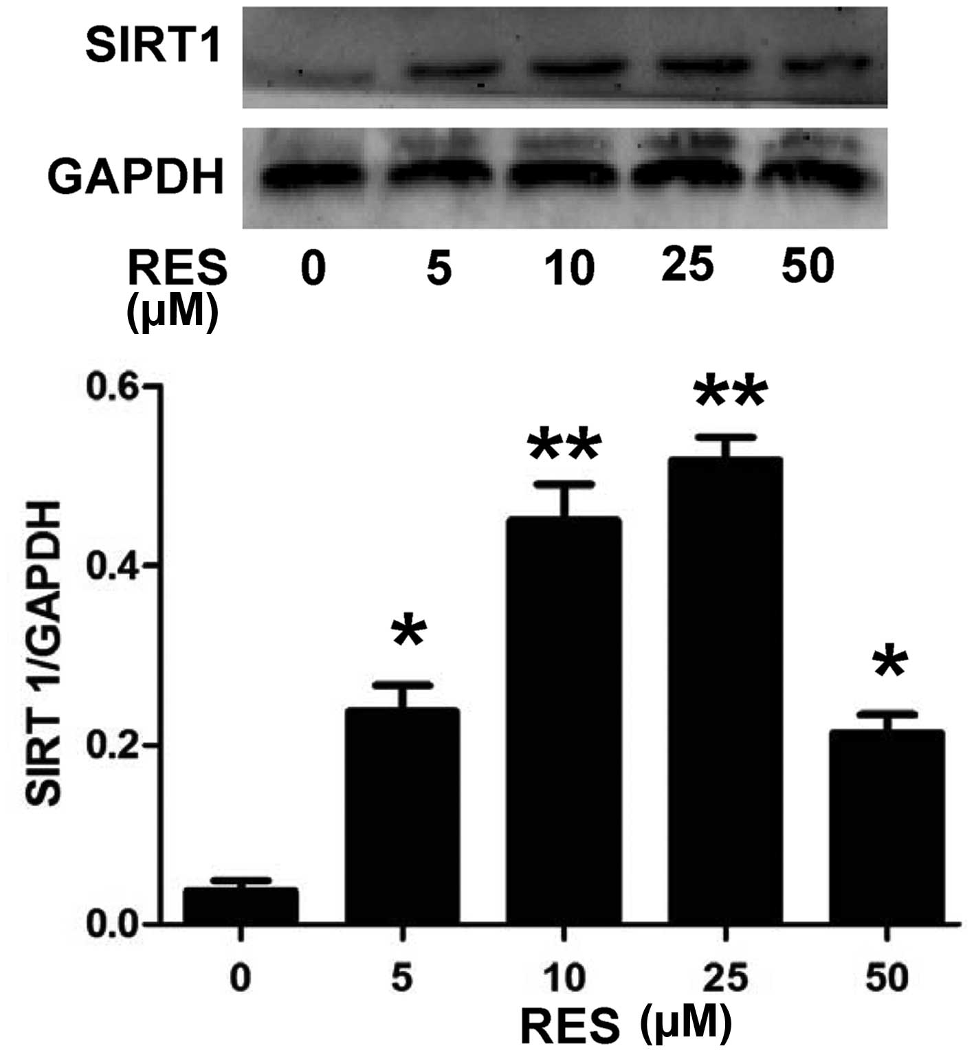
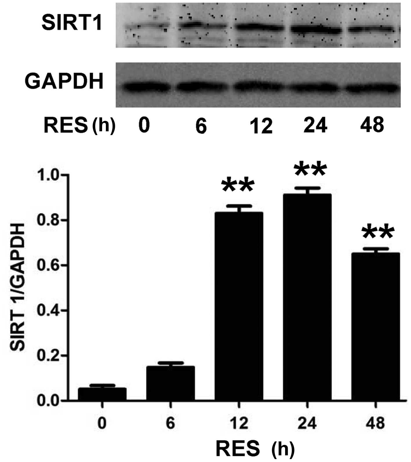
RES activates SIRT1 in H9c2 cells
In order to investigate the underlying mechanism of
the anti-apoptotic effect of RES, the effect of RES on SIRT1
expression levels in H9c2 cells was investigated. As presented in
Fig. 1, H9c2 cell pretreatment with
RES at different concentrations (5, 10, 25 and 50 µM) for 24 h,
prior to exposure to 5 µM DOX for 24 h, significantly increased the
expression levels of SIRT1 (P<0.05 or P<0.01). The expression
levels of SIRT1 reached a peak following treatment with 25 µM RES,
and then declined when 50 µM RES was used. In addition, as
indicated in Fig. 2, exposure of
H9c2 cells to 25 µM RES for time periods ranging between 6 and 48 h
resulted in the rapid activation of SIRT1. The SIRT1 expression was
significantly higher at 12, 24 and 48 h (P<0.01) compared with
the control (0 µM RES), with the most notable effect at 24 h.
Therefore, the concentration of 25 µM RES and incubation time of 24
h were selected for subsequent experiments.
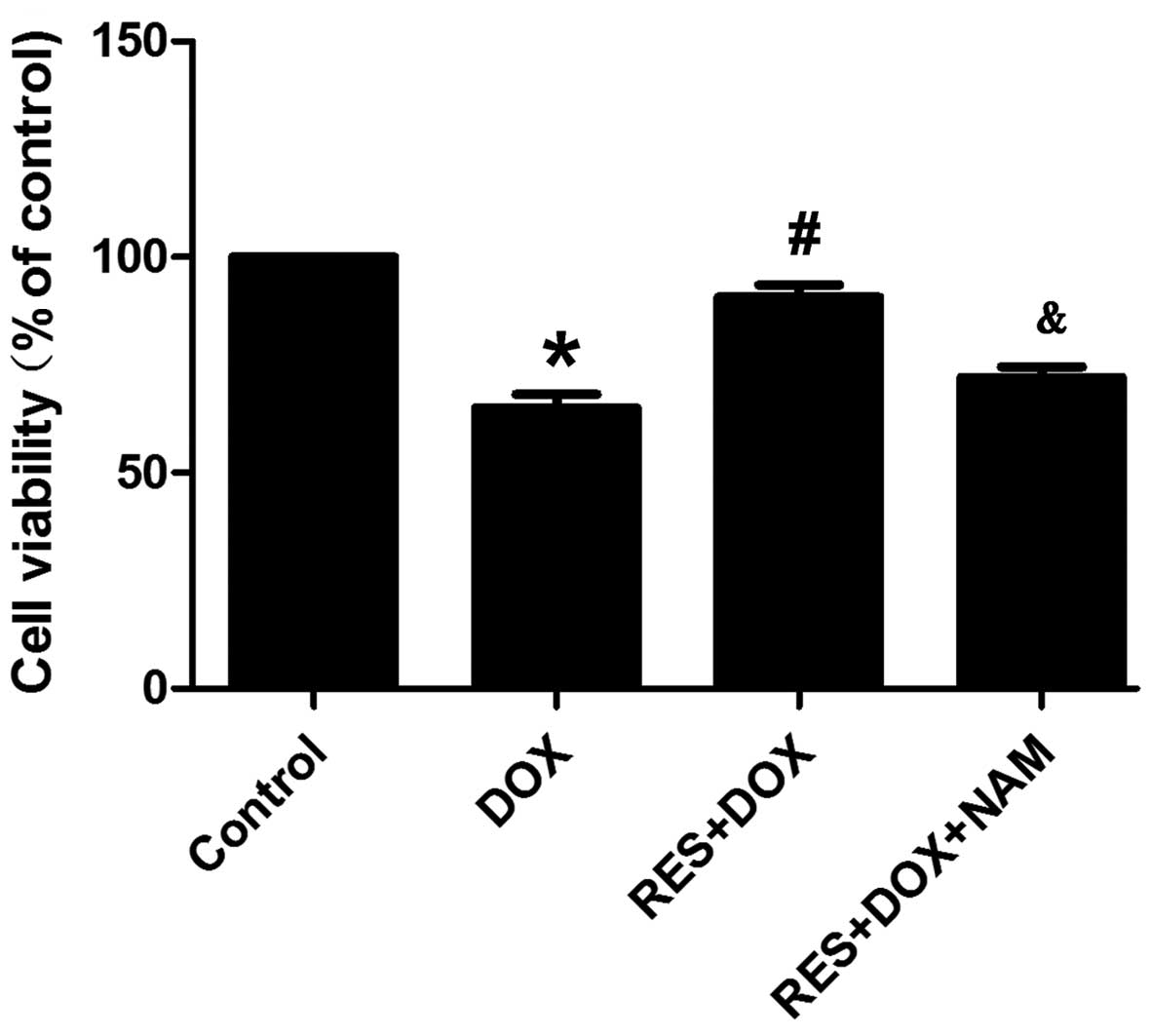
RES inhibits DOX-induced
cytotoxicity
Cell viability was investigated using MTT assay. As
Fig. 3 demonstrates, the exposure of
H9c2 cells to DOX for 24 h induced significant cytotoxicity
(P<0.05), resulting in a decrease in cell viability. However,
cell pretreatment with 25 µM RES for 24 h prior to exposure to DOX
significantly ameliorated DOX-induced cytotoxicity (P<0.05), as
evidenced by an increase in cell viability. Pretreatment of the
H9c2 cells with 40 mM NAM for 60 min prior to the exposure to RES
plus DOX reversed the protective effect of RES, resulting in a
significant decrease in cell viability (P<0.05). These results
suggest that RES exerts a protective effect against DOX-induced
cytotoxicity.
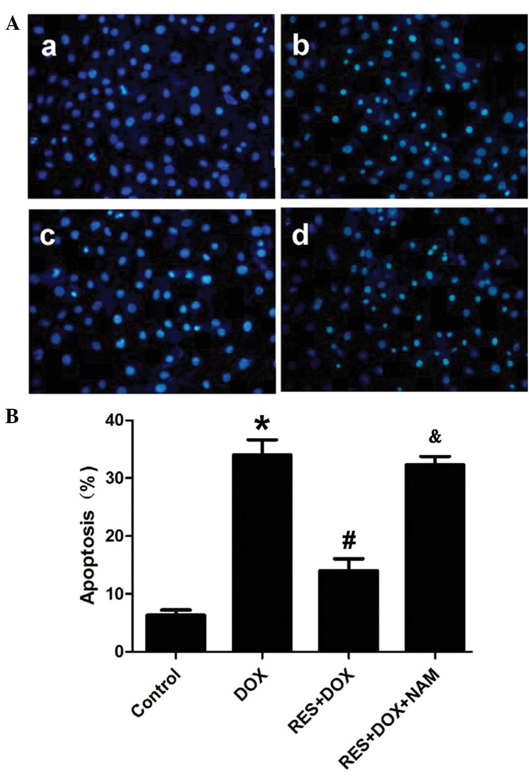
RES reduces DOX-induced apoptosis in
H9c2 cells
The effects of RES on DOX-induced apoptosis were
also observed using Hoechst 33258 nuclear staining. Fig. 4 demonstrates that H9c2 cells treated
with DOX for 24 h took up more Hoechst 33258 stain, as compared
with the control group. However, cell pretreatment with RES for 24
h prior to DOX exposure significantly decreased the DOX-induced
uptake of Hoechst 33258 (P<0.05). Pretreatment of the H9c2 cells
with 40 mM NAM for 60 min prior to the exposure of cells to RES
plus DOX significantly reduced the protective effect of RES
(P<0.05).
SIRT1 inhibits DOX-induced apoptosis
via the SIRT1 signaling pathway
Studies have demonstrated that FoxO proteins induce
the expression of cell death genes, such as Bim. In addition, FoxO
proteins are primarily expressed in the cytoplasm, and are
relocated to the nucleus when cells are subjected to various
stresses; however, they can only interact with SIRT1 in the
nucleus. Thus, SIRT1 can affect FoxO-induced transcription
(20,21). In the present study, pretreatment
with 25 µM RES for 24 h before administration of DOX was found to
decrease the expression levels of FoxO1 and P53 (Fig. 5; P<0.05). However, this effect was
reversed by the administration of 40 mM NAM, prior to RES and DOX
treatment. The expression of the FoxO target gene, Bim, was also
investigated. It was observed that pretreatment of cells with RES
inhibited the expression of Bim, and this effect was reversed by
pretreatment with 40 mM NAM, prior to RES and DOX.
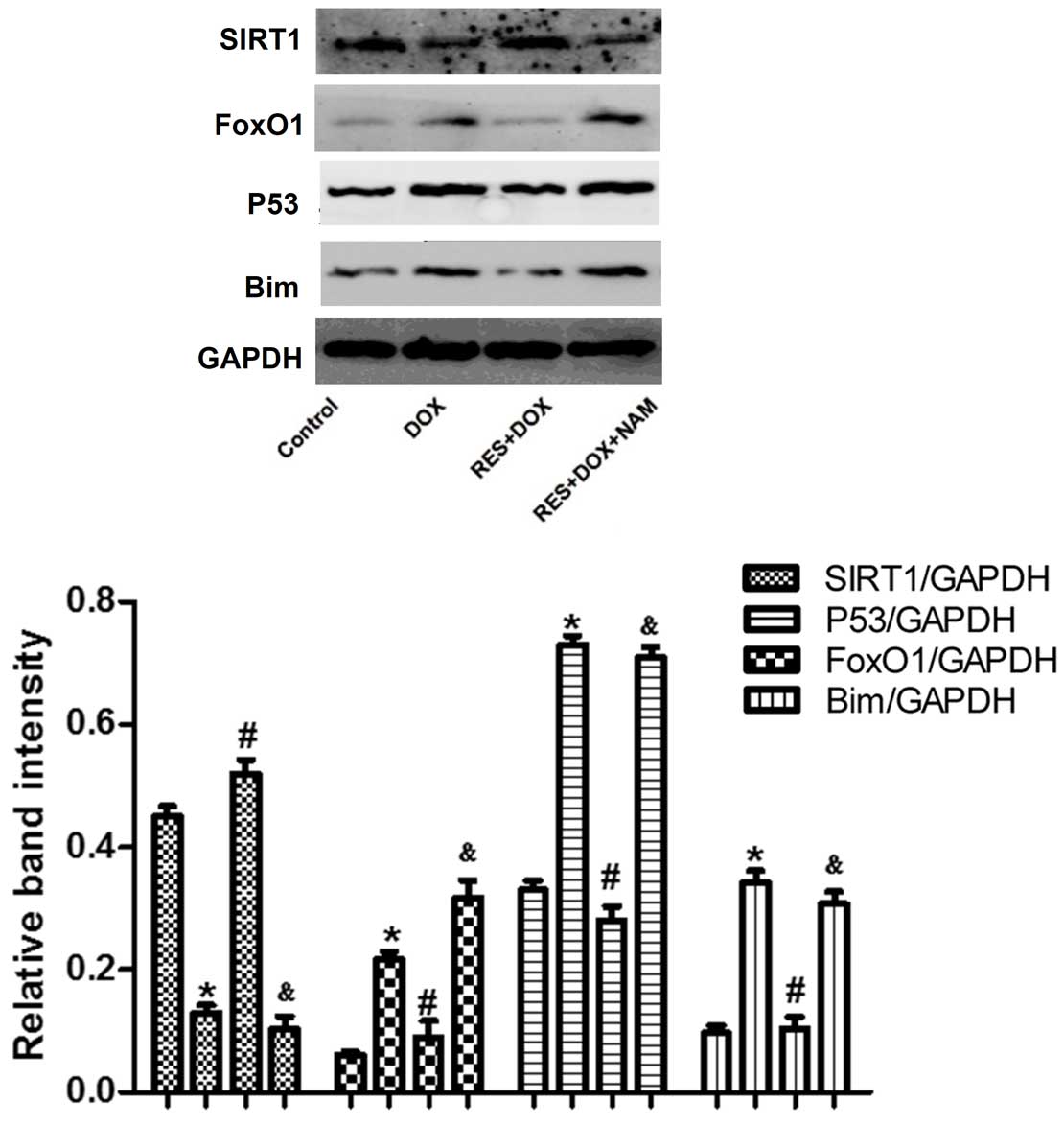 | Figure 5.Protein expression levels of SIRT1,
FoxO1, P53 and Bim in H9c2 cells treated with DOX, RES + DOX or NAM
+ RES + DOX, as analyzed by western blot analysis and quantified by
densitometric analysis. Data are shown as the mean ± standard error
(n=3). *P<0.05 vs. the control group; #P<0.05 vs.
the DOX-treated group; &P<0.05 vs. the RES+DOX
group. SIRT1, sirtuin 1; FoxO1, forkhead box protein O1; Bim,
Bcl-2-like protein 11; DOX, doxorubicin; RES, resveratrol; NAM,
nicotinamide. |
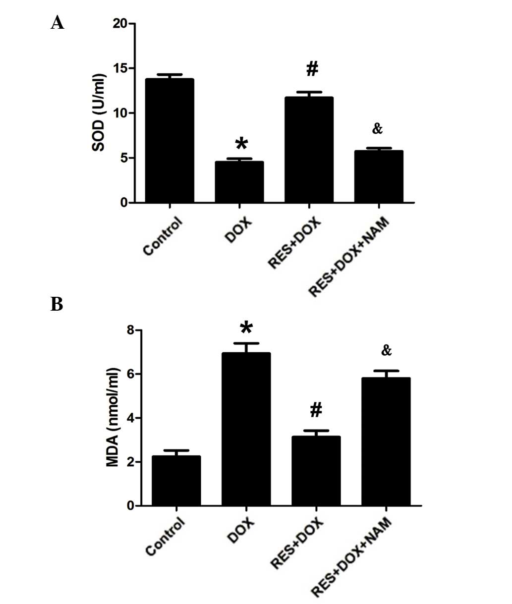
RES restores SOD activity and
decreases MDA expression levels in H9c2 cells
In order to investigate the protective effects of
RES, SOD activity and MDA expression levels in DOX-treated H9c2
cells were investigated as indicators of cell toxicity. As
presented in Fig. 6, following
treatment with DOX, the SOD activity was significantly decreased
and the MDA expression level was significantly increased (both
P<0.05). However, pretreatment with RES significantly increased
SOD activity and decreased the MDA expression (both P<0.05).
This protective effect of RES was reversed by the addition of 40 mM
NAM (P<0.05).
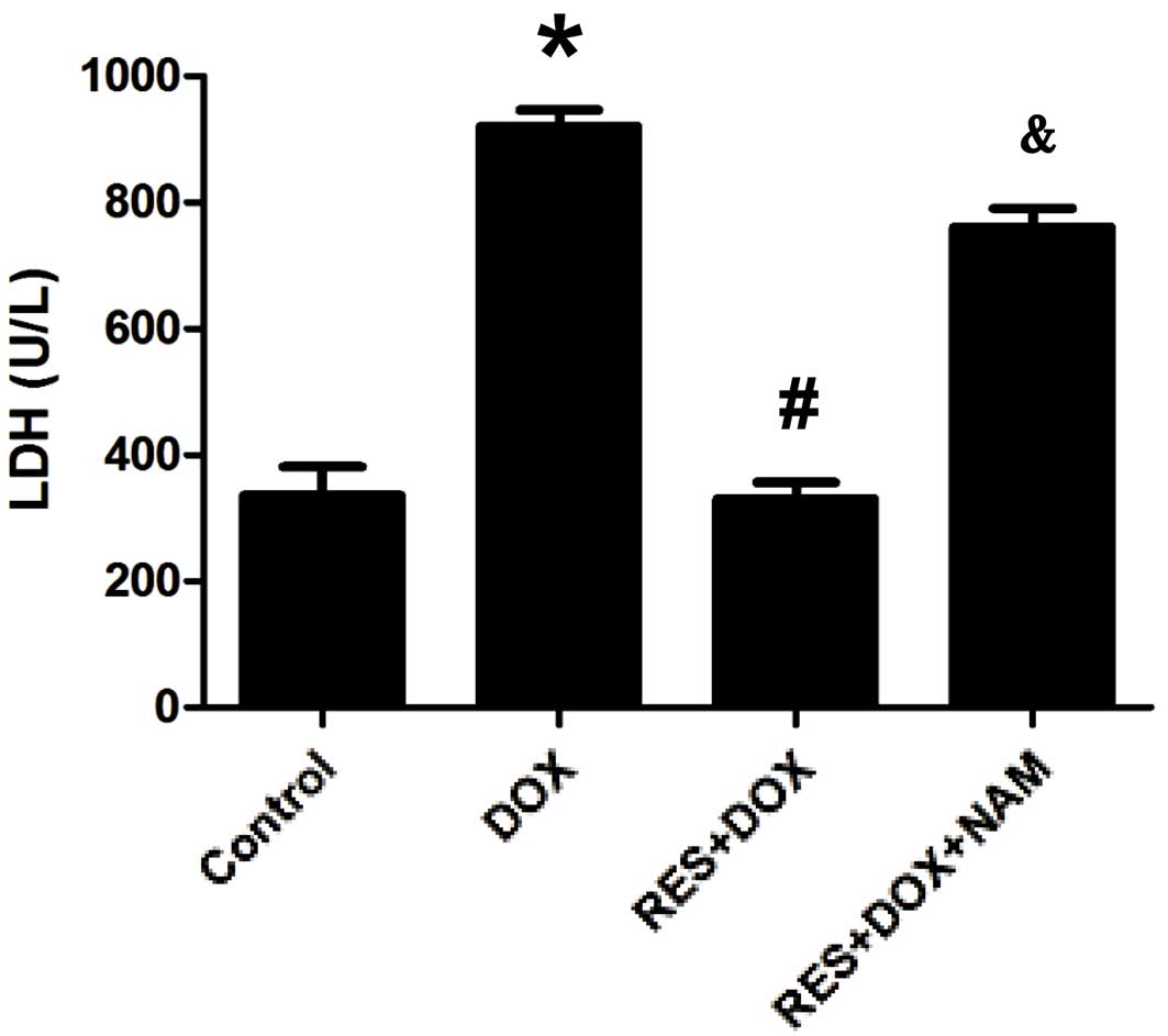
RES decreases LDH levels in H9c2
cells
In order to investigate the protective effects of
RES against DOX-induced cytotoxicity, the amount of LDH released
into the culture medium was measured. As shown in Fig. 7, when cells were exposed to DOX, LDH
release was significantly increased (P<0.05); however, this was
significantly reduced by pretreatment with RES (P<0.05). This
protective effect of RES was significantly reversed following
pretreatment with 40 mM NAM (P<0.05; Fig. 5).
Discussion
DOX is a widely used and successful anti-tumor drug;
however, its clinical use is limited due to its severe cumulative
dose-associated cardiotoxicity (2).
Numerous studies have demonstrated that the primary molecular
mechanism involved in DOX-induced cardiotoxicity is free
radical-induced oxidative stress, and cardiomyocyte death by
apoptosis and necrosis (5,22). In accordance with previous studies
(23,24), in the present study it was observed
that the exposure of H9c2 cells to DOX significantly induced
cellular injuries, including decreases in cell viability and the
expression of SIRT1, as well as increases in cell apoptosis and in
the expression levels of FoxO1, P53 and Bim.
RES is a polyphenol that is primarily found in red
wine, and occurs naturally in grapes, mulberries and peanuts. RES
has numerous protective features; it can reduce the risk of
cardiovascular disease and protect cardiomyocytes from apoptosis
(25). It has also been demonstrated
to protect cardiomyocytes against DOX-induced apoptosis (26). In addition, RES has been shown to
enhance the anti-cancer activity of DOX, and protect against
DOX-induced cardiac toxicity in vitro and in mice (11). However, the mechanism underlying the
effect of RES in protecting cardiomyocytes against apoptosis
remains unclear.
It has been suggested that decreased SIRT1
expression is associated with DOX-induced myocardial apoptosis
(27). As SIRT1 activity protects
against increased oxidative stress and enhances cell survival, the
SIRT1 signaling pathway is a plausible mechanism of action for the
protective effect of RES against the effects of DOX. In the present
study, the results demonstrated that SIRT1 expression was decreased
following DOX treatment, and it was observed that RES
preconditioning increased SIRT1 expression while significantly
attenuating DOX-induced apoptosis. However, the effect of RES on
SIRT1 expression levels was reduced in the presence of NAM.
Activated SIRT1 has been shown to protect the heart
from oxidative stress by activating FoxO-dependent mechanisms
(18). In addition, SIRT1 prevents
p53-dependent apoptosis resistance to oxidative stress (28). Li et al (29) observed that RES protects
cardiomyocytes from oxidative stress through the SIRT1 and
mitochondrial biogenesis signaling pathways. Lou et al
(30) reported that RES protects
H9c2 cells against DOX-induced ER stress through ER stabilization,
and significantly increased the activation of SIRT1, thereby
leading to cardiac cell survival. To the best of our knowledge, the
present study is the first to demonstrate that RES decreases the
expression levels of FoxO1 and P53, and that this effect is largely
reversed upon pretreatment with 40 mM NAM. Furthermore, the effect
of SIRT1 on a FoxO1-induced target gene expression was investigated
in the current study. Consistently, RES reduced the expression of
Bim and cell apoptosis. In addition, it was demonstrated that
pretreatment of H9c2 cells with 40 mM NAM promoted the expression
of Bim and H9c2 cell apoptosis.
In conclusion, the present study demonstrated that
RES pretreatment is able to inhibit DOX-induced apoptosis in H9c2
cells and that its effects are mediated by SIRT1 activation. The
current study investigated the mechanisms underlying the
anti-apoptotic effect of RES in cardiomyocytes, and provided
valuable evidence for identifying RES as a candidate for the
treatment of cardiovascular diseases.
Acknowledgements
The present study was supported by the Graduate
Student Research Innovation project of Hunan province (grant no.
CX2013B397).
References
|
1
|
Menna P, Recalcati S, Cairo G and Minotti
G: An introduction to the metabolic determinants of anthracycline
cardiotoxicity. Cardiovasc Toxicol. 7:80–85. 2007. View Article : Google Scholar : PubMed/NCBI
|
|
2
|
Lipshultz SE, Karnik R, Sambatakos P,
Franco VI, Ross SW and Miller TL: Anthracycline-related
cardiotoxicity in childhood cancer survivors. Curr Opin Cardiol.
29:103–112. 2014. View Article : Google Scholar : PubMed/NCBI
|
|
3
|
Spallarossa P, Garibaldi S, Altieri P,
Fabbi P, Manca V, Nasti S, Rossettin P, Ghigliotti G, Ballestrero
A, Patrone F, et al: Carvedilol prevents doxorubicin-induced free
radical release and apoptosis in cardiomyocytes in vitro. J Mol
Cell Cardiol. 37:837–846. 2004. View Article : Google Scholar : PubMed/NCBI
|
|
4
|
Lee DH, Kim S and Nam KS: Protective
effects of deep sea water against doxorubicin-induced
cardiotoxicity in H9c2 cardiac muscle cells. Int J Oncol.
45:2569–2575. 2014.PubMed/NCBI
|
|
5
|
Al-Harthi SE, Alarabi OM, Ramadan WS,
Alaama MN, Al-Kreathy HM, Damanhouri ZA, Khan LM and Osman AM:
Amelioration of doxorubicin-induced cardiotoxicity by resveratrol.
Mol Med Rep. 10:1455–1460. 2014.PubMed/NCBI
|
|
6
|
Goyal SN, Mahajan UB, Chandrayan G,
Kumawat VS, Kamble S, Patil P, Agrawal YO, Patil CR and Ojha S:
Protective effect of oleanolic acid on oxidative injury and
cellular abnormalities in doxorubicin induced cardiac toxicity in
rats. Am J Transl Res. 8:60–69. 2016.PubMed/NCBI
|
|
7
|
Renaud J, Bournival J, Zottig X and
Martinoli MG: Resveratrol protects DAergic PC12 cells from high
glucose-induced oxidative stress and apoptosis: Effect on p53 and
GRP75 localization. Neurotox Res. 25:110–123. 2014. View Article : Google Scholar : PubMed/NCBI
|
|
8
|
Liu MH, Yuan C, He J, Tan TP, Wu SJ, Fu
HY, Liu J, Yu S, Chen YD, Le QF, et al: Resveratrol protects PC12
cells from high glucose-induced neurotoxicity via PI3K/Akt/FoxO3a
pathway. Cell Mol Neurobiol. 35:513–522. 2014. View Article : Google Scholar : PubMed/NCBI
|
|
9
|
Tatlidede E, Sehirli O, Velioğlu-Oğünc A,
Cetinel S, Yeğen BC, Yarat A, Süleymanoğlu S and Sener G:
Resveratrol treatment protects against doxorubicin-induced
cardiotoxicity by alleviating oxidative damage. Free Radic Res.
43:195–205. 2009. View Article : Google Scholar : PubMed/NCBI
|
|
10
|
Shankar S, Singh G and Srivastava RK:
Chemoprevention by resveratrol: Molecular mechanisms and
therapeutic potential. Front Biosci. 12:4839–4854. 2007. View Article : Google Scholar : PubMed/NCBI
|
|
11
|
Rezk YA, Balulad SS, Keller RS and Bennett
JA: Use of resveratrol to improve the effectiveness of cisplatin
and doxorubicin: Study in human gynecologic cancer cell lines and
in rodent heart. Am J Obstet Gynecol. 194:e23–e26. 2006. View Article : Google Scholar : PubMed/NCBI
|
|
12
|
Park DG: Antichemosensitizing effect of
resveratrol in cotreatment with oxaliplatin in HCT116 colon cancer
cell. Ann Surg Treat Res. 86:68–75. 2014. View Article : Google Scholar : PubMed/NCBI
|
|
13
|
Wang Y: Molecular links between caloric
restriction and Sir2/SIRT1 Activation. Diabetes Metab J.
38:321–329. 2014. View Article : Google Scholar : PubMed/NCBI
|
|
14
|
Bitterman KJ, Anderson RM, Cohen HY,
Latorre-Esteves M and Sinclair DA: Inhibition of silencing and
accelerated aging by nicotinamide, a putative negative regulator of
yeast sir2 and human SIRT1. J Biol Chem. 277:45099–45107. 2002.
View Article : Google Scholar : PubMed/NCBI
|
|
15
|
Alcendor RR, Gao S, Zhai P, Zablocki D,
Holle E, Yu X, Tian B, Wagner T, Vatner SF and Sadoshima J: Sirt1
regulates aging and resistance to oxidative stress in the heart.
Circ Res. 100:1512–1521. 2007. View Article : Google Scholar : PubMed/NCBI
|
|
16
|
Yamamoto T and Sadoshima J: Protection of
the heart against ischemia/reperfusion by silent information
regulator 1. Trends Cardiovasc Med. 21:27–32. 2011. View Article : Google Scholar : PubMed/NCBI
|
|
17
|
Luo J, Nikolaev AY, Imai S, Chen D, Su F,
Shiloh A, Guarente L and Gu W: Negative control of p53 by Sir2alpha
promotes cell survival under stress. Cell. 107:137–148. 2001.
View Article : Google Scholar : PubMed/NCBI
|
|
18
|
Chen CJ, Yu W, Fu YC, Wang X, Li JL and
Wang W: Resveratrol protects cardiomyocytes from hypoxia-induced
apoptosis through the SIRT1-FoxO1 pathway. Biochem Biophys Res
Commun. 378:389–393. 2009. View Article : Google Scholar : PubMed/NCBI
|
|
19
|
Guo R, Lin J, Xu W, Shen N, Mo L, Zhang C
and Feng J: Hydrogen sulfide attenuates doxorubicin-induced
cardiotoxicity by inhibition of the p38 MAPK pathway in H9c2 cells.
Int J Mol Med. 31:644–650. 2013.PubMed/NCBI
|
|
20
|
Shukla S, Rizvi F, Raisuddin S and Kakkar
P: FoxO proteins' nuclear retention and BH3-only protein Bim
induction evoke mitochondrial dysfunction-mediated apoptosis in
berberine-treated HepG2 cells. Free Radic Biol Med. 76:185–199.
2014. View Article : Google Scholar : PubMed/NCBI
|
|
21
|
Li F, Qu H, Cao HC, Li MH, Chen C, Chen
XF, Yu B, Yu L, Zheng LM and Zhang W: Both FOXO3a and FOXO1 are
involved in the HGF-protective pathway against apoptosis in
endothelial cells. Cell Biol Int. 39:1131–1137. 2015. View Article : Google Scholar : PubMed/NCBI
|
|
22
|
Angsutararux P, Luanpitpong S and
Issaragrisil S: Chemotherapy-Induced Cardiotoxicity: Overview of
the Roles of Oxidative Stress. Oxid Med Cell Longev.
2015:7956022015. View Article : Google Scholar : PubMed/NCBI
|
|
23
|
Wang X, Wang XL, Chen HL, Wu D, Chen JX,
Wang XX, Li RL, He JH, Mo L, Cen X, et al: Ghrelin inhibits
doxorubicin cardiotoxicity by inhibiting excessive autophagy
through AMPK and p38-MAPK. Biochem Pharmacol. 88:334–350. 2014.
View Article : Google Scholar : PubMed/NCBI
|
|
24
|
Guo R, Wu K, Chen J, Mo L, Hua X, Zheng D,
Chen P, Chen G, Xu W and Feng J: Exogenous hydrogen sulfide
protects against doxorubicin-induced inflammation and cytotoxicity
by inhibiting p38MAPK/NFκB pathway in H9c2 cardiac cells. Cell
Physiol Biochem. 32:1668–1680. 2013.PubMed/NCBI
|
|
25
|
Das DK, Mukherjee S and Ray D: Erratum to:
Resveratrol and red wine, healthy heart and longevity. Heart Fail
Rev. 16:425–435. 2011. View Article : Google Scholar : PubMed/NCBI
|
|
26
|
Oktem G, Uysal A, Oral O, Sezer ED,
Olukman M, Erol A, Akgur SA and Bilir A: Resveratrol attenuates
doxorubicin-induced cellular damage by modulating nitric oxide and
apoptosis. Exp Toxicol Pathol. 64:471–479. 2012. View Article : Google Scholar : PubMed/NCBI
|
|
27
|
Danz ED, Skramsted J, Henry N, Bennett JA
and Keller RS: Resveratrol prevents doxorubicin cardiotoxicity
through mitochondrial stabilization and the Sirt1 pathway. Free
Radic Biol Med. 46:1589–1597. 2009. View Article : Google Scholar : PubMed/NCBI
|
|
28
|
Shang L, Zhou H, Xia Y, Wang H, Gao G,
Chen B, Liu Q, Shao C and Gong Y: Serum withdrawal up-regulates
human SIRT1 gene expression in a p53-dependent manner. J Cell Mol
Med. 13:4176–4184. 2009. View Article : Google Scholar : PubMed/NCBI
|
|
29
|
Li YG, Zhu W, Tao JP, Xin P, Liu MY, Li JB
and Wei M: Resveratrol protects cardiomyocytes from oxidative
stress through SIRT1 and mitochondrial biogenesis signaling
pathways. Biochem Biophys Res Commun. 438:270–276. 2013. View Article : Google Scholar : PubMed/NCBI
|
|
30
|
Lou Y, Wang Z, Xu Y, Zhou P, Cao J, Li Y,
Chen Y, Sun J and Fu L: Resveratrol prevents doxorubicin-induced
cardiotoxicity in H9c2 cells through the inhibition of
endoplasmicreticulum stress and the activation of the Sirt1
pathway. Int J Mol Med. 36:873–880. 2015.PubMed/NCBI
|