Introduction
In India, the plant Asparagus racemosus
Willd. (Liliaceae) is commonly called Satavari, Satawar or Satmuli
in Hindi, Satavari in Sanskrit and Shatamuli in Bengali. In
Thailand, it is called Sam-Sib or Sam-Roi-Rak. The plant is a
spinous undershrub with tuberous short rootstock bearing numerous
succulent tuberous roots (30–100 cm long and 1–2 cm thick) that are
silvery white or ash-colored externally and white internally
(1). Its pharmacological
applications, particularly from the root extracts, have recently
been found to possess a phytoestrogenic effect, an effect on
neurodegenerative disorders, as well as antidiarrhoeal,
antidyspepsia, adaptogenic, cardioprotective, antibacterial,
immunoadjuvant and antitussive effects. The root extracts of
Asparagus racemosus have been employed in two major forms as
methanolic and aqueous extracts, the products of which include root
extract in tablet form, root powder in tablet form and root extract
in syrup form (1).
Among the many biological activities of Asparagus
racemosus, it has been reported to have antioxidant activity.
Methanolic root extracts were found to markedly increase superoxide
dismutase, catalase and ascorbic acid, while decreasing lipid
peroxidase product (malondialdehyde) in rats (2,3). In
addition, Asparagus racemosus has recently been shown to
contain ten steroidal saponins (4)
and racemofuran (5). The aqueous
extracts have also been shown to exhibit an antioxidant effect in
rat liver mitochondria by protecting against the radiation-induced
loss of protein thiols and inactivation of superoxide dismutase
(6), and the amelioration of
oxidative stress and hepatotoxicity (7).
Cationic liposomes are among the non-viral vectors
considered to be promising tools for gene delivery through their
formation of a lipoplex with nucleic acids, such as RNA or DNA
(8,9). Their advantages include
biocompatibility, a plasmid-independent structure, the opportunity
for chemical or physical manipulation, and large-scale production
and cost-effectiveness, while drawbacks include low transfection
efficiency (10) and cytotoxicity
(11).
Toxicity studies of cationic liposomes revealed a
significant effect when the positive charge density was increased.
Lipofectamine (commercially available polyvalent cationic
liposomes) was reported to possess a higher transfection efficiency
than Lipofectin and DOTAP (monovalent cationic liposomes) in the
macrophage RAW264.7 cell line (12,13).
Nonetheless, cationic lipids with a higher positive charge density
are generally more toxic (14).
Moreover, some reports on toxicity have found that cationic
liposomes with a monovalent positive charge (stearylamine) induced
apoptosis and the production of reactive oxygen species (ROS)
(15) in the RAW264.7 (16) and mouse immature B (WEHI 231) cell
lines (17). In in vivo
toxicity studies of differently charged liposomes by intratracheal
instillation in mice, Lipofectamine was found to cause much greater
toxicity than DOTAP in a dose-dependent manner, which was
associated with ROS generation, while neutral and negative
liposomes were not toxic at the relevant concentration (18). Therefore, ROS are considered to
play a key role in cationic liposome-mediated cytotoxicity. This
effect was found to be charge-dependent and was prevented by ROS
scavengers.
Toxicity and apoptosis induced by cationic liposomes
as well as their mechanisms have been found to be definitely
associated with ROS generation. Thus, the antioxidant effect of
Asparagus racemosus root extract may be applicable for
protecting cells from apoptosis related to ROS generation. Such an
application would provide an alternative for scavenging ROS, and
hence improve transfection efficiency. In this study, different
fractions of Asparagus racemosus root extracts from
successive extraction with different solvents were investigated in
regards to their antioxidant activity and protective effect against
Lipofectamine-induced apoptosis in human lung epithelial H460
cells.
Materials and methods
Chemicals and reagents
Lipofectamine was obtained from Invitrogen
(Carlsbad, CA, USA). This liposomal formulation is composed of a
3:1 mixture of cationic lipid DOSPA and neutral lipid DOPE.
Oleanolic acid, diosgenin, 2,2-diphenyl-1-picrylhydrazyl (DPPH),
and Hoechst® 33342 dye were purchased from Sigma
Chemical Co. (St. Louis, MO, USA). Thin layer chromatography silica
gel plates (60F254), ascorbic acid, hexane, ethanol,
acetone, methanol, ethyl acetate, chloroform and dichloromethane
were purchased from Merck KG (Darmstadt, Germany).
Plant materials
The roots of Asparagus racemosus were
collected from Tak Province, Thailand. The plants were identified
by the botanical staff, and the voucher specimen of the plant was
deposited in the herbarium of the Faculty of Pharmaceutical
Sciences, Naresuan University, Phitsanulok, Thailand.
Successive extraction of Asparagus
racemosus roots
Asparagus racemosus roots were dried and
minced into powder, and then successively extracted with solvents
starting from non-polar (hexane) to more polar (water) solvents.
The hexane fraction was filtered and evaporated by a rotary
evaporator (Eyela® A-3S; Tokyo Rikakikai, Japan), after
which soft yellow paste was obtained and named AR1-1. The marc was
further extracted with 95% ethanol, filtrated and evaporated, after
which the dark brown viscous liquid was obtained and named AR1-2.
The residual marc was further extracted with water at 80°C. After
filtration, the aqueous fraction was divided into two portions, one
of which was evaporated and freeze-dried until brown powder was
obtained and named AR1-3. The other portion was added to acetone
and the resulting precipitate was separated out from the solution.
Both the solution and precipitate were evaporated, freeze-dried,
and named AR1-4 and AR1-5, respectively.
Thin layer chromatography (TLC)
fingerprint
TLC is one of several techniques useful for the
identification of phytochemical compounds (19,20).
In this study, solutions of oleanolic acid and diosgenin (0.5
mg/ml) in methanol were used as known references for triterpene and
steroid structure, respectively (21,22).
The samples of the Asparagus racemosus extracts were
prepared by dissolving fractions of AR1-1, AR1-2 and AR1-3 to AR1-5
(10 mg/ml) in ethyl acetate, methanol and water, respectively. All
samples were filtered through a 0.2-μm nylon membrane filter, after
which 8 μl was spotted by Autospot equipped with a scanner and
camera (Camag®; Limonat V, Switzerland) on TLC silica
gel plates and air-dried for 5 min. The plates with the oleanolic
acid and AR1-1 samples were developed with system I (2:1,
hexane:ethyl acetate) (19), while
those of diosgenin and AR1-2 to AR1-5 samples were developed with
system II (6.4:5:1, chloroform:methanol:water) (23). After completion, the plates were
dried at 100–110°C for 5 min, sprayed with 10 ml
anisaldehyde-sulfuric acid reagent, heated at 105°C for 5–10 min
and visualized under white and UV light (366 nm) for TLC
fingerprints. This experiment was performed in duplicate.
Total saponin quantification
The total saponin content of Asparagus
racemosus extracts was determined as described by Makkar et
al (21) and Xi et al
(24). Sample solutions of
diosgenin (0.5 mg/ml) in methanol and those of AR1-1, AR1-2 and
AR1-3 to AR1-5 (4 mg/ml) in dichloromethane, methanol and water,
respectively, were prepared and filtered through a 0.2-μm nylon
membrane filter before use. A 250-μl portion of 8% w/v vanillin
solution in absolute ethanol was added to 250 μl of sample, which
was subsequently mixed with 2.5 ml 72% v/v sulfuric acid, incubated
in a water bath at 60°C for 10 min and finally cooled in an ice
bath for 5 min. The absorbance of this mixture was measured at 544
nm (Perkin Elmer Lambda 35; Perkin Elmer, Waltham, MA, USA). The
calibration curve of diosgenin at a high degree of linearity with a
correlation coefficient (r2) of 0.9991 was achieved
across the specified range of 8.3–41.6 μg/ml. The diosgenin
equivalent (DGE) (μg/mg extract) was used as reference in
determining the total amount of saponins in each fraction of the
extracts. This experiment was performed in triplicate.
Antioxidant activity testing by DPPH
assay
The DPPH assay for assessing the antioxidant
activity of the Asparagus racemosus extracts was carried out
as described by Brand-Williams et al (25), with minor modifications. The
absorbance of the DPPH solution in methanol was measured at 515 nm
(Perkin Elmer Lambda 35), and a calibration curve at a very high
degree of linearity with a correlation coefficient (r2)
of 0.9999 was achieved across the specified range of 10–100 μM.
Solutions of AR1-1, AR1-2 and AR1-3 to AR1-5 (4 mg/ml) were
prepared in dichloromethane, methanol and water, respectively, 200
μl of which was mixed with 1,400 μl of 60 μM DPPH solution. The
decrease in absorbance was determined at 0, 1 and every 15 min
until 60 min. The percentage of DPPH remaining at 60 min was
determined and plotted as a function of the AR1-4 concentration.
L-ascorbic acid was used as a positive control. The antioxidant
activity was expressed in terms of the mean effective concentration
(EC50), which was defined as the amount of antioxidant
that could decrease the initial DPPH concentration by 50%.
Cell culture
Human lung epithelial NCI-H460 cells were obtained
from the American Type Culture Collection (Manassas, VA, USA). The
cells were cultured in RPMI-1640 medium (Invitrogen) supplemented
with 5% fetal bovine serum, 2 mM L-glutamine, 100 U/ml of
penicillin and 100 μg/ml of streptomycin. The cells were grown in a
humidified atmosphere of 5% CO2 at 37°C until they
reached over 80% confluence. The cells were subsequently passaged
by the use of a solution containing 0.05% trypsin and 0.5 mM
EDTA.
Antiapoptotic effect in H460 cells
The aqueous fraction AR1-4 (10 mg/ml) was dissolved
in water and filtered through a 0.2-μm nylon membrane filter before
use. Cells (3×10⁴/well) were seeded in a 96-well plate
in 100 μl of culture medium and grown for 24 h. The subconfluent
monolayer of cells was washed once with PBS and once with
RPMI-1640. Subsequently, 100 μl of RPMI-1640 was added, and the
cells were untreated or pre-treated with various concentrations of
AR1-4 (100, 500 and 1,000 μg/ml) for 1 h, and then treated with
Lipofectamine (20 μg/ml) for 6 h. The untreated cells were used as
a control. Apoptosis was determined by the Hoechst 33342 assay.
For Hoechst 33342 staining, cells were incubated
with 10 μg/ml of Hoechst 33342 dye at 37°C for 30 min. Analysis for
apoptosis was carried out by scoring the percentage of cells having
intensely condensed chromatin and/or fragmented nuclei by
fluorescence microscopy (Axiovert 25 CFI; Carl Zeiss, USA).
Approximately 1,000 nuclei from random fields were analyzed for
each sample. The percentage of apoptosis was calculated as
(apoptotic nuclei/total nuclei) × 100%. The experiment was
performed in triplicate.
Results and Discussion
Standardization of Asparagus racemosus
extracts
Asparagus racemosus extracts contain saponins
as the main constituents (26).
The major constituents of Asparagus racemosus in methanolic
extract are steroidal saponins, which have been separated, purified
and termed ‘Shatavarin’. Ten derivatives of Shatavarin have been
defined as Shatavarin I to X (1,4).
Gautam et al (23) found
that an Asparagus racemosus aqueous decoction contained
steroidal saponin, alkaloids, proteins, starch, tannin and
mucilage. Agrawal et al (7)
carried out screening tests and reported positive results for
steroids, phytosterols, carbohydrates, tannins, anthraquinones,
saponins, glycosides and flavonoids, and negative results for
terpenoids, amino acids and alkaloids. Most recently, Visavadiya
and Narasimhacharya (26) found
that most of the phytoconstituents in Asparagus racemosus
root are saponins (8.83%), while the remaining constituents are
polyphenols (1.69%), phytosterols (0.79%), ascorbic acid (0.76%)
and flavonoids (0.48%).
Since saponins are found in both aqueous and
methanolic extracts of Asparagus racemosus and can be
purified successfully to pure compounds, they were considered as
markers in this study. Standardization of Asparagus
racemosus in this study involved the confirmation of the
presence of saponins and control of the quality of Asparagus
racemosus extracts by examining the TLC fingerprints and total
saponin content.
Thin layer chromatographic
fingerprints
The anisaldehyde-sulfuric acid reagent is generally
used to detect phenol, sugar, steroid and terpene, which will turn
violet, blue, red, grey or green, respectively. It was found that
the TLC fingerprints of oleanolic acid and AR1-1 developed with
system I exhibited a blue color, while those of diosgenin and AR1-2
to AR1-5 developed with system II exhibited yellow-green and green
colors under white light. Detailed information regarding the
Rf values and color zones is listed in Table I, and indicates that the compounds
detected in AR1-1 to AR1-5 using this reagent may contain the
structure of phenol, sugar, steroid and/or terpene in their
molecules. Oleanolic acid, as triterpene, showed only a blue
stripe, whereas AR1-1 exhibited many stripes with a blue color, one
being on top of the lane. This indicates that AR1-1 may contain
some saponins, including triterpenoid saponin. The more polar
developing system II was applied to diosgenin and to fractions
AR1-2 to AR1-5, which appeared to contain some steroidal saponins
since green stripes similar to that of diosgenin appeared.
 | Table I.Rf values and color zones
of TLC fingerprints of oleanolic acid and AR1-1 (hexane fraction)
developed with system I, and diosgenin, AR1-2 (ethanol fraction)
and AR1-3 to AR1-5 (aqueous fractions) developed with system II,
visualized under white and UV light (366 nm). |
Table I.
Rf values and color zones
of TLC fingerprints of oleanolic acid and AR1-1 (hexane fraction)
developed with system I, and diosgenin, AR1-2 (ethanol fraction)
and AR1-3 to AR1-5 (aqueous fractions) developed with system II,
visualized under white and UV light (366 nm).
| Compound | Rf | Color under white
light | Color under UV light
(366 nm) |
|---|
| Oleanolic acid | 0.31 | Blue | Brown |
| AR1-1 (hexane
fraction) | 0.38 | Blue | Violet |
| 0.44 | Light blue | Light brown |
| 0.56 | Light blue | Pink |
| 0.69 | Blue | Red |
| 0.81 | Dark blue | Red |
| Diosgenin | 1.00 | Yellow-green | Brown |
| AR1-2 (ethanol
fraction) | 0.24 | Light green | Light brown |
| 0.34 | Light green | Light brown |
| 0.45 | Dark green | Dark brown |
| AR1-3 (aqueous
fraction) | 0.24 | Light green | Light brown |
| 0.34 | Light green | Light brown |
| 0.45 | Green | Brown |
| AR1-4 (aqueous
fraction) | 0.24 | Light green | Light brown |
| 0.34 | Light green | Light brown |
| 0.45 | Green | Brown |
| AR1-5 (aqueous
fraction) | 0.24 | Light green | Light brown |
| 0.34 | Light green | Light brown |
| 0.45 | Green | Brown |
Among all of the fractions of the Asparagus
racemosus extracts, the compounds of AR1-1 differed from those
of AR1-2 to AR1-5. Fractions AR1-2 to AR-5 consisted of similar
constituents as evidenced by the same pattern of TLC fingerprints
with three main bands and a zone near the starting spot. Moreover,
the band at an Rf value of 0.45 was prominent in AR1-2,
while a zone near the starting spot was more pronounced in
fractions AR1-3 to AR1-5. As a result, solubility of the former was
increased using 95% ethanol as an extracting solvent, whereas
solubility of the latter was increased using water as an extracting
solvent. This was determined by the successive extraction using
non-polar to more polar solvents.
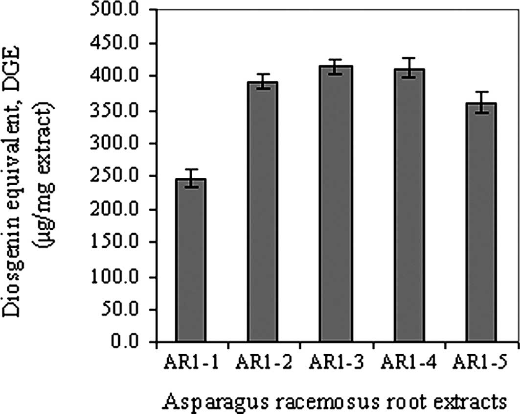
Total saponin quantification
Vanillin-sulfuric acid reagent was used for
derivatization of the saponins having an OH group at their C-3
position to give chromogens. The chromogens formed were not
dependent on the nature of the sugar moieties (21). Among all of the Asparagus
racemosus extracts, the amount of DGE could be ranked in order
as: AR1-2 ∼ AR1-3 ∼ AR1-4 > AR1-5 > AR1-1, as shown in
Fig. 1. It is apparent that the
highest amount of saponins was found in the 95% ethanol and aqueous
fractions, but with a difference in the type of saponins.
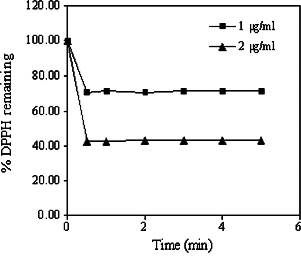
Antioxidant activity testing by DPPH
assay
In this study, ascorbic acid was used as a positive
control, and AR1-4 was chosen as a representative based on its
highest saponin content. Firstly, the reaction kinetics in the
antioxidant activity of both ascorbic acid and AR1-4 with DPPH were
carried out to determine the suitable incubation time before
measurement. These were found to reach a steady state by 1 and 60
min, respectively (Fig. 2). The
time to reach steady state, as classified by Brand-William et
al (25), for reaction
kinetics of ascorbic acid was found to be much more rapid (<1
min) than that of AR1-4 (60 min). Active compounds having
antioxidant activity in AR1-4 would consist` of a large complex
molecule with slow reaction kinetics, while small molecule of
ascorbic acid could easily be subject to interaction with DPPH.
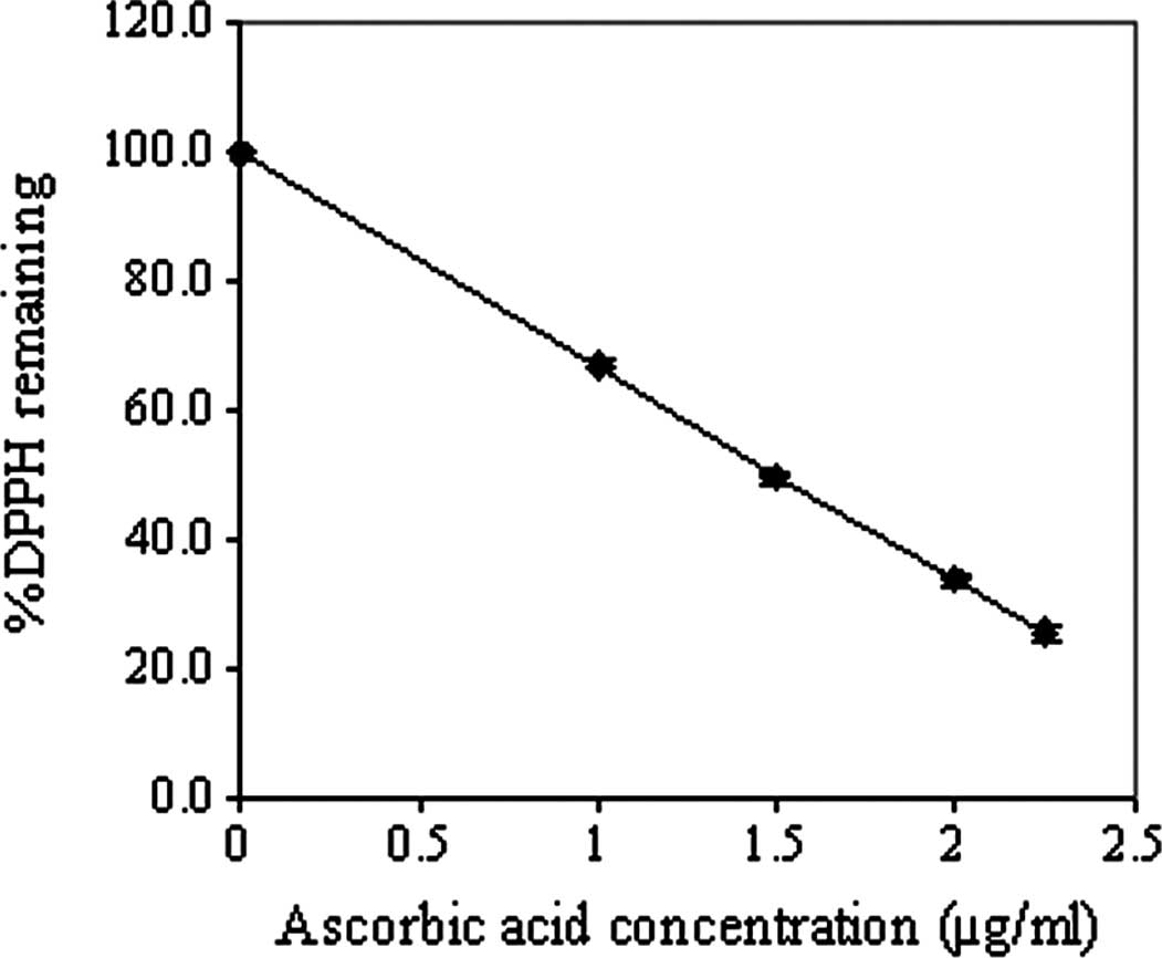
The mean effective concentrations (EC50)
of ascorbic acid and AR1-4, as shown in Fig. 3, were found to be 1.5 and 600
μg/ml, respectively. Recently, Potduang et al (27) showed that the EC50 of a
crude ethanol extract of Asparagus racemosus dry powdered
root was 381.91 μg/ml, which was relatively lower than that of the
aqueous fraction AR1-4 from successive extraction in this study.
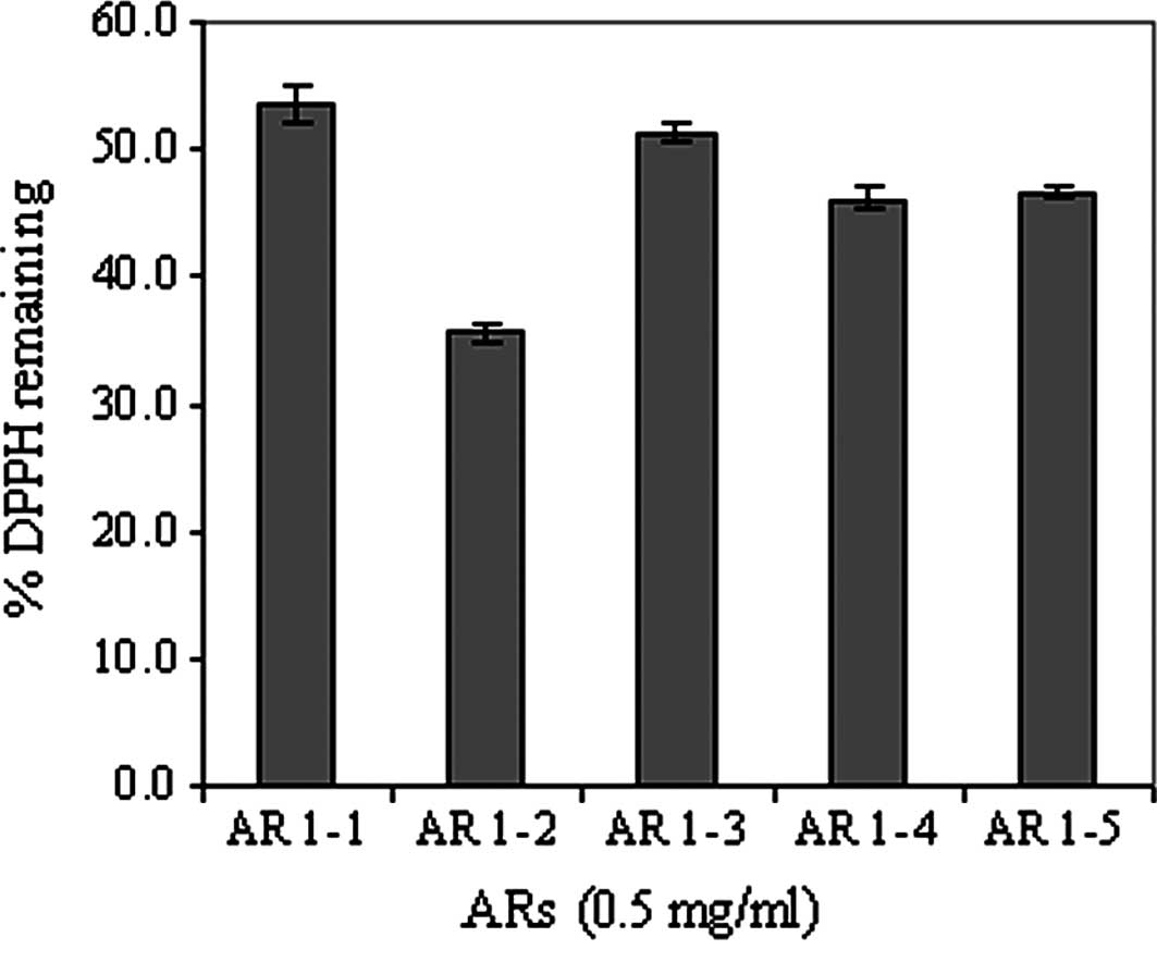
The antioxidant activities in terms of the % DPPH remaining of the
various fractions of Asparagus racemosus extracts at a fixed
concentration of 500 μg/ml are compared in Fig. 4; AR1-2 (95% ethanol fraction)
significantly exhibited the highest antioxidant activity compared
to the other fractions. These results were consistent with those
reported above. In addition, the antioxidant activities of
fractions other than AR1-2 were almost in the same order of
magnitude.
Although Asparagus racemosus extracts exhibit
far less antioxidant activity than ascorbic acid when tested in
vitro with the DPPH method, numerous studies have reported
their substantial antioxidant effect when tested in vivo
(2,3,6,26).
According to these studies, antioxidant enzymes (superoxide
dismutase, catalase) and ascorbic acid were increased by the
effects of Asparagus racemosus aqueous and methanolic
extracts, although the underlying mechanism has not yet been
elucidated. To date, there are no reports indicating that saponins
of Asparagus racemosus extracts are the active compound
responsible for this antioxidant effect.
Antiapoptotic effect in H460 cells
The AR1-4 aqueous fraction was selected for the
preliminary test to assess the antioxidant effect in cells, since
its main constituents shown in the TLC fingerprint were similar to
those of fractions AR1-3 and AR1-5 in terms of both main bands and
intensity. Additionally, AR1-4 was more readily dissolved in water
compared to the other fractions, which was more appropriate for the
treatment of cells due to the non-toxicity of the water solvent.
The results of the Hoechst assay showed that treatment of the H460
cells with Lipofectamine (20 μg/ml) caused extensive apoptosis
compared to the control (Fig. 5).
Upon pre-treatment of the cells with AR1-4, Lipofectamine-induced
apoptosis was significantly decreased with an increasing
concentration of AR1-4.
Toxicity and apoptosis induced by cationic liposomes
as well as their mechanisms have been found to be clearly
associated with ROS generation, which could effectively be
inhibited by ROS scavengers or antioxidant enzymes, particularly
superoxide dismutase (11,16,28).
The protective effect of AR1-4 against Lipofectamine-induced
apoptosis in this study can probably be attributed to the reduction
in oxidative stress-induced toxicity imposed by its antioxidant
activity. However, it is interesting to note that the antioxidant
activity of AR1-4 with DPPH in this study was 400 times less than
that of ascorbic acid, while it effectively protected the cells
from apoptosis as compared to the untreated cells (Fig. 5). The results suggest that this
antiapoptotic effect may be caused by the indirect enhancement of
antioxidant enzymes, such as superoxide dismutase or catalase,
rather than a direct antioxidant effect against ROS in the cells.
This is supported by several studies showing that root extracts of
Asparagus racemosus are capable of markedly increasing and
protecting the inactivation of superoxide dismutase and catalase in
rats (2,3,6,7).
In conclusion, Asparagus racemosus extracts
appear to be composed of saponins, the contents of which are richer
in extracts from polar compared to non-polar solvents. Successive
extraction resulted in fractions having different antioxidant
activities. In addition, aqueous fraction AR1-4 was shown to
exhibit a significant protective effect against
Lipofectamine-induced apoptosis in human lung epithelial H460
cells.
Acknowledgements
Financial support provided by the Thai
Herbal Nano-Cosmeceuticals Coordinated Research Program, the
National Nanotechnology Center, and the National Science and
Technology Development Agency, Thailand, is gratefully
acknowledged.
References
|
1.
|
Bopana N and Saxena S: Asparagus
racemosus – ethnopharmacological evaluation and conservation
needs. J Ethnopharmacol. 110:1–15. 2007. View Article : Google Scholar
|
|
2.
|
Bhatnagar M, Sisodia SS and Bhatnagar R:
Antiulcer and antioxidant activity of Asparagus racemosus
Willd and Withania somnifera Dunal in rats. Ann NY Acad Sci.
1056:261–278. 2005.PubMed/NCBI
|
|
3.
|
Sairam K, Priyambada S, Aryya NC and Goel
RK: Gastroduodenal ulcer protective activity of Asparagus
racemosus: an experimental, biochemical and histological study.
J Ethnopharmacol. 86:1–10. 2003. View Article : Google Scholar : PubMed/NCBI
|
|
4.
|
Hayes PY, Jahidin AH, Lehmann R, Penman K,
Kitching W and Voss JJD: Steroidal saponins from the roots of
Asparagus racemosus. Phytochemistry. 69:796–804. 2008.
View Article : Google Scholar : PubMed/NCBI
|
|
5.
|
Wiboonpun N, Phuwapraisirisan P and
Tip-pyang S: Identification of antioxidant compound from
Asparagus racemosus. Phytother Res. 18:771–773. 2004.
View Article : Google Scholar
|
|
6.
|
Kamat JP, Boloor KK, Devasagayam TPA and
Venkatachalam SR: Antioxidant properties of Asparagus
racemosus against damage induced by γ-radiation in rat liver
mitochondria. J Ethnopharmacol. 71:425–435. 2000.
|
|
7.
|
Agrawal A, Sharma M, Rai SK, Singh B,
Tiwari M and Chandra R: The effect of the aqueous extract of the
roots of Asparagus racemosus on hepatocarcinogenesis
initiated by diethylnitrosamine. Phytother Res. 22:1175–1182.
2008.PubMed/NCBI
|
|
8.
|
Gao X, Kim KS and Liu D: Nonviral gene
delivery: what we know and what is next. AAPS J. 9:E92–E104. 2007.
View Article : Google Scholar : PubMed/NCBI
|
|
9.
|
Niidome T and Huang L: Gene therapy
progress and prospects: nonviral vectors. Gene Ther. 9:1647–1652.
2002. View Article : Google Scholar : PubMed/NCBI
|
|
10.
|
Strom G and Crommelin DJA: Liposomes:
quo vadis? PSTT. 1:19–31. 1998.
|
|
11.
|
Dokka S, Toledo D, Shi X, Castronova V and
Rojanasakul Y: Oxygen radical-mediated pulmonary toxicity induced
by some cationic liposomes. Pharm Res. 17:521–525. 2000. View Article : Google Scholar : PubMed/NCBI
|
|
12.
|
Dokka S, Toledo D, Shi X, Ye J and
Rojanasakul Y: High-efficiency gene transfection of macrophages by
lipoplexes. Int J Pharm. 2006:97–104. 2000. View Article : Google Scholar
|
|
13.
|
Wolf BB and Green DR: Suicidal tendencies:
apoptotic cell death by caspase family proteinases. J Biol Chem.
274:20049–20052. 1999. View Article : Google Scholar : PubMed/NCBI
|
|
14.
|
Lv H, Zhang S, Wang B, Cui S and Yan J:
Toxicity of cationic lipids and cationic polymers in gene delivery.
J Control Release. 114:100–109. 2006. View Article : Google Scholar : PubMed/NCBI
|
|
15.
|
Almofti MR, Harashima H, Shinohara Y,
Almofti A, Baba Y and Kiwada H: Cationic liposome-mediated gene
delivery: biophysical study and mechanism of internalization. Arch
Biochem Biophys. 410:246–253. 2003. View Article : Google Scholar : PubMed/NCBI
|
|
16.
|
Aramaki Y, Takano S and Tsuchiya S:
Induction of apoptosis in macrophages by cationic liposomes. FEBS
Lett. 460:472–476. 1999. View Article : Google Scholar : PubMed/NCBI
|
|
17.
|
Aramaki Y, Takano S, Arima H and Tsuchiya
S: Induction of apoptosis in WEHI 231 cells by cationic liposomes.
Pharm Res. 17:515–520. 2000. View Article : Google Scholar : PubMed/NCBI
|
|
18.
|
Dokka S, Toledo D, Shi X, Ye J and
Rojanasakul Y: High-efficiency gene transfection of macrophages by
lipoplexes. Int J Pharm. 206:97–104. 2000. View Article : Google Scholar : PubMed/NCBI
|
|
19.
|
Uematsu Y, Hirata K, Suzuki K, Iida K and
Kamata K: Investigation of spectrophotometrically determined
substances in Yucca extracts by GC/MS, TLC and on-column injection
GC. J Food Hyg Soc Japan. 45:141–145. 2004. View Article : Google Scholar : PubMed/NCBI
|
|
20.
|
Zhao L, Huang C, Shan Z, Xiang B and Mei
L: Fingerprint analysis of Psoralea corylifolia L. by HPLC
and LC-MS. J Chromatogr B Analyt Technol Biomed Life Sci.
B821:67–74. 2005.
|
|
21.
|
Makkar HPS, Siddhuraju P and Becker K:
Plant Secondary Metabolites. Serries Methods in Molecular Biology.
393. Humana Press; New Jersey: 2007
|
|
22.
|
Wojciak-Kosior M: Application of high
performance thin-layer chromatography to separation of oleanolic
acid, ursolic and belulinic acids. J Pre-Clin Clin Res. 1:176–178.
2007.
|
|
23.
|
Gautam M, Diwanay S, Gairola S, Shinde Y,
Patki P and Patwardhan B: Immunoadjuvant potential of Asparagus
racemosus aqueous extract in experimental system. J
Ethnopharmacol. 91:251–255. 2004.
|
|
24.
|
Xi M, Hai C, Tang H, Chen M, Fang K and
Liang X: Antioxidant and antiglycation properties of total saponins
extracted from traditional Chinese medicine used to treat diabetes
mellitus. Phytother Res. 22:228–237. 2008. View Article : Google Scholar : PubMed/NCBI
|
|
25.
|
Brand-Williams W, Cuvelier ME and Berset
C: Use of a free radical method to evaluate antioxidant activity.
Lebensm-Wiss U-Technol. 28:25–30. 1995. View Article : Google Scholar
|
|
26.
|
Visavadiya NP and Narasimhacharya AVRL:
Asparagus root regulates cholesterol metabolism and improves
antioxidant status in hypercholesteremic rats. Evid Based
Complement Altern Med. 6:219–226. 2009. View Article : Google Scholar : PubMed/NCBI
|
|
27.
|
Potduang B, Meeploy M, Giwanon R, Benmart
Y, Kaewduang M and Supatanakul W: Biological activities of
Asparagus racemosus. Afr J Tradit Complement Altern Med.
5:230–237. 2008.
|
|
28.
|
Kongkaneramit L, Sarisuta N, Azad N, Lu Y,
Iyer AKV, Wang L and Rojanasakul Y: Dependence of reactive oxygen
species and FLICE inhibitory protein on Lipofectamine-induced
apoptosis in human lung epithelial cells. J Pharmacol Exp Ther.
325:969–977. 2008. View Article : Google Scholar : PubMed/NCBI
|