Introduction
Intracerebral hemorrhage (ICH) refers to a
non-traumatic cerebral parenchymal hemorrhage with high mortality
and morbidity rates, which can have a considerable impact on human
health. Post-ICH secondary cerebral edema damages the blood-brain
barrier, triggers disorders of the sodium pumps in the brain,
causes cellular edema, increases intracranial pressure and
eventually leads to nerve cell necrosis (1,2). As a
result, post-ICH edema is the main cause of disease progression and
mortality. Thus, effective control of cerebral edema could
significantly reduce ICH-induced neurological damage.
Hyperbaric oxygen (HBO) therapy refers to the
exposure of the body to a high-pressure environment (>1 standard
atmospheric pressure), so that the patient breathes in HBO or
hyperbaric mixed oxygen (97% O2 + >3%
CO2), in order to achieve therapeutic effects against
various diseases (3). Numerous
animal experiments have confirmed that HBO preconditioning (HBO-PC)
can significantly reduce hypoxic-ischemic injuries in the brain,
spinal cord and myocardium (4,5);
however, few reports have described its application in ICH
treatment (6,7).
Aquaporin-4 (AQP-4) is a membrane protein that
mediates the transmembrane water transportation of various types of
cells. The protein is composed of four active subunits that form a
heterotetrameric structure, as confirmed by three-dimensional
technology (8). AQP-4 is mainly
distributed in astrocytes, and the cells that most intensively
express the protein lie on the glial limiting membrane, which is
formed by the subarachnoid astrocyte foot processes and the surface
of the perivascular astrocytes. AQP-4 can also be expressed in the
ependymal cells, choroid plexus and pia mater, as well as in the
paraventricular and supraoptic nucleus of the hypothalamus
(9). AQP-4 has been found to be the
only cell membrane transportation protein that is permeable to
water molecules and other small molecules. It comprises, therefore,
the structural basis of water transportation and regulation among
the cerebrospinal fluid, glial cells and blood vessels, has a close
association with the development of the blood-brain barrier and
plays a key role in the regulation of the cerebral water balance
(10). It remains unclear whether
HBO reduces cerebral edema by affecting the AQP-4 expression in the
brain tissues of patients with ICH.
The present study examined the peri-hematoma edema
and AQP-4 expression in experimental ICH rats following HBO-PC and
aimed to investigate the effects and mechanism of HBO-PC in the
treatment of ICH.
Materials and methods
Animals
Healthy adult male Sprague Dawley® rats (n=156),
weighing 350–380 g, were provided by the Experimental Animal Center
of Hebei Medical University (Shijiazhuang, China). Prior to the
experiment, the rats were cage-bred separately in the Experimental
Animal Center of the Second Hospital of Hebei Medical University
(Shijiazhuang, China) at a constant temperature of 20–25°C and with
a standard diet and drinking water available ad libitum. The
present study was carried out in strict accordance with the
recommendations of the Guide for the Care and Use of Laboratory
Animals of the National Institutes of Health. The animal use
protocol has been reviewed and approved by the Institutional Animal
Care and Use Committee of Hebei Medical University.
Animal grouping
Sixty-six of the rats were randomly divided into
three groups: The sham-surgery group (SHG; n=6); the control group
(A-ICH; n=30), in which the rats were injected with autologous
blood; and the experimental group (P-HBO; n=30), in which the rats
underwent a 5-day period of HBO-PC before being prepared as an ICH
model. The latter two groups were then randomly divided into five
subgroups, namely the postoperative 24-h, 48-h, 72-h, 5-day and
7-day subgroups, with 6 rats in each subgroup.
ICH model
The preparation of an autologous blood-injected ICH
model was performed as previously described (11). Following anesthesia with chloral
hydrate, the rats were fixed on a stereotactic frame (Stoelting
Co., Wood Dale, IL, USA); the bregma was exposed and a small hole
was then drilled 0.5 mm anterior to the bregma and 3 mm to the
right of the midline. A total of 50 µl arterial blood was
subsequently extracted with a micro-syringe and inserted into the
drilled hole. The needle depth was 5.8 mm (to approximate the
position of the caudate nucleus), and the injection lasted 10 min
and was followed by needle-standing for 5 min. Bone wax was used to
seal the pinhole and the skin was disinfected and sutured. The same
method was used for the SHG model preparation but an equal volume
of saline, instead of blood, was used.
HBO-PC
Prior to the preparation of the ICH model, a single
infant oxygen chamber (type YL0.5/1.2; Wuhan Second Ship Design
Institute, Wuhan, China) was used to perform HBO exposure on the
rats in the experimental group. The pressurization time was 15 min,
the pressure was regulated at 0.10 MPa and the oxygen concentration
was maintained at >90% for 60 min of oxygen aspiration. The
decompression time was 15 min and the cabin temperature was
maintained at ~24°C. HBO-PC was performed once a day for 5
consecutive days and then the ICH model was prepared using the
aforementioned method.
Assessment of neurological
function
Twenty-four hours after the successful preparation
of the animal models, the behavior of the rats of each group was
scored according to the improved Longa classification method
(12). The scores were as follows: 0
points, no symptoms of neurological deficit; 1 point, inability to
extend the contralateral forelimb; 2 points, tonic flexion of the
contralateral forelimb; 3 points, mild circling around the
contralateral side; 4 points, severe circling around the
contralateral side; 5 points, falling toward the contralateral
side. Scores >1 point indicated the successful preparation of
the model.
Determination of cerebral water
content
The rats in each group were sacrificed at the
appropriate time-points by spinal dislocation and the wet and dry
weight method was used to measure the water content in the
peri-hemorrhagic cerebral tissues. Following the removal of the
frontal pole, a 2-mm-thick sample of brain tissue was extracted
from the lesion side for the purpose of determining the water
content. The brain tissue was placed into pre-weighed tin foil (A),
and the combined weight of the foil and brain tissue (B) was
obtained. The result of B-A was the wet weight of the brain tissue.
The brain tissue was then wrapped with the tin foil and placed into
an electric oven (WH-43; Tianjin Taisite Instrument Co., Ltd.,
Tianjin, China) and dried at 100°C for 24 h. The brain tissue and
foil (C) were then reweighed upon returning to room temperature.
The result of C-A was the dry weight. Finally, the data were
entered into the following equation: Brain water content = (wet
weight-dry weight)/wet weight × 100% [(B-C)/(B-A) × 100%].
Determination of AQP-4
Following anesthesia with chloral hydrate, the rat
brain tissues were obtained, fixed in 4% paraformaldehyde, rinsed
in 0.01 M phosphate-buffered saline, dehydrated with a conventional
ethanol gradient, hyalinized with xylene and embedded in paraffin.
A tissue slicer (Leica Microsystems, Wetzlar, Germany) was then
used to prepare 5-µm tissue sections. After enzyme closure with 3%
hydrogen peroxide (Sigma-Aldrich, St. Louis, MO, USA) and antigen
retrieval using citrate buffer (Sigma-Aldrich), the staining was
performed using the immunohistochemical Avidin Biotin Complex
method using a primary rabbit anti-rat polyclonal AQP-4 antibody
(cat. no. sc20812; dilution, 1:300; Santa Cruz Biotechnology, Inc.,
Dallas, TX, USA) and a horseradish peroxidase-labeled goat
anti-rabbit IgG secondary antibody (cat. no. ab67203; dilution,
1:200; Beijing Zhongshan Jinqiao Biotechnology Co., Ltd., Beijing,
China), according to the manufacturer's instructions. PBS was used
instead of primary antibody as a control. Three brain slices were
selected for each rat and 5 different randomly selected fields of
view were observed with a medical optical microscope (Olympus
Corp., Tokyo, Japan) at x400 magnification. The positive cells
(positive appearance of AQP-4 exhibited as brownish-yellow
cytoplasm) were counted in order to calculate the rate of positive
cells in the brain tissues.
Statistical analysis
All data were entered into the computer to generate
a database, and SPSS 13.0 statistical software (SPSS Inc., Chicago,
IL, USA) was used for the statistical analysis. Data are expressed
as the mean ± standard deviation. P<0.05 was considered to
indicate a statistically significant difference. Analysis of
variance was used for the analysis of the mean values of the
measurement data of multiple groups.
Results
Success rate of animal model
establishment
The establishment of the ICH model had a 60% success
rate, and the experimental animals typically died 48–72 h after the
lesioning. The rats that died were replenished in a timely manner
to ensure that the number of rats in each experimental group did
not change.
Scoring of neurological function
The mean 24-h postoperative neurological scores of
the SHG, A-ICH and P-HBO were 0, 4.12±0.41 and 3.91±0.37,
respectively. The A-ICH animals exhibited significant neurological
dysfunctions compared with the SHG animals (P<0.05); however, no
significant differences where observed in the degree of
neurological dysfunction between the HBO-PC and A-ICH
(P>0.05).
Cerebral water content
The water content in the brains of the A-ICH animals
was higher than that in the brains of the SHG animals at all
time-points (P<0.05). In the A-ICH, the cerebral edema was most
obvious 48 h postoperatively. Despite the fact that the edema
showed a tendency towards alleviation as the time passed, the water
content remained significantly higher in the A-ICH than that in the
SHG 7 days postoperatively (P<0.05). The cerebral edema in the
P-HBO was significantly more severe 24 and 48 h postoperatively
than that at 7 days postoperatively (P<0.05). After 48 h, the
edema gradually reduced and essentially returned to the level of
SHG on postoperative day 7. Compared with the A-ICH, the edema was
reduced in P-HBO animals, particularly at 48 and 72 h
postoperatively, when the difference between the groups was
significant (P<0.05) (Table
I).
 | Table I.Postoperative cerebral water content
among the different groups. |
Table I.
Postoperative cerebral water content
among the different groups.
| Group | 24 h (%) | 48 h (%) | 72 h (%) | 5 days (%) | 7 days (%) |
|---|
| SHG |
76.83±2.61 |
76.83±2.61 |
76.83±2.61 |
76.83±2.61 |
76.83±2.61 |
| A-ICH |
83.14±3.29a |
86.12±2.10a |
85.58±4.74a |
82.78±3.84a |
81.91±2.67a |
| P-HBO |
81.31±3.03a |
83.04±3.43a,b |
80.86±4.04a,b |
79.07±2.03a |
77.64±4.07 |
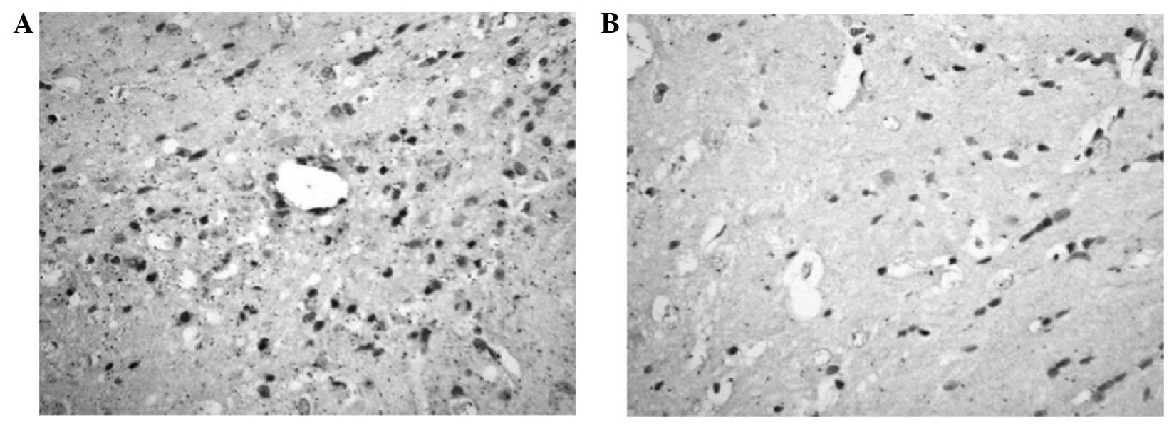
AQP-4 expression
Following the ICH, AQP-4 was expressed in the brain
tissues of all groups. The lowest AQP-4 expression was observed in
the SHG. The AQP-4 expression of the A-ICH started to increase 24 h
postoperatively, peaked 48 h postoperatively and began its gradual
decrease at 72 h. The expression in the A-ICH was close to but
still higher than that of the SHG on postoperative day 7
(P<0.05). The AQP-4 expression in the P-HBO at each time-point
was consistently lower than that in the A-ICH, with significant
differences between the two groups at 24 h, 48 h and 7 days
postoperatively (P<0.05) (Table
II and Fig. 1A and B).
| Table II.Aquaporin-4 expression among the
different groups. |
Table II.
Aquaporin-4 expression among the
different groups.
| Group | 24 h (%) | 48 h (%) | 72 h (%) | 5 days (%) | 7 days (%) |
|---|
| SHG |
39.45±5.67 |
39.45±5.67 |
39.45±5.67 |
39.45±5.67 |
39.45±5.67 |
| A-ICH |
45.06±3.36a |
52.76±5.16a |
49.07±2.80a |
47.59±4.82a |
44.68±3.77a |
| P-HBO |
44.27±4.30a,b |
48.72±3.96a,b |
45.30±4.01a |
43.29±4.03a |
40.05±2.11b |
Discussion
The results of the present study showed that the
success rate of ICH establishment was 60%, which was lower than the
71 and 79% reported previously (11,12). It
is generally believed that the factors affecting the success rate
of modeling are the following (13):
The amount of narcotic drugs and blood injected, the insertion
depth of the micro-syringe needle, the time and speed of liquid
injection and the living conditions of the animals. The mortality
rate in the present study was high and the animals typically died
48–72 h after the model preparation, at which time the cerebral
edema was at its most severe form. Since this experiment was
carried out in the hot summer months and there was a 30-min
distance from the animal laboratory to the HBO treatment site, it
may have been that, besides the aforementioned laboratory factors,
the environmental factors were the key reason for the high levels
of animal mortality in the present study.
With regard to the treatment of ICH, common clinical
strategies comprise medical and surgical approaches; however, HBO
therapy has recently been started to be assessed in trials as a
potential ICH treatment method. In a previous clinical study HBO
has exhibited significant effects in relieving post-ICH secondary
cerebral edema; however its specific mechanism of action remains
unclear (14). Cerebral edema is a
common pathological alteration that follows cerebral cell injuries
induced by trauma, hemorrhage, ischemia and cancer, and is a key
factor affecting the prognosis and lives of the patients. The
cerebral edema formation-related factors have been suggested to
include the following: Toxic effects of hemoglobin on the brain
tissues generated by the brain damage-induced red blood cell lysis
and rupture; neuronal apoptosis and proinflammatory reactions
induced by prothrombin activation; complement activation effects
caused by the inflammatory response; and water balance disorders in
the brain tissues caused by damage to the blood-brain barrier
(15–17). In short, the formation of cerebral
edema is a complex process, which includes and is influenced by a
variety of factors. In 2000, Manley et al (18) used AQP-4 gene-knockout mice for the
water intoxication experiment, and the survival rate of
gene-knockout mice was found to be significantly higher than that
of the mice of the control group. The brain water content and the
angioedema in the capillary ultrastructural observation of these
animals were lower than those in the control group, which confirmed
the association between AQP-4 and cerebral edema.
AQP-4 is a functional protein, which was first
separated from the red blood cell Rh proteins by Agre in 1998
(19) and was confirmed to be a
membrane protein that could mediate extra- and intracellular water
transportation in 1992 (20). The
characteristic distribution of AQP-4 in brain tissues is that it is
most intensively expressed on the glial limiting membrane, formed
by the astrocyte foot processes and the surface of the perivascular
astrocytes (21). AQP-4 exhibits a
highly polar expression distribution on the glial foot process
membranes, and its density in the pia mater region has been shown
to be several times the density in the neuropil (22,23). The
distribution of AQP-4 on the brain tissue membrane has been found
to be consistent with the polarity distribution of K+
channels. The expression sites of AQP-4 in the choroid plexus
epithelial cells, periventricular ependymal cells and pia mater are
consistent with its reabsorption sites (24). The above distribution characteristics
of AQP in the brain could suggest that AQP-4 is the structural base
of water transportation between the cerebrospinal fluid and cells
and that it plays a role in maintaining the intracellular and
extracellular balance of water and K+ concentrations and
participating in the regulation of osmotic pressure. Thus, AQP-4 is
the key factor that affects the water and electrolyte balance in
the central nervous system; this finding provides a theoretical
basis for further studies on the association between AQP-4 brain
edema.
HBO therapy, an effective means of treating cerebral
edema, was previously reported to reduce secondary cerebral edema
caused by subarachnoid hemorrhages and traumatic brain injury
through the inhibition of AQP-4 expression; however, it remained
unclear whether HBO therapy could affect the AQP-4 expression
(25) in post-ICH cerebral tissues.
As a special type of HBO therapy, HBO-PC is usually used in the
study of the incidence of high altitude reactions. It has been
shown that HBO-PC does not only improve the body's capacity for
oxidation resistance and reduce the incidence of high-altitude
reactions, but it also plays a neuroprotective role in rats with
altitude-induced traumatic brain injuries and improves their
neurological functions (26). Other
studies have shown that HBO-PC can enhance the ischemic tolerance
of the spinal cord and promote functional nerve recovery following
spinal cord injury (27–29). HBO-PC has been increasingly used in
clinical surgeries as a novel method that could improve the success
rate of surgery. For example, the application of conventional
HBO-PC on surgical patients several days before their surgery could
effectively reduce the side effects of anesthesia and improve the
hypoxic tolerance of the heart, brain and other vital organs. It
has also been shown that HBO-PC can improve myocardial function
following coronary bypass surgery and reduce myocardial injury
(30). A clinical study confirmed
that HBO could significantly improve the early clinical symptoms of
allergic vasculitis, and HBO-PC could effectively prevent or reduce
its complications (31).
Regarding the mechanism underlying the protective
effect of HBO-PC in hypoxic-ischemic encephalopathy, the results of
previous experimental studies (32–36) on
an ICH animal model under a high-pressure oxygen environment
revealed that HBO-PC could reduce apoptosis in the early stage of
ICH and inhibit the apoptotic transformation of damaged brain cells
in the late stage of ischemia. Its brain-protective effect was
associated with the upregulation of the brain-derived neurotrophic
factor expression level, as well as with the inhibition of
mitogen-activated protein kinase p38 activities. Another study
suggested that HBO-PC could reduce post-cerebral hypoxic nerve
damage by upregulating the activities of antioxidant enzymes, such
as catalase, superoxide dismutase and cellular hypoxia-inducible
factor-1α, among others (34). This
could promote the generation of erythropoietin in the cerebral
cortex and hippocampus, change the permeability of the blood-brain
barrier, reduce cerebral edema and, thus, promote the recovery of
neurological function. In the aforementioned studies, the role of
HBO-PC in the treatment of hypoxic-ischemic encephalopathy was
further clarified; however, reports on the application of HBO-PC in
ICH are still rare. Qin et al (37,38)
applied HBO-PC to experimental ICH rats and found that the
activation of p44/42 mitogen-activated protein kinase in the brain
tissue was associated with the degree of cerebral edema. HBO-PC was
shown to be involved in the synthesis of heat shock proteins by
activating p70 S6 kinases, thereby inducing the protective effect
in post-ICH brain tissues (38).
This study introduced novel ideas for the effect of HBO in ICH.
The present experimental results showed that,
despite the reduction in the postoperative neurological dysfunction
of the P-HBO rats at 24 h after ICH, the difference between the
neurological dysfunction of the P-HBO and A-ICH rats was not
significant, indicating that HBO-PC could not alleviate the
symptoms at the onset of ICH; however, the cerebral edema and AQP-4
expression levels around the hemorrhagic focus in the P-HBO were
significantly lower than those in the A-ICH at various time-points,
suggesting that HBO-PC downregulated AQP-4 expression. This
downregulation reduced the cerebral edema, thus playing a
neuroprotective role and strengthening the resistance to ICH. The
present study provided evidence for the clinical application of
HBO-PC in the prevention of ICH-associated diseases; however,
large, randomized, controlled studies are required for the
confirmation of the treatment effects and mechanisms of HBO-PC
against ICH.
References
|
1
|
Mun-Bryce S, Kroh FO, White J and
Rosenberg GA: Brain lactate and pH dissociation in edema: 1H- and
31P-NMR in collagenase-induced hemorrhage in rats. Am J Physiol.
265:R697–R702. 1993.PubMed/NCBI
|
|
2
|
Wagner KR, Xi G, Hua Y, Kleinholz M, de
Courten-Myers GM and Myers RE: Early metabolic alterations in
edematous perihematomal brain regions following experimental
intracerebral hemorrhage. J Neurosurg. 88:1058–1065. 1998.
View Article : Google Scholar : PubMed/NCBI
|
|
3
|
Schäbitz WR, Schade H, Heiland S, et al:
Neuroprotection by hyperbaric oxygenation after experimental focal
cerebral ischemia monitored by MRI. Stroke. 35:1175–1179. 2004.
View Article : Google Scholar : PubMed/NCBI
|
|
4
|
Gu GJ, Li YP, Peng ZY, et al: Mechanism of
ischemic tolerance induced by hyperbaric oxygen preconditioning
involves upregulation of hypoxia-inducible factor-1alpha and
erythropoietin in rats. J Appl Physiol. (1985)104:1185–1191. 2008.
View Article : Google Scholar : PubMed/NCBI
|
|
5
|
Huang G, Xu J, Xu L, et al: Hyperbaric
oxygen preconditioning induces tolerance against oxidative injury
and oxygen-glucose deprivation by up-regulating heat shock protein
32 in rat spinal neurons. PLoS One. 9:e859672014. View Article : Google Scholar : PubMed/NCBI
|
|
6
|
Qin Z, Karabiyikoglu M, Hua Y, Silbergleit
R, He Y, Keep RF and Xi G: Hyperbaric oxygen-induced attenuation of
hemorrhagic transformation after experimental focal transient
cerebral ischemia. Stroke. 38:1362–1367. 2007. View Article : Google Scholar : PubMed/NCBI
|
|
7
|
Peng ZR, Yang AL and Yang QD: The effect
of hyperbaric oxygen on intracephalic angiogenesis in rats with
intracerebral hemorrhage. J Neurol Sci. 342:114–123. 2014.
View Article : Google Scholar : PubMed/NCBI
|
|
8
|
Sunami K, Takeda Y, Hashimoto M and
Hirakawa M: Hyperbaric oxygen reduces infarct volume in rats by
increasing oxygen supply to the ischemic periphery. Crit Care Med.
28:2831–2836. 2000. View Article : Google Scholar : PubMed/NCBI
|
|
9
|
Wada K, Miyazawa T, Nomura N, et al:
Mn-SOD and Bcl-2 expression after repeated hyperbaric oxygenation.
Acta Neurochir Suppl. 76:285–290. 2000.PubMed/NCBI
|
|
10
|
Kalns J, Lane J, Delgado A, et al:
Hyperbaric oxygen exposure temporarily reduces Mac-1 mediated
functions of human neutrophils. Immunol Lett. 83:125–131. 2002.
View Article : Google Scholar : PubMed/NCBI
|
|
11
|
Lin S, Yin Q, Zhong Q, et al: Heme
activates TLR4-mediated inflammatory injury via MyD88/TRIF
signaling pathway in intracerebral hemorrhage. J Neuroinflammation.
9:462012. View Article : Google Scholar : PubMed/NCBI
|
|
12
|
Longa EZ, Weinstein PR, Carlson S and
Cummins R: Reversible middle cerebral artery occlusion without
craniectomy in rats. Stroke. 20:84–91. 1989. View Article : Google Scholar : PubMed/NCBI
|
|
13
|
Del Bigio MR, Yan HJ, Buist R and Peeling
J: Experimental intracerebral hemorrhage in rats. Magnetic
resonance imaging and histopathological correlates. Stroke.
27:2312–2320. 1996. View Article : Google Scholar : PubMed/NCBI
|
|
14
|
Qin Z, Xi G, Keep RF, Silbergleit R, He Y
and Hua Y: Hyperbaric oxygen for experimental intracerebral
hemorrhage. Acta Neurochir Suppl. 105:113–117. 2008.PubMed/NCBI
|
|
15
|
Murakami K, Kondo T, Yang G, Chen SF,
Morita-Fujimura Y and Chan PH: Cold injury in mice: A model to
study mechanisms of brain edema and neuronal apoptosis. Prog
Neurobiol. 57:289–299. 1999. View Article : Google Scholar : PubMed/NCBI
|
|
16
|
Zhang C, Lee JY, Keep RF, Pandey A,
Chaudhary N, Hua Y and Xi G: Brain edema formation and complement
activation in a rat model of subarachnoid hemorrhage. Acta
Neurochir Suppl. 118:157–161. 2013.PubMed/NCBI
|
|
17
|
Zador Z, Bloch O, Yao X and Manley GT:
Aquaporins: Role in cerebral edema and brain water balance. Prog
Brain Res. 161:185–194. 2007.PubMed/NCBI
|
|
18
|
Manley GT, Fujimura M, Ma T, et al:
Aquaporin-4 deletion in mice reduces brain edema after acute water
intoxication and ischemic stroke. Nat Med. 6:159–163. 2000.
View Article : Google Scholar : PubMed/NCBI
|
|
19
|
Agre P, Bonhivers M and Borgnia MJ: The
aquaporins, blueprints for cellular plumbing systems. J Biol Chem.
273:14659–14662. 1998. View Article : Google Scholar : PubMed/NCBI
|
|
20
|
Preston GM, Carroll TP, Guggino WB and
Agre P: Appearance of water channels in Xenopus oocytes
expressing red cell CHIP28 protein. Science. 256:385–387. 1992.
View Article : Google Scholar : PubMed/NCBI
|
|
21
|
Neely JD, Christensen BM, Nielsen S and
Agre P: Heterotetrameric composition of aquaporion-4 water
channels. Biochemistry. 38:11156–11163. 1999. View Article : Google Scholar : PubMed/NCBI
|
|
22
|
Warth A, Mittelbronn M and Wolburg H:
Redistribution of the water channel protein aquaporin-4 and the
K+ channel protein Kir4.1 differs in low- and high-grade
human brain tumors. Acta Neuropathol. 109:418–426. 2005. View Article : Google Scholar : PubMed/NCBI
|
|
23
|
Verkman AS, Binder DK, Bloch O, Auguste K
and Papadopoulos MC: Three distinct roles of aquaporin-4 in brain
function revealed by knockout mice. Biochim Biophys Acta.
1758:1085–1093. 2006. View Article : Google Scholar : PubMed/NCBI
|
|
24
|
Venero JL, Vizuete ML, Machado A and Cano
J: Aquaporins in the central nervous system. Prog Neurobiol.
63:321–336. 2001. View Article : Google Scholar : PubMed/NCBI
|
|
25
|
Nida TY, Biros MH, Pheley AM, Bergman TA
and Rockswold GL: Effect of hypoxia or hyperbaric oxygen on
cerebral edema following moderate fluid percussion or cortical
impact injury in rats. J Neurotrauma. 12:77–85. 1995. View Article : Google Scholar : PubMed/NCBI
|
|
26
|
Thom SR: Hyperbaric oxygen: Its mechanisms
and efficacy. Plast Reconstr Surg. 127 (Suppl 1):131S–141S. 2011.
View Article : Google Scholar : PubMed/NCBI
|
|
27
|
Nie H, Xiong L, Lao N, Chen S, Xu N and
Zhu Z: Hyperbaric oxygen preconditioning induces tolerance against
spinal cord ischemia by upregulation of antioxidant enzymes in
rabbits. J Cereb Blood Flow Metab. 26:666–674. 2006. View Article : Google Scholar : PubMed/NCBI
|
|
28
|
Lu PG, Hu SL, Hu R, et al: Functional
recovery in rat spinal cord injury induced by hyperbaric oxygen
preconditioning. Neurol Res. 34:944–951. 2012. View Article : Google Scholar : PubMed/NCBI
|
|
29
|
Wang L, Li W, Kang Z, et al: Hyperbaric
oxygen preconditioning attenuates early apoptosis after spinal cord
ischemia in rats. J Neurotrauma. 26:55–66. 2009. View Article : Google Scholar : PubMed/NCBI
|
|
30
|
Yogaratnam JZ, Laden G, Guvendik L, Cowen
M, Cale A and Griffin S: Hyperbaric oxygen preconditioning improves
myocardial function, reduces length of intensive care stay and
limits complications post coronary artery bypass graft surgery.
Cardiovasc Revasc Med. 11:8–19. 2010. View Article : Google Scholar : PubMed/NCBI
|
|
31
|
Godman CA, Chheda KP, Hightower LE,
Perdrizet G, Shin DG and Giardina C: Hyperbaric oxygen induces a
cytoprotective and angiogenic response in human microvascular
endothelial cells. Cell Stress Chaperones. 15:431–442. 2010.
View Article : Google Scholar : PubMed/NCBI
|
|
32
|
Ostrowski RP, Graupner G, Titova E, et al:
The hyperbaric oxygen preconditioning-induced brain protection is
mediated by a reduction of early apoptosis after transient global
cerebral ischemia. Neurobiol Dis. 29:1–13. 2008. View Article : Google Scholar : PubMed/NCBI
|
|
33
|
Yamashita S, Hirata T, Mizukami Y, et al:
Repeated preconditioning with hyperbaric oxygen induces
neuroprotection against forebrain ischemia via suppression of p38
mitogen activated protein kinase. Brain Res. 1301:171–179. 2009.
View Article : Google Scholar : PubMed/NCBI
|
|
34
|
Li J, Liu W, Ding S, et al: Hyperbaric
oxygen preconditioning induces tolerance against brain
ischemia-reperfusion injury by upregulation of antioxidant enzymes
in rats. Brain Res. 1210:223–229. 2008. View Article : Google Scholar : PubMed/NCBI
|
|
35
|
Peng Z, Ren P, Kang Z, et al: Up-regulated
HIF-1alpha is involved in the hypoxic tolerance induced by
hyperbaric oxygen preconditioning. Brain Res. 1212:71–78. 2008.
View Article : Google Scholar : PubMed/NCBI
|
|
36
|
Yan W, Fang Z, Yang Q, et al: SirT1
mediates hyperbaric oxygen preconditioning-induced ischemic
tolerance in rat brain. J Cereb Blood Flow Metab. 33:396–406. 2013.
View Article : Google Scholar : PubMed/NCBI
|
|
37
|
Qin Z, Song S, Xi G, et al:
Preconditioning with hyperbaric oxygen attenuates brain edema after
experimental intracerebral hemorrhage. Neurosurg Focus. 22:E132007.
View Article : Google Scholar : PubMed/NCBI
|
|
38
|
Qin Z, Hua Y, Liu W, et al: Hyperbaric
oxygen preconditioning activates ribosomal protein S6 kinases and
reduces brain swelling after intracerebral hemorrhage. Acta
Neurochir Suppl. 102:317–320. 2008.PubMed/NCBI
|