Introduction
Lomatogonium rotatum Fries ex Nym., which is
referred to as ‘Habirigan-Digeda’ in Mongolian, is the whole plant
of a Gentian family herb with a height of 15–40 cm. L.
rotatum is contained within the Mongolian volumes of Chinese
Materia Medica. As an annual herb growing on hillsides, grasslands
and near ditches, L. rotatum is preferentially distributed
in the mountain areas of Inner Mongolia in China. L. rotatum
contains flavonoids including cynaroside and orientin (1), which previous studies have shown to be
useful in the treatment of dyspepsia, gall bladder swelling,
jaundice and inflammation of the liver and gallbladder (2,3).
Previous studies evaluating long-term treatment with L.
rotatum have demonstrated that it may lower blood lipid levels
and prevent obesity (1,4); however its exact mechanism of action
remains to be fully elucidated. Daily foodstuff contains some
natural fructose and in recent years foods with considerable
refined fructose content have become fairly popular. This is a
worrying trend as a high intake of foods rich in fructose is
associated with obesity and metabolic problems (5).
The lipogenic capacity exhibited by liver and
adipose tissue is controlled by fatty acid synthase (FAS), and it
has previously been demonstrated that FAS gene expression is vital
for the conversion of fructose into lipids (6). At present, individuals often suffer
from hyperlipidemia due to changes in the metabolism of lipids, and
hyperlipidemia contributes to arteriosclerosis and coronary heart
disease (CHD). In addition, it has been reported that high levels
of triglyceride (TG) may increase the risk of CHD (7).
Previous studies have determined the effects of
flavonoids extracted from L. rotatum, particularly
decussatin, which has been shown to inhibit the growth of HEP-G2
hepatoma cells, resulting in reduced blood glucose levels and a
decrease in depression, leukemia and tuberculosis (8,9).
However, studies regarding the hypolipidemic effects of the
following flavonoids: 1-hydroxy-3,5,8-trimethoxyxanthone, methyl
swertiamarin and 6,8-dihydroxy-1,2-dimethoxyxanthone are rare
(10–12).
Previous studies have demonstrated that L.
rotatum is able to decrease blood glucose; therefore, L.
rotatum may potentially be useful as a prophylactic treatment
for atherosclerosis (13). In
addition, following the long-term consumption of high-fructose
foods, adenosine monophosphate-activated protein kinase (AMPK) in
L. rotatum may continuously suppress hyperlipidemia,
hyperleptinemia and FAS (14).
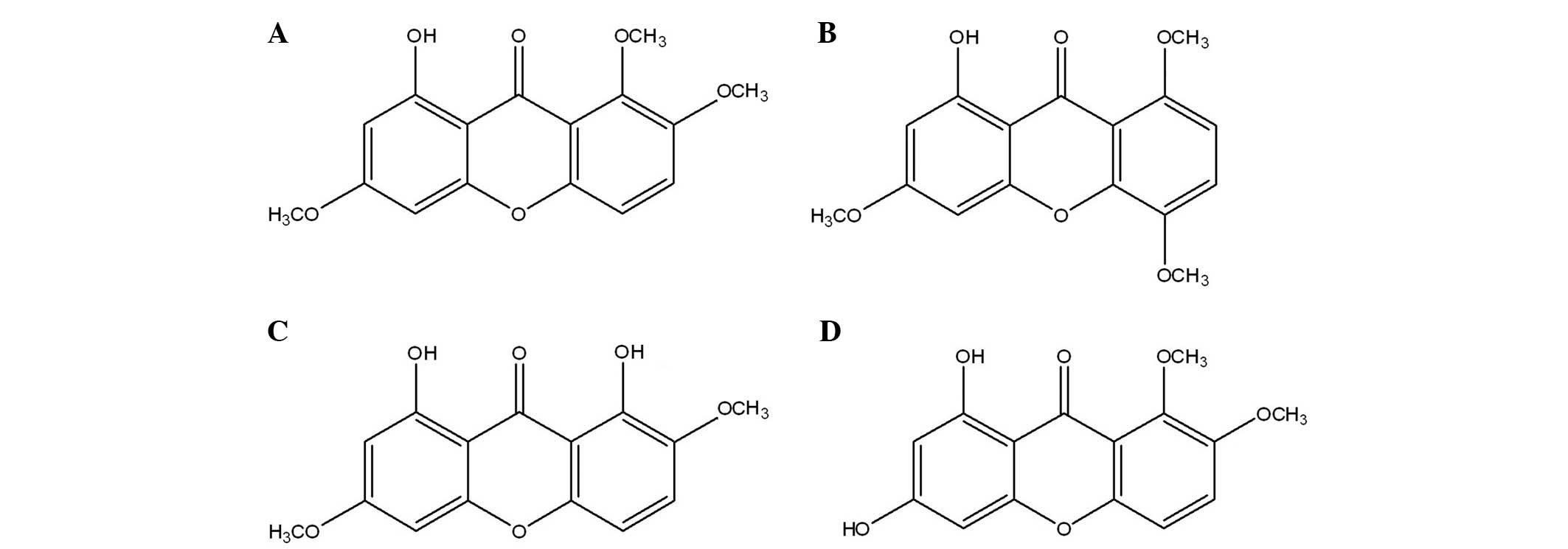
Therefore, in the present study the hypolipidemic and
obesity-inhibiting effects of four flavonoids isolated from L.
rotatum (Fig. 1) were
investigated in Wistar rats.
Materials and methods
Materials
Enzyme assay total cholesterol (TC; #4220),
triglyceride (TG; #4240), polyethylene sulfate precipitation method
low-density lipoprotein-cholesterol (LDL-C; #4210) and
phosphotungstic acid-magnesium precipitation method high-density
lipoprotein-cholesterol (HDL-C; #4200) kits were purchased from
Biosino Bio-Technology and Science, Inc. (Beijing, China).
Flavonoids A-D were yellow needle-like crystals with the following
molecular weights: A, 302 g/mol; B, 302 g/mol; C, 288 g/mol and D,
288 g/mol. The four flavonoids were extracted and synthesized at
the Inner Mongolia Medical University (Hohhot, China) (15), and uniformly mixed into the basic
feeds of the experimental rats. Anti-FAS (MHCD9528) was purchased
from Thermo Fisher Scientific, Inc. (Carlsbad, CA, USA), and
anti-phospho AMPK (AA393), anti-AMPK (AA393-1; Beyotime Institute
of Biotechnology, Shanghai, China), and horseradish
peroxidase-conjugated anti-mouse (NCAM-1/CD56; R&D Systems,
Inc., Minneapolis, MN, USA) and anti-rabbit (ab191866) antibodies
were purchased from Abcam (Cambridge, UK).
Animals and treatment
The present study complied with all protocols and
policies outlined by the Animal Care and Use Committee of Inner
Mongolia Medical University. The present study complied with all
protocols and policies outlined by the Animal Care and Use
Committee of Inner Mongolia Medical University (Huhhot, China). A
total of 60 six-week-old male Wistar rats (weight, 150–200 g), were
provided by the Experimental Animal Center of Inner Mongolia
Medical University, and were housed at the Inner Mongolia Medical
University in stainless steel wire-bottomed cages. The rats were
acclimated to the laboratory conditions (19–23°C, 60% humidity and
12-h light/dark cycle) for at least 1 week prior to the initiation
of the present study, during which the rats received ad
libitum access to water and Purina® rat food. Following one
week of acclimation, the rats were divided into the following
groups, and subsequently weighed every week for 12 weeks (16–18):
Control group, basic diet (Purina® rat food); Model group,
high-fructose diet (40% Purina® rat food + 60% fructose); Group A,
high-fructose diet + 20 mg/kg flavonoid A; Group B, high-fructose
diet + 20 mg/kg flavonoid B; Group C, high-fructose diet + 20 mg/kg
flavonoid C; Group D, high-fructose diet + 20 mg/kg flavonoid D.
Blood samples were collected from the femoral arteries of the rats,
and the serum was separated in order to detect the levels of TC,
TG, HDL-C and LDL-C. Hepatic, kidney and epididymal adipose tissue
samples were harvested, weighed and stored at −70°C.
Determination of TG
Serum levels of TG were analyzed using the
glycerol-3-phosphate oxidase/phenol and aminophenazone method
(Biosino Bio-Technology and Science Inc.) (19,20). TG
was enzymatically hydrolyzed to glycerol and free fatty acids using
specific lipases, which were subsequently oxidized into
H2O2 using glycerol kinase and glycerol
phosphatase. H2O2 was then converted into
colored quinonimine via a peroxidase-catalyzed reaction with
4-aminoantipyrine and phenol. The levels of TG in the serum samples
were determined at 520 nm and expressed as mg/100 ml (21).
Determination of TC
Serum levels of TC were measured using the
cholesterol oxidase/phenol and aminophenazone (CHOD-PAP) method
(Biosino Bio-Technology and Science Inc.). Using detergents,
cholesterol and its esters were released from lipoproteins prior to
hydrolyzation of the esters using cholesterol esterase, which were
subsequently enzymatically oxidized into
H2O2. H2O2 was then
converted into colored quinonimine via a peroxidase-catalyzed
reaction with 4-aminoantipyrine and phenol. The levels of TC in the
serum samples were determined at 520 nm and expressed as mg/100 ml
(21).
Determination of HDL-C
LDL was precipitated by phosphotungstic acid and
magnesium ions, which were subsequently removed by centrifugation
in order to suspend the HDL in the supernatant. HDL-C was then
measured using the CHOD-PAP method (Biosino Bio-Technology And
Science Inc.). The levels of HDL-C in the serum samples were
expressed as mg/100 ml.
Determination of LDL-C
LDL was precipitated by heparin at its isoelectric
point (pH 5.12). HDL remained in the supernatant following
centrifugation and the levels of LDL-C were subsequently determined
by enzymatic methods: LDL-C level = TC level - level of cholesterol
in the supernatant. The content of LDL-C in the serum samples was
expressed as mg/100 ml.
Determination of blood glucose
Glucose levels were measured following 12 weeks of
feeding. The tails of the rats were stored in a water bath at 45°C
prior to the collection of blood samples from 1 mm of the tail end.
Glucose detection was conducted according to the ortho-tolidine
method (22).
Determination of alanine
aminotransferase (ALT), aspartate aminotransferase (AST),
non-esterified fatty acid (NEFA), insulin and leptin levels
Serum ALT, AST and NEFA levels were determined
according to the manufacturer's instructions (Biosino
Bio-Technology And Science Inc.). The insulin concentrations of the
serum samples were determined using a Rat Insulin ELISA kit
(Mercodia, Uppsala, Sweden), and the tests were 100% cross-reactive
with human and rat insulin. During incubation, insulin in the
sample reacted with peroxidase-conjugated anti-insulin and bound to
the microtitration well. The unbound enzyme-labeled antibodies were
subsequently removed by a facile washing step, and the bound
conjugate was detected following the addition of
3,3′,5,5′-tetra-methylbenzidine. Acids were added to terminate the
reaction and the absorbance was measured at 450 nm. Serum leptin
levels were determined using a Rat Leptin TiterZyme Enzyme
Immunometric Assay kit (Applied Biosystems; Thermo Fisher
Scientific, Inc., Foster City, CA, USA), under normal conditions.
Leptin was immobilized by a polyclonal antibody on a microtiter
plate and, after a short incubation, the excessive sample was
rinsed, and leptin polyclonal antibody labeled with horseradish
peroxidase was added. Subsequently, the excessive antibody was
rinsed and the substrate was added prior to incubation for 30 min.
The absorbance of the resultant colored solutions was detected at
450 nm, which was directly proportional to the concentrations of
leptin in the respective samples (23).
Determination of total lipids, TG and TC in the
liver. To detect the levels of hepatic total lipids, TG and TC, the
rat livers were homogenized using a mixed chloroform/methanol/water
(8:4:3) solution (LabGEN 7 Homogenizer; Cole-Parmer, Vernon Hills,
IL, USA). The resultant mixture was shaken at 37°C for 1 h and
centrifuged at 2,000 × g for 10 min. Subsequently, the bottom layer
was collected and resuspended in order to analyze the hepatic
lipids. The levels of TG, TC and total lipids were measured
according to the enzymatic protocol outlined by Biosino
Bio-Technology And Science Inc. (24).
Reverse transcription polymerase chain
reaction (RT-PCR) analysis
Total RNA was isolated from liver tissue samples
using ISOGEN® reagent (Nippon Gene Co., Ltd., Toyama, Japan).
Briefly, cDNA was prepared from 5 µg total RNA, oligo (dT)18 primer
and Moloney murine leukemia virus reverse transcriptase (Promega
Biotech Co., Ltd., Beijing, China) by incubation at 40°C for 90
min. The total PCR volume was 50 µl containing dNTPs, 1 µl reaction
buffer, 1 µM primers (FAS, forward 5′-CCACTAGAAGCGTCTGCTGATCTG-3′;
reverse, 5′-TGCTATGTCCTACATATCGAGGACGC-3′), 2 µl reverse
transcriptase and 50 Uml-1 Taq DNA polymerase. A total of 5
µl PCR products were subsequently separated by 2% agarose gel
electrophoresis and were visualized via staining with ethidium
bromide.
Western blotting
Each liver sample was homogenized in lysis buffer
containing 50 mM Tris-HCl buffer, 50 mM NaF, 5 mM sodium
pyrophosphate, 0.25 M sucrose, 1 mM ethylenediaminetetraacetic
acid, 1 mM ethylenediaminetetraacetic acid, 1 mM dithiothreitol,
0.1 mM phenylmethylsulfonyl fluoride and 0.5% sodium dodecyl
sulfate (pH 7.5). The tissue lysates were then centrifuged in order
to remove insoluble substances and the respective protein contents
were measured using a Bio-Rad Protein Assay kit (Bio-Rad
Laboratories, Inc., Hercules, CA, USA). The proteins were
subsequently separated by 10% SDS-PAGE (Bio-Rad Laboratories, Inc.)
and electro-transferred onto a polyvinylidene difluoride membrane,
which was then pre-incubated overnight at 4°C in phosphate-buffered
saline containing 0.01% Tween-20, 1% bovine serum albumin (Nanjing
SenBeiJia Biological Technology Co., Ltd., Nanjing, China) and 0.2%
NaN3. Following this, the proteins were successively
incubated with various primary antibodies and secondary
anti-rabbit/goat/mouse IgG conjugated to horseradish peroxidase.
The immunoreactive bands were visualized using enhanced
chemiluminescent reagents.
Statistical analysis
All data were analyzed using SPSS 16.0 software
(SPSS Inc., Chicago, IL, USA) and expressed as the mean ± standard
deviation. The mean values were derived from three independent
experiments. Two groups were compared by Students t-test
(Mann-Whitney test was used in the case of heterogeneity of
variance), and multiple groups were compared by one-way analysis of
variance with Student-Newman-Keuls post-hoc analysis. P<0.05 was
considered to indicate a statistically significant difference.
Results
Effects of flavonoids extracted from
L. rotatum on body weight and diet intake
The metabolism of Wistar rats may be affected by 60%
fructose intake, which is associated with as glucose tolerance,
high serum TG levels and increased insulin resistance; therefore,
rats in the model group were fed foodstuff containing 60% fructose
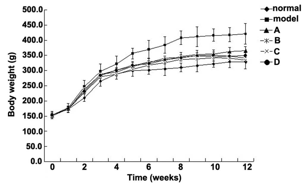
(25,26). The present study aimed to compare the
effects of various flavonoids from L. rotatum on 6-week-old
Wistar rats with high fructose-induced hyperlipidemia. Experimental
rats, weighing ~150 g each, were divided into six groups (n=10),
and fed various diets for 12 weeks. The rats were weighed weekly
and the observed weight changes are outlined in Fig. 2. The body weight of the rats in the
model group was significantly increased from the 3rd week, whereas
the weight of the rats in the control and flavonoid groups
increased steadily without significant differences. The flavonoid D
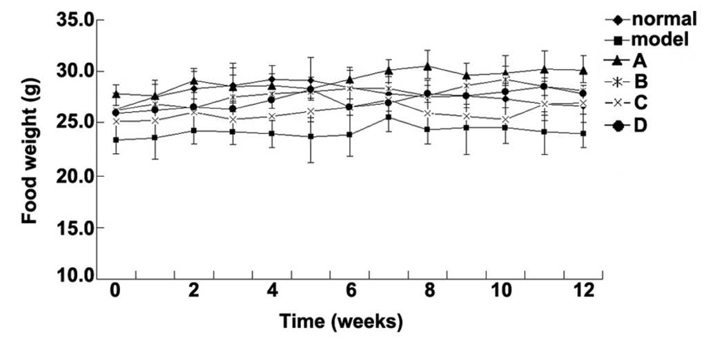
group exhibited the lowest mean weight. The food intake data for
all of the experimental groups are presented in Fig. 3. Food intake volume and body weight
were linearly correlated in all groups except the model group.
Effects of flavonoids extracted from
L. rotatum on liver, kidney and adipose tissue weight
The weight of the rat hepatic, renal and epididymal
adipose tissues are outlined in Table
I. L. rotatum flavonoids did not influence the liver
weight of the rats; whereas the epididymal adipose tissues of the
rats fed various flavonoids weighed less, as compared with those
only fed fructose. In addition, the weight of the kidney tissues
harvested from rats fed L. rotatum flavonoids were also
lower, as compared with the model group, with the exception of the
flavonoid B group.
 | Table I.Effects of flavonoids extracted from
Lomatogonium rotatum on the weight of liver, kidney and
adipose tissues. |
Table I.
Effects of flavonoids extracted from
Lomatogonium rotatum on the weight of liver, kidney and
adipose tissues.
|
| Liver | Epididymal adipose
tissue | Kidney |
|---|
|
|
|
|
|
|---|
| Group | Weight (g) | Relative
weight | Weight (g) | Relative
weight | Weight (g) | Relative
weight |
|---|
| Control | 15.8±2.2 | 3.6±0.3 |
7.2±0.3 |
1.5±0.2a | 3.3±0.2 | 0.77±0.2 |
| Model | 16.5±0.9 | 3.8±0.4 |
6.8±0.5 |
1.7±0.4a |
3.1±0.5 |
0.82±0.3a |
| A | 14.9±2.4 | 4.1±0.5 |
5.0±1.1 |
1.3±0.4b |
2.7±0.3 |
0.74±0.4b |
| B | 13.8±1.5 |
4.3±0.3b |
5.2±0.4 |
1.3±0.5b |
2.5±0.1b | 0.77±0.3 |
| C | 14.7±1.5 | 4.1±0.2 |
4.9±1.0* |
1.4±0.2b |
2.5±0.4b |
0.71±0.1b |
| D | 14.2±2.3 | 4.2±0.4 |
4.7±0.6* |
1.4±0.3b | 2.6±0.2 |
0.72±0.2b |
Effects of flavonoids extracted from
L. rotatum on lipid metabolism
It has been revealed that rats fed a high-fructose
diet may suffer from increased insulin resistance,
hyperinsulinemia, hypertriglyceridemia and hypertension. To verify
the effects of L. rotatum flavonoids on lipid metabolism,
serum and liver tissue samples from Wistar rats were analyzed. In
the present study, a high-fructose diet significantly elevated the
levels of serum insulin, leptin and NEFA (Table II). Following a 12-week
administration of L. rotatum flavonoids, the levels of
fasting blood glucose, feeding blood glucose and leptin in the
serum had decreased to normal. In addition, the rats in flavonoid
groups A and D exhibited reduced levels of serum cholesterol,
insulin, leptin and NEFA (Table
II). Rats in flavonoid B group demonstrated improved liver
function; however, function was not completely recovered. In
addition, in the flavonoid C group feeding blood glucose levels did
not return to normal.
| Table II.Relative clinical and biochemical
indices following a 12-week administration. |
Table II.
Relative clinical and biochemical
indices following a 12-week administration.
| Group | ALT
(U·l−1) | AST
(U·l−1) | NEFA
(mmol·l−1) | Fasting blood
glucose (mg·dl−1) | Feeding blood
glucose (mg·dl−1) | Insulin
(pmol·l−1) | Leptin
(ngm·l−1) |
|---|
| Control | 31.5±0.26 | 105.3±1.41 | 0.17±0.02 | 63.2±4.7 |
95.6±10.1 | 142.6±36.5 | 20.04±1.2 |
| Model |
26.3±0.34a |
95.4±0.97a |
0.33±0.01a |
73.5±3.6a |
122.6±20.3a |
188.7±42.2a |
25.4±2.2a |
| A |
23.7±0.65b |
99.8±0.45b |
0.23±0.05b |
59.6±3.5b |
92.6±16.8b |
109.6±33.1b |
19.6±2.5b |
| B | 25.2±0.42 |
92.4±0.87 | 0.26±0.03 |
60.1±4.5b |
95.8±17.6b |
121.4±35.6b |
22.3±0.8b |
| C |
24.1±0.24b |
99.3±0.64b |
0.24±0.01b |
64.5±1.2b | 101.3±15.4 |
133.4±45.6b |
20.4±1.5b |
| D |
22.8±0.76b |
91.5±0.75 |
0.24±0.05b |
63.3±6.0b |
97.8±11.9b |
136.8±26.1b |
21.8±3.3b |
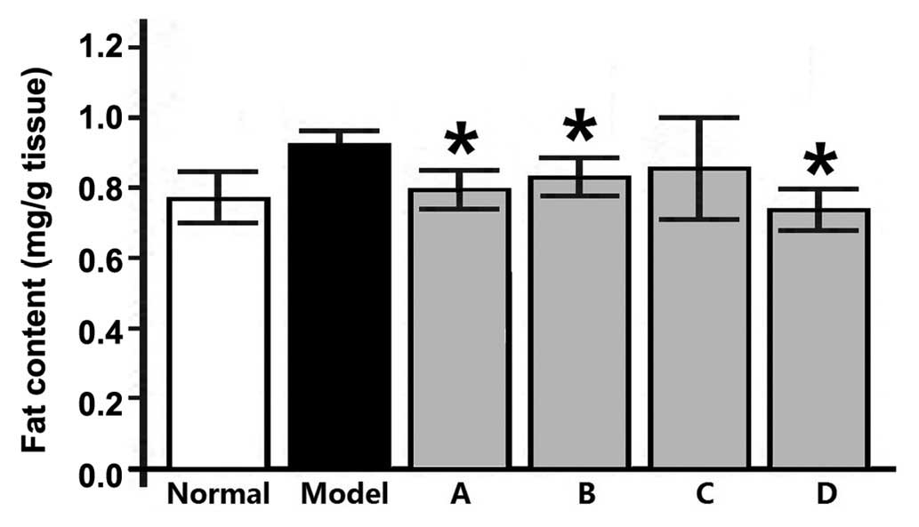
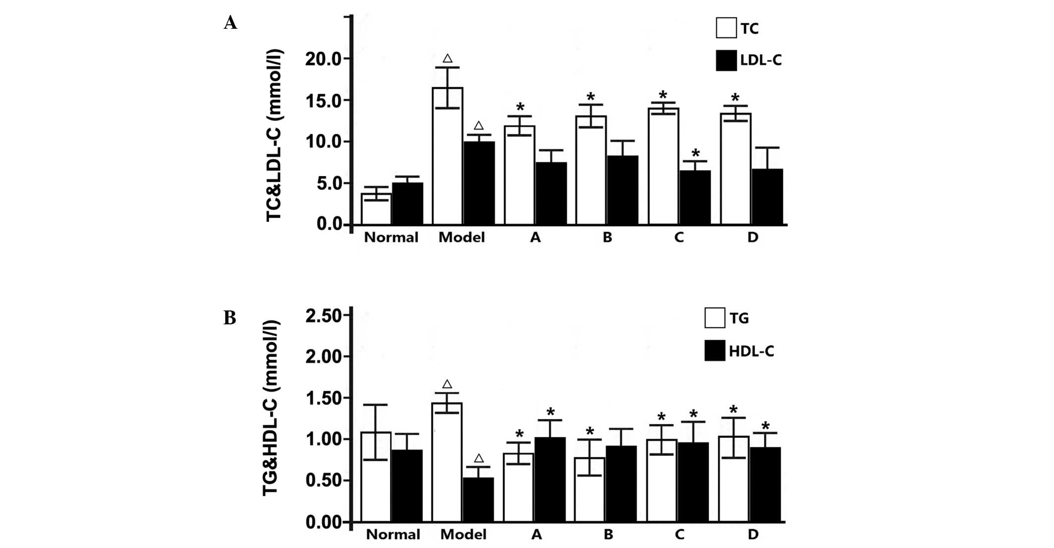
To determine the effects of L. rotatum
flavonoids on the homeostasis of liver fat, hepatic fat
accumulation was investigated in rats. The resultant liver fat
content (Fig. 4) and TG and
cholesterol levels (Fig. 5) indicate
that fructose-rich foods may significantly increase fat
accumulation in the liver. The fat content, and serum TG and
cholesterol levels were all significantly lowered after 12-week
administration of L. rotatum flavonoids, as compared with
rats fed only a high-fructose diet. Thus suggesting that L.
rotatum flavonoids successfully reduced hepatic fat content,
and blood TG and cholesterol levels, as well as increasing serum
HDL-C levels. As outlined in Fig.5,
flavonoid A demonstrated good therapeutic action in reducing total
cholesterol; however, the LDL-C levels were not significantly
altered. Furthermore, following flavonoid C administration the
resultant serum lipid parameters demonstrated good pharmacological
activity, with a significant reduction in LDL-C levels (P<0.05),
and flavonoids B and D functioned similarly.
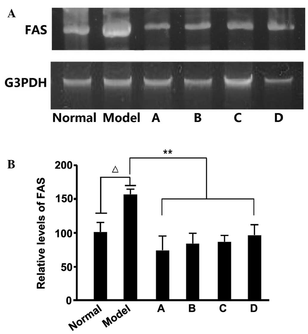
Effects of flavonoids extracted from
L. rotatum on the expression levels of FAS and pAMPK
Previous studies have demonstrated that
fructose-rich foods may elevate TG levels in the liver and blood,
and a high fructose intake may result in hypertriglyceridemia in
rats. Fructose-induced hypertriglyceridemia is considered to
originate from the excessive secretion of TG in the liver (27), with the enhanced expression of
several enzymes, including FAS, associated with the promotion of
hepatic TG secretion. After detecting the FAS levels in rat liver
samples, we hypothesize that the hypolipidemic effects of L.
rotatum flavonoids are realized by inhibiting the expression of
FAS. We also estimate that FAS activity is controlled by enzyme
content rather than changes in chemical bond or enzymatic
structure. RNA and protein samples were extracted from the liver of
rats fed various L. rotatum flavonoids, in order to conduct
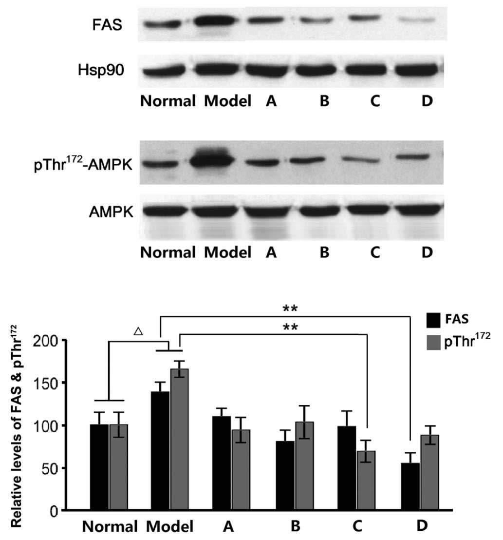
RT-PCR and western blotting. In the present study, the
administration of a high-fructose diet significantly increased FAS
mRNA and protein expression levels (Figs. 6 and 7) in rats, as compared with the other
groups. FAS mRNA expression levels were significantly decreased
(P<0.01) in the rats fed L. rotatum flavonoids, and the
four flavonoids appeared to function identically. In addition, the
protein levels of FAS were successfully controlled in all of the
flavonoid groups; however, the effects of flavonoid D were most
evident (P<0.01). Another important indicator is the
phosphorylation of AMPK in the liver of the rats. Western blot
analysis demonstrated that flavonoid C significantly decreased the
threonine-172 phosphorylation of AMPK in the liver lysates
(P<0.01; Fig. 7).
Discussion
Hypercholesterolemia and hypertriglyceridemia are
considered important factors associated with the incidence of
atherosclerosis and CHD. In the present study, rats fed a
high-fructose diet demonstrated hyperlipidemia, increased body
weight, adipose tissue and blood insulin levels, and decreased
glucose tolerance. A previous study demonstrated that Wistar rats
experienced a significant increase in lipid levels after being fed
a high-fructose diet for two weeks, and the effect became more
apparent after 10 weeks (28).
Flavonoid A, which is the most frequently assessed of the four
flavonoids, is capable of preventing atherosclerosis by decreasing
blood pressure. Flavonoids A-D are obtained from the same plant and
have similar structures; therefore, their common effects should be
considered (9,29). In the present study, the levels of
TC, LDL-C and TG were significantly increased, and the HDL-C levels
were significantly decreased in the model group, as compared with
the control group, suggesting that the model was successfully
established. In the flavonoid C group, the four serum lipid
parameters were improved following treatment, and LDL-C was the
only parameter to not demonstrate a significant change in the
flavonoid D group. Furthermore, TC, TG and HDL-C levels were
significantly improved in the flavonoid A group, and TC and TG were
significantly decreased in the flavonoid B group.
Following a 12-week administration of L.
rotatum flavonoids, serum levels of fasting blood glucose and
feeding blood glucose were significantly decreased. The rats in the
flavonoid A and D groups demonstrated reduced levels of serum
insulin; however, feeding blood glucose levels did not return to
normal in the flavonoid C group. These results suggested that
flavonoid C may produce insulin resistance in rats, which should be
the focus of more detailed studies in the future (30). Leptin is encoded by the Ob gene, and
the amount of adipose tissue and insulin secretion can increase the
levels of serum leptin. Therefore, the leptin levels of rats fed a
high-fructose diet should be higher, as compared with those in the
control group. The present study demonstrated that high-fructose
foods may lead to excessive insulin secretion, thereby stimulating
the production of leptin (29,31). All
L. rotatum flavonoids were able to downregulate leptin and,
owing to their potential hypolipidemic effects, further studies
investigating the flavonoids of various Mongolian medicines are
recommended in the future.
Epidemiological studies have demonstrated that
flavonoid intake is inversely related to CHD. FAS is involved in
energy metabolism, and is associated with various human diseases,
including obesity, cardiovascular disease and cancer. Previous
studies have reported that epigallocatechin 3-gallate (EGCG) in
flavonoids may inhibit FAS in the liver of broiler chickens
(32,33). In the present study, the majority of
the flavonoids successfully reduced the relative weight of
epididymal adipose tissue. Therefore, the authors of the present
study infer that flavonoids A, B and D have a similar effect to
EGCG in inhibiting the activity of FAS. A previous in-depth study
investigated the molecular dynamic mechanisms of L. rotatum
flavonoids on the expression of FAS (6), and such studies should be continued in
the future. All L. rotatum flavonoids may inhibit the
expression of FAS in the liver by stimulating AMPK activity in
hepatocyte cells via the liver kinase B1 pathway (34). Furthermore, the majority of
flavonoids decreased the threonine-172 phosphorylation of AMPK in
the liver lysate, which suggested that flavonoids stimulate AMPK
activity, thereby reducing fatty acid synthesis in the liver and
fat accumulation (35). Furthermore,
the activities of acetyl-CoA carboxylase and FAS may be inhibited;
however, the mechanisms of FAS inhibition and anti-obesity remain
unclear, and future studies are required.
In conclusion, the present study demonstrated that
certain flavonoids extracted from L. rotatum may mitigate
hypercholesterolemia and hypertriglyceridemia induced by the intake
of high-fructose foods, as well as improve the metabolism of lipids
and leptin in rats with insulin resistance. The four flavonoids
that were investigated decreased the levels of TG, total
cholesterol, leptin and insulin within a certain range, and
improved insulin resistance, liver function, and HDL-C levels. The
present study also demonstrated that flavonoid C exhibited the best
overall effects and inhibited FAS in the liver tissue of rats.
However, the metabolic mechanisms investigated in the present study
are not identical between humans and experimental rats, therefore
it remains unclear whether the flavonoids may also prevent fat
accumulation in the human liver. As such, the pharmacological
effects of flavonoids should be further investigated in humans.
These findings may contribute to the treatment of fatty liver and
obesity-associated diseases in humans.
Acknowledgements
The present study was financially supported by the
Nature Science Foundation of Inner Mongolia Autonomous Region
(grant no. 2013MS1224), the Scientific Project of the Affiliated
Hospital of Inner Mongolia Medical University (grant no.
NYFY2010YB006), the Inner Mongolia Medical University Youth
Innovation Fund (grant no. NY2010QN002), and the Key Scientific
Fund of the Affiliated Hospital of Inner Mongolia Medical
University (grant no. NYFYZD20130158).
References
|
1
|
Li YL, Suo YR, Liao ZX and Ding LS: The
glycosides from Lomatogonium rotatum. Nat Prod Res.
22:198–202. 2008. View Article : Google Scholar : PubMed/NCBI
|
|
2
|
Zhao N, Wang M and Li XE: Biological
macro-idea and criterion of osteopathic fracture immobilization in
China's traditional Mongolian medicine. J Tradit Chin Med.
32:114–118. 2012. View Article : Google Scholar : PubMed/NCBI
|
|
3
|
Burie: Preliminary study on classification
and nomenclative system of Mongolian traditional medicine. Zhong
Guo Zhong Yao Za Zhi. 36:1539–1541. 2011.(In Chinese).
|
|
4
|
Li ZH, Zhang AH, Yun XH, Zhang CH, Zhu SD,
Zou DZ, Bi YQ and Li MH: Ecology suitability study of Lomatogonium
rotatum in Inner Mongolia. Zhong Guo Zhong Yao Za Zhi. 40:778–784.
2015.(In Chinese).
|
|
5
|
Schultz A, Barbosa-da-Silva S, Aguila MB
and Mandarim-de-Lacerda CA: Differences and similarities in hepatic
lipogenesis, gluconeogenesis and oxidative imbalance in mice fed
diets rich in fructose or sucrose. Food Funct. 6:1684–1691. 2015.
View Article : Google Scholar : PubMed/NCBI
|
|
6
|
Huang HC and Lin JK: Pu-erh tea, green
tea, and black tea suppresses hyperlipidemia, hyperleptinemia and
fatty acid synthase through activating AMPK in rats fed a
high-fructose diet. Food Funct. 3:170–177. 2012. View Article : Google Scholar : PubMed/NCBI
|
|
7
|
Hada DS, Garg S, Ramteke GB and Ratre MS:
Effect of Nonsurgical Periodontal Treatment on Clinical and
Biochemical Risk Markers of Cardiovascular Disease: A Randomized
Trial. J Periodontol. 86:1–16. 2015. View Article : Google Scholar : PubMed/NCBI
|
|
8
|
Pant N, Jain DC and Bhakuni RS: Some
chemical constituents of Swertia chirata. Indian J Chem B.
41:1980–1986. 2002.
|
|
9
|
Jia J, Chen T, Wang P, Chen G, You J, Liu
Y and Li Y: Preparative separation of methylswertianin, swerchirin
and decussatin from the Tibetan medicinal plant Swertia
mussotii using high-speed counter-current chromatography.
Phytochem Anal. 23:332–336. 2012. View
Article : Google Scholar : PubMed/NCBI
|
|
10
|
Chakravarty AK, Mukhopadhyay S, Moitra SK
and Das B: Syringaresinol, a hepatoprotective agent and other
constituents from Swertia chirata. India J Chem B.
33:405–408. 1994.
|
|
11
|
Wang J, Liu Y, Cai Y, Zhang F, Xia G and
Xiang F: Cloning and functional analysis of geraniol
10-hydroxylase, a cytochrome P450 from Swertia mussotii
Franch. Biosci Biotechnol Biochem. 74:1583–1590. 2010. View Article : Google Scholar : PubMed/NCBI
|
|
12
|
Chiba K, Yamazaki M and Kikuchi M, Kakuda
R and Kikuchi M: New physiological function of secoiridoids:
Neuritogenic activity in PC12h cells. J Nat Med. 65:186–190. 2011.
View Article : Google Scholar : PubMed/NCBI
|
|
13
|
Lv Y, Zhang HT, Wang YF, Zhu H, Long P,
Wang ZW, Zhang N and Zhang CH: Preliminary comparative study of
swertiamarin and swertisin on three kinds of Digeda-species
Mongolian medicinal materials. Zhong Guo Zhong Yao Za Zhi.
40:804–806. 2015.(In Chinese).
|
|
14
|
Marques MB, Ribeiro-Oliveira A Jr,
Guimarães J, Nascimento GF, Anjos AP, Vilas-Boas WW, Santos RA,
Thomas JD, Igreja SM, Grossman AB, et al: Modifications in basal
and stress-induced hypothalamic AMP-activated protein kinase (AMPK)
activity in rats chronically treated with an angiotensin II
receptor blocker. Stress. 15:554–561. 2012. View Article : Google Scholar : PubMed/NCBI
|
|
15
|
Alaribe CS, Shode F, Coker HA, Ayoola G,
Sunday A, Singh N and Iwuanyanwu S: Antimicrobial activities of
hexane extract and decussatin from stembark extract of Ficus
congensis. Int J Mol Sci. 12:2750–2756. 2011. View Article : Google Scholar : PubMed/NCBI
|
|
16
|
Reddy SS, Ramatholisamma P, Karuna R and
Saralakumari D: Preventive effect of Tinospora cordifolia
against high-fructose diet-induced insulin resistance and oxidative
stress in male Wistar rats. Food Chem Toxicol. 47:2224–2229. 2009.
View Article : Google Scholar : PubMed/NCBI
|
|
17
|
Uluışık D and Keskin E: Effects of ginseng
and echinacea on cytokine mRNA expression in rats.
ScientificWorldJournal. 2012:9420252012. View Article : Google Scholar : PubMed/NCBI
|
|
18
|
Moura LP, Figueredo GA, Bertolini NO,
Ceccato M, Pereira JR, Sponton AC and de Mello MA: Dietary
restriction, caloric value and the accumulation of hepatic fat.
Lipids Health Dis. 11:2012. View Article : Google Scholar : PubMed/NCBI
|
|
19
|
Li Y, Yang H, Han WW, Liao MX and Lu YQ:
An enzyme sensor for phenolic compounds analysis. Guang Pu Xue Yu
Guang Pu Fen Xi. 30:571–574. 2010.(In Chinese). PubMed/NCBI
|
|
20
|
Araújo AN, Lima JL, Pinto PC and Saraiva
ML: Enzymatic determination of glucose in milk samples by
sequential injection analysis. Anal Sci. 25:687–692. 2009.
View Article : Google Scholar : PubMed/NCBI
|
|
21
|
Shahzad F, Tawwab S and Ahsan U: Lipid
profiles of non-diabetic healthy and ischaemic heart disease
patients. J Coll Physicians Surg Pak. 23:242–246. 2013.PubMed/NCBI
|
|
22
|
Leguit P Jr: Compartment syndrome of the
upper arm. Neth J Surg. 34:123–126. 1982.PubMed/NCBI
|
|
23
|
Wang X, Li S and Zhou Z: A rapid one-step
method of EIA for detection of circulating antigen of
Schistosoma japonicum. Chin Med J (Engl). 112:124–128.
1999.PubMed/NCBI
|
|
24
|
Diraison F, Pachiaudi C and Beylot M: In
vivo measurement of plasma cholesterol and fatty acid synthesis
with deuterated water: Determination of the average number of
deuterium atoms incorporated. Metabolism. 45:817–821. 1996.
View Article : Google Scholar : PubMed/NCBI
|
|
25
|
Reddy SS, Ramatholisamma P, Ramesh B,
Baskar R and Saralakumari D: Beneficiary effect of Tinospora
cordifolia against high-fructose diet induced abnormalities in
carbohydrate and lipid metabolism in Wistar rats. Horm Metab Res.
41:741–746. 2009. View Article : Google Scholar : PubMed/NCBI
|
|
26
|
Barbosa CR, Albuquerque EM, Faria EC,
Oliveira HC and Castilho LN: Opposite lipemic response of Wistar
rats and C57BL/6 mice to dietary glucose or fructose
supplementation. Braz J Med Biol Res. 40:323–331. 2007. View Article : Google Scholar : PubMed/NCBI
|
|
27
|
Shrestha S, Ehlers SJ, Lee JY, Fernandez
ML and Koo SI: Dietary green tea extract lowers plasma and hepatic
triglycerides and decreases the expression of sterol regulatory
element-binding protein-1c mRNA and its responsive genes in
fructose-fed, ovariectomized rats. J Nutr. 139:640–645. 2009.
View Article : Google Scholar : PubMed/NCBI
|
|
28
|
Collino M, Aragno M, Castiglia S, Miglio
G, Tomasinelli C, Boccuzzi G, Thiemermann C and Fantozzi R:
Pioglitazone improves lipid and insulin levels in overweight rats
on a high cholesterol and fructose diet by decreasing hepatic
inflammation. Br J Pharmacol. 160:1892–1902. 2010. View Article : Google Scholar : PubMed/NCBI
|
|
29
|
Chintalwar GJ and Chattopadhyay S:
Structural confirmation of decussatin, a Swertia decussata
xanthone. Nat Prod Res. 20:53–56. 2006. View Article : Google Scholar : PubMed/NCBI
|
|
30
|
Frangioudakis G, Gyte AC, Loxham SJ and
Poucher SM: The intravenous glucose tolerance test in cannulated
Wistar rats: A robust method for the in vivo assessment of
glucose-stimulated insulin secretion. J Pharmacol Toxicol Methods.
57:106–113. 2008. View Article : Google Scholar : PubMed/NCBI
|
|
31
|
Zhang X, Zhao Y, Zhang M, Pang X, Xu J,
Kang C, Li M, Zhang C, Zhang Z, Zhang Y, et al: Structural changes
of gut microbiota during berberine-mediated prevention of obesity
and insulin resistance in high-fat diet-fed rats. PLoS One.
7:e425292012. View Article : Google Scholar : PubMed/NCBI
|
|
32
|
Zhao YX, Liang WJ, Fan HJ, Ma QY, Tian WX,
Dai HF, Jiang HZ, Li N and Ma XF: Fatty acid synthase inhibitors
from the hulls of Nephelium lappaceum L. Carbohydr Res.
346:1302–1306. 2011. View Article : Google Scholar : PubMed/NCBI
|
|
33
|
Kim GS, Park HJ, Woo JH, Kim MK, Koh PO,
Min W, Ko YG, Kim CH, Won CK and Cho JH: Citrus aurantium
flavonoids inhibit adipogenesis through the Akt signaling pathway
in 3T3-L1 cells. BMC Complement Altern Med. 12:312012. View Article : Google Scholar : PubMed/NCBI
|
|
34
|
Hwang Pil Y, Kim Gyun H, Choi JH, Do
Truong M, Tran TP, Chun HK, Chung YC, Jeong TC and Jeong HG:
3-Caffeoyl, 4-dihydrocaffeoylquinic acid from Salicornia herbacea
attenuates high glucose-induced hepatic lipogenesis in human HepG2
cells through activation of the liver kinase B1 and silent
information regulator T1/AMPK-dependent pathway. Mol Nutr Food Res.
57:471–482. 2013. View Article : Google Scholar : PubMed/NCBI
|
|
35
|
Do MT, Kim HG, Choi JH, Khanal T, Park BH,
Tran TP, Hwang YP, Na M and Jeong HG: Phillyrin attenuates high
glucose-induced lipid accumulation in human HepG2 hepatocytes
through the activation of LKB1/AMP-activated protein
kinase-dependent signalling. Food Chem. 136:415–425. 2013.
View Article : Google Scholar : PubMed/NCBI
|