Introduction
Non-alcoholic fatty liver disease (NAFLD) has been
thought to be a benign disease, which is often associated with the
central obesity, insulin resistance (IR) and metabolic syndrome.
However, studies showed that (1,2) NAFLD is
a chronic disease, including benign lesions and significant liver
damage, such as the lobular inflammation, ballooning degeneration
of liver cells, liver fibrosis and cirrhosis (3). According to statistics, more than 30%
of adults in the United States and other Western countries suffer
from NAFLD (4), and the morbidity
rate of those NAFLD patients with diabetes mellitus (DM) and
transaminase is not included (5–7). NAFLD
often occurs in patients with obesity and type 2 DM (T2DM), and its
incidence rate is 57% in patients with obesity and 70% in patients
with T2DM (4,8). In addition to hepatic complications,
the risk of cardiac metabolic complications, such as T2DM and
cardiovascular disease (CVD), is also increased in NAFLD patients
(4,9). Coronary heart disease (CHD) is a common
complication. The incidence of NAFLD combined with CHD is
increasing gradually. It affects the digestive system and
cardiovascular system including the liver, kidney and heart. The
factors that cause CHD complicated NAFLD are various, influenced by
external environment and diet habits, and are related to genetic
factors of family (6). At present,
there are few reports on the treatment of complications, mainly in
the combination of diet control and cardiovascular drugs. The
prognosis is poor, with easy relapse, and the treatment effect is
not satisfactory (7). Endothelial
dysfunction is an independent predictor of prognosis of CHD,
including endothelium derived vasodilation, endothelium derived
vasoconstrictor function generation, inflammatory and immune
responses to reactive oxygen and nitrogen activation. In this
study, the effects of liver function, IR and inflammatory factors
on vascular endothelial dilation function and prognosis of CHD
patients complicated with NAFLD were investigated.
Patients and methods
General materials
A total of 80 patients with CHD treated in Jinan
Central Hospital (Jinan, China) from October 2016 to July 2017 were
randomly enrolled, including 42 males and 38 females aged 52.5±8.8
years on average. The diagnostic criteria of CHD: i) typical angina
pectoris or suspected angina pectoris and oral nitroglycerin
remission within 5 min; ii) hypertensive history with left
ventricular enlargement and aortic node calcification by X-ray,
left ventricular high voltage by ECG; iii) patients with old
myocardial infarction, myocardial infarction graphics by ECG,
except those with suspected myocardial infarction graphics caused
by other causes; iv) left axis deviation, left precordial lead with
ST-T change by ECG; and v) left axis deviation is not more than
30°, with A2>P2 or apical systolic murmur, and it cannot be
explained by other reasons. All patients were divided into the
NAFLD group (n=41), including 21 males and 20 females aged 49.6±8.3
years on average, and the simple CHD group (n=39), including 20
males and 19 females aged 50.2±9.0 years on average according to
whether they were complicated with NAFLD. There were no
statistically significant differences in the sex and age between
the two groups (P>0.05). The diagnosis of NAFLD met the
diagnostic criteria in the Guidelines for the Diagnosis and
Treatment of NAFLD (revised in 2010). The exclusion criteria i)
standard: alcohol and other clear liver injury caused by serious
liver disease, such as fatty liver, hepatitis, hepatocirrhosis,
liver cancer, and liver ascites; ii) diabetics; iii) other organs
with obvious inflammation; and iv) patients with mental disorders
who cannot cooperate with the study. The study was approved by the
Ethics Committee of Jinan Central Hospital and informed consents
were signed by the patients or guardians.
Research methods
Determination of laboratory
indexes
The height and weight of all subjects were measured,
the body mass index (BMI) was calculated, and the venous blood was
drawn to detect the fasting blood glucose (FBG), liver functions
[alanine aminotransferase (ALT), aspartate aminotransferase (AST)
and total bilirubin (TBIL)] and triglyceride (TG).
IR assessment
The fasting insulin (FINS) was detected using the
full-automatic biochemical detector; IR was evaluated using the
homeostasis model assessment-IR (HOMA-IR): HOMA-IR = (FINS ×
FBG)/22.5; the islet β-cell basic function was expressed as the
insulin secretion index (HOMA-β): HOMA-β = 20 × FINS/(FBG-3.5).
Determination of vascular endothelial
dilation function
LOGIQ-500 color ultrasound and 7.0 linear array
probe (GE Healthcare, Chicago, IL, USA) with the detection depth of
4 cm was used to detect the brachial artery diameter under resting
state after oral administration of nitroglycerin to reflect the
blood flow changes caused by the vascular reactive hyperemia
(vascular endothelium-dependent dilation function, FMV). The
ultrasonic scanning of brachial artery was performed to measure the
diameter of vessel in the three cases: i) in rest state, namely
resting for at least 10 min; ii) after the reactive hyperemia test
(vascular compression and expansion caused by inflation and
deflation of cuff of sphygmomanometer), namely after the cuff
compression to 250 mmHg or after discharge of cuff; and iii) after
the sublingual administration of anti-angina doses of nitroglycerin
(vascular endothelium-independent dilation function), namely after
the sublingual administration of 400 µg nitroglycerin after
resting. The ultrasonic scanning cycle is 15–60 sec before the cuff
discharge.
Determination of related inflammatory
factors
The tumor necrosis factor-α (TNF-α),
high-sensitivity C-reactive protein (hs-CRP) and nitric oxide (NO)
levels were detected via enzyme-linked immunosorbent assay (ELISA).
The serum NO content was detected using the Griess reagent staining
method; after standardized test, hs-CRP was detected using the
LX-20 full-automatic biochemical analyzer manufactured by Beckman
Coulter, Inc. (Brea, CA, USA); TNF-α was detected via ELISA using
the microplate reader (the kit was from R&D Systems, Inc.,
Minneapolis, MN, USA). The reagents were placed in the constant
temperature water bath until it reached the room temperature, and
the experiment was performed in strict accordance with the
instructions of the kit. After reagent preparation and sample
loading, the plate was washed with ZM X-988B full-automatic plate
washer 3 times; the optical density (OD) value at 450 nm was
detected using the an-thos2010 full-automatic microplate reader
(Bio-Rad Laboratories, Inc., Hercules, USA); the standard curve was
drawn using the computer software; OD value of the sample = OD
value measured - OD value of the blank control; TNF-α concentration
of the corresponding sample was checked in the OD value curve of
the standard sample.
Incidence of CVDs
The incidence of CVDs within 10 years was summarized
and the incidence rates were scored using the Framingham equation,
prospective cardiovascular Munster study (PROCAM) and National
Cholesterol Education Program Adult Treatment Expert Panel III
(NCEP-ATPIII) score. Framingham equation includes the sex, age, DM,
hypertension, smoking, total cholesterol and high-density
lipoprotein (HDL) cholesterol, and electrocardiogram of left
ventricular hypertrophy (Y/N); PROCAM is based on the sex, age,
blood pressure, smoking, total cholesterol and low-density
lipoprotein cholesterol, TG, family history of myocardial
infarction, DM and angina; ATPIII score is based on the sex, age,
basal blood pressure, DM, smoking, total cholesterol and HDL
cholesterol, and family history of early-onset CVDs.
Statistical analysis
Statistical Product and Service Solutions (SPSS)
v.19.0 software (SPSS, Inc., Chicago, IL, USA) was used for data
processing. Measurement data are presented as (mean ± standard
deviation); Chi-square test was used for the enumeration data, and
the t-test was used for the intergroup comparison. Pearsons
correlation analysis was performed for the correlation analysis.
P<0.05 was considered to indicate a statistically significant
difference.
Results
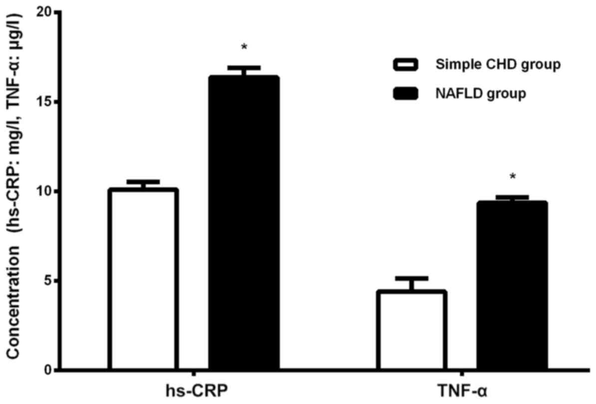
Comparison of serum TNF-α and hs-CRP
between the two groups
The level of hs-CRP and TNF-α in the NAFLD group was
significantly increased compared with that in the simple CHD group
(P<0.05; Fig. 1).
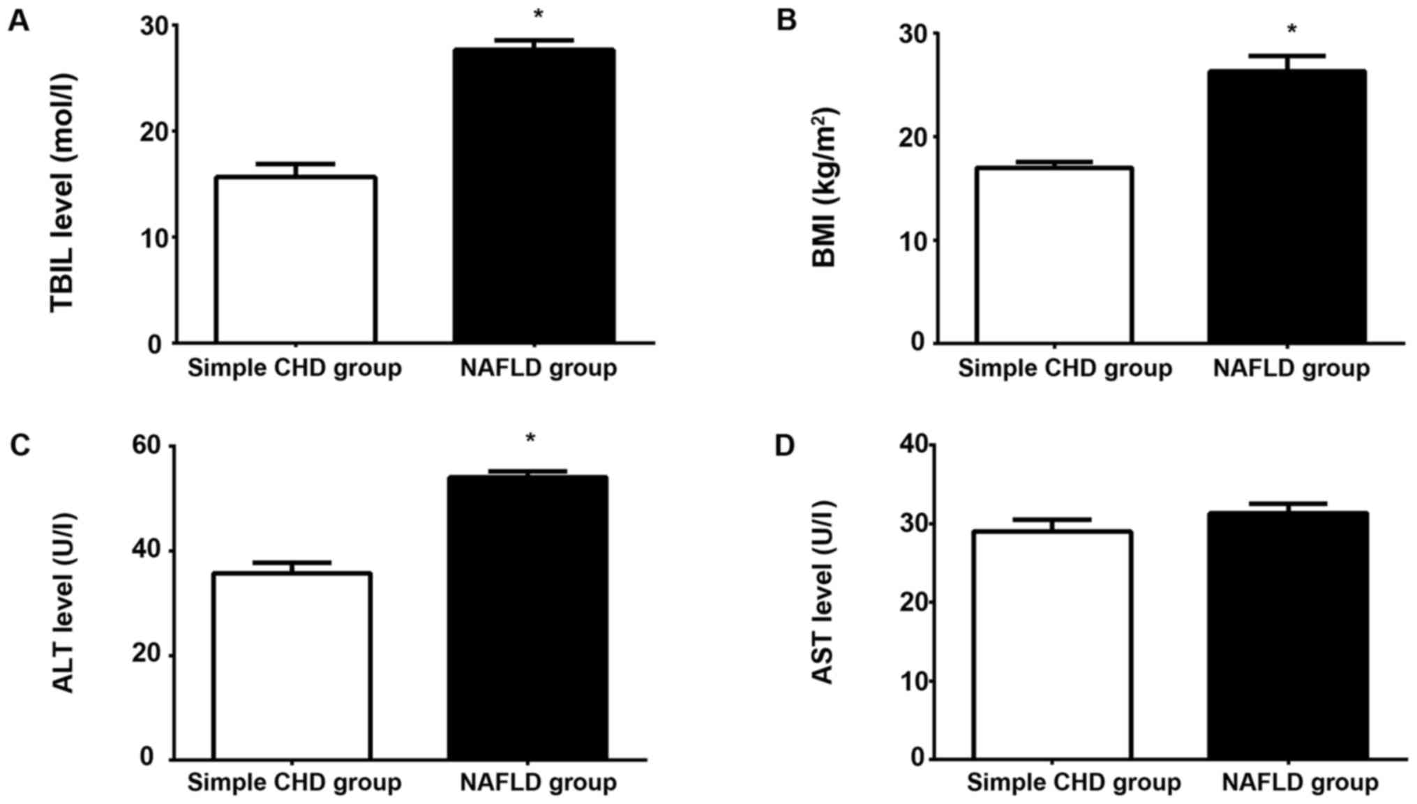
Comparison of general conditions and
liver function between the two groups
The results of comparisons of BMI and liver function
between the two groups showed that the serum TBIL and ALT levels
and BMI in the NAFLD group were significantly higher than those in
the simple CHD group, and the differences were statistically
significant (P<0.05; Fig. 2A-C).
There was no statistically significant difference in the serum AST
level between the two groups (P>0.05; Fig. 2A-D).
Correlation between liver function
(ALT) and vascular endothelial dilation function
The Pearsons correlation analysis revealed that
there was a linearly positive correlation between ALT and brachial
artery diameter in the NAFLD group (r=0.311, P<0.05); there was
a statistically significant difference in the correlation between
HOMA-IR and HOMA-β indexes and brachial artery diameter in the
groups (r=−0.128, r=0.219, P<0.05); there was also a
statistically significant difference in the correlation between
hs-CRP and brachial artery diameter in the groups (r=−0.312,
P<0.05), but TNF-α had no correlation with brachial artery
diameter, and the difference was not statistically significant
(r=3.286, P>0.05; Fig. 3A-E).
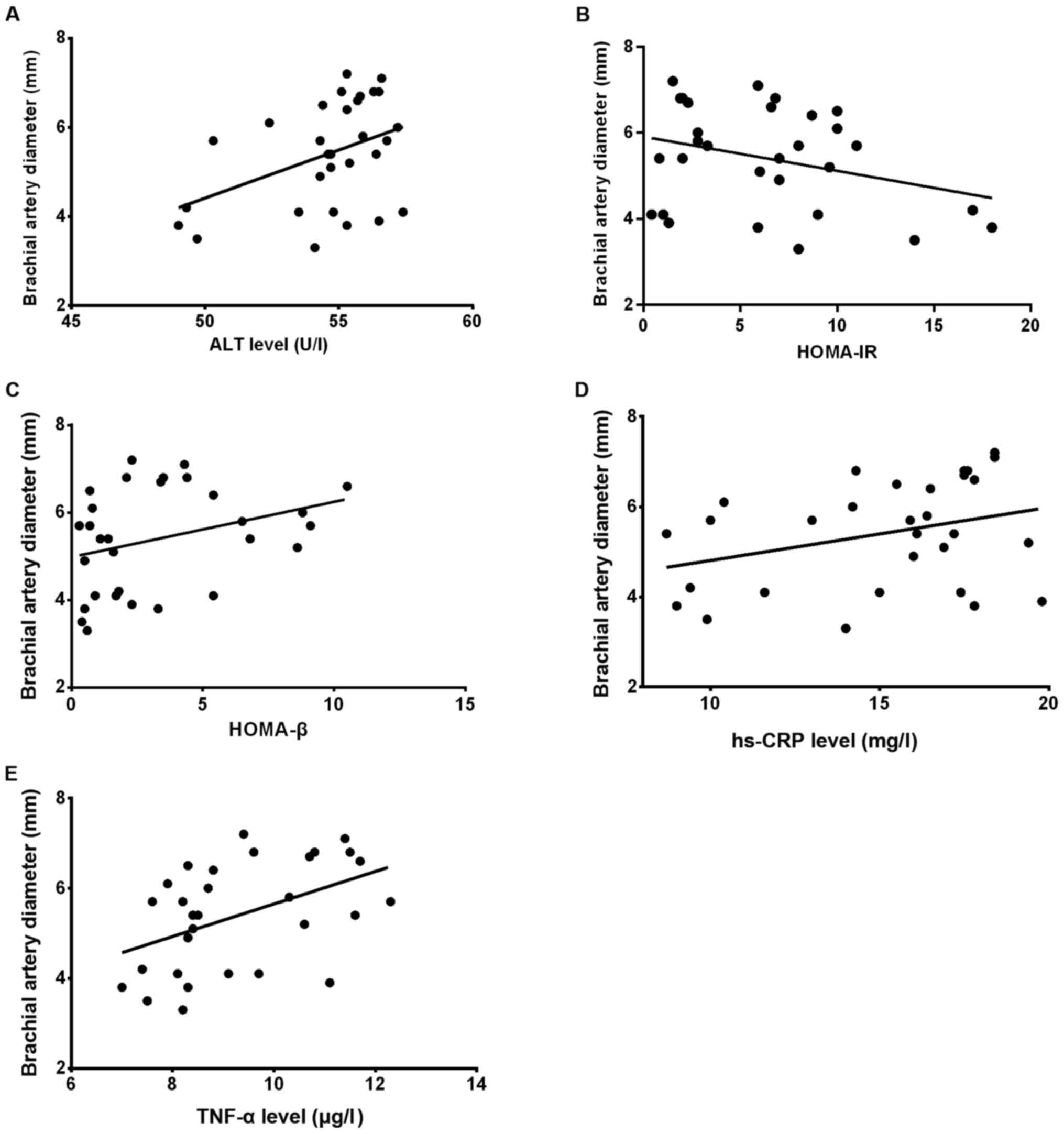 | Figure 3.(A) Correlation analysis between serum
ALT and brachial artery diameter in the NAFLD group showed that
there was a statistically significant difference (r=0.311,
P<0.05). (B and C) There was a statistically significant
difference in the correlation between HOMA-IR and HOMA-β indexes
and brachial artery diameter in both groups (r=−0.128, r=0.219,
P<0.05). (D and E) There was a statistically significant
difference in the correlation between hs-CRP and brachial artery
diameter in both groups (r=−0.312, P<0.05); TNF-α had no
correlation with brachial artery diameter, and the difference was
not statistically significant (r=3.286, P>0.05). ALT, alanine
aminotransferase; NAFLD, non-alcoholic fatty liver disease;
HOMA-IR, homeostasis model assessment-insulin resistance; hs-CRP,
high-sensitivity C-reactive protein; TNF-α, tumor necrosis
factor-α. |
Comparison of islet function and
vascular endothelial dilation function between the two groups
The HOMA-IR and HOMA-β indexes in the NAFLD group
were obviously decreased compared with those in the simple CHD
group, and the differences were statistically significant
(P<0.01), but FBG had no statistically significant difference
between the two groups (P>0.05; Table
I).
 | Table I.Comparison of islet function between
the two groups. |
Table I.
Comparison of islet function between
the two groups.
| Groups | FBG (mmol/l) | HOMA-IR | HOMA-β | FMV (mm) |
|---|
| NAFLD group | 7.9±3.8 | 4.0±3.2 | 26.2±18.3 | 3.36±0.62 |
| Simple CHD group | 8.3±3.4 | 2.6±3.5 | 19.8±10.1 | 4.27±0.54 |
| t | 1.16 | 3.86 | 4.21 | 3.48 |
| P-value | 0.162 | <0.001 | 0.001 | 0.062 |
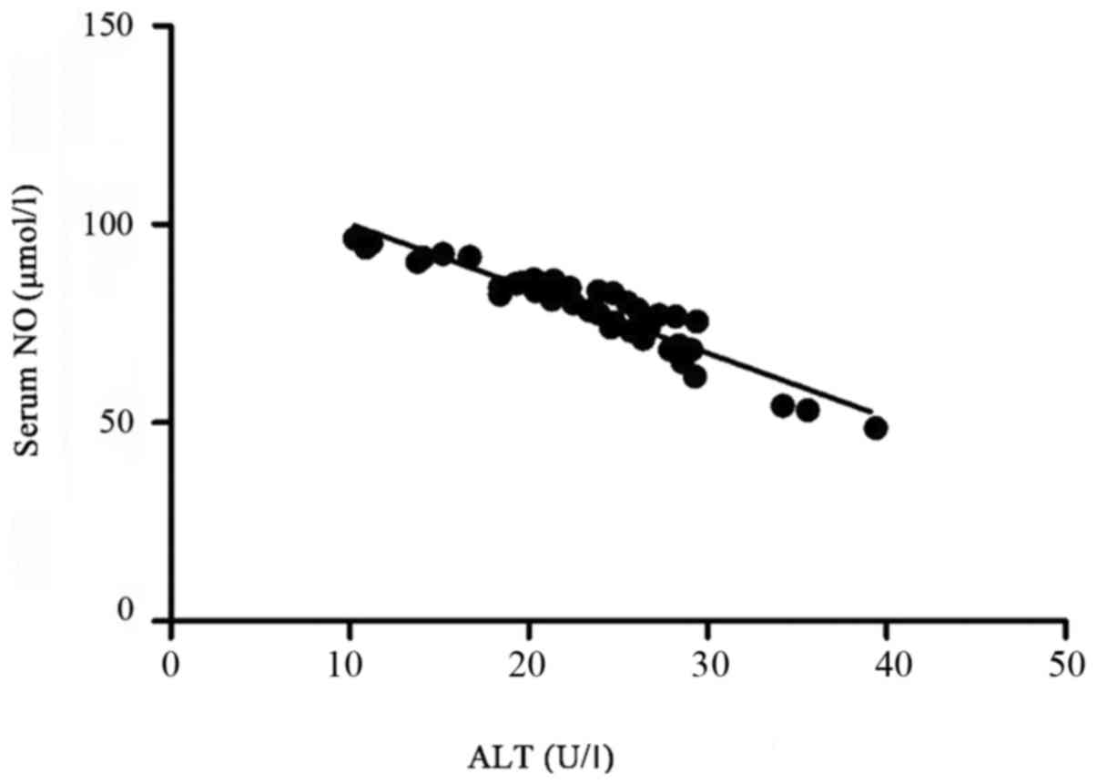
Correlation between serum NO and ALT
levels
The Pearsons correlation analysis of serum ALT and
NO levels revealed that there was a linearly negative correlation
between them (r=−0.325, P<0.05; Fig.
4).
Comparison of intima-media thickness
(IMT), number of carotid plaques and detection rate of carotid
plaques between the two groups
There were significant differences in the IMT,
number of carotid plaques and detection rate of carotid plaques
between the two groups (P<0.05; Table II).
| Table II.Comparisons of IMT, number of carotid
plaques and detection rate of carotid plaques between the two
groups. |
Table II.
Comparisons of IMT, number of carotid
plaques and detection rate of carotid plaques between the two
groups.
| Groups | n | IMT (mm) | Number of carotid
plaques | Detection rate of
carotid plaques (%) |
|---|
| NAFLD group | 41 | 1.21±0.05 | 21.3±1.52 | 72.2% |
| Simple CHD group | 39 | 0.29±0.03 | 1.30±1.56 | 55.4% |
| P-value |
| 0.041 | 0.032 | 0.028 |
Prognoses of the two groups
The incidence rate of CVDs within 10 years was
divided into very low (<1%), low (1–3%), moderate (4–6%), high
(7–10%) and very high (>10%). The risk of cardiovascular events
within 10 years in the NAFLD group was slightly increased compared
with that in the simple CHD group. According to the Framingham
equation, PROCAM and NCEP-ATPIII score, the differences were
statistically significant between the two groups (P<0.05;
Table III).
| Table III.Comparison of cardiovascular risk
between the two groups. |
Table III.
Comparison of cardiovascular risk
between the two groups.
|
| Framingham score | PROCAM score | ATPIII score |
|---|
|
|
|
|
|
|---|
| Risk of
cardiovascular events within 10 years | NAFLD group | Simple CHD group | NAFLD group | Simple CHD group | NAFLD group | Simple CHD group |
|---|
| Median, %(IQR) | 1.2 (5.1) | 3.7 (6.7) | 0.5 (1.1) | 0.8 (2.2) | <1.0 (2.5) | 1.5 (4.0) |
| <1% | 43 | 13 | 57 | 37 | 54 | 29 |
| 1–3% | 21 | 42 | 36 | 33 | 25 | 40 |
| 4–6% | 18 | 12 | 013 | 14 | 11 |
|
| 7–10% | 7 | 23 | 74 | 7 | 10 |
|
| >10% | 11 | 10 | 013 | 0 | 10 |
|
| P-value | 0.017 | 0.045 | 0.129 |
|
|
|
Discussion
NAFLD is a clinically pathological syndrome
characterized by parenchymal liver cells and fat accumulation in
the absence of excessive drinking. The main pathological feature of
CHD is coronary atherosclerosis, and it mainly occurs in the
middle-aged and elderly people, which may be caused by genetic and
environmental factors. Abnormal lipid metabolism is often
considered to be the main factors of increased blood lipids, and
hyperlipidemia is also considered as the common cause of CHD and
NAFLD. The clinical diagnosis rate of CHD complicated with NAFLD is
very high. Recently it is confirmed that NAFLD is very closely
related to the incidence of CHD (10,11). The
pathogenesis of NAFLD and CHD is mainly systemic chronic
inflammation, gradually enhanced oxidative stress and vascular
endothelial dysfunction (12). In
addition, several studies have shown that IR refers to the fact
that the tissues and organs can not produce normal physiological
and biological reactions to a certain amount of insulin, which is
the common metabolic defect of many metabolic diseases and
considered as an idiopathic feature of NAFLD (13,14),
even for subjects without obesity (15). Compared with simple CHD patients, CHD
patients complicated with NAFLD will produce IR, cause the release
of cytokines and a series of inflammatory reactions, promote the
release of serum APN, TNF-α, hs-CRP and other factors and increase
the mitochondrial oxidative burden of liver cells. A large number
of activated oxygen-free radicals bind to the corresponding factors
to form peroxides, manifested as coronary atherosclerosis. In
addition, CHD patients complicated with NAFLD suffer from vascular
endothelial dilation dysfunction, IR and abnormal liver function to
some extent, significantly slowing down the blood vessel velocity
combined with the common role of inflammatory factors, which is
manifested directly as platelet aggregation, thus affecting the
vascular endothelial dilation function and prognosis (16).
The results of comparisons of BMI and liver function
between the two groups showed that the serum TBIL and ALT levels
and BMI in the NAFLD group were significantly higher than those in
the simple CHD group, and the differences were statistically
significant; there was no statistically significant difference in
the serum AST level between the two groups. The Pearsons
correlation analysis revealed that there was a linearly positive
correlation between ALT and brachial artery diameter in the NAFLD
group (r=0.311, P<0.05). The HOMA-IR and HOMA-β indexes in the
NAFLD group were obviously increased compared with those in the
simple CHD group, and the differences were statistically
significant (P<0.01); but the FBG had no statistically
significant difference between the two groups (P>0.05).
Moreover, the base value of carotid diameter (Do) had no
statistically significant difference between the two groups
(P>0.05), and it was significantly decreased in the NAFLD group
compared with that in the simple CHD group. There were significant
differences in the IMT, number of carotid plaques and detection
rate of carotid plaques between the two groups (P<0.05). There
was a statistically significant difference in the correlation
between HOMA-IR and HOMA-β indexes and brachial artery diameter in
both groups (r=−0.128, r=0.219, P<0.05). There was also a
statistically significant difference in the correlation between
hs-CRP and brachial artery diameter in the groups (r=−0.312,
P<0.05), but TNF-α had no correlation with brachial artery
diameter, and the difference was not statistically significant
(r=3.286, P>0.05). Epidemiological data show that the mortality
of CVD is significantly increased in patients diagnosed with NAFLD
(17). Similarly, a recent study
showed that compared with the control group, the average IMT and
plaque detection rate are higher in NAFLD patients with carotid
atherosclerotic, which is consistent with this study. The risk of
cardiovascular events within 10 years in the NAFLD group was
slightly increased compared with that in the simple CHD group.
According to the Framingham equation, PROCAM and NCEP-ATPIII score,
the differences were statistically significant between the two
groups (P<0.05). The results of this study showed that the level
of hs-CRP and TNF-α in the NAFLD group was significantly increased
compared with that in the simple CHD group (P<0.05), suggesting
that TNF-α and hs-CRP are of great significance in the occurrence
and development of CHD complicated with NAFLD. Lomonaco et
al (18) found that the serum
TNF-α level is an independent predictor for the acute myocardial
infarction and death of CHD patients. Some scholars have proposed
(19) that TNF-α is related to the
formation of atherosclerosis in CHD patients, which mainly acts on
the endothelial cells to produce the platelet-derived growth factor
and promote the smooth muscle cell migration and proliferation,
ultimately leading to the endothelial cell dysfunction and intima
thickening, so it is involved in the occurrence and development of
atherosclerosis.
Pearsons correlation analysis of serum ALT and NO
levels showed that there was a linearly positive correlation
between them (r=0.325, P<0.05). The serum NO is considered to be
an important index of endothelial function; its correlation with
the vascular endothelial dilatation function is not clearly
reported yet, but the NO content is decreased with the increase of
serum ALT level in NAFLD patients (20). Therefore, the changes in liver
function indexes, IR and related inflammatory factors in CHD
patients complicated with NAFLD significantly affect the vascular
endothelial dilation function, which also have some effects on the
occurrence of CVDs.
Acknowledgements
Not applicable.
Funding
No funding was received.
Availability of data and materials
The datasets used and/or analyzed during the current
study are available from the corresponding author on reasonable
request.
Authors' contributions
BW wrote the manuscript. BW and FL performed ELISA.
JG and CW worked on IR assessment. DX and CL were responsible for
statistical analysis. All authors read and approved the final
manuscript.
Ethics approval and consent to
participate
The study was approved by the Ethics Committee of
Jinan Central Hospital (Jinan, China) and signed informed consents
were obtained by the patients or guardians.
Patient consent for publication
Not applicable.
Competing interests
The authors declare that they have no competing
interests.
References
|
1
|
Szczepaniak LS, Babcock EE, Schick F,
Dobbins RL, Garg A, Burns DK, McGarry JD and Stein DT: Measurement
of intracellular triglyceride stores by H spectroscopy: validation
in vivo. Am J Physiol. 276:E977–E989. 1999.PubMed/NCBI
|
|
2
|
Browning JD, Szczepaniak LS, Dobbins R,
Nuremberg P, Horton JD, Cohen JC, Grundy SM and Hobbs HH:
Prevalence of hepatic steatosis in an urban population in the
United States: Impact of ethnicity. Hepatology. 40:1387–1395. 2004.
View Article : Google Scholar : PubMed/NCBI
|
|
3
|
Day CP: Pathogenesis of steatohepatitis.
Best Pract Res Clin Gastroenterol. 16:663–678. 2002. View Article : Google Scholar : PubMed/NCBI
|
|
4
|
Chalasani N, Younossi Z, Lavine JE, Diehl
AM, Brunt EM, Cusi K, Charlton M and Sanyal AJ: American
Gastroenterological Association; American Association for the Study
of Liver Diseases; American College of Gastroenterologyh: The
diagnosis and management of non-alcoholic fatty liver disease:
practice guideline by the American Gastroenterological Association,
American Association for the Study of Liver Diseases, and American
College of Gastroenterology. Gastroenterology. 142:1592–1609. 2012.
View Article : Google Scholar : PubMed/NCBI
|
|
5
|
Fracanzani AL, Valenti L, Bugianesi E,
Andreoletti M, Colli A, Vanni E, Bertelli C, Fatta E, Bignamini D,
Marchesini G, et al: Risk of severe liver disease in nonalcoholic
fatty liver disease with normal aminotransferase levels: a role for
insulin resistance and diabetes. Hepatology. 48:792–798. 2008.
View Article : Google Scholar : PubMed/NCBI
|
|
6
|
Kotronen A, Juurinen L, Hakkarainen A,
Westerbacka J, Cornér A, Bergholm R and Yki-Järvinen H: Liver fat
is increased in type 2 diabetic patients and underestimated by
serum alanine aminotransferase compared with equally obese
nondiabetic subjects. Diabetes Care. 31:165–169. 2008. View Article : Google Scholar : PubMed/NCBI
|
|
7
|
Gastaldelli A, Cusi K, Pettiti M, Hardies
J, Miyazaki Y, Berria R, Buzzigoli E, Sironi AM, Cersosimo E,
Ferrannini E, et al: Relationship between hepatic/visceral fat and
hepatic insulin resistance in nondiabetic and type 2 diabetic
subjects. Gastroenterology. 133:496–506. 2007. View Article : Google Scholar : PubMed/NCBI
|
|
8
|
Bellentani S, Saccoccio G, Masutti F,
Crocè LS, Brandi G, Sasso F, Cristanini G and Tiribelli C:
Prevalence of and risk factors for hepatic steatosis in Northern
Italy. Ann Intern Med. 132:112–117. 2000. View Article : Google Scholar : PubMed/NCBI
|
|
9
|
Bhatia LS, Curzen NP, Calder PC and Byrne
CD: Non-alcoholic fatty liver disease: a new and important
cardiovascular risk factor? Eur Heart J. 33:1190–1200. 2012.
View Article : Google Scholar : PubMed/NCBI
|
|
10
|
Sunny NE, Parks EJ, Browning JD and
Burgess SC: Excessive hepatic mitochondrial TCA cycle and
gluconeogenesis in humans with nonalcoholic fatty liver disease.
Cell Metab. 14:804–810. 2011. View Article : Google Scholar : PubMed/NCBI
|
|
11
|
Souza MR, Diniz MF, Medeiros-Filho JE and
Araújo MS: Metabolic syndrome and risk factors for non-alcoholic
fatty liver disease. Arq Gastroenterol. 49:89–96. 2012. View Article : Google Scholar : PubMed/NCBI
|
|
12
|
Chapman MJ, Ginsberg HN, Amarenco P,
Andreotti F, Borén J, Catapano AL, Descamps OS, Fisher E, Kovanen
PT, Kuivenhoven JA, et al: European Atherosclerosis Society
Consensus Panel: Triglyceride-rich lipoproteins and high-density
lipoprotein cholesterol in patients at high risk of cardiovascular
disease: evidence and guidance for management. Eur Heart J.
32:1345–1361. 2011. View Article : Google Scholar : PubMed/NCBI
|
|
13
|
Sanyal AJ, Campbell-Sargent C, Mirshahi F,
Rizzo WB, Contos MJ, Sterling RK, Luketic VA, Shiffman ML and Clore
JN: Nonalcoholic steatohepatitis: Association of insulin resistance
and mitochondrial abnormalities. Gastroenterology. 120:1183–1192.
2001. View Article : Google Scholar : PubMed/NCBI
|
|
14
|
Yki-Järvinen H: Liver fat in the
pathogenesis of insulin resistance and type 2 diabetes. Dig Dis.
28:203–209. 2010. View Article : Google Scholar : PubMed/NCBI
|
|
15
|
Fabbrini E, Magkos F, Mohammed BS, Pietka
T, Abumrad NA, Patterson BW, Okunade A and Klein S: Intrahepatic
fat, not visceral fat, is linked with metabolic complications of
obesity. Proc Natl Acad Sci USA. 106:15430–15435. 2009. View Article : Google Scholar : PubMed/NCBI
|
|
16
|
Bugianesi E, Gastaldelli A, Vanni E,
Gambino R, Cassader M, Baldi S, Ponti V, Pagano G, Ferrannini E and
Rizzetto M: Insulin resistance in non-diabetic patients with
non-alcoholic fatty liver disease: sites and mechanisms.
Diabetologia. 48:634–642. 2005. View Article : Google Scholar : PubMed/NCBI
|
|
17
|
Wald D, Teucher B, Dinkel J, Kaaks R,
Delorme S, Boeing H, Seidensaal K, Meinzer HP and Heimann T:
Automatic quantification of subcutaneous and visceral adipose
tissue from whole-body magnetic resonance images suitable for large
cohort studies. J Magn Reson Imaging. 36:1421–1434. 2012.
View Article : Google Scholar : PubMed/NCBI
|
|
18
|
Lomonaco R, Ortiz-Lopez C, Orsak B, Webb
A, Hardies J, Darland C, Finch J, Gastaldelli A, Harrison S, Tio F,
et al: Effect of adipose tissue insulin resistance on metabolic
parameters and liver histology in obese patients with nonalcoholic
fatty liver disease. Hepatology. 55:1389–1397. 2012. View Article : Google Scholar : PubMed/NCBI
|
|
19
|
Machado MV, Ferreira DM, Castro RE,
Silvestre AR, Evangelista T, Coutinho J, Carepa F, Costa A,
Rodrigues CM and Cortez-Pinto H: Liver and muscle in morbid
obesity: the interplay of fatty liver and insulin resistance. PLoS
One. 7:e317382012. View Article : Google Scholar : PubMed/NCBI
|
|
20
|
Gastaldelli A: Role of beta-cell
dysfunction, ectopic fat accumulation and insulin resistance in the
pathogenesis of type 2 diabetes mellitus. Diabetes Res Clin Pract.
93 Suppl 1:S60–S65. 2011. View Article : Google Scholar : PubMed/NCBI
|