Introduction
Osteoporosis is a common chronic metabolic bone
disease characterized by low bone mineral density and low-traumata
fractures (1,2). As a common disease among postmenopausal
women, the main mechanism for the development of postmenopausal
osteoporosis is the imbalance between bone formation and bone
resorption resulting from a decreased estrogen level (3,4). China
is the country with the largest number of elderly people worldwide
(5). In China, it is estimated that
at least 90 million people suffer from osteoporosis and by 2050
this will increase to 221 million (6,7).
Osteoporosis is a chromic disease that predominantly affects the
elderly worldwide, especially women (8), and therefore improvements in the
prevention and treatment of osteoporosis are required.
Restoring and maintaining the balance between bone
formation and bone resorption is an effective way to treat
postmenopausal osteoporosis (9,10).
However, due to the complex molecular mechanisms underlying
osteogenic bone formation, and the lack of osteogenic drug targets,
the current treatment of postmenopausal osteoporosis is focused on
the inhibition of osteoclast activity and bone resorption capacity.
Therefore, a greater understanding of the molecular mechanism
underlying osteogenic bone formation, as well as identifying
therapeutic targets with potential osteogenic effects, will provide
novel strategies for the treatment of postmenopausal
osteoporosis.
MicroRNAs (miRNAs or miRs) are a family of small
non-coding single stranded RNAs, which can negatively regulate the
expression of target genes during various cellular events,
including proliferation, apoptosis and differentiation by binding
to the 3′untranslated region (UTR) of target genes (11–14).
Increasing evidence suggests that miRNAs are involved in the
development of osteoporosis, including postmenopausal osteoporosis
(15–18). However, the cellular function of
miR-135a-5p in postmenopausal osteoporosis remains unknown.
The aim of the present study was to investigate
miR-135a-5p expression and the cellular function of miR-135a-5p and
its underlying mechanism in postmenopausal osteoporosis.
Materials and methods
Clinical samples
Bone fragments were obtained from 20 postmenopausal
female patients (54–71 years old) with osteoporosis who underwent
hip replacement for osteoporotic fractures (OP) at Ningbo First
Hospital between April 2015 and April 2017. The control group were
recruited at the same time and consisted of 20 postmenopausal
female patients (52–74 years old) with osteoarthritis only and not
osteoporosis, according to BMD and T-score measurements
(0.791±0.081 and −0.26±0.831, respectively). Bone fragments, which
were extracted from the transcervical region of the femoral neck,
were dissected into smaller fragments, washed three times in PBS
and stored at −80°C until further use. The present study was
approved by the Ethics Committee of Ningbo First Hospital (Ningbo,
China) and written informed consent was obtained from each
patient.
To extract total RNA, bone fragments in
TRIzol® reagent (Invitrogen; Thermo Fisher Scientific,
Inc., Waltham, MA, USA) were homogenized using a tissue homogenizer
(Omni International, Kennesaw, GA, USA) and total RNA was extracted
using the RNA RNeasy kit (Qiagen GmbH, Hilden, Germany) according
to the manufacturer's protocol.
Cell culture and treatment
Mouse myoblast cell line C2C12 (ATCC®
CRL-1772™) was purchased from the American Type Culture Collection
(Manassas, VA, USA). Cells were cultured in Dulbecco's modified
Eagle medium (Invitrogen; Thermo Fisher Scientific, Inc.)
supplemented with 10% fetal bovine serum (Invitrogen; Thermo Fisher
Scientific, Inc.) and 1% streptomycin and penicillin mix solution,
and maintained at 37°C in a 5% CO2-humidified
incubator.
The C2C12 cell line is a typical pluripotent
mesenchymal precursor cell line that possesses the potential to
differentiate into myoblasts, chondroblasts and osteoblasts
(19,20). In the current study, the C2C12 cell
line was used as a cellular model of osteogenic differentiation.
Osteogenic differentiation was induced following treatment with 2
nM bone morphogenetic protein 2 (BMP2; Invitrogen; Thermo Fisher
Scientific, Inc.) for 24 h, as previously described (21).
Cell transfection
C2C12 cells were seeded in 6-well plates at a
density of 1×10⁶ cells/well and cultured at 37°C for 24
h. miRNA mimic and inhibitor were obtained from Shanghai GenePharma
Co., Ltd. (Shanghai, China). Cells were subsequently transfected
with 100 nM miR-135a-5p mimic (5′-UAUGGCUUUUUAUUCCUAUGUGA-3′), 100
nM mimic control (5′-UCUCCAAACGUGUCACCUTT-3′), 100 nM miR-135a-5p
inhibitor (5′-UCACAUAGGAAUAAAAAGCCAUA-3′), 100 nM inhibitor control
(5′-CAGUACUUUUGUGUAGUACAA-3′), 2 µl control-plasmid (cat. no.
sc-108083), 2 µl RUNX2-plasmid (cat. no. sc-400183-ACT; both Santa
Cruz Biotechnology, Inc., Santa Cruz, CA, USA), miR-135a-5p
mimic+control-plasmid or miR-135a-5p mimic+RUNX2-plasmid using
Lipofectamine® 2000 reagent (Invitrogen; Thermo Fisher
Scientific, Inc.), according to the manufacturer's protocol. Cells
without any treatment were considered as the control group.
Transfection efficiency was detected following 24-h transfection
using RT-qPCR.
Alkaline phosphatase (ALP)
activity
Following a 24-h transfection with miR-135a-5p
mimic, mimic control, miR-135a-5p inhibitor, inhibitor control,
miR-135a-5p inhibitor+control-siRNA or miR-135a-5p inhibitor+RUNX2-
siRNA, the ALP activity of C2C12 cells was detected. As previously
described (19), the Alkaline
Phosphatase Assay kit (cat. no. P0321; Beyotime Institute of
Biotechnology, Shanghai, China) was used to detect the ALP activity
according to the manufacturer's protocol. ALP activity was
determined by measuring the absorbance at a wavelength of 405 nm
using a microplate reader (BD Biosciences, Franklin Lakes, NJ,
USA).
Reverse transcription-quantitative
polymerase chain reaction (RT-qPCR)
Total RNA was extracted from C2C12 cells using
TRIzol reagent, according to the manufacturer's protocol. Total RNA
was reverse transcribed into cDNA using the High-Capacity cDNA
Reverse Transcription kit (Applied Biosystems; Thermo Fisher
Scientific, Inc.), according to the manufacturer's protocol. qPCR
was subsequently performed using the 2X SYBR Green PCR Master mix
(Applied Biosystems; Thermo Fisher Scientific, Inc.). Primer
sequences used for the qPCR were as follows: GAPDH forward,
5′CTTTGGTATCGTGGAAGGACTC3′ and reverse, 5′GTAGAGGCAGGGATGATGTTCT3′;
U6 forward, 5′GCTTCGGCAGCACATATACTAAAAT3′ and reverse,
5′CGCTTCACGAATTTGCGTGTCAT3′; miR-135a-5p forward,
5′TTGGTCTTGTTTCCCGGTCC3′ and reverse, 5′TCACAGCTCCACAGGCTAAC3′;
osteocalcin (OC) forward, 5′CTGACCTCACAGATCCCAAGC3′ and reverse,
5′TGGTCTGATAGCTCGTCACAAG3′; Osterix forward, 5′ACCAGGTCCAGGCAACAC3′
and reverse, 5′-GCAAAGTCAGATGGGTAAGTAG-3′; ALP forward,
5′CTTGACTGTGGTTACTGCTGATCA3′ and reverse,
5′GTATCCACCGAATGTGAAAACGT3′; and RUNX2 forward,
5′AGTCCCAACTTCCTGTGCTCC3′ and reverse, 5′CGGTAACCACAGTCCCATCTG3′.
The thermocycling conditions were as follows: Initial denaturation
at 95°C for 10 min; 35 cycles of 95°C for 15 sec and 55°C for 40
sec. Relative mRNA expression was quantified using the
2−ΔΔCq method and normalized to the internal reference
gene U6 or GAPDH, respectively (22).
Western blot analysis
Following treatment with BMP2, total protein was
extracted from C2C12 cells using RIPA buffer (cat. no. P0013E;
Beyotime Institute of Biotechnology). Total protein was quantified
using a bicinchoninic acid assay kit (cat. no. BCA1-1KT;
Sigma-Aldrich; Merck KGaA) and 30 µg protein/lane was separated via
SDS-PAGE on a 10% gel. The separated proteins were subsequently
transferred onto polyvinylidene difluoride membranes (EMD
Millipore, Billerica, MA, USA) and blocked for 1.5 h at room
temperature with 5% non-fat milk. The membranes were incubated with
primary antibodies ALP (cat. no. sc-365765), Osterix (cat. no.
sc-393060; both Santa Cruz Biotechnology, Inc.), OC (cat. no.
Ab93876; Abcam), Runx2 (cat. no. 12556) and β-actin (cat. no. 4970;
both Cell Signaling Technology, Inc., Danvers, MA, USA; all
1:1,000) overnight at 4°C. Subsequently, membranes were incubated
with a horseradish peroxidase-conjugated anti-rabbit immunoglobulin
G secondary antibodies (1:2,000; cat. no. 7074; Cell Signaling
Technology, Inc.) for 3 h at room temperature. Protein bands were
visualized using an enhanced chemiluminescence reagent (Thermo
Fisher Scientific, Inc.), according to the manufacturer's
protocol.
Dual-luciferase reporter assay
TargetScan bioinformatics software (www.targetscan.org/vert_72) was used to search
for potential targets of miR-135a-5p, which identified RUNX2 as a
potential target gene of miR-135a-5p. To confirm the direct binding
between miR-135a-5p and RUNX2, the wild type (WT-RUNX2,
3′-GCAAUACAUUAUUAUAGCCAUAA-5′) and mutant (MUT-RUNX2,
3′-GCAAUACAUUAUUAUCCGGCAAA-5′) 3′UTR of RUNX2 was cloned into the
pmiR-RB-Report™ luciferase reporter vector (Guangzhou RiboBio Co.,
Ltd., Guangzhou, China). Point mutations in the binding site for
miR-135a-5p in the 3′UTR of RUNX2 were generated using the
QuikChange Site-Directed Mutagenesis kit (Stratagene; Agilent
Technologies, Inc., Santa Clara, CA, USA), according to the
manufacturer's protocol. C2C12 cells were co-transfected with
WT-RUNX2 or MUT-RUNX2 and miR-135a-5p or mimic control using
Lipofectamine® 2000 (Invitrogen; Thermo Fisher
Scientific, Inc.), according to the manufacturer's protocol.
Following 24-h transfection, luciferase activity was detected using
the Dual-Luciferase® Assay system (Promega Corporation,
Madison, WI, USA), according to the manufacturer's protocol.
Firefly luciferase activity was normalized to Renilla
luciferase activity.
Statistical analysis
Data are presented as the mean ± standard deviation.
All statistical analyses were performed using SPSS statistical
software (version 18.0; SPSS, Inc., Chicago, IL, USA). The
statistical significance of differences between two groups was
analyzed using Student's t-test. One-way analysis of variance
followed by Tukey's post hoc test was used to analyze differences
among multiple groups. All experiments were repeated three times.
P<0.05 was considered to indicate a statistically significant
difference.
Results
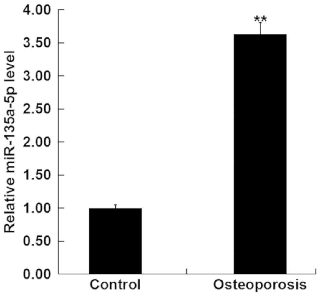
Enhanced miR-135a-5p expression in
postmenopausal women with osteoporosis
To determine whether miR-135a-5p was involved in the
development of osteoporosis, the expression level of miR-135a-5p
was detected using RT-qPCR in the bone tissue fragments from
postmenopausal women with and without osteoporosis. The results
revealed that the expression level of miR-135a-5p was significantly
increased in postmenopausal women with osteoporosis compared with
the postmenopausal women without osteoporosis (Fig. 1).
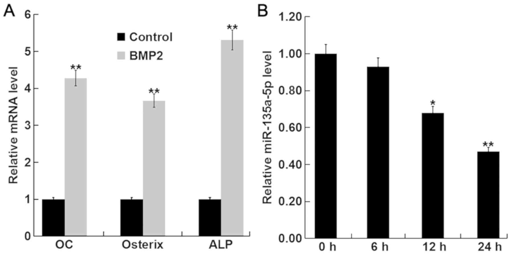
miR-135a-5p is downregulated during
osteogenic differentiation induced by BMP2
To investigate the function of miR-135a-5p in
osteoporosis, the expression level of miR-135a-5p was detected
during osteogenic differentiation induced by BMP2 in C2C12 cells.
To confirm the successful induction of osteogenic differentiation,
the relative mRNA expression level of key osteoblast markers OC,
osterix, and ALP were detected using RT-qPCR. The relative mRNA
expression levels of OC, osterix and ALP were significantly
increased following treatment with BMP compared with the control
group (Fig. 2A), indicating the
successful induction of osteogenic differentiation. In addition,
BMP2 treatment significantly decreased the expression level of
miR-135a-5p in a time-dependent manner (Fig. 2B). Taken together, these results
suggest that miR-135a-5p was downregulated during osteogenic
differentiation.
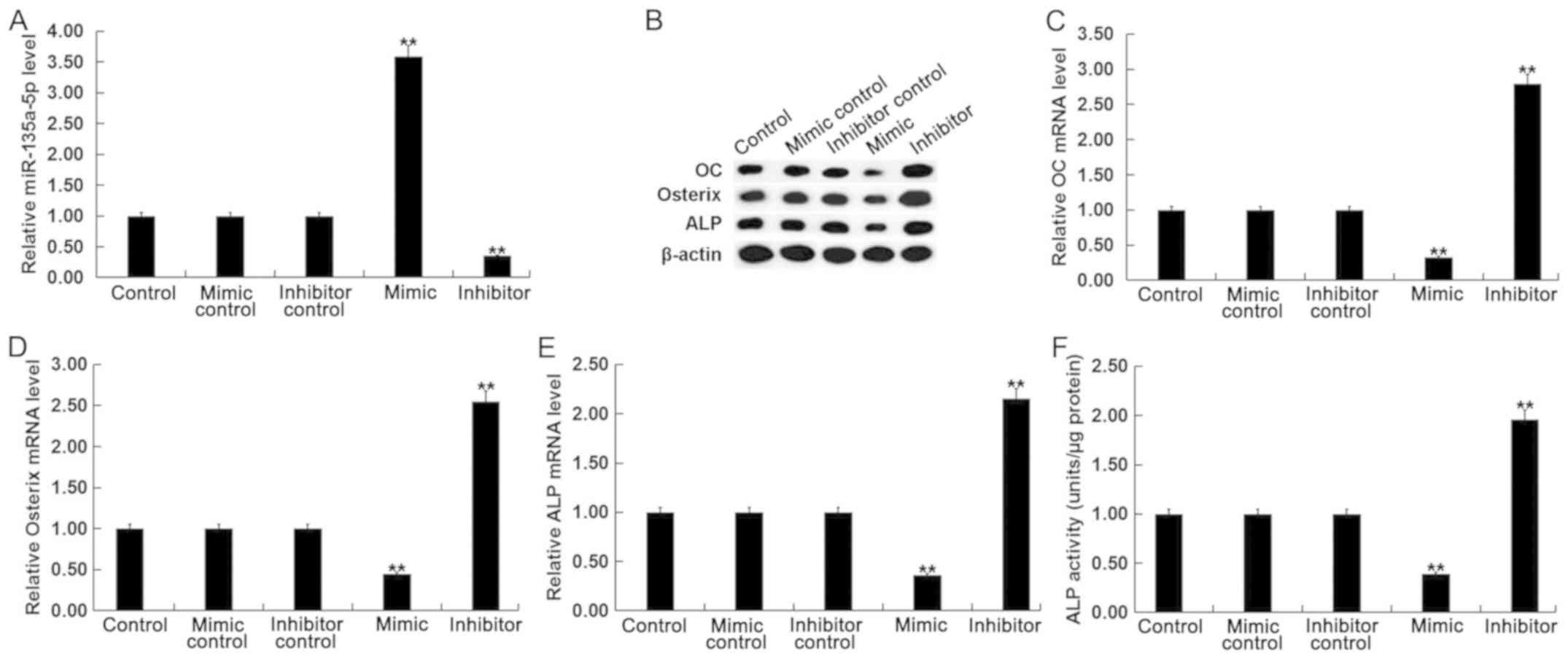
Effects of miR-135a-5p in osteogenic
differentiation in C2C12 cells
To further investigate the function of miR-135a-5p
in osteogenic differentiation, C2C12 cells were treated with 2 nM
BMP2 following transfection with miR-135a-5p mimic, mimic control,
miR-135a-5p inhibitor or inhibitor control. Transfection efficiency
was detected using RT-qPCR following 24-h transfection. The
expression level of miR-135a-5p was significantly increased in
BMP-treated C2C12 cells following transfection with miR-135a-5p
mimic compared with the control (Fig.
3A).
 | Figure 3.Effect of miR-135a-5p in osteogenic
differentiation. To explore the function of miR-135a-5p in
osteoblast differentiation, C2C12 cells were treated with 2 nM BMP2
for 24 h following transfection with miR-135a-5p mimic, mimic
control, miR-135a-5p inhibitor or inhibitor control, respectively,
for 2 h. (A) The relative miR-135a-5p expression level was
determined by RT-qPCR in C2C12 cells following transfection with
and treatment with BMP2. (B) The relative protein expression level
of OC, Osterix and ALP were determined by western blot analysis in
C2C12 cells following transfection and treatment with BMP2. The
relative mRNA expression level of (C) OC, (D) Osterix and (E) ALP
were determined by RT-qPCR in C2C12 cells following transfection
and treatment with BMP2. (F) ALP activity was examined in C2C12
cells following transfection and treatment with BMP2. Data are
presented as the mean ± standard deviation from three independent
experiments. **P<0.01 vs. Control. miR, microRNA; BMP2, bone
morphogenetic protein 2; RT-qPCR, reverse
transcription-quantitative polymerase chain reaction; OC,
osteocalcin; ALP, alkaline phosphatase. |
To determine the effect of miR-135a-5p in osteogenic
differentiation, changes in mRNA and protein expression levels of
specific osteoblast markers were examined. The mRNA and protein
expression levels of OC, osterix and ALP were significantly
decreased in BMP-treated C2C12 cells following transfection with
miR-135a-5p mimic compared with the control, whilst the opposite
effect was observed following transfection with miR-135a-5p
(Fig. 3B-E). In addition, it was
observed that compared with the control group, the ALP activity in
BMP2-treated cells was significantly suppressed following
miR-135a-5p overexpression, whilst ALP activity was significantly
enhanced following miR-135a-5p knockdown (Fig. 3F). Taken together, these results
suggest that miR-135a-5p can inhibit osteogenic
differentiation.
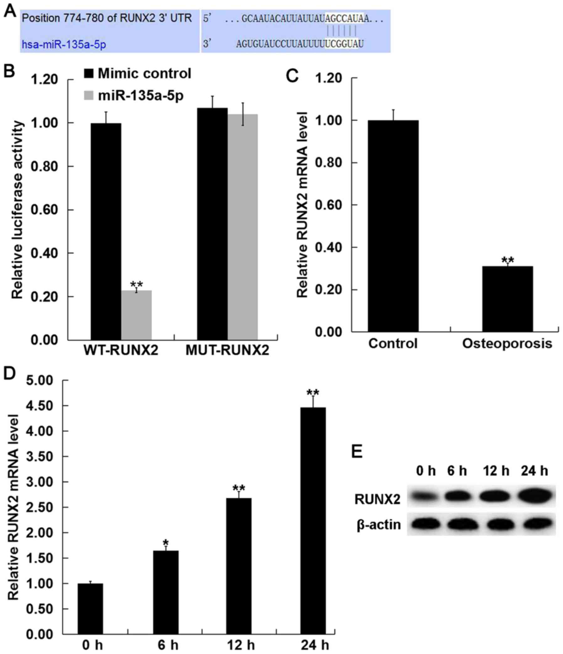
RUNX2 is a direct target of
miR-135a-5p
To investigate the molecular mechanism of
miR-135a-5p in osteogenic differentiation, TargetScan (http://www.targetscan.org) was used to predict the
potential target genes of miR-135a-5p. RUNX2 was identified as a
potential target of miR-135a-5p (Fig.
4A). To confirm the prediction, luciferase reporter assays were
performed to validate the direct interaction between miR-135a-5p
and RUNX2. Luciferase activity was significantly decreased
following co-transfection with miR-135a-5p mimic and WT-RUNX2
compared with MUT-RUNX2, which had no significant effect on
luciferase activity and miR-135a-5p mimic (Fig. 4B), indicating that miR-135a-5p
directly targets RUNX2.
The mRNA expression level of RUNX2 was detected
using RT-qPCR in the bone tissue fragments from postmenopausal
women with and without osteoporosis. The current study demonstrated
that the mRNA expression level of RUNX2 was significantly decreased
in postmenopausal women with osteoporosis compared with the
postmenopausal women without osteoporosis (Fig. 4C). Furthermore, BMP2 treatment
significantly increased the mRNA and protein expression levels of
RUNX2 in C2C12 cells in a time-dependent manner (Fig. 4D and E).
miR-135a-5p overexpression inhibits osteogenic
differentiation in C2C12 cells by targeting RUNX2. To investigate
whether miR-135a-5p serves a role in osteogenic differentiation by
direct targeting of RUNX2, C2C12 cells were treated with 2 nM BMP2
following transfection with miR-135a-5p mimic, mimic control,
control-plasmid, RUNX2-plasmid, miR-135a-5p mimic+control-plasmid
or miR-135a-5p mimic+RUNX2-plasmid. Transfection efficiency was
detected using RT-qPCR following 24-h transfection. The expression
level of RUNX2 was significantly increased in BMP-treated C2C12
cells following transfection with RUNX2-plasmid compared with the
control (Fig. 5A and B). The
relative protein and mRNA expression levels of RUNX2 were decreased
in BMP2-treated C2C12 cells following transfection with miR-135a-5p
mimic, however the miR-135a-5p-induced effects were reversed
following transfection with RUNX2-plasmid (Fig. 5C and D).
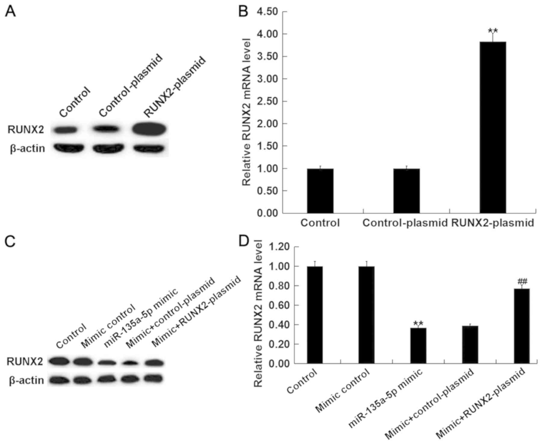 | Figure 5.miR-135a-5p regulates RUNX2 expression
in BMP2 treated C2C12 cells. C2C12 cells were treated with 2 nM
BMP2 for 24 h following transfection with miR-135a-5p mimic, mimic
control, control-plasmid, RUNX2-plasmid, miR-135a-5p
mimic+control-plasmid or miR-135a-5p mimic+RUNX2-plasmid,
respectively, for 2 h. The relative RUNX2 (A) protein and (B) mRNA
expression levels were detected by western blot and RT-qPCR
analysis, respectively, in C2C12 cells following transfection with
control-plasmid and RUNX2-plasmid. The relative RUNX2 (C) protein
and (D) mRNA expression level was detected by western blot and
RT-qPCR analysis, respectively, in C2C12 cells following
transfection with mimic control, miR-135a-5p mimic, miR-135a-5p
mimic+control-plasmid or miR-135a-5p mimic+RUNX2-plasmid. Data are
presented as the mean ± standard deviation from three independent
experiments. **P<0.01 vs. Control; ##P<0.01 vs.
Mimic group. miR, microRNA; RUNX2, runt related transcription
factor 2; BMP2, bone morphogenetic protein 2; RT-qPCR, reverse
transcription-quantitative polymerase chain reaction. |
The mRNA and protein expression levels of OC,
osterix and ALP were decreased in BMP-treated C2C12 cells following
transfection with miR-135a-5p mimic compared with the control,
however these effects were reversed following transfection with
RUNX2-plasmid (Fig. 6A-D). In
addition, ALP activity was suppressed by miR-135a-5p overexpression
however the addition of RUNX2-plasmid reversed the effect and
significantly enhanced ALP activity (Fig. 6E).
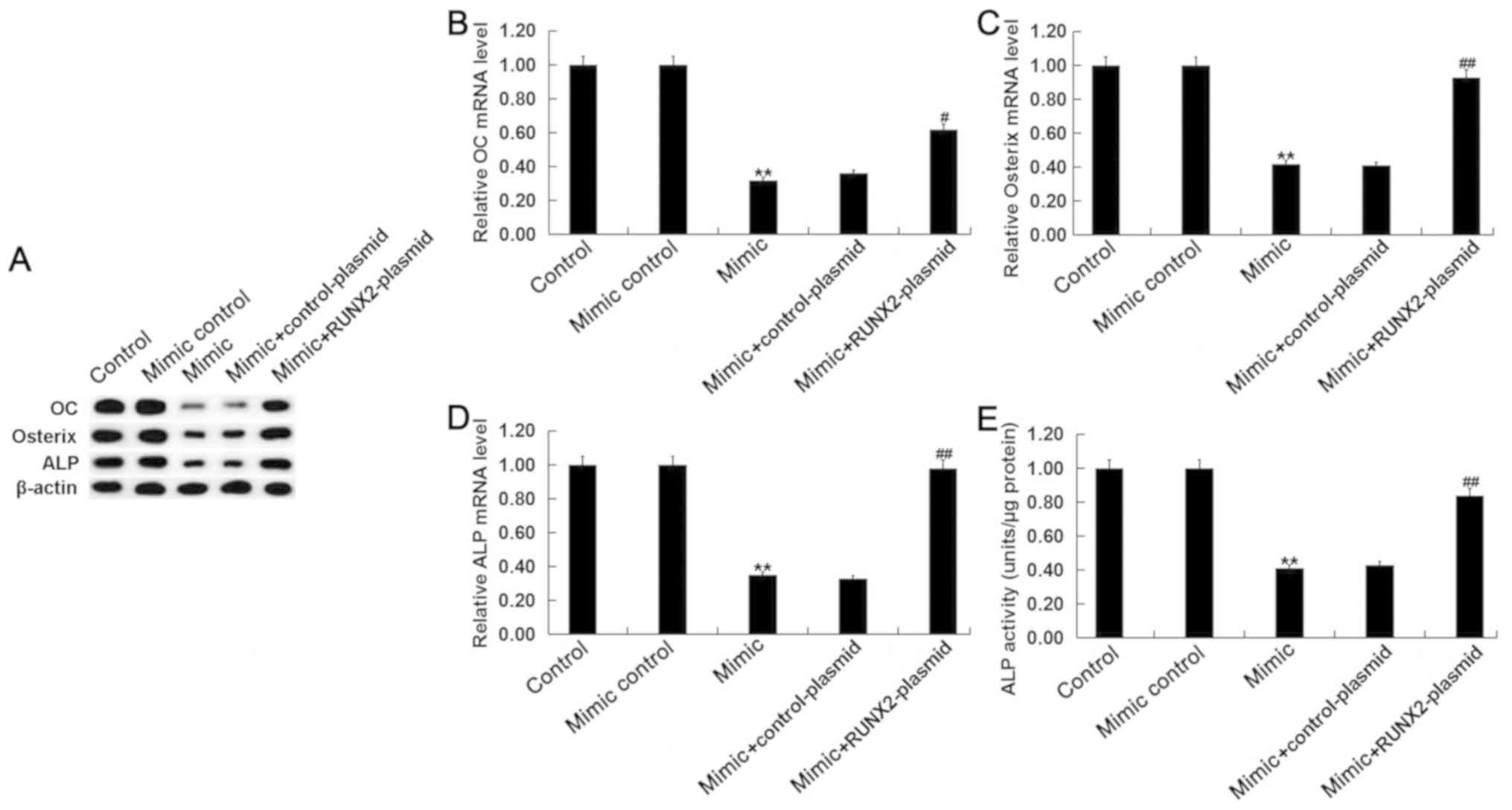 | Figure 6.miR-135a-5p inhibits osteogenic
differentiation by targeting RUNX2. C2C12 cells were treated with 2
nM BMP2 for 24 h following transfection with miR-135a-5p mimic,
mimic control, control-plasmid, RUNX2-plasmid, miR-135a-5p
mimic+control-plasmid or miR-135a-5p mimic+RUNX2-plasmid,
respectively, for 2 h. (A) The relative protein expression level of
OC, Osterix and ALP were determined by western blot analysis in
C2C12 cells following transfection and treatment with BMP2. The
relative mRNA expression level of (B) OC, (C) Osterix and (D) ALP
were determined by reverse transcription-quantitative polymerase
chain reaction in C2C12 cells following transfection and treatment
with BMP2. (E) ALP activity was examined in C2C12 cells following
transfection and treatment with BMP2. Data are presented as the
mean ± standard deviation from three independent experiments.
**P<0.01 vs. Control; #P<0.05 vs. Mimic;
##P<0.01 vs. Mimic; miR, microRNA; RUNX2, runt
related transcription factor 2; BMP2, bone morphogenetic protein 2;
OC, osteoclacin; ALP, alkaline phosphatase. |
Discussion
In the present study, the miR-135a-5p expression
level was significantly increased in postmenopausal women with
osteoporosis. However, during osteogenic differentiation in
vitro, the miR-135a-5p expression level was significantly
decreased. miR-135a-5p overexpression decreased the expression of
osteoblast makers, OC, osterix and ALP, as well as suppressing ALP
activity. By contrast, miR-135a-5p knockdown increased the
expression of osteoblast makers, and enhanced ALP activity.
Therefore, the current study demonstrated the inhibitory effect of
miR-135a-5p on osteogenic differentiation. In addition,
bioinformatics analysis was used to predict RUNX2 as a direct
target of miR-135a-5p. The RUNX2 expression level was significantly
decreased in postmenopausal women with osteoporosis. However,
during osteogenic differentiation in vitro, the RUNX2
expression level was significantly increased. Taken together, the
results suggest that miR-135a-5p inhibited osteogenic
differentiation by targeting RUNX2 and therefore miR-135a-5p may be
a promising therapeutic target for the treatment of
osteoporosis.
In China, osteoporosis is a common bone-related
disease (23). The incidence of
osteoporosis is high in postmenopausal women due to several
independent risk factors which include, estrogen deficiency,
persistent calcium loss and aging (24). Osteoporosis occurs due to an
imbalance between osteoclastic bone resorption and osteoblastic
bone formation (25). Restoring and
maintaining the balance between bone formation and bone resorption
is an effective way to treat postmenopausal osteoporosis (9,10).
Although some progress has been made in the treatment of
postmenopausal osteoporosis, there are currently no effective
therapies (26). Therefore,
understanding of the molecular mechanism underlying osteogenic bone
formation, as well as identifying therapeutic targets with
potential osteogenic effects, may provide novel strategies for the
treatment of postmenopausal osteoporosis.
Several studies have indicated that miRNAs serve an
important role in the regulation of osteoblastic differentiation or
bone formation (27,28). The study of miRNAs in postmenopausal
osteoporosis presents a novel direction for the diagnosis and
treatment of postmenopausal osteoporosis (15–18).
There have been relatively few studies investigating the cellular
function of miR-135a-5p. Chen et al (29) reported that miR-135a-5p could inhibit
3T3-L1 adipogenesis by activating the Wnt/β-catenin signaling
pathway. Wang et al (30)
demonstrated that serum miR-135a-5p expression was upregulated in
colorectal cancer and suggested that miR-135a-5p expression may be
used as a diagnostic biomarker for colorectal cancer. Yao et
al (31) demonstrated that
miR-135a-5p promoted proliferation and metastasis in hepatocellular
carcinoma cells via direct targeting of Kruppel-like factor 4. In
addition, miR-135a-5p is thought to serve an inhibitory role in
several types of cancer including thyroid carcinoma, head and neck
squamous cell carcinoma and glioblastoma (32–34).
Furthermore, miR-135a-5p is involved in the neuroprotective effects
of hydrogen sulfide against Parkinson's disease (35). TargetScan was used to predict
potential target genes of miR-135a-5p. TargetScan revealed hundreds
of target genes of miR-135a-5p, including RUNX2. RUNX2, a
transcription factor that belongs to the runt homology domain
protein family, is the predominant transcription factor for
osteoblast differentiation and bone formation and it may therefore
be a potential target for the treatment of osteoporosis (36,37).
These findings suggest that miR-135a-5p may serve a role in
osteogenic differentiation and osteoporosis. However, the role of
miR-135a-5p in postmenopausal osteoporosis remain unknown.
Therefore, the aim of the present study was to investigate
miR-135a-5p expression in postmenopausal women with osteoporosis as
well as the cellular function of miR-135a-5p and its underlying
mechanism in osteoblast differentiation.
The relative miR-135a-5p expression level in the
bone tissue fragments of postmenopausal women with and without
osteoporosis was examined. The current study demonstrated that the
expression level of miR-135a-5p was significantly upregulated in
postmenopausal women with osteoporosis. Further analysis indicated
that miR-135a-5p expression was downregulated during osteogenic
differentiation. Overexpression of miR-135a-5p inhibited osteogenic
differentiation, whilst miR-135a-5p knockdown enhanced osteogenic
differentiation. To further investigate the underlying molecular
mechanism of miR-135a-5p in osteogenic differentiation, the direct
interaction between miR-135a-5p and RUNX2 was predicted using
TargetScan and confirmed using dual-luciferase reporter assay. To
investigate whether miR-135a-5p serves a role in osteogenic
differentiation by direct targeting of RUNX2, rescue experiments
were performed. The inhibitory effect of miR-135a-5p on osteogenic
differentiation was reversed by RUNX2 overexpression, indicating
that miR-135a-5p can inhibit osteogenic differentiation via
RUNX2.
In conclusion, the current study demonstrated that
miR-135a-5p expression was upregulated in postmenopausal women with
osteoporosis, and miR-135a-5p inhibited osteogenic differentiation
via direct targeting of RUNX2. Therefore, miR-135a-5p may be a
promising therapeutic target for the treatment of postmenopausal
osteoporosis.
Acknowledgements
Not applicable.
Funding
The present study was supported by the Ningbo
Natural Science Foundation Project (grant no. 2017A610278).
Availability of data and materials
The datasets used and/or analyzed during the current
study are available from the corresponding author on reasonable
request.
Authors' contributions
XS contributed to study design, data collection,
statistical analysis and data interpretation. ZZ contributed to
data collection, manuscript preparation and the literature
search.
Ethics approval and consent to
participate
The present study was approved by the Ethics
Committee of Ningbo First Hospital (Ningbo, China) and written
informed consent was obtained from each patient.
Patient consent for publication
Not applicable.
Competing interests
The authors declare that they have no competing
interests.
References
|
1
|
Dalle Carbonare L, Valenti MT, Zanatta M,
Donatelli L and Lo Cascio V: Circulating mesenchymal stem cells
with abnormal osteogenic differentiation in patients with
osteoporosis. Arthritis Rheum. 60:3356–3365. 2009. View Article : Google Scholar : PubMed/NCBI
|
|
2
|
Rachner TD, Khosla S and Hofbauer LC:
Osteoporosis: Now and the future. Lancet. 377:1276–1287. 2011.
View Article : Google Scholar : PubMed/NCBI
|
|
3
|
Kikuta S, Tanaka N, Kazama T, Kazama M,
Kano K, Ryu J, Tokuhashi Y and Matsumoto T: Osteogenic effects of
dedifferentiated fat cell transplantation in rabbit models of bone
defect and ovariectomy-induced osteoporosis. Tissue Eng Part A.
19:1792–1802. 2013. View Article : Google Scholar : PubMed/NCBI
|
|
4
|
Lubkowska A, Dobek A, Mieszkowski J,
Garczynski W and Chlubek D: Adiponectin as a biomarker of
osteoporosis in postmenopausal women: Controversies. Dis Markers
2014. 9751782014.
|
|
5
|
He W, Goodkind D and Kowal P: U.S. Census
Bureau, International population reports, P95/16-1, An Aging World;
2015 Washington DC: U.S.: Government Publishing Office; 2016
|
|
6
|
Wang Y, Tao Y, Hyman ME, Li J and Chen Y:
Osteoporosis in china. Osteoporos Int. 20:1651–1662. 2009.
View Article : Google Scholar : PubMed/NCBI
|
|
7
|
Li Y, Xuan M, Wang B, Yang J, Zhang H,
Zhang XZ, Guo XH, Lü XF, Xue QY, Yang GY, et al: Comparison of
parathyroid hormone (1–34) and elcatonin in postmenopausal women
with osteoporosis: An 18-month randomized, multicenter controlled
trial in China. Chin Med J (Engl). 126:457–463. 2013. View Article : Google Scholar : PubMed/NCBIPubMed/NCBI
|
|
8
|
Pouresmaeili F, Kamalidehghan B, Kamarehei
M and Goh YM: A comprehensive overview on osteoporosis and its risk
factors. Ther Clin Risk Manag. 14:2029–2049. 2018. View Article : Google Scholar : PubMed/NCBI
|
|
9
|
Marie PJ and Kassem M: Osteoblasts in
osteoporosis: Past, emerging, and future anabolic targets. Eur J
Endocrinol. 165:1–10. 2011. View Article : Google Scholar : PubMed/NCBI
|
|
10
|
Ruiz-Gaspà S, Blanch-Rubió J,
Ciria-Recasens M, Monfort J, Tío L, Garcia-Giralt N, Nogués X,
Monllau JC, Carbonell-Abelló J and Pérez-Edo L: Reduced
proliferation and osteocalcin expression in osteoblasts of male
idiopathic osteoporosis. Calcif Tissue Int. 86:220–226. 2010.
View Article : Google Scholar : PubMed/NCBI
|
|
11
|
Hammond SM: An overview of microRNAs. Adv
Drug Deliv Rev. 87:3–14. 2015. View Article : Google Scholar : PubMed/NCBI
|
|
12
|
Soifer HS, Rossi JJ and Saetrom P:
MicroRNAs in disease and potential therapeutic applications. Mol
Ther. 15:2070–2079. 2017. View Article : Google Scholar
|
|
13
|
Krol J, Loedige I and Filipowicz W: The
widespread regulation of microRNA biogenesis, function and decay.
Nat Rev Genet. 11:597–610. 2010. View
Article : Google Scholar : PubMed/NCBI
|
|
14
|
O'Connell RM, Rao DS, Chaudhuri AA and
Baltimore D: Physiological and pathological roles for microRNAs in
the immune system. Nat Rev Immunol. 10:111–122. 2010. View Article : Google Scholar : PubMed/NCBI
|
|
15
|
Jin D, Wu X, Yu H, Jiang L, Zhou P, Yao X,
Meng J, Wang L, Zhang M and Zhang Y: Systematic analysis of
lncRNAs, mRNAs, circRNAs and miRNAs in patients with postmenopausal
osteoporosis. Am J Transl Res. 10:1498–1510. 2018.PubMed/NCBI
|
|
16
|
Mandourah AY, Ranganath L, Barraclough R,
Vinjamuri S, Hof RV, Hamill S, Czanner G, Dera AA, Wang D and
Barraclough DL: Circulating microRNAs as potential diagnostic
biomarkers for osteoporosis. Sci Rep. 8:84212018. View Article : Google Scholar : PubMed/NCBI
|
|
17
|
Feng Q, Zheng S and Zheng J: The emerging
role of microRNAs in bone remodeling and its therapeutic
implications for osteoporosis. Biosci Rep. 38(pii):
BSR201804532018. View Article : Google Scholar : PubMed/NCBI
|
|
18
|
Li H, Wang Z, Fu Q and Zhang J: Plasma
miRNA levels correlate with sensitivity to bone mineral density in
postmenopausal osteoporosis patients. Biomarkers. 19:553–556. 2014.
View Article : Google Scholar : PubMed/NCBI
|
|
19
|
Katagiri T, Yamaguchi A, Komaki M, Abe E,
Takahashi N, Ikeda T, Rosen V, Wozney JM, Fujisawa-Sehara A and
Suda T: Bone morphogenetic protein-2 converts the differentiation
pathway of C2C12 myoblasts into the osteoblast lineage. J Cell
Biol. 127:1755–1766. 1994. View Article : Google Scholar : PubMed/NCBI
|
|
20
|
Shin CS, Lecanda F, Sheikh S, Weitzmann L,
Cheng SL and Civitelli R: Relative abundance of different cadherins
defines differentiation of mesenchymal precursors into osteogenic,
myogenic, or adipogenic pathways. J Cell Biochem. 78:566–577. 2000.
View Article : Google Scholar : PubMed/NCBI
|
|
21
|
Zhang Y, Gao Y, Cai L, Li F, Lou Y, Xu N,
Kang Y and Yang H: MicroRNA-221 is involved in the regulation of
osteoporosis through regulates RUNX2 protein expression and
osteoblast differentiation. Am J Transl Res. 9:126–135.
2017.PubMed/NCBI
|
|
22
|
Livak KJ and Schmittgen TD: Analysis of
relative gene expression data using real-time quantitative PCR and
the 2(-Delta Delta C(T)) method. Methods. 25:402–408. 2001.
View Article : Google Scholar : PubMed/NCBI
|
|
23
|
Feng Z, Liu C, Guan X and Mor V: China's
rapidly aging population creates policy challenges in shaping a
viable long-term care system. Health Aff (Millwood). 31:2764–2773.
2012. View Article : Google Scholar : PubMed/NCBI
|
|
24
|
Black DM and Rosen CJ: Postmenopausal
osteoporosis. N Engl J Med. 374:2096–2097. 2016. View Article : Google Scholar : PubMed/NCBI
|
|
25
|
Raisz LG: Pathogenesis of osteoporosis:
Concepts, conflicts, and prospects. J Clin Invest. 115:3318–3325.
2005. View
Article : Google Scholar : PubMed/NCBI
|
|
26
|
Tella SH and Gallagher JC: Prevention and
treatment of postmenopausal osteoporosis. J Steroid Biochem Mol
Biol. 142:155–170. 2014. View Article : Google Scholar : PubMed/NCBI
|
|
27
|
Lian JB, Stein GS, van Wijnen AJ, Stein
JL, Hassan MQ, Gaur T and Zhang Y: MicroRNA control of bone
formation and homeostasis. Nat Rev Endocrinol. 8:212–227. 2012.
View Article : Google Scholar : PubMed/NCBI
|
|
28
|
Valenti MT, Dalle Carbonare L and Mottes
M: Role of microRNAs in progenitor cell commitment and osteogenic
differentiation in health and disease (Review). Int J Mol Med.
41:2441–2449. 2018.PubMed/NCBI
|
|
29
|
Chen C, Peng Y, Peng Y, Peng J and Jiang
S: miR-135a-5p inhibits 3T3-L1 adipogenesis through activation of
canonical Wnt/β-catenin signaling. J Mol Endocrinol. 52:311–320.
2014. View Article : Google Scholar : PubMed/NCBI
|
|
30
|
Wang Q, Zhang H, Shen X and Ju S: Serum
microRNA-135a-5p as an auxiliary diagnostic biomarker for
colorectal cancer. Ann Clin Biochem. 54:76–85. 2017. View Article : Google Scholar : PubMed/NCBI
|
|
31
|
Yao S, Tian C, Ding Y, Ye Q, Gao Y, Yang N
and Li Q: Down-regulation of Krüppel-like factor-4 by
microRNA-135a-5p promotes proliferation and metastasis in
hepatocellular carcinoma by transforming growth factor-β1.
Oncotarget. 7:42566–42578. 2016. View Article : Google Scholar : PubMed/NCBI
|
|
32
|
Zhao X, Sun Z, Li H, Jiang F, Zhou J and
Zhang L: MiR-135a-5p modulates biological functions of thyroid
carcinoma cells via targeting VCAN 3′-UTR. Cancer Biomark.
20:207–216. 2017. View Article : Google Scholar : PubMed/NCBI
|
|
33
|
Guo LM, Ding GF, Xu W, Ge H, Jiang Y, Chen
XJ and Lu Y: MiR-135a-5p represses proliferation of HNSCC by
targeting HOXA10. Cancer Biol Ther. 1–28. 2018.
|
|
34
|
Lin J, Wen X, Zhang X, Sun X, Yunzhi L,
Peng R, Zhu M, Wang M, Zhang Y, Luo W, et al: miR-135a-5p and
miR-124-3p inhibit malignancy of glioblastoma by downregulation of
syndecan binding protein. J Biomed Nanotechnol. 14:1317–1329.
2018.
|
|
35
|
Liu Y, Liao S, Quan H, Lin Y, Li J and
Yang Q: Involvement of microRNA-135a-5p in the protective effects
of hydrogen sulfide against parkinson's disease. Cell Physiol
Biochem. 40:18–26. 2016. View Article : Google Scholar : PubMed/NCBI
|
|
36
|
Chen D, Zhao M and Mundy GR: Bone
morphogenetic proteins. Growth Factors. 22:233–241. 2004.
View Article : Google Scholar : PubMed/NCBI
|
|
37
|
Kim JY, Cheon YH, Kwak SC, Baek JM, Yoon
KH, Lee MS and Oh J: Emodin regulates bone remodeling by inhibiting
osteoclastogenesis and stimulating osteoblast formation. J Bone
Miner Res. 29:1541–1553. 2014. View Article : Google Scholar : PubMed/NCBI
|