Introduction
Abdominal aortic aneurysm (AAA), defined as a focal
dilation of the abdominal aorta by ≥1.5 times the normal diameter
or to a diameter of >3 cm (1),
is a common, progressive and lethal vascular disease in adults aged
>65 years (2). It was estimated
that there were 2,347,339 cases and 41,374 deaths attributable to
AAA in 2013 in the USA (3). The
incidence of AAA was 27.9 per 100,000 in men and 3.3 per 100,000 in
women, and the mortality due to rupture was 59.2% in men and 48.8%
in women in Germany in 2014(4).
Currently, surgical repair (including open and endovascular) is the
mainstay treatment for AAA, but long-term prognosis remains
unsatisfactory, with an 8-year survival rate of 50% (5). Therefore, it is essential to develop
novel therapeutic options.
Although the pathogenesis is complex, loss of aortic
vascular smooth muscle cells (VSMCs) is an evident pathological
feature of AAA (6). Accumulating
studies have found that microRNAs (miRNAs/miRs), small non-coding
RNA molecules comprised of 18-25 nucleotides, are aberrantly
expressed in AAA and could control the proliferation and apoptosis
of aortic VSMCs by binding to the 3'-untranslated region (3'-UTR)
of target mRNAs and then inhibiting their expression
post-transcriptionally. Liang et al (7) detected that the expression of miR-195
was significantly increased in the aortic media of patients with
AAA compared with normal non-aneurysmal tissues. Overexpression of
miR-195 in human aortic VSMCs led to a significant decrease in cell
proliferation and an increase in cell apoptosis, while transfection
with anti-miR-195 obtained the opposite results. Luciferase
reporter assays and rescue studies indicated that SMAD family
member 3 (Smad3) may be a downstream target of miR-195(7). Zhao et al (8) identified that the expression levels of
miR-7 were upregulated in aortic specimens obtained from patients
with AAA compared with aortic specimens from controls. Knockdown of
miR-7 increased the proliferation and suppressed the apoptosis of
primary VSMCs isolated from aortic specimens of patients with AAA
(8). Luciferase reporter, reverse
transcription-quantitative PCR (RT-qPCR) and western blot assays
suggested that miR-7 may function by directly targeting
cytoskeleton associated protein 4(8). Cao et al (9) observed that miR-504 was less expressed
in the aortic cells derived from patients with AAA compared with
that in the controls. Cell growth was significantly increased in
VSMCs transfected with miR-504 vectors compared with those with
non-targeting controls (9). The
tumor suppressor p53 was the direct target of miR-504(9). Furthermore, miR-129-5p, miR-155-5p and
miR-28-5p were also reported to be apoptosis-related drivers for
AAA by targeting Wnt family member 5A (10), Fos proto-oncogene/Zic family member
3 (ZIC3) (11) and glutamate
ionotropic receptor AMPA type subunit 4/LY6/PLAUR domain containing
3(12), respectively. By contrast,
miR-21a/miR-26a and miR-126 may exert protective effects on the
development of AAA by modulating phosphatase and tensin homolog
(PTEN) (13,14) and ADAM metallopeptidase domain
9(15), respectively. These
findings implied that miRNAs may represent important targets for
the development of anti-AAA agents.
A previous study from our group integrated the miRNA
and mRNA expression profile datasets of aortic samples from
patients with AAA and identified several novel miRNA/mRNA
interaction pairs that may be crucial in the development of human
AAA. These previous results included the predicted interaction pair
of miR-15b (which was upregulated in AAA) with the mRNA for
acyl-CoA synthetase short chain family member 2 (ACSS2; which was
downregulated in AAA) (16).
Therefore, the present study aimed to validate the regulatory
relationship between miR-15b and ACSS2 by using a luciferase
reporter assay, and to explore their roles in the proliferation and
apoptosis of aortic VSMCs by in vitro overexpression or
knockdown experiments. The present results may provide preliminary
evidence to suggest that miR-15b/ACSS2 may be a potential
therapeutic target for AAA.
Materials and methods
Cell culture
A human aortic VSMC line (T/G HA-VSMC) was purchased
from Procell Life Science & Technology Co., Ltd.. Human aortic
VSMCs were cultured in smooth muscle cell medium (SMCM; ScienCell
Research Laboratories, Inc.) which contained basic medium, 2% fetal
bovine serum, 1% smooth muscle cell growth supplement and 1%
penicillin/streptomycin solution. A human embryonic kidney
(HEK)-293T cell line was purchased from the Institute of Basic
Medical Sciences, Chinese Academy of Medical Sciences (Beijing,
China) and maintained in Dulbecco's modified Eagle's medium
(Hyclone; Cytiva) containing 10% fetal calf serum (Gibco; Thermo
Fisher Scientific, Inc.). Both cell lines were kept in a humidified
atmosphere of 5% CO2 at 37˚C.
Cell transfection
Empty pcDNA3.1 vector, ACSS2 overexpression plasmid
(pcDNA3.1/ACSS2), miR-15b mimics/inhibitors and mimic/inhibitor
negative controls (NC) were obtained from RiboBio Co., Ltd.. At 24
h before the transient transfections, an appropriate number of
human aortic VSMCs were seeded into six-well plates with 2 ml of
SMCM and grown to 60-70% confluency at 37˚C in an atmosphere
containing 5% CO2. Then, the aortic VSMCs were divided
into six groups, and miR-15b-5p mimic NC
(5'-UCACAACCUCCUAGAAAGAGUAGA-3'; double stranded; 100 nM),
miR-15b-5p mimic (5'-UAGCAGCACAUCAUGGUUUACA-3'; double stranded;
100 nM), miR-15b-5p inhibitor NC (5'-UCUACUCUUUCUAGGAGGUUGUGA-3';
single stranded; 200 nM), miR-15b-5p inhibitor
(5'-AUCGUCGUGUAGUACCAAAUGU-3'; single stranded; 200 nM), pcDNA3.1
plasmid (4 µg/well), pcDNA3.1/ACSS2 plasmid (4 µg/well), miR-15b-5p
mimics+pcDNA3.1 plasmid, or miR-15b-5p mimics+pcDNA3.1/ACSS2
plasmid were added into the plates. The transient transfections of
these mimics or inhibitors were performed using the Exfect
Transfection reagent (Vazyme Biotech Co.) according to the
manufacturer's instructions.
Cell proliferation assay
At 60-70% confluence, human aortic VSMCs
(2x10³/well) were plated into 96-well plates with 200 µl
of SMCM and underwent the different transfection treatments as
aforementioned. Cell viability was determined at 0, 24, 48 and 72 h
post-transfection using the Cell Counting Kit-8 (CCK-8; Beyotime
Institute of Biotechnology). The absorbance of each well was
measured using a microplate reader (MK3; Thermo Fisher Scientific,
Inc.) at 450 nm.
Cell apoptosis assay
At 60-70% confluence, human aortic VSMCs
(2x10³/well) were plated into six-well plates with 2 ml
of SMCM and subjected to various transfection treatments as
aforementioned. At 48 h after transfection, human aortic VSMCs were
digested with 0.25% trypsin for 3-5 min, centrifuged at 1,000 x g
for 5 min and washed with precooled phosphate-buffered saline
twice. Cell pellets were resuspended with 1X binding buffer at a
density of 106 cells/ml. A total of 105 cells
were then stained with 5 µl Annexin V and 5 µl propidium iodide for
15 min at room temperature in the dark (MultiSciences Biotech Co.,
Ltd.). Cell apoptosis was analyzed by flow cytometry using
CytExpert software (version 2.0; Beckman Coulter, Inc.).
RT-qPCR
At 48 h after transfection, human aortic VSMCs were
treated with TRIzol® reagent (Tiangen Biotech Co., Ltd.)
to isolate the total RNA. For amplification of ACSS2, total RNA was
reverse transcribed to cDNA using the FastQuant cDNA synthesis kit
(Tiangen Biotech Co., Ltd.), and then qPCR was performed using
SuperReal PreMix (Tiangen Biotech Co., Ltd.). For amplification of
miR-15b-5p, first strand cDNA synthesis was conducted with the
miRNA 1st-Strand cDNA Synthesis kit (by stem-loop; Vazyme Biotech
Co., Ltd.), and qPCR was run according to the instructions of the
miRNA Universal SYBR qPCR Master Mix (Vazyme Biotech Co., Ltd.).
Primers were synthesized by the Beijing Dingguo Changsheng
Biotechnology Co., Ltd., and were as follows: ACSS2,
CGTGATGGGGCTTCCTGAG (forward) and GTTGTACCGAAGGAATGGGC (reverse);
miR-15b-5p, CGCGTAGCAGCACATCATGG (forward) and
GTCGTATCCAGTGCAGGGTCCGAGGTATTCGCACTGGATACGACTGTAAA (reverse);
β-actin, ACACTGTGCCCATCTACG (forward) and TGTCACGCACGATTTCC
(reverse); and U6, CTCGCTTCGGCAGCACA (forward) and
AACGCTTCACGAATTTGCGT (reverse). β-actin and U6 served as an
endogenous reference for mRNAs and miRNAs, respectively. The PCR
procedure was: Denaturation at 95˚C for 15 min, followed by 40
cycles of 95˚C for 10 sec, 60˚C for 20 sec and 72˚C for 30 sec. The
relative expression levels of mRNAs and miRNAs were determined
using the 2-ΔΔCq method (17).
Western blot analysis
At 48 h after transfection, human aortic VSMCs were
treated with the RIPA lysis buffer (Solarbio Science &
Technology Co., Ltd.) to extract the total proteins. The
concentration of total proteins was determined using the BCA
Protein Assay kit (Beyotime Institute of Biotechnology). The
protein samples (30 µg) were separated by 10% sodium dodecyl
sulfonate-polyacrylamide gel electrophoresis and then
electrophoretically transferred to polyvinylidene fluoride membranes
(EMD Millipore). After being blocked with 5% skim milk for 1 h at
room temperature, the membranes were incubated with primary
antibodies against ACSS2 (1:2,000; cat. no. 16087-1-AP; ProteinTech
Group, Inc.), prostaglandin-endoperoxide synthase 2 (PTGS2;
1:2,000; cat. no. 27308-1-AP; ProteinTech Group, Inc.) and GAPDH
(1:1,000; cat. no. ab181602; Abcam) overnight at 4˚C. The membranes
were then incubated with the horseradish peroxidase-conjugated
secondary antibody at room temperature for 1 h (1:5,000; cat. no.
E030120; EarthOx Life Sciences). Protein bands were visualized
using an enhanced chemiluminescence kit (Beyotime Institute of
Biotechnology) and quantified using ImageJ software (version 1.46;
National Institutes of Health). GAPDH was used as an internal
control. Protein expression levels were calculated as relative to
the mimic NC group, which was set as 1.0.
Luciferase reporter assay
TargetScan software (version 7.0; targetscan.org/) (18)
was used to identify the miR-15b-5p binding site in the 3'-UTR of
ACSS2. The wild-type (WT) and mutant (MUT) 3'-UTR sequences of
ACSS2 at the miR-15b binding site were chemically synthesized by
General Biosystems, Inc., and inserted into the
XhoI/NotI site of psiCHECK-2 luciferase vector
(Promega Corporation) to construct the recombinant plasmids
psiCHECK2-ACSS2-WT and psiCHECK2-ACSS2-MUT, respectively.
miR-15b-5p mimics or miR-15b-5p mimic NC were co-transfected with
psiCHECK2-ACSS2-WT and psiCHECK2-ACSS2-MUT into HEK-293T cells
using the Exfect Transfection reagent (Vazyme Biotech Co., Ltd.).
After 48 h, firefly and Renilla luciferase activities were
measured on an EnSpire reader (PerkinElmer, Inc.) using the Dual
Luciferase Assay System (Dalian Meilun Biology Technology Co.,
Ltd.).
Statistical analysis
All experiments were performed at least two
independent times. Data were expressed as mean ± standard
deviation. Differences between two groups were analyzed by
Student's t-test. Differences among more than two groups were
tested using one-way analysis of variance followed by post hoc
Tukey's (equal variances) or Dunnett's T3 (unequal variances) test.
Statistical analysis was conducted using SPSS 18.0 (SPSS, Inc.) and
graphs were generated in GraphPad Prism 5.0 (GraphPad Software,
Inc.). P<0.05 was considered to indicate a statistically
significant difference.
Results
Construction of in vitro transfection
models
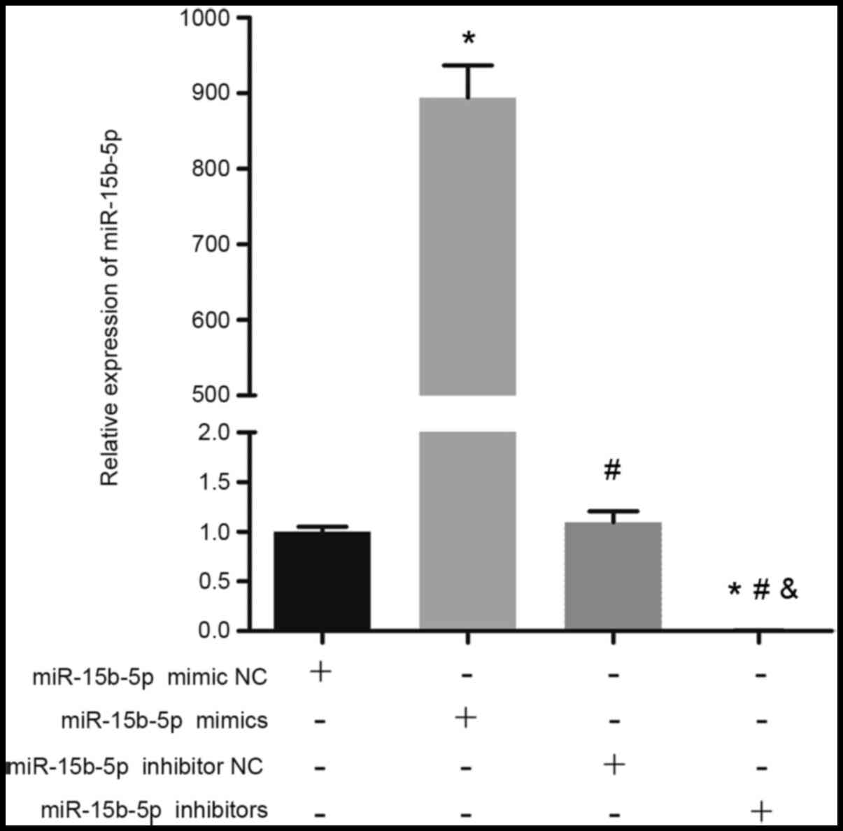
To explore the potential roles of miR-15-5p, in
vitro overexpression and knockdown models were first
established by transfecting the human aortic VSMCs with miR-15b-5p
mimics and miR-15b-5p inhibitors, respectively. As shown in
Fig. 1, the expression levels of
miR-15b-5p were significantly increased in the miR-15b-5p mimic
group compared with those in the mimic NC group. By contrast, the
expression levels of miR-15b-5p were significantly lower in the
miR-15b-5p inhibitor group compared with those in the inhibitor NC
group (Fig. 1). These findings
indicated that overexpression and knockdown of miR-15b-5p were
successfully established in human aortic VSMCs.
miR-15b-5p has an antiproliferative
role in aortic VSMCs
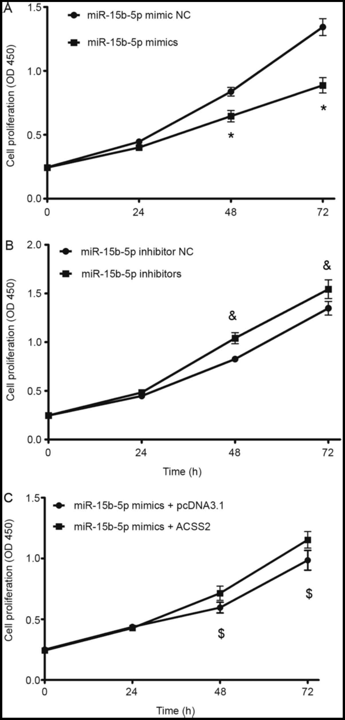
CCK-8 assay was used to evaluate the impact of
miR-15b-5p on the proliferation of human aortic VSMCs. The results
demonstrated that the absorbance value of aortic VSMCs was
significantly lower at 48 and 72 h after transfection with
miR-15b-5p mimics compared with the mimic NC group, but no
statistical difference was observed at 24 h (Fig. 2A). By contrast, transfection with
miR-15b-5p inhibitors resulted in a significant increase in cell
proliferation compared with the inhibitor NC group, showing a
higher absorbance value at 48 and 72 h after transfection (Fig. 2B). These findings suggested that
miR-15b-5p may have an antiproliferative role in aortic VSMCs. The
time point of 48 h post-transfection was selected for subsequent
analyses.
miR-15b-5p exerts proapoptotic
activity in aortic VSMCs
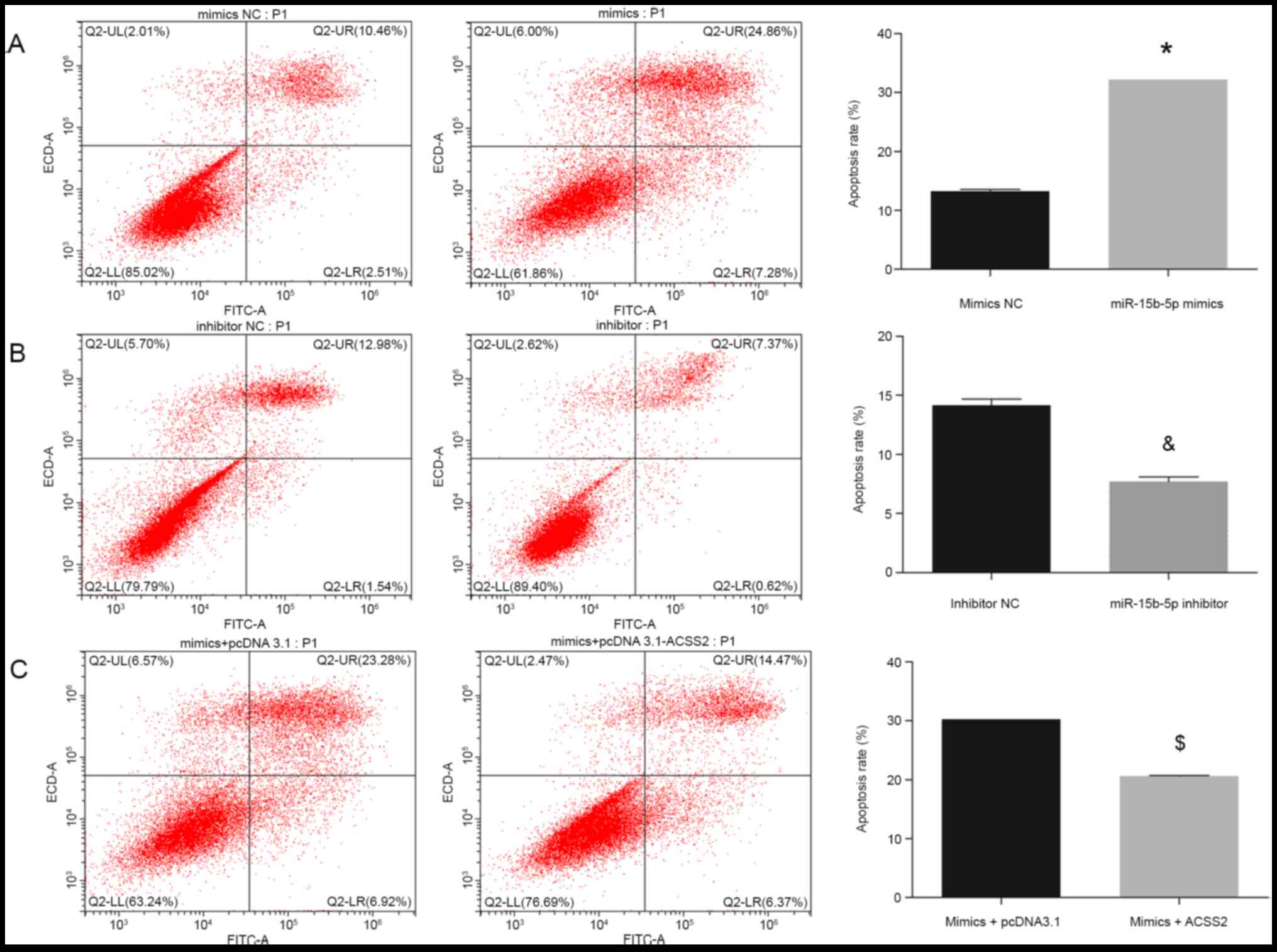
Annexin V-FITC/PI staining and flow cytometry
analysis were used to investigate the effects of miR-15b-5p on the
apoptosis of aortic VSMCs. As shown in Fig. 3A, the apoptosis rate of aortic VSMCs
transfected with miR-15b-5p mimics was found to be increased by
~2.5-fold compared with that in cells transfected with the mimic
NC. By contrast, transfection with the miR-15b-5p inhibitors was
associated with a 1.8-fold decrease in the proportion of apoptotic
cells in aortic VSMCs when compared with the inhibitor NC group
treatment (Fig. 3B). These findings
indicated that miR-15b-5p may exert proapoptotic activity in aortic
VSMCs.
miR-15b-5p regulates the expression of
ACSS2 in aortic VSMCs
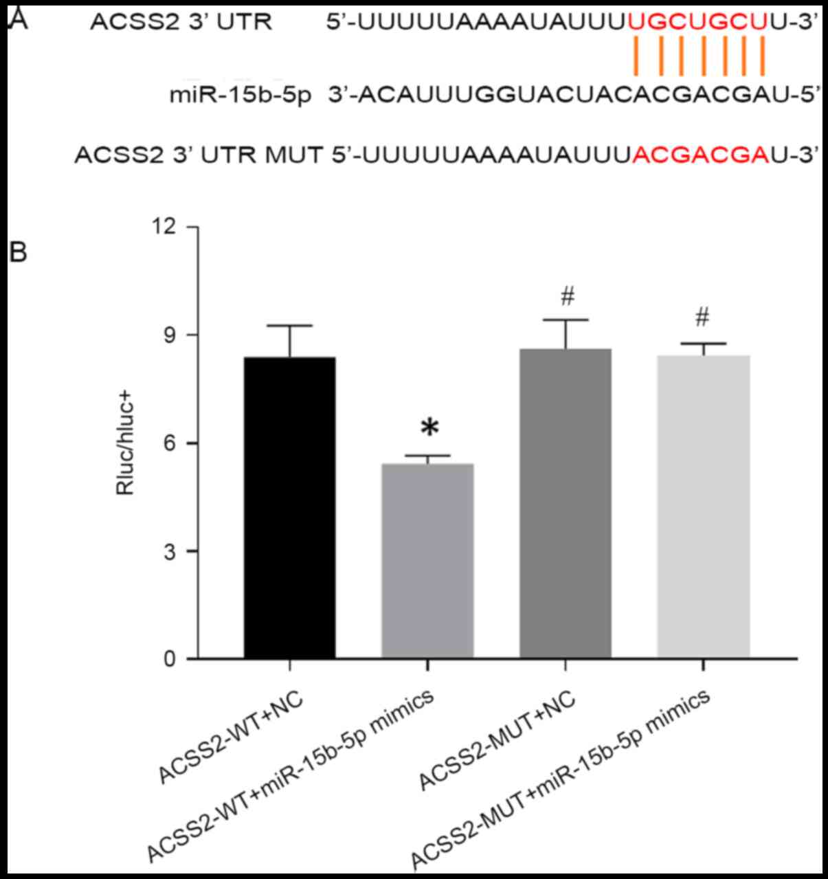
Bioinformatic analysis predicted that the 3'-UTR
region of ACSS2 had the binding site for miR-15b-5p (Fig. 4A). To explore the regulatory
relationship of ACSS2 and miR-15b-5p, a dual-luciferase reporter
assay in HEK-293T cells was conducted. As expected, the luciferase
activity of the ACSS2-WT vector was significantly inhibited by
miR-15b-5p mimics compared with the mimic NC, while no significant
change was detected in the luciferase activity of the ACSS2-MUT
reporter between the miR-15b-5p mimics and mimic NC groups
(Fig. 4B), implying the ACSS2 may
be a direct target of miR-15b-5p.
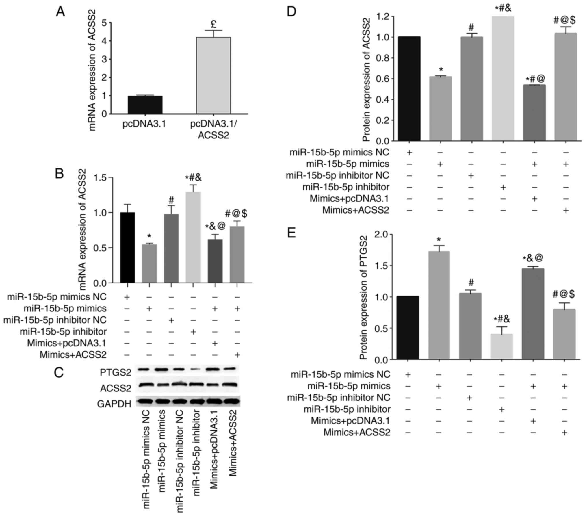
To further confirm their interaction, the expression
levels of ACSS2 were measured in aortic VSMCs following different
transfections. In line with the dual-luciferase reporter assay,
both the mRNA and protein expression levels of ACSS2 in aortic
VSMCs were found to be significantly reduced after transfection
with miR-15b-5p mimics, but significantly increased after
transfection with miR-15b-5p inhibitors (Fig. 5B-D).
miR-15b-5p regulates the proliferation
and apoptosis of aortic VSMCs via targeting ACSS2
To further explore whether the proapoptotic and
antiproliferative effects of miR-15b-5p was achieved through ACSS2,
rescue experiments were performed. Firstly, pcDNA3.1/ACSS2 was
confirmed to have an effective transfection efficiency (Fig. 5A). Then, miR-15b-5p-mediated
suppression of ACSS2 was demonstrated to be rescued by simultaneous
overexpression of ACSS2 in aortic VSMCs (Fig. 5B-D). Functional assays demonstrated
that overexpression of ACSS2 reversed the effects of miR-15b-5p
mimics on aortic VSMCs, resulting in an increase in cell
proliferation (Fig. 2C), and a
decrease in cell apoptosis (Fig.
3C), compared with the miR-15b-5p+empty pcDNA3.1 vector group.
These findings suggested that miR-15b-5p-induced apoptosis may be
rescued by ACSS2.
Furthermore, our previous bioinformatics prediction
analysis revealed that miR-15b-5p may function in the development
of AAA by regulating ACSS2 and then affecting the lipid
biosynthetic process (16). PTGS2
(also known as COX-2) is a key regulatory enzyme in lipid
metabolism, which was also upregulated in AAA (16). Thus, it can be hypothesized that
PTGS2 may be a downstream target of the miR-15b-5p/ACSS2
interaction. To validate this hypothesis, the protein expression
levels of PTGS2 were detected in all six transfection groups. In
accordance with its expression in AAA, PTGS2 was demonstrated to be
highly expressed in the miR-15b-5p mimic group, but significantly
reduced in the miR-15b-5p inhibitor group compared with their
corresponding controls. Additionally, overexpression of ACSS2 could
partially block the production of PTGS2 protein (Fig. 5C and E).
Discussion
The present study, for the first time, demonstrated
that overexpression of miR-15b-5p promoted the apoptosis and
inhibited the proliferation of human aortic VSMCs by downregulating
the expression of ACSS2 and subsequently upregulating PTGS2. Of
note, silencing of miR-15b-5p obtained the opposite results. The
present findings were in line with the pathological feature of
aortic VSMCs loss in AAA (6,19,20)
and the observed expression levels of miR-15b-5p (upregulated),
ACSS2 (downregulated) and PTGS2 (upregulated) in AAA samples
(16). Therefore, the
hsa-miR-15b-5p/ACSS2 interaction axis may be an important
contributor to the development of AAA by regulating the
proliferation and apoptosis of human aortic VSMCs and may represent
attractive therapeutic molecules for AAA.
Although there were no studies to explore the role
of miR-15b-5p in AAA, its function in cancer may indirectly explain
the importance of miR-15b-5p for the development and progression of
AAA. Dong et al (21)
reported that miR-15b-5p was upregulated in liver cancer tissues
and cell lines, and that miR-15b-5p levels were associated with
high tumor-node-metastasis stage, tumor capsular infiltration and
shorter overall survival rates. Overexpression of miR-15b-5p
promoted liver cancer cell proliferation and invasion (21). Zhao et al (22) identified that miR-15b-5p was highly
expressed in gastric cancer cell lines, tissues, and plasma samples
in comparison with normal controls. Ectopic expression of
miR-15b-5p in gastric cancer cells accelerated cell proliferation,
migration, invasion and epithelial-mesenchymal transition (22). Wu et al (23) observed that knockdown of miR-15b-5p
inhibited growth and invasiveness, and induced apoptosis of breast
cancer cells and significantly restrained tumor growth in a
xenograft model in mice. These findings indicated that miR-15b-5p
may be an oncogenic driver. Because depletion of aortic VSMCs is a
common pathological feature in AAA, the present study speculated
that miR-15b-5p may also be upregulated in AAA samples similar to
cancer, and then it may promote the apoptosis of aortic VSMCs. The
present study confirmed this hypothesis, demonstrating that miR-15b
mimics significantly reduced the viability and facilitated the
apoptosis of human VSMCs, and that opposite effects were observed
following miR-15b inhibitor transfection. Further confirmation of
this hypothesis was reported in a recent study by Sun et al
(24).
The known downstream target genes of miR-15b-5p
include axin 2(21), progestin and
adipoQ receptor family member 3(22), heparanase 2(23), and reversion inducing cysteine rich
protein with kazal motifs (25) in
cancer cells, and the insulin like growth factor 1 receptor
(24) in human VSMCs. The present
study, for the first time, demonstrated that ACSS2 may also be a
direct target of miR-15b-5p using the dual-luciferase reporter
system. In addition, ACSS2 overexpression could reverse the
proapoptotic and antiproliferative effects of miR-15b-5p on aortic
VSMCs. In consideration of the roles of miR-15b-5p in AAA and
cancer, it can be speculated that ACSS2 may serve as a tumor
suppressor gene. This hypothesis has been supported by several
previous studies: Hur et al (26) and Bae et al (27) identified that downregulation of ACSS2
is an independent factor to predict poor survival of patients with
gastric cancer and colorectal carcinoma, respectively. Knockdown of
ACSS2 was shown to increase the invasion and migration ability of
hepatocellular carcinoma cells by promoting the
epithelial-mesenchymal transition (28). However, the tumor suppressor
mechanisms of ACSS2 were rarely reported because more scholars
believe that ACSS2 may be oncogenic (29-31)
by catalyzing the production of acetyl-CoA from acetate, CoA and
ATP and then promoting fatty acid synthesis as cell membrane
component, single molecular or energy source in tumor cells
(32-34).
The present study demonstrated that ACSS2 may exert tumor
suppressor roles by inhibiting the expression of PTGS2. This
negative regulatory relationship between ACSS2 and PTGS2 may be
associated with the ability of ACSS2 to drive acetylation of the
target genes and then reduce their activity (28). Furthermore, considerable evidence
revealed that the expression of PTGS2 was increased in an AAA
animal model and patients (35);
the use of a PTGS2 inhibitor (36-38)
or genetic deficiency of PTGS2(39)
significantly reduced the incidence, severity, aortic rupture and
mortality in AAA model mice. PTGS2 may be involved in the
development of AAA due to its ability to convert arachidonic acid
into prostaglandins. Prostaglandin E2 was found to inhibit DNA
synthesis, proliferation and death of aortic VSMCs derived from
aneurysmal aorta by activation of a serial of inflammatory pathways
(40). In line with these studies,
the current findings also revealed that the expression of PTGS2 was
increased in VSMCs with higher apoptosis rates.
There were several limitations in the present study.
Firstly, the present study provided preliminary evidence to show
that the miR-15b-5p/ACSS2/PTGS2 axis may be a potential target for
the treatment of AAA by changing the apoptosis and proliferation of
aortic VSMCs. In vivo animal models need to be established
in order to confirm the therapeutic effects of anti-miR-15b-5p in
AAA prior to clinical application. Secondly, it will be valuable to
collect aortic samples from patients with AAA from our hospital to
validate the expression of miR-15b-5p/ACSS2/PTGS2 and their
association with clinical features (such as aneurysm diameter and
rupture incidence). Thirdly, the downstream mechanisms of ACSS2
need to be further explored in the future, by overexpression and
knockdown of ACSS2 and by miR-15b-5p and PTGS2 (or other genes)
rescue experiments.
In conclusion, the present study suggested that
miR-15b-5p may promote the apoptosis and inhibit the proliferation
of aortic VSMCs via targeting the ACSS2/PTGS2 axis. Targeted
regulation of the miR-15b-5p/ACSS2/PTGS2 axis may serve as a
potential approach for the treatment of AAA, a disease
characterized pathologically by loss of VSMCs.
Acknowledgements
Not applicable.
Funding
Funding: The present study was supported by the Special Disease
of Vasculitis in Pudong New Area (grant no. PWZzb2017-07), the
Outstanding Clinical Discipline Project of Shanghai Pudong (grant
no. PWYgy-2018-08) and the Program for Medical Key Department of
Shanghai (grant no. ZK2019A10).
Availability of data and materials
All data generated or analyzed during this study are
included in this published article.
Authors' contributions
SG, JT and ZQ contributed to the conception and
design of this study. SG performed the experiments and the
statistical analyses. JM and YP were involved in the interpretation
of the data. SG and JT drafted the manuscript. ZQ edited and
revised the manuscript. SG and ZQ confirm the authenticity of all
the raw data. All authors read and approved the final
manuscript.
Ethics approval and consent to
participate
Not applicable.
Patient consent for publication
Not applicable.
Competing interests
The authors declare that they have no competing
interests.
References
|
1
|
Kent KC: Clinical practice. Abdominal
aortic aneurysms. N Engl J Med. 371:2101–2108. 2014.PubMed/NCBI View Article : Google Scholar
|
|
2
|
Dereziński TL, Fórmankiewicz B, Migdalski
A, Brazis P, Jakubowski G, Woda Ł and Jawień A: The prevalence of
abdominal aortic aneurysms in the rural/urban population in central
Poland-Gniewkowo aortic study. Kardiol Pol. 75:705–710.
2017.PubMed/NCBI View Article : Google Scholar
|
|
3
|
Stuntz M: Modeling the burden of abdominal
aortic aneurysm in the USA in 2013. Cardiology. 135:127–131.
2016.PubMed/NCBI View Article : Google Scholar
|
|
4
|
Kühnl A, Erk A, Trenner M, Salvermoser M,
Schmid V and Eckstein HH: Incidence, treatment and mortality in
patients with abdominal aortic aneurysms. Dtsch Arztebl Int.
114:391–398. 2017.PubMed/NCBI View Article : Google Scholar
|
|
5
|
Lederle FA, Kyriakides TC, Stroupe KT,
Freischlag JA, Padberg FT Jr, Matsumura JS, Huo Z and Johnson GR:
OVER Veterans Affairs Cooperative Study Group. Open versus
endovascular repair of abdominal aortic aneurysm. N Engl J Med.
380:2126–2135. 2019.PubMed/NCBI View Article : Google Scholar
|
|
6
|
Thompson RW, Liao S and Curci JA: Vascular
smooth muscle cell apoptosis in abdominal aortic aneurysms. Coron
Artery Dis. 8:623–631. 1997.PubMed/NCBI View Article : Google Scholar
|
|
7
|
Liang B, Che J, Zhao H, Zhang Z and Shi G:
MiR-195 promotes abdominal aortic aneurysm media remodeling by
targeting Smad3. Cardiovasc Ther. 35(e12286)2017.PubMed/NCBI View Article : Google Scholar
|
|
8
|
Zhao F, Chen T and Jiang N:
CDR1as/miR-7/CKAP4 axis contributes to the pathogenesis of
abdominal aortic aneurysm by regulating the proliferation and
apoptosis of primary vascular smooth muscle cells. Exp Ther Med.
19:3760–3766. 2020.PubMed/NCBI View Article : Google Scholar
|
|
9
|
Cao X, Cai Z, Liu J, Zhao Y, Wang X, Li X
and Xia H: miRNA-504 inhibits p53-dependent vascular smooth muscle
cell apoptosis and may prevent aneurysm formation. Mol Med Rep.
16:2570–2578. 2017.PubMed/NCBI View Article : Google Scholar
|
|
10
|
Zhang Y, Liu Z, Zhou M and Liu C:
MicroRNA-129-5p inhibits vascular smooth muscle cell proliferation
by targeting Wnt5a. Exp Ther Med. 12:2651–2656. 2016.PubMed/NCBI View Article : Google Scholar
|
|
11
|
Zhao L, Ouyang Y, Bai Y, Gong J and Liao
H: miR-155-5p inhibits the viability of vascular smooth muscle cell
via targeting FOS and ZIC3 to promote aneurysm formation. Eur J
Pharmacol. 853:145–152. 2019.PubMed/NCBI View Article : Google Scholar
|
|
12
|
Yue J, Zhu T, Yang J, Si Y, Xu X, Fang Y
and Fu W: CircCBFB-mediated miR-28-5p facilitates abdominal aortic
aneurysm via LYPD3 and GRIA4. Life Sci. 253(117533)2020.PubMed/NCBI View Article : Google Scholar
|
|
13
|
Maegdefessel L, Azuma J, Toh R, Deng A,
Merk DR, Raiesdana A, Leeper NJ, Raaz U, Schoelmerich AM, McConnell
MV, et al: MicroRNA-21 blocks abdominal aortic aneurysm development
and nicotine-augmented expansion. Sci Transl Med.
4(122ra22)2012.PubMed/NCBI View Article : Google Scholar
|
|
14
|
Peng J, He X, Zhang L and Liu P:
MicroRNA-26a protects vascular smooth muscle cells against
H2O2-induced injury through activation of the PTEN/AKT/mTOR
pathway. Int J Mol Med. 42:1367–1378. 2018.PubMed/NCBI View Article : Google Scholar
|
|
15
|
Shen G, Sun Q, Yao Y, Li S, Liu G, Yuan C,
Li H, Xu Y and Wang H: Role of ADAM9 and miR-126 in the development
of abdominal aortic aneurysm. Atherosclerosis. 297:47–54.
2020.PubMed/NCBI View Article : Google Scholar
|
|
16
|
Gan S, Pan Y and Mao J: miR-30a-GNG2 and
miR-15b-ACSS2 interaction pairs may be potentially crucial for
development of abdominal aortic aneurysm by influencing
inflammation. DNA Cell Biol. 38:1540–1556. 2019.PubMed/NCBI View Article : Google Scholar
|
|
17
|
Livak KJ and Schmittgen TD: Analysis of
relative gene expression data using real-time quantitative PCR and
the 2(-Delta Delta C(T)) method. Methods. 25:402–408.
2001.PubMed/NCBI View Article : Google Scholar
|
|
18
|
Agarwal V, Bell GW, Nam JW and Bartel DP:
Predicting effective microRNA target sites in mammalian mRNAs.
Elife. 4(e05005)2015.PubMed/NCBI View Article : Google Scholar
|
|
19
|
Zhang Z, Zou G, Chen X, Lu W, Liu J, Zhai
S and Qiao G: Knockdown of lncRNA PVT1 inhibits vascular smooth
muscle cell apoptosis and extracellular matrix disruption in a
murine abdominal aortic aneurysm model. Mol Cells. 42:218–227.
2019.PubMed/NCBI View Article : Google Scholar
|
|
20
|
Xue M, Li G, Li D, Wang Z, Mi L, Da J and
Jin X: Up-regulated MCPIP1 in abdominal aortic aneurysm is
associated with vascular smooth muscle cell apoptosis and MMPs
production. Biosci Rep. 39(BSR20191252)2019.PubMed/NCBI View Article : Google Scholar
|
|
21
|
Dong Y, Zhang N, Zhao S, Chen X, Li F and
Tao X: miR-221-3p and miR-15b-5p promote cell proliferation and
invasion by targeting Axin2 in liver cancer. Oncol Lett.
18:6491–6500. 2019.PubMed/NCBI View Article : Google Scholar
|
|
22
|
Zhao C, Li Y, Chen G, Wang F, Shen Z and
Zhou R: Overexpression of miR-15b-5p promotes gastric cancer
metastasis by regulating PAQR3. Oncol Rep. 38:352–358.
2017.PubMed/NCBI View Article : Google Scholar
|
|
23
|
Wu B, Liu G, Jin Y, Yang T, Zhang D, Ding
L, Zhou F, Pan Y and Wei Y: miR-15b-5p promotes growth and
metastasis in breast cancer by targeting HPSE2. Front Oncol.
10(108)2020.PubMed/NCBI View Article : Google Scholar
|
|
24
|
Sun Y, Gao Y, Song T, Yu C, Nie Z and Wang
X: MicroRNA-15b participates in the development of peripheral
arterial disease by modulating the growth of vascular smooth muscle
cells. Exp Ther Med. 18:77–84. 2019.PubMed/NCBI View Article : Google Scholar
|
|
25
|
Chen R, Sheng L, Zhang HJ, Ji M and Qian
WQ: miR-15b-5p facilitates the tumorigenicity by targeting RECK and
predicts tumour recurrence in prostate cancer. J Cell Mol Med.
22:1855–1863. 2018.PubMed/NCBI View Article : Google Scholar
|
|
26
|
Hur H, Kim YB, Ham IH and Lee D: Loss of
ACSS2 expression predicts poor prognosis in patients with gastric
cancer. J Surg Oncol. 112:585–591. 2015.PubMed/NCBI View Article : Google Scholar
|
|
27
|
Bae JM, Kim JH, Oh HJ, Park HE, Lee TH,
Cho NY and Kang GH: Downregulation of acetyl-CoA synthetase 2 is a
metabolic hallmark of tumor progression and aggressiveness in
colorectal carcinoma. Mod Pathol. 30:267–277. 2017.PubMed/NCBI View Article : Google Scholar
|
|
28
|
Sun L, Kong Y, Cao M, Zhou H, Li H, Cui Y,
Fang F, Zhang W, Li J, Zhu X, et al: Decreased expression of
acetyl-CoA synthase 2 promotes metastasis and predicts poor
prognosis in hepatocellular carcinoma. Cancer Sci. 108:1338–1346.
2017.PubMed/NCBI View Article : Google Scholar
|
|
29
|
Yao L, Guo X and Gui Y: Acetyl-CoA
synthetase 2 promotes cell migration and invasion of renal cell
carcinoma by upregulating lysosomal-associated membrane protein 1
expression. Cell Physiol Biochem. 45:984–992. 2018.PubMed/NCBI View Article : Google Scholar
|
|
30
|
Zhang S, He J, Jia Z, Yan Z and Yang J:
Acetyl-CoA synthetase 2 enhances tumorigenesis and is indicative of
a poor prognosis for patients with renal cell carcinoma. Urol
Oncol. 36:243.e9–243.e20. 2018.PubMed/NCBI View Article : Google Scholar
|
|
31
|
Mi L, Zhou Y, Wu D, Tao Q, Wang X, Zhu H,
Gao X, Wang J, Ling R, Deng J, et al: ACSS2/AMPK/PCNA
pathway-driven proliferation and chemoresistance of esophageal
squamous carcinoma cells under nutrient stress. Mol Med Rep.
20:5286–5296. 2019.PubMed/NCBI View Article : Google Scholar
|
|
32
|
Huang Z, Zhang M, Plec AA, Estill SJ, Cai
L, Repa JJ, McKnight SL and Tu BP: ACSS2 promotes systemic fat
storage and utilization through selective regulation of genes
involved in lipid metabolism. Proc Natl Acad Sci USA.
115:E9499–E9506. 2018.PubMed/NCBI View Article : Google Scholar
|
|
33
|
Lee MY, Yeon A, Shahid M, Cho E, Sairam V,
Figlin R, Kim KH and Kim J: Reprogrammed lipid metabolism in
bladder cancer with cisplatin resistance. Oncotarget.
9:13231–13243. 2018.PubMed/NCBI View Article : Google Scholar
|
|
34
|
Xu H, Luo J, Ma G, Zhang X, Yao D, Li M
and Loor JJ: Acyl-CoA synthetase short-chain family member 2
(ACSS2) is regulated by SREBP-1 and plays a role in fatty acid
synthesis in caprine mammary epithelial cells. J Cell Physiol.
233:1005–1016. 2018.PubMed/NCBI View Article : Google Scholar
|
|
35
|
Chapple KS, Parry DJ, McKenzie S,
MacLennan KA, Jones P and Scott DJ: Cyclooxygenase-2 expression and
its association with increased angiogenesis in human abdominal
aortic aneurysms. Ann Vasc Surg. 21:61–66. 2007.PubMed/NCBI View Article : Google Scholar
|
|
36
|
Ghoshal S and Loftin CD: Cyclooxygenase-2
inhibition attenuates abdominal aortic aneurysm progression in
hyperlipidemic mice. PLoS One. 7(e44369)2012.PubMed/NCBI View Article : Google Scholar
|
|
37
|
King VL, Trivedi DB, Gitlin JM and Loftin
CD: Selective cyclooxygenase-2 inhibition with celecoxib decreases
angiotensin II-induced abdominal aortic aneurysm formation in mice.
Arterioscler Thromb Vasc Biol. 26:1137–1143. 2006.PubMed/NCBI View Article : Google Scholar
|
|
38
|
Mukherjee K, Gitlin JM and Loftin CD:
Effectiveness of cyclooxygenase-2 inhibition in limiting abdominal
aortic aneurysm progression in mice correlates with a
differentiated smooth muscle cell phenotype. J Cardiovasc
Pharmacol. 60:520–529. 2012.PubMed/NCBI View Article : Google Scholar
|
|
39
|
Gitlin JM, Trivedi DB, Langenbach R and
Loftin CD: Genetic deficiency of cyclooxygenase-2 attenuates
abdominal aortic aneurysm formation in mice. Cardiovasc Res.
73:227–236. 2007.PubMed/NCBI View Article : Google Scholar
|
|
40
|
Walton LJ, Franklin IJ, Bayston T, Brown
LC, Greenhalgh RM, Taylor GW and Powell JT: Inhibition of
prostaglandin E2 synthesis in abdominal aortic aneurysms:
Implications for smooth muscle cell viability, inflammatory
processes, and the expansion of abdominal aortic aneurysms.
Circulation. 100:48–54. 1999.PubMed/NCBI View Article : Google Scholar
|