China ranks first in the world in terms of incidence
of diabetes and the age of patients diagnosed with diabetes
exhibits a decreasing trend (1).
Diabetes is a metabolic disorder characterized by hyperglycemia
caused by insufficient insulin secretion, insulin resistance or
both (2). In recent years,
exosomes have been demonstrated not only to be biomarkers of
diabetic complications but also to participate in the etiology of
diabetes (3,4). Therefore, exosomes are helpful for
identifying diabetic complications at an early stage and may be
used to diagnose and treat diabetes at the molecular level. The
present article reviews the role of exosomes in the pathogenesis of
diabetic complications and discusses the prospects of using
exosomes in the diagnosis and treatment of diabetic
complications.
Exosomes are cell vesicles with lipid bilayer
membranes with a diameter measuring 30-120 nm that contain specific
proteins, related metabolites, lipids and nucleic acids (5). Exosomes not only transmit information
between cells in autocrine, paracrine and telecrine states but also
correspond to cells (6). Exosomes
may be released by most cells in the body and are present in most
bodily fluids, such as plasma, urine, saliva, cerebrospinal fluid
and breast milk, bronchoalveolar lavage fluid and epididymal fluid
(7). Exosomes may be separated by
various methods, including ultrahigh-speed centrifugation,
filtration, precipitation and immune enrichment (8). They have complex components,
including DNA fragments, circular RNA, mRNA, microRNA (miRNA/miR),
functional proteins, transcription factors and other substances.
Internal vesicles are secreted into the extracellular environment
through the fusion of vesicle endosomes and the plasma membrane and
then bind with receptor cells to exchange information between cells
by releasing substances into receptor cells (9). In addition, exosomes have been
demonstrated to have an important role in tumor diagnosis,
migration and growth, as well as tissue damage repair, immune
antigen presentation and neurodegenerative diseases (10).
Diabetes is a group of metabolic diseases
characterized by hyperglycemia. The etiology, pathogenesis and
complexity of diabetes have remained to be fully clarified
(11). Diabetes may be divided
into two categories according to its pathogenesis as follows: Type
1 diabetes mellitus (T1DM), in which there is a lack of insulin due
to the destruction of islet β cells, and type 2 diabetes mellitus
(T2DM), in which insulin resistance is accompanied by insulin
deficiency or impaired insulin secretion (12). Persistent hyperglycemia may induce
diabetic complications by regulating the epigenetics of target
cells (13). Exosomes have
important roles in the pathology of T1DM and T2DM, including the
destruction of pancreatic tissue by mediating immune reactions and
inducing adipocytes to reduce insulin resistance (14). In recent years, studies have
indicated that exosomes carrying miRNAs and long noncoding RNAs
have important roles in the development of diabetes by regulating
metabolic signals, insulin signaling and the interaction between
inflammatory pancreatic cells (15). In addition, miRNAs carried by
exosomes may be used as biomarkers in diabetes and diabetic
complications (16), which may
help to identify diabetic complications at an early stage, thereby
allowing patients to receive effective treatment in time.
Diabetic complications include microangiopathy,
atherosclerotic cardiovascular disease, neurological complications
and diabetic foot ulcers (28).
Microvascular disease is a specific complication of diabetes and
its typical changes are basement membrane thickening and
microcirculation disease. Microvascular disease may affect all
tissues and organs in the whole body, and diabetic nephropathy and
retinopathy are the most common (29). Experiments in rats have indicated
that MSC-Exos reduce oxidative stress by inhibiting C-C motif
chemokine receptor 2 and reduce the damage from the inflammatory
response to its microvascular endothelium (30); therefore, exosomes may promote
angiogenesis and enhance the repair of vascular endothelium damaged
by oxidative stress. Exosomes derived from adipose mesenchymal stem
cells (AMSC-Exos) are rich in miR-125a, which may inhibit the
expression of the angiogenesis inhibitor delta-like 4 protein and
promote angiogenesis in rats (31). It has been indicated that the
complement system has an important role in the development of
vascular damage (32).
Furthermore, Huang et al (33) reported that a lack of IgG in
exosomes results in a reduction in vascular damage in diabetic
mice. Another study suggested that miR-21 carried by exosomes
derived from glioma stem cells upregulates vascular endothelial
growth factor expression, which promotes the angiogenic ability of
endothelial cells (30).
Therefore, exosomes may be a promising drug candidate for treating
microvascular damage caused by high glucose.
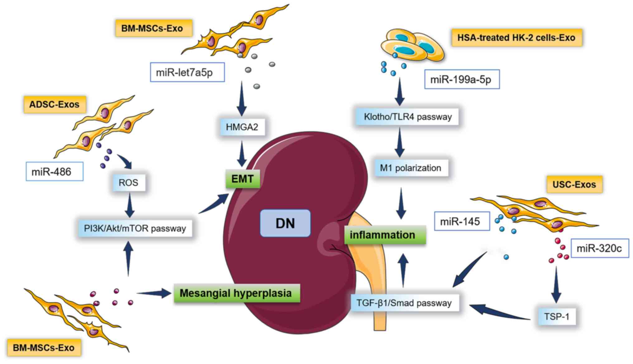
DN is a complex and common chronic kidney disease,
and it is one of the primary causes of end-stage renal disease
(34). The clinical features of DN
are persistent albuminuria and the thickening of the glomerular
basement membrane. DN causes an accumulation of the extracellular
matrix and induces autophagy, causing podocyte damage and
progressive renal insufficiency (35). The traditional method for
diagnosing DN is to detect protein in urine; however, as the
deterioration of renal function usually occurs earlier than
proteinuria, DN cannot be prevented at an early stage (36). It has been reported that podocytes
induced by high glucose secrete exosomes carrying early flowering 3
(Elf3), which may only be detected in the urine of patients with
DN, suggesting that measuring the level of Elf3 protein in exosomes
derived from the human urine stem cells (USC-Exos) of patients with
DN may be used as an early, non-invasive marker of cell damage in
podocytes (37). The expression of
Wilms tumor protein 1 (WT1) in USC-Exos of patients increases with
the decline in the patient's renal function, suggesting that WT1
may be used as a monitoring indicator of early renal function in DN
(38). The expression of
miR-19b-3p in the USC-Exos of patients with DN is significantly
increased and it is positively correlated with the severity of
renal interstitial inflammatory reactions in patients with DN,
suggesting that miR-19b-3p can be used as a molecular marker for
monitoring the progression of DN (39). Fibrosis is a key step in the
pathology of DN (40). Exosomes
derived from bone marrow MSCs (BM-MSCs-Exo) carry miR-let7a-5p,
which acts on high mobility group AT-hook 2 to promote the
transformation of DN renal tubular epithelial cells and renal
fibrosis in rats (41). In
addition, USC-Exos carry miR-145 and miR-320c, which directly or
indirectly activate the TGF-β1/Smad pathway of renal cells and
promote renal fibrosis in rats (42). High glucose and proteinuria
synergistically stimulate renal podocytes and renal tubular cells
to produce chemokines, stimulate the release of inflammatory
factors, increase the synthesis of extracellular matrix and
accelerate glomerular sclerosis (43). In addition, exosomes secreted from
human serum albumin-treated HK-2 cells released miR-199a-5p, which
targeted the Klotho/Toll-like receptor (TLR)4 pathway to induce
M1-type macrophage (M1) polarization, accelerating the inflammatory
reaction and promoting the progression of DN (44). In clinical patients, molecules
derived from BM-MSCs-Exo were observed to induce glomerular
hypertrophy by promoting the PI3K/Akt/mTOR pathway and participate
in the proliferation of mesangial cells and the extracellular
matrix (45). Adipose stem
cell-derived exosomes (ADSC-Exos) containing miR-486 activate
reactive oxygen species (ROS), thereby targeting the PI3K/Akt/mTOR
pathway to accelerate the apoptosis of podocytes, and they were
observed to participate in the occurrence of DN in rats (46). Exosomes not only serve as related
markers of DN but also participate in the pathophysiological
processes of DN. Fig. 1 provides a
schematic of the mechanisms of the involvement of exosomes in the
pathogenesis of DN.
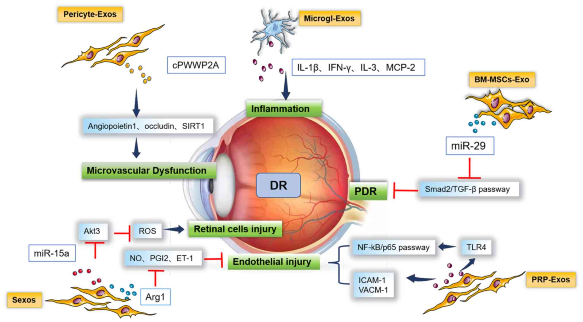
Diabetic retinopathy (DR) is a common complication
of diabetes. Long-term chronic hyperglycemia may cause retinal
microcirculation disorders, which manifest as vascular degeneration
in the early stage and develop into proliferative DR during the
late stage, which is accompanied by proliferative
neovascularization, leading to vitreous bodies and even retinal
detachment (47). In recent years,
studies have indicated that exosomes reflect the relationship
between cell secretion and the progression of DR (33). Mazzeo et al (48) compared the miRNA expression
profiles of plasma extracellular vesicles (including exocytosis)
between patients with DR and healthy individuals and observed that
the expression of miR-150-5p, miR-21-3p and miR-30b-5p was
increased in patients with DR, indicating that exosomes carrying
these miRNAs may be used as biomarkers for DR. The pathogenesis of
DR is complex and remains to be fully elucidated, but it is
primarily related to damage to the vascular endothelium and the
progression of vascular inflammation and oxidative stress (49). Fig.
2 provides a schematic of the mechanisms of the involvement of
exosomes in the pathogenesis of DR. A previous study (50) has shown that the arginase 1
contained in the Sexos of patients with DR inhibits nitric oxide
(NO), endothelin 1 and prostacyclin but increases prostaglandin 2
after being taken up by endothelial cells to cause vascular
endothelial damage in DR. In addition, miR-15a carried by Sexos
inhibits PKB3/Akt3 to activate ROS, which destroy retinal cells in
patients, including pigment epithelial cells, Müller cells and
photoreceptor cells, representing a pathogenic mechanism of DR
(51). Zhang et al
(52) indicated that exosomes
derived from platelet plasma target TLR4, which activates the
NF-κB/p65 pathway, causing damage to human retinal endothelial
cells, and they determined that these exosomes upregulate the
expression of retinal intercellular adhesion molecule 1 and
vascular cell adhesion molecule 1, inducing retinal degradation.
Experiments in rats have indicated that miR-29 carried by
BM-MSCs-Exo inhibited the Smad2/TGF-β pathway, which caused
proliferative retinopathy and further detachment of the retina
(53). Microglia are immune cells
of the retina that are closely related to the progression of DR.
Exosomes derived from microglia release interleukin-1β (IL-1β),
IL-3, interferon-γ and macrophage chemoattractant protein 2,
resulting in a significant inflammatory reaction in patients
(54). In addition, exosomes
derived from peripheral cells carry
proline-tryptophan-tryptophan-proline domain-containing protein 2A
and act on angiopoietin 1, occludin, and sirtuin1, which regulates
endothelial cells and aggravates retinal vascular dysfunction in
rats (55). The abovementioned
studies demonstrated that exosomes affect and participate in the
pathophysiological processes of DR via various mechanisms.
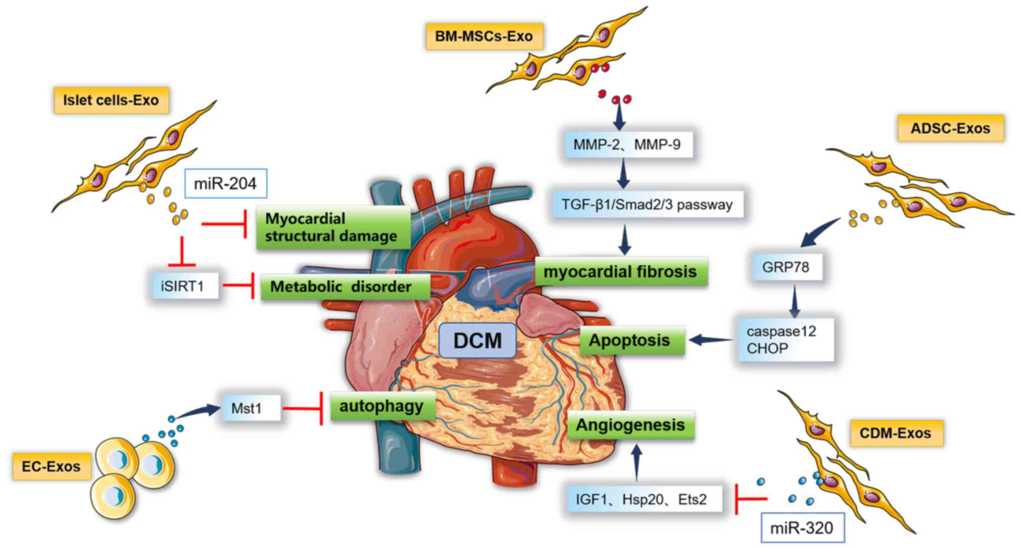
DCM is caused by extensive necrosis of the
myocardium due to cardiac microvascular disease and myocardial
metabolic disorders (56). The
prevalence of myocardial damage caused by high glucose in the
population is increasing and the progression of this damage is
becoming increasingly more rapid. The pathogenesis of DCM is
primarily related to metabolic disorders, myocardial fibrosis,
myocardial cell apoptosis and microvascular disease (57). Fig.
3 provides a schematic of the mechanisms of the involvement of
exosomes in the pathogenesis of DCM. In recent years, studies have
indicated that exosomes are closely related to the pathogenesis of
DCM. Cardiac fibrosis is the primary cause of myocardial
dysfunction in patients with diabetes. The transdifferentiation of
fibroblasts into myofibroblasts is key to the process of cardiac
fibrosis (58). Experiments in
rats suggested that BM-MSCs-Exo carry molecular targeting
proteases, such as plasmin, MMP-2 and MMP-9, to activate the
TGF-β1/Smad2/3 signaling pathway to promote cardiac muscle
fibrosis, causing myocardial damage (59). Furthermore, miR-204 carried by
exosomes derived from islet cells inhibits the expression of
insulin receptor substrate 1 in rats, causing insulin resistance.
In addition to aggravating myocardial energy mechanism disorders,
miR-204 also directly damages the structure and function of the
left ventricle (60). In rats, it
was demonstrated that ADSC-Exos activate the key promoters of the
cardiomyocyte apoptosis pathway, caspase12 and C/EBP-homologous
protein, by targeting 78-kD glucose-regulated protein and mediating
the endoplasmic reticulum response. ADSC-Exos induce cell apoptosis
and eventually lead to DCM (61).
Observations in rats have also indicated that miR-320 carried by
cardiomyocyte exosomes inhibits target gene receptors such as
insulin-like growth factor 1 (IGF1), heat shock protein 20 and E26
transformation-specific proto-oncogene 2. The inhibition of cardiac
endothelial cell migration and blood vessel formation may be one of
the mechanisms of microangiogenesis in DCM (62). Exosomes derived from endothelial
cells increase the content of mammalian ste20-like kinase-1 (Mst1),
a key protein in the Hippo pathway that regulates organ size,
apoptosis and autophagy and enhance the binding of Mst1 and death
domain-associated protein to disrupt GLUT4 membrane translocation,
thereby aggravating insulin resistance. Furthermore, Mst1 inhibits
autophagy of cardiomyocytes, enhances cell apoptosis and promotes
the formation of DCM in rats (63). Although exosomes have important
roles in the mechanisms of DCM, the evidence available is limited
to animal experiments (64) and
corresponding clinical studies are lacking. The diabetes-mediated
regulation of exosomes in the cardiovascular system is currently
under further investigation, which may be a promising treatment
approach that will benefit numerous clinical patients.
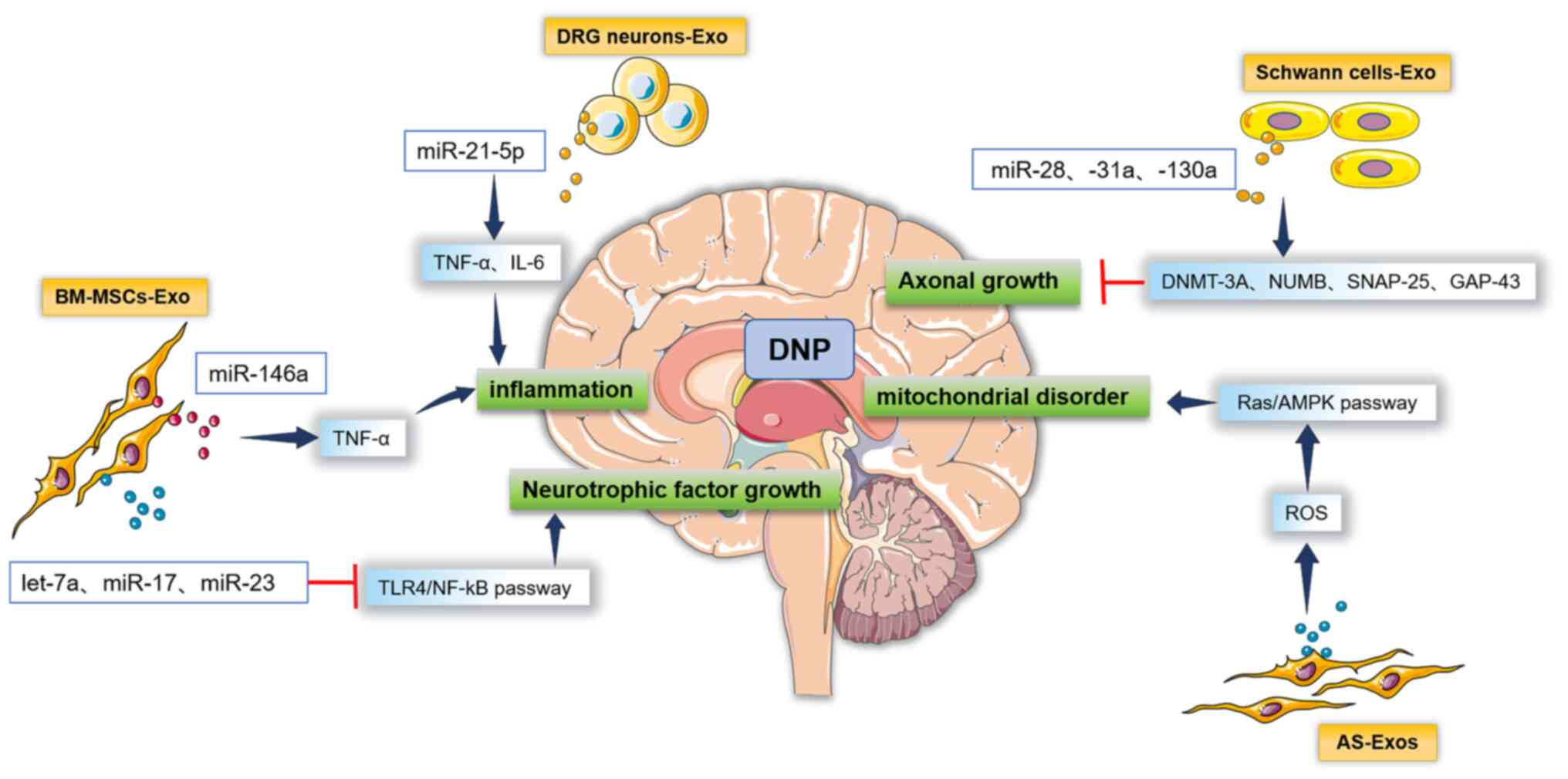
DPN may affect any part of the nervous system and
includes central nervous system complications, peripheral
neuropathy and autonomic neuropathy (65). The pathogenesis of DPN is complex
and is primarily related to inflammation under high-glucose
conditions. The activation of factors is related to intracranial
microvascular disease (66).
Fig. 4 provides a schematic of the
mechanisms of the involvement of exosomes in the pathogenesis of
DPN. It has been indicated that certain exosomes are inflammatory
markers of DPN. The miR-21-5p carried by exosomes originating from
dorsal root ganglia neurons increases TNF-α, IL-6 and other
inflammatory factors, which induces the polarization of macrophages
to the proinflammatory M1 phenotype, accelerating the occurrence
and development of DPN in rats (67). miR-146a carried by BM-MSCs-Exo also
increases TNF-α and mediates the inflammatory reaction (68). Jia et al (69) report that the expression of miR-28,
-31a and -130a in Schwann cells-derived exosome is increased by the
action of target proteins in axons, such as DNA methyltransferase
3A, Numb protein, synaptosome protein 25 and growth-related protein
43, which causes rat axon ischemia, hypoxia and abnormal
metabolism, inhibits axon growth and aggravates DPN damage.
BM-MSCs-Exo carry let-7a, miR-17, miR-23 and other molecules that
inhibit the TLR4/NF-κB signaling pathway, leading to a decrease in
neurotrophic factors and ultimately participating in the
pathogenesis of DPN in mice (70).
Part of the reason for the damage to the nervous system is the
disturbance of nervous system homeostasis caused by oxidative
stress and increased apoptosis (71). Based on this observation, studies
on nerve cells have indicated that exosomes derived from astrocytes
activate ROS and target the Ras/adenosine monophosphate-activated
protein kinase signaling pathway, which causes mitochondrial
dysfunction and apoptosis, ultimately leading to neuronal damage
(72). The above mechanistic study
suggests the potential of using exosomes in treating DPN.
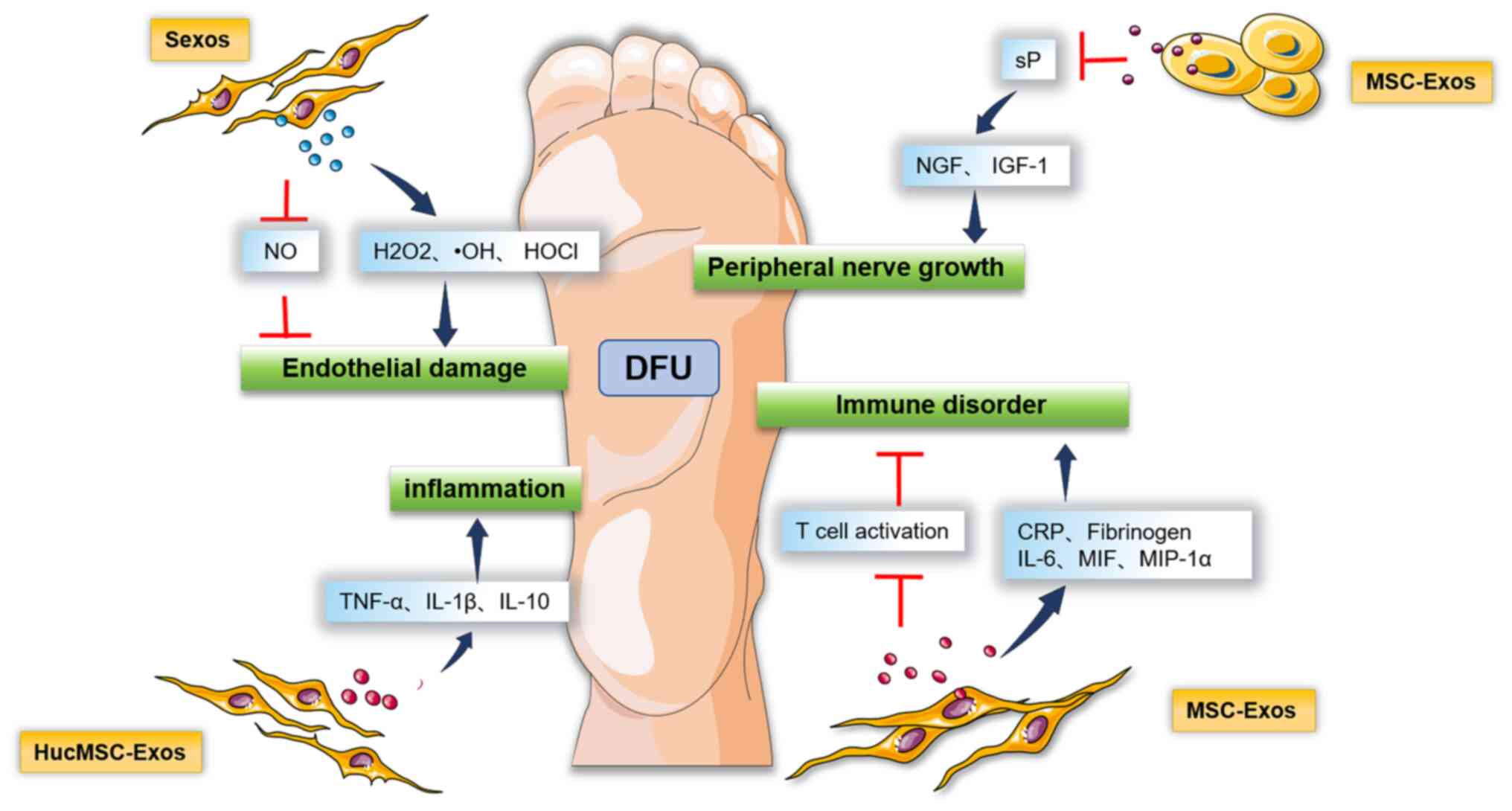
DFU refers to foot ulcers, infections and/or deep
tissue destruction associated with abnormalities in the distal
nerves of the lower extremities and varying degrees of peripheral
vascular disease (73). DFU is one
of the common complications of diabetes and a chronic disease
caused by multiple factors related to skin susceptibility, vascular
disease, neuropathy and immune factors under high-glucose
conditions (74). In recent years,
it has been indicated that exosomes participate in the occurrence
of DFU through a variety of mechanisms (75) (Fig.
5). For instance, HucMSC-Exos increase inflammatory factors,
such as TNF-α, IL-1β and IL-10, leading to pathological
manifestations in mice, such as poor granulation formation, tissue
fragility, delayed epithelialization, tissue rupture and necrosis
(76). In addition, exosomes
derived from the Sexos of diabetic patients induce the accumulation
of hydrogen peroxide, hydroxyl radicals and hypochlorous acid, as
well as the reduction of endothelial relaxing factors, such as NO,
causing damage to the vascular endothelium, which slows down the
repair of diabetic wounds in mice (77). MSC-Exos significantly increase
C-reactive protein, fibrinogen, IL-6, macrophage migration
inhibitory factor and macrophage inflammatory protein-1α but
decrease the expression and secretion of activated T cells, which
participate in the pathogenesis of DFU in rats (78). The sensory neuropeptide substance P
(sP) secreted by nerve endings is a vasoactive substance (79) that mediates nerve regulation in
wound repair. The delayed healing of diabetic wounds is closely
related to the lack of local sP secretion (80). The molecules carried by MSC-Exos
induce the destruction of ulcers under high-glucose conditions and
their mechanism of action is to inhibit sP production. The
expression of neurotrophic factors, such as nerve growth factor and
IGF-1, decreases, which leads to peripheral nerve regeneration
disorders, thereby inhibiting the healing of ulcer wounds in rats
(81). Thus, diagnosis and
treatment plans may be formulated based on the pathogenesis of DFU
resulting from exosomes.
Due to the complexity of the clinical manifestations
underlying different types of diabetes complications, it is
challenging to identify them in time by using current laboratory
methods (82). However,
physiological and pathological changes may be monitored by
analyzing the contents of lipids, proteins, nucleic acids and
exosomes. In addition, exosomes are simple, cheap and easy to
obtain and may meet the demand for novel biological markers to
diagnose diabetes in the future (18). In addition, as a natural endogenous
carrier of drugs, exosomes have unique advantages: They have good
immunocompatibility and low immunogenicity and are able to avoid
the rapid clearance of mononuclear macrophages (83). Exosomes may also deliver drugs to
specific tissues or organs (84).
Therefore, exosomes may overcome the difficulties faced by the
current encapsulation of nucleic acid drugs based on miRNAs, which
will make it possible for nucleic acid drugs to be used widely in
treating diabetic complications in the future (85). However, the pathogenesis of
diabetic complications caused by exosomes from different sources
has remained to be fully elucidated and more research is required
to identify the mechanisms of exosome regulation as a therapeutic
target (16). Research focused on
exosomes as the entry point will provide novel ideas and methods
for the prevention and treatment of diabetic complications in the
future and is expected to become a novel strategy for curing
diabetic complications.
Not applicable.
Funding: This study was supported by grants from the National
Natural Science Foundation of China (grant nos. 81802446 and
82173371), the Tai Shan Young Scholar Foundation of Shandong
Province (grant no. tsqn201909192), the Shandong Provincial Natural
Science Foundation (grant nos. ZR2019BH050, ZR2020YQ59 and
ZR202103020202) and the Project of Medicine Health and Technology
Development Plan of Shandong Province (grant nos. 202003031182 and
202003031183).
Data sharing is not applicable to this article, as
no datasets were generated or analyzed during the current
study.
BZ and QB conceived the study. SS performed the
literature search. XuL was involved in the writing of the
manuscript. DJ and XiL reviewed and revised the article. All the
authors read and approved the final version of the manuscript. Data
authentication is not applicable.
Not applicable.
Not applicable.
The authors declare that they have no competing
interests.
|
1
|
Saeedi P, Petersohn I, Salpea P, Malanda
B, Karuranga S, Unwin N, Colagiuri S, Guariguata L, Motala AA,
Ogurtsova K, et al: Global and regional diabetes prevalence
estimates for 2019 and projections for 2030 and 2045: Results from
the international diabetes federation diabetes atlas, 9th edition.
Diabetes Res Clin Pract. 157(107843)2019.PubMed/NCBI View Article : Google Scholar
|
|
2
|
Thomas CC and Philipson LH: Update on
diabetes classification. Med Clin North Am. 99:1–16.
2015.PubMed/NCBI View Article : Google Scholar
|
|
3
|
Müller G: Microvesicles/exosomes as
potential novel biomarkers of metabolic diseases. Diabetes Metab
Syndr Obes. 5:247–282. 2012.PubMed/NCBI View Article : Google Scholar
|
|
4
|
Ying W, Riopel M, Bandyopadhyay G, Dong Y,
Birmingham A, Seo JB, Ofrecio JM, Wollam J, Hernandez-Carretero A,
Fu W, et al: Adipose tissue macrophage-derived exosomal miRNAs can
modulate in vivo and in vitro insulin sensitivity. Cell.
171:372–384.e12. 2017.PubMed/NCBI View Article : Google Scholar
|
|
5
|
Lin J, Li J, Huang B, Liu J, Chen X, Chen
XM, Xu YM, Huang LF and Wang XZ: Exosomes: Novel biomarkers for
clinical diagnosis. ScientificWorldJournal.
2015(657086)2015.PubMed/NCBI View Article : Google Scholar
|
|
6
|
Kalluri R and LeBleu VS: The biology,
function, and biomedical applications of exosomes. Science.
367(eaau6977)2020.PubMed/NCBI View Article : Google Scholar
|
|
7
|
Théry C, Zitvogel L and Amigorena S:
Exosomes: Composition, biogenesis and function. Nat Rev Immunol.
2:569–579. 2002.PubMed/NCBI View
Article : Google Scholar
|
|
8
|
Doyle LM and Wang MZ: Overview of
extracellular vesicles, their origin, composition, purpose, and
methods for exosome isolation and analysis. Cells.
8(727)2019.PubMed/NCBI View Article : Google Scholar
|
|
9
|
Pegtel DM and Gould SJ: Exosomes. Annu Rev
Biochem. 88:487–514. 2019.PubMed/NCBI View Article : Google Scholar
|
|
10
|
Zhang J, Li S, Li L, Li M, Guo C, Yao J
and Mi S: Exosome and exosomal microRNA: Trafficking, sorting, and
function. Genomics Proteomics Bioinformatics. 13:17–24.
2015.PubMed/NCBI View Article : Google Scholar
|
|
11
|
Lebovitz HE: Etiology and pathogenesis of
diabetes mellitus. Pediatr Clin North Am. 31:521–530.
1984.PubMed/NCBI View Article : Google Scholar
|
|
12
|
Kaul K, Tarr JM, Ahmad SI, Kohner EM and
Chibber R: Introduction to diabetes mellitus. Adv Exp Med Biol.
771:1–11. 2012.PubMed/NCBI View Article : Google Scholar
|
|
13
|
Forbes JM and Cooper ME: Mechanisms of
diabetic complications. Physiol Rev. 93:137–188. 2013.PubMed/NCBI View Article : Google Scholar
|
|
14
|
Jin J, Shi Y, Gong J, Zhao L, Li Y, He Q
and Huang H: Exosome secreted from adipose-derived stem cells
attenuates diabetic nephropathy by promoting autophagy flux and
inhibiting apoptosis in podocyte. Stem Cell Res Ther.
10(95)2019.PubMed/NCBI View Article : Google Scholar
|
|
15
|
Chang W and Wang J: Exosomes and their
noncoding RNA cargo are emerging as new modulators for diabetes
mellitus. Cells. 8(853)2019.PubMed/NCBI View Article : Google Scholar
|
|
16
|
Castaño C, Novials A and Párrizas M:
Exosomes and diabetes. Diabetes Metab Res Rev.
35(e3107)2019.PubMed/NCBI View Article : Google Scholar
|
|
17
|
Kakleas K, Soldatou A, Karachaliou F and
Karavanaki K: Associated autoimmune diseases in children and
adolescents with type 1 diabetes mellitus (T1DM). Autoimmun Rev.
14:781–797. 2015.PubMed/NCBI View Article : Google Scholar
|
|
18
|
Cianciaruso C, Phelps EA, Pasquier M,
Hamelin R, Demurtas D, Alibashe Ahmed M, Piemonti L, Hirosue S,
Swartz MA, De Palma M, et al: Primary human and rat β-cells release
the intracellular autoantigens GAD65, IA-2, and proinsulin in
exosomes together with cytokine-induced enhancers of immunity.
Diabetes. 66:460–473. 2017.PubMed/NCBI View Article : Google Scholar
|
|
19
|
Rahman MJ, Regn D, Bashratyan R and Dai
YD: Exosomes released by islet-derived mesenchymal stem cells
trigger autoimmune responses in NOD mice. Diabetes. 63:1008–1020.
2014.PubMed/NCBI View Article : Google Scholar
|
|
20
|
Garcia-Contreras M, Brooks RW, Boccuzzi L,
Robbins PD and Ricordi C: Exosomes as biomarkers and therapeutic
tools for type 1 diabetes mellitus. Eur Rev Med Pharmacol Sci.
21:2940–2956. 2017.PubMed/NCBI
|
|
21
|
Tsukita S, Yamada T, Takahashi K, Munakata
Y, Hosaka S, Takahashi H, Gao J, Shirai Y, Kodama S, Asai Y, et al:
MicroRNAs 106b and 222 improve hyperglycemia in a mouse model of
insulin-deficient diabetes via pancreatic β-cell proliferation.
EBioMedicine. 15:163–172. 2017.PubMed/NCBI View Article : Google Scholar
|
|
22
|
Malone JI and Hansen BC: Does obesity
cause type 2 diabetes mellitus (T2DM)? Or is it the opposite?
Pediatr Diabetes. 20:5–9. 2019.PubMed/NCBI View Article : Google Scholar
|
|
23
|
Heydemann A: An overview of murine high
fat diet as a model for type 2 diabetes mellitus. J Diabetes Res.
2016(2902351)2016.PubMed/NCBI View Article : Google Scholar
|
|
24
|
Zhao H, Shang Q, Pan Z, Bai Y, Li Z, Zhang
H, Zhang Q, Guo C, Zhang L and Wang Q: Exosomes from
adipose-derived stem cells attenuate adipose inflammation and
obesity through polarizing m2 macrophages and beiging in white
adipose tissue. Diabetes. 67:235–247. 2018.PubMed/NCBI View Article : Google Scholar
|
|
25
|
Castaño C, Kalko S, Novials A and Párrizas
M: Obesity-associated exosomal miRNAs modulate glucose and lipid
metabolism in mice. Proc Natl Acad Sci USA. 115:12158–12163.
2018.PubMed/NCBI View Article : Google Scholar
|
|
26
|
Brown AE and Walker M: Genetics of insulin
resistance and the metabolic syndrome. Curr Cardiol Rep.
18(75)2016.PubMed/NCBI View Article : Google Scholar
|
|
27
|
Sun Y, Shi H, Yin S, Ji C, Zhang X, Zhang
B, Wu P, Shi Y, Mao F, Yan Y, et al: Human mesenchymal stem cell
derived exosomes alleviate type 2 diabetes mellitus by reversing
peripheral insulin resistance and relieving β-cell destruction. ACS
Nano. 12:7613–7628. 2018.PubMed/NCBI View Article : Google Scholar
|
|
28
|
Cole JB and Florez JC: Genetics of
diabetes mellitus and diabetes complications. Nat Rev Nephrol.
16:377–390. 2020.PubMed/NCBI View Article : Google Scholar
|
|
29
|
Barrett EJ, Liu Z, Khamaisi M, King GL,
Klein R, Klein BEK, Hughes TM, Craft S, Freedman BI, Bowden DW, et
al: Diabetic microvascular disease: An endocrine society scientific
statement. J Clin Endocrinol Metab. 102:4343–4410. 2017.PubMed/NCBI View Article : Google Scholar
|
|
30
|
Shen B, Liu J, Zhang F, Wang Y, Qin Y,
Zhou Z, Qiu J and Fan Y: CCR2 positive exosome released by
mesenchymal stem cells suppresses macrophage functions and
alleviates ischemia/reperfusion-induced renal injury. Stem Cells
Int. 2016(1240301)2016.PubMed/NCBI View Article : Google Scholar
|
|
31
|
Liang X, Zhang L, Wang S, Han Q and Zhao
RC: Exosomes secreted by mesenchymal stem cells promote endothelial
cell angiogenesis by transferring miR-125a. J Cell Sci.
129:2182–2189. 2016.PubMed/NCBI View Article : Google Scholar
|
|
32
|
Chimenti MS, Ballanti E, Triggianese P and
Perricone R: Vasculitides and the complement system: A
comprehensive review. Clin Rev Allergy Immunol. 49:333–346.
2015.PubMed/NCBI View Article : Google Scholar
|
|
33
|
Huang C, Fisher KP, Hammer SS, Navitskaya
S, Blanchard GJ and Busik JV: Plasma exosomes contribute to
microvascular damage in diabetic retinopathy by activating the
classical complement pathway. Diabetes. 67:1639–1649.
2018.PubMed/NCBI View Article : Google Scholar
|
|
34
|
Martínez-Castelao A, Navarro-González JF,
Górriz JL and de Alvaro F: The concept and the epidemiology of
diabetic nephropathy have changed in recent years. J Clin Med.
4:1207–1216. 2015.PubMed/NCBI View Article : Google Scholar
|
|
35
|
Zhang L, Li R, Shi W, Liang X, Liu S, Ye
Z, Yu C, Chen Y, Zhang B, Wang W, et al: NFAT2 inhibitor
ameliorates diabetic nephropathy and podocyte injury in db/db mice.
Br J Pharmacol. 170:426–439. 2013.PubMed/NCBI View Article : Google Scholar
|
|
36
|
Ioannou K: Diabetic nephropathy: Is it
always there? Assumptions, weaknesses and pitfalls in the
diagnosis. Hormones (Athens). 16:351–361. 2017.PubMed/NCBI View Article : Google Scholar
|
|
37
|
Sakurai A, Ono H, Ochi A, Matsuura M,
Yoshimoto S, Kishi S, Murakami T, Tominaga T, Nagai K, Abe H and
Doi T: Involvement of Elf3 on Smad3 activation-dependent injuries
in podocytes and excretion of urinary exosome in diabetic
nephropathy. PLoS One. 14(e0216788)2019.PubMed/NCBI View Article : Google Scholar
|
|
38
|
Abe H, Sakurai A, Ono H, Hayashi S,
Yoshimoto S, Ochi A, Ueda S, Nishimura K, Shibata E, Tamaki M, et
al: Urinary exosomal mRNA of WT1 as diagnostic and prognostic
biomarker for diabetic nephropathy. J Med Invest. 65:208–215.
2018.PubMed/NCBI View Article : Google Scholar
|
|
39
|
Kim H, Bae YU, Jeon JS, Noh H, Park HK,
Byun DW, Han DC, Ryu S and Kwon SH: The circulating exosomal
microRNAs related to albuminuria in patients with diabetic
nephropathy. J Transl Med. 17(236)2019.PubMed/NCBI View Article : Google Scholar
|
|
40
|
Calle P and Hotter G: Macrophage phenotype
and fibrosis in diabetic nephropathy. Int J Mol Sci.
21(2806)2020.PubMed/NCBI View Article : Google Scholar
|
|
41
|
Wang T, Zhu H, Yang S and Fei X: Let-7a-5p
may participate in the pathogenesis of diabetic nephropathy through
targeting HMGA2. Mol Med Rep. 19:4229–4237. 2019.PubMed/NCBI View Article : Google Scholar
|
|
42
|
Ding Y and Choi ME: Regulation of
autophagy by TGF-β: Emerging role in kidney fibrosis. Semin
Nephrol. 34:62–71. 2014.PubMed/NCBI View Article : Google Scholar
|
|
43
|
Tervaert TW, Mooyaart AL, Amann K, Cohen
AH, Cook HT, Drachenberg CB, Ferrario F, Fogo AB, Haas M, de Heer
E, et al: Pathologic classification of diabetic nephropathy. J Am
Soc Nephrol. 21:556–563. 2010.PubMed/NCBI View Article : Google Scholar
|
|
44
|
Jia Y, Zheng Z, Xue M, Zhang S, Hu F, Li
Y, Yang Y, Zou M, Li S, Wang L, et al: Extracellular vesicles from
albumin-induced tubular epithelial cells promote the M1 macrophage
phenotype by targeting klotho. Mol Ther. 27:1452–1466.
2019.PubMed/NCBI View Article : Google Scholar
|
|
45
|
Ding Y and Choi ME: Autophagy in diabetic
nephropathy. J Endocrinol. 224:R15–R30. 2015.PubMed/NCBI View Article : Google Scholar
|
|
46
|
Lu Q, Wang WW, Zhang MZ, Ma ZX, Qiu XR,
Shen M and Yin XX: ROS induces epithelial-mesenchymal transition
via the TGF-β1/PI3K/Akt/mTOR pathway in diabetic nephropathy. Exp
Ther Med. 17:835–846. 2019.PubMed/NCBI View Article : Google Scholar
|
|
47
|
Antonetti DA, Klein R and Gardner TW:
Diabetic retinopathy. N Engl J Med. 366:1227–1239. 2012.PubMed/NCBI View Article : Google Scholar
|
|
48
|
Mazzeo A, Beltramo E, Lopatina T, Gai C,
Trento M and Porta M: Molecular and functional characterization of
circulating extracellular vesicles from diabetic patients with and
without retinopathy and healthy subjects. Exp Eye Res. 176:69–77.
2018.PubMed/NCBI View Article : Google Scholar
|
|
49
|
Heng LZ, Comyn O, Peto T, Tadros C, Ng E,
Sivaprasad S and Hykin PG: Diabetic retinopathy: Pathogenesis,
clinical grading, management and future developments. Diabet Med.
30:640–650. 2013.PubMed/NCBI View Article : Google Scholar
|
|
50
|
Shosha E, Xu Z, Narayanan SP, Lemtalsi T,
Fouda AY, Rojas M, Xing J, Fulton D, Caldwell RW and Caldwell RB:
Mechanisms of diabetes-induced endothelial cell senescence: Role of
arginase 1. Int J Mol Sci. 19(1215)2018.PubMed/NCBI View Article : Google Scholar
|
|
51
|
Naruse R, Suetsugu M, Terasawa T, Ito K,
Hara K, Takebayashi K, Morita K, Aso Y and Inukai T: Oxidative
stress and antioxidative potency are closely associated with
diabetic retinopathy and nephropathy in patients with type 2
diabetes. Saudi Med J. 34:135–141. 2013.PubMed/NCBI
|
|
52
|
Zhang W, Dong X, Wang T and Kong Y:
Exosomes derived from platelet-rich plasma mediate
hyperglycemia-induced retinal endothelial injury via targeting the
TLR4 signaling pathway. Exp Eye Res. 189(107813)2019.PubMed/NCBI View Article : Google Scholar
|
|
53
|
Shao L, Zhang Y, Lan B, Wang J, Zhang Z,
Zhang L, Xiao P, Meng Q, Geng YJ, Yu XY and Li Y: MiRNA-sequence
indicates that mesenchymal stem cells and exosomes have similar
mechanism to enhance cardiac repair. Biomed Res Int.
2017(4150705)2017.PubMed/NCBI View Article : Google Scholar
|
|
54
|
Vujosevic S, Micera A, Bini S, Berton M,
Esposito G and Midena E: Proteome analysis of retinal glia
cells-related inflammatory cytokines in the aqueous humour of
diabetic patients. Acta Ophthalmol. 94:56–64. 2016.PubMed/NCBI View Article : Google Scholar
|
|
55
|
Liu C, Ge HM, Liu BH, Dong R, Shan K, Chen
X, Yao MD, Li XM, Yao J, Zhou RM, et al: Targeting
pericyte-endothelial cell crosstalk by circular RNA-cPWWP2A
inhibition aggravates diabetes-induced microvascular dysfunction.
Proc Natl Acad Sci USA. 116:7455–7464. 2019.PubMed/NCBI View Article : Google Scholar
|
|
56
|
Dillmann WH: Diabetic cardiomyopathy. Circ
Res. 124:1160–1162. 2019.PubMed/NCBI View Article : Google Scholar
|
|
57
|
Bugger H and Abel ED: Molecular mechanisms
of diabetic cardiomyopathy. Diabetologia. 57:660–671.
2014.PubMed/NCBI View Article : Google Scholar
|
|
58
|
Jia G, Demarco VG and Sowers JR: Insulin
resistance and hyperinsulinaemia in diabetic cardiomyopathy. Nat
Rev Endocrinol. 12:144–153. 2016.PubMed/NCBI View Article : Google Scholar
|
|
59
|
Liu X, Song X, Lu J, Chen X, Liang E, Liu
X, Zhang M, Zhang Y, Du Z and Zhao Y: Neferine inhibits
proliferation and collagen synthesis induced by high glucose in
cardiac fibroblasts and reduces cardiac fibrosis in diabetic mice.
Oncotarget. 7:61703–61715. 2016.PubMed/NCBI View Article : Google Scholar
|
|
60
|
Schenk S, McCurdy CE, Philp A, Chen MZ,
Holliday MJ, Bandyopadhyay GK, Osborn O, Baar K and Olefsky JM:
Sirt1 enhances skeletal muscle insulin sensitivity in mice during
caloric restriction. J Clin Invest. 121:4281–4288. 2011.PubMed/NCBI View Article : Google Scholar
|
|
61
|
Tao S, Chen L, Song J, Zhu N, Song X, Shi
R, Ge G and Zhang Y: Tanshinone IIA ameliorates diabetic
cardiomyopathy by inhibiting Grp78 and CHOP expression in
STZ-induced diabetes rats. Exp Ther Med. 18:729–734.
2019.PubMed/NCBI View Article : Google Scholar
|
|
62
|
Wang X, Huang W, Liu G, Cai W, Millard RW,
Wang Y, Chang J, Peng T and Fan GC: Cardiomyocytes mediate
anti-angiogenesis in type 2 diabetic rats through the exosomal
transfer of miR-320 into endothelial cells. J Mol Cell Cardiol.
74:139–150. 2014.PubMed/NCBI View Article : Google Scholar
|
|
63
|
Hu J, Wang S, Xiong Z, Cheng Z, Yang Z,
Lin J, Wang T, Feng X, Gao E, Wang H and Sun D: Exosomal Mst1
transfer from cardiac microvascular endothelial cells to
cardiomyocytes deteriorates diabetic cardiomyopathy. Biochim
Biophys Acta Mol Basis Dis. 1864:3639–3649. 2018.PubMed/NCBI View Article : Google Scholar
|
|
64
|
Quinaglia T, Oliveira DC, Matos-Souza JR
and Sposito AC: Diabetic cardiomyopathy: Factual or factoid? Rev
Assoc Med Bras (1992). 65:61–69. 2019.PubMed/NCBI View Article : Google Scholar
|
|
65
|
Feldman EL, Callaghan BC, Pop-Busui R,
Zochodne DW, Wright DE, Bennett DL, Bril V, Russell JW and
Viswanathan V: Diabetic neuropathy. Nat Rev Dis Primers.
5(41)2019.PubMed/NCBI View Article : Google Scholar
|
|
66
|
Ma J, Yu H, Liu J, Chen Y, Wang Q and
Xiang L: Metformin attenuates hyperalgesia and allodynia in rats
with painful diabetic neuropathy induced by streptozotocin. Eur J
Pharmacol. 764:599–606. 2015.PubMed/NCBI View Article : Google Scholar
|
|
67
|
Yin Z, Han Z, Hu T, Zhang S, Ge X, Huang
S, Wang L, Yu J, Li W, Wang Y, et al: Neuron-derived exosomes with
high miR-21-5p expression promoted polarization of M1 microglia in
culture. Brain Behav Immun. 83:270–282. 2020.PubMed/NCBI View Article : Google Scholar
|
|
68
|
Feng Y, Chen L, Luo Q, Wu M, Chen Y and
Shi X: Involvement of microRNA-146a in diabetic peripheral
neuropathy through the regulation of inflammation. Drug Des Devel
Ther. 12:171–177. 2018.PubMed/NCBI View Article : Google Scholar
|
|
69
|
Jia L, Chopp M, Wang L, Lu X, Szalad A and
Zhang ZG: Exosomes derived from high-glucose-stimulated Schwann
cells promote development of diabetic peripheral neuropathy. FASEB
J. 32(fj201800597R)2018.PubMed/NCBI View Article : Google Scholar
|
|
70
|
Alomar SY, Gheit R, Enan ET, El-Bayoumi
KS, Shoaeir MZ, Elkazaz AY, Al Thagfan SS, Zaitone SA and El-Sayed
RM: Novel mechanism for memantine in attenuating diabetic
neuropathic pain in mice via downregulating the spinal
HMGB1/TRL4/NF-kB inflammatory axis. Pharmaceuticals (Basel).
14(307)2021.PubMed/NCBI View Article : Google Scholar
|
|
71
|
Chen Q, Zhang D, Wang L, Zhang Y, Chen H,
Chen F and He Z: Effect of intermittent high glucose on
oxygen-glucose deprivation/refurnish neuronal survival. Zhonghua
Wei Zhong Bing Ji Jiu Yi Xue. 31:61–66. 2019.PubMed/NCBI View Article : Google Scholar : (In Chinese).
|
|
72
|
Bonomelli B, Martegani E and Colombo S:
Lack of SNF1 induces localization of active Ras in mitochondria and
triggers apoptosis in the yeast saccharomyces cerevisiae. Biochem
Biophys Res Commun. 523:130–134. 2020.PubMed/NCBI View Article : Google Scholar
|
|
73
|
Boulton AJ: Diabetic neuropathy and foot
complications. Handb Clin Neurol. 126:97–107. 2014.PubMed/NCBI View Article : Google Scholar
|
|
74
|
Noor S, Zubair M and Ahmad J: Diabetic
foot ulcer-a review on pathophysiology, classification and
microbial etiology. Diabetes Metab Syndr. 9:192–199.
2015.PubMed/NCBI View Article : Google Scholar
|
|
75
|
Li X, Xie X, Lian W, Shi R, Han S, Zhang
H, Lu L and Li M: Exosomes from adipose-derived stem cells
overexpressing Nrf2 accelerate cutaneous wound healing by promoting
vascularization in a diabetic foot ulcer rat model. Exp Mol Med.
50:1–14. 2018.PubMed/NCBI View Article : Google Scholar
|
|
76
|
Zhu B, Zhang L, Liang C, Liu B, Pan X,
Wang Y, Zhang Y, Zhang Y, Xie W, Yan B, et al: Stem cell-derived
exosomes prevent aging-induced cardiac dysfunction through a novel
exosome/lncRNA MALAT1/NF-κB/TNF-α signaling pathway. Oxid Med Cell
Longev. 2019(9739258)2019.PubMed/NCBI View Article : Google Scholar
|
|
77
|
Harrell CR, Jovicic N, Djonov V,
Arsenijevic N and Volarevic V: Mesenchymal stem cell-derived
exosomes and other extracellular vesicles as new remedies in the
therapy of inflammatory diseases. Cells. 8(1605)2019.PubMed/NCBI View Article : Google Scholar
|
|
78
|
Li M, Wang T, Tian H, Wei G, Zhao L and
Shi Y: Macrophage-derived exosomes accelerate wound healing through
their anti-inflammation effects in a diabetic rat model. Artif
Cells Nanomed Biotechnol. 47:3793–3803. 2019.PubMed/NCBI View Article : Google Scholar
|
|
79
|
Chen O, Donnelly CR and Ji RR: Regulation
of pain by neuro-immune interactions between macrophages and
nociceptor sensory neurons. Curr Opin Neurobiol. 62:17–25.
2020.PubMed/NCBI View Article : Google Scholar :
Qing L, Chen H,
Tang J and Jia X: Exosomes and their MicroRNA cargo: New players in
peripheral nerve regeneration. Neurorehabil Neural Repair 32,
765-776, 2018.
|
|
80
|
Qing L, Chen H, Tang J and Jia X: Exosomes
and Their MicroRNA Cargo: New Players in Peripheral Nerve
Regeneration. Neurorehabil Neural Repair. 32:765–776.
2018.PubMed/NCBI View Article : Google Scholar
|
|
81
|
Dalirfardouei R, Jamialahmadi K, Jafarian
AH and Mahdipour E: Promising effects of exosomes isolated from
menstrual blood-derived mesenchymal stem cell on wound-healing
process in diabetic mouse model. J Tissue Eng Regen Med.
13:555–568. 2019.PubMed/NCBI View Article : Google Scholar
|
|
82
|
DeFronzo RA, Ferrannini E, Groop L, Henry
RR, Herman WH, Holst JJ, Hu FB, Kahn CR, Raz I, Shulman GI, et al:
Type 2 diabetes mellitus. Nat Rev Dis Primers.
1(15019)2015.PubMed/NCBI View Article : Google Scholar
|
|
83
|
Cho JA, Yeo DJ, Son HY, Kim HW, Jung DS,
Ko JK, Koh JS, Kim YN and Kim CW: Exosomes: A new delivery system
for tumor antigens in cancer immunotherapy. Int J Cancer.
114:613–622. 2005.PubMed/NCBI View Article : Google Scholar
|
|
84
|
Rani S, Ryan AE, Griffin MD and Ritter T:
Mesenchymal stem cell-derived extracellular vesicles: Toward
cell-free therapeutic applications. Mol Ther. 23:812–823.
2015.PubMed/NCBI View Article : Google Scholar
|
|
85
|
Zhang Y, Bi J, Huang J, Tang Y, Du S and
Li P: Exosome: A review of its classification, isolation
techniques, storage, diagnostic and targeted therapy applications.
Int J Nanomedicine. 15:6917–6934. 2020.PubMed/NCBI View Article : Google Scholar
|