Introduction
Acute promyelocytic leukemia (APL) is a curable
subtype of acute myeloid leukemia (1). All-trans-retinoic acid (ATRA) and
arsenic trioxide (ATO) are first line treatment options for APL
(1). Although contemporary
combined ATRA and ATO therapy is effective in inducing complete
remission (CR) in the majority of patients, the use of this regimen
remains controversial in pregnant women. ATO has a significant
transplacental transport and is highly embryotoxic (2,3).
Therefore, its use in pregnancy is not recommended.
In addition, the management of APL is challenging
because of the inherent hyperfibrinolytic state, treatment-induced
differentiation syndrome and infection (2). Pregnancy associated with APL is
extremely rare and makes its management more difficult (4-6).
APL and its treatment during pregnancy may lead to maternal
complications, such as coagulopathy and abortions, as well as fetal
complications, such as preterm birth and intrauterine growth
restriction (3). The current study
reported the case of a young patient delivering two healthy infants
while undergoing treatment for APL.
Case report
A female patient (31-year-old) had no children for
~5 years after marriage and had two miscarriages within 6 weeks of
pregnancy. After in vitro fertilization treatment in
Zhongshan Boai Hospital Affiliated to Southern Medical University
(Zhongshan, China), the patient successfully conceived and the
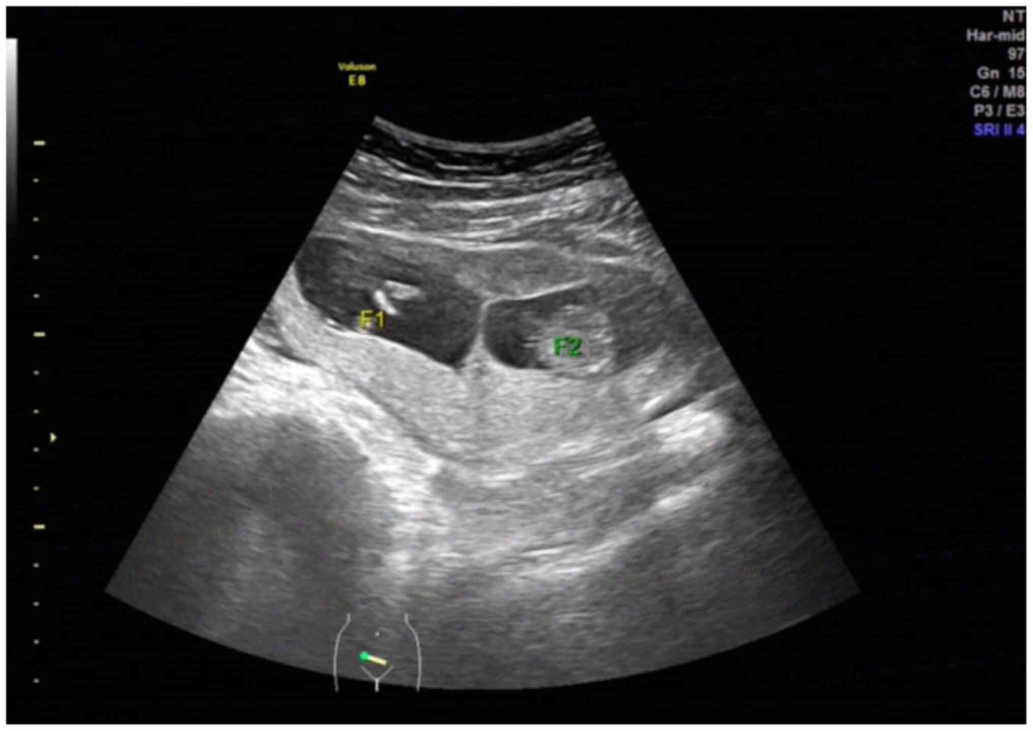
postoperative color Doppler ultrasound showed a twin pregnancy
(Fig. 1). In October 2017, the
patient presented to the antenatal service of the aforementioned
hospital at 13-week gestational age with vaginal bleeding for 4
days and lower abdominal pain for 1 day accompanied by bleeding
gums and skin ecchymosis.
Laboratory blood tests showed hemoglobin (HGB) 110
g/l (normal range, 110-150 g/l), white blood cell (WBC) count
1.70x10⁹/l (normal range, 4-10x109/l),
neutrophils 0.86x10⁹/l (normal range,
1.80-6.30x10⁹/l), platelets (PLT) 77x109/l
(normal range, 100-300x109/l), prothrombin time (PT)
15.20 sec (normal range, 11-13 sec) and D-dimer 8.53 µg/ml (normal
range, <0.5 µg/ml). Serum biochemical tests revealed normal
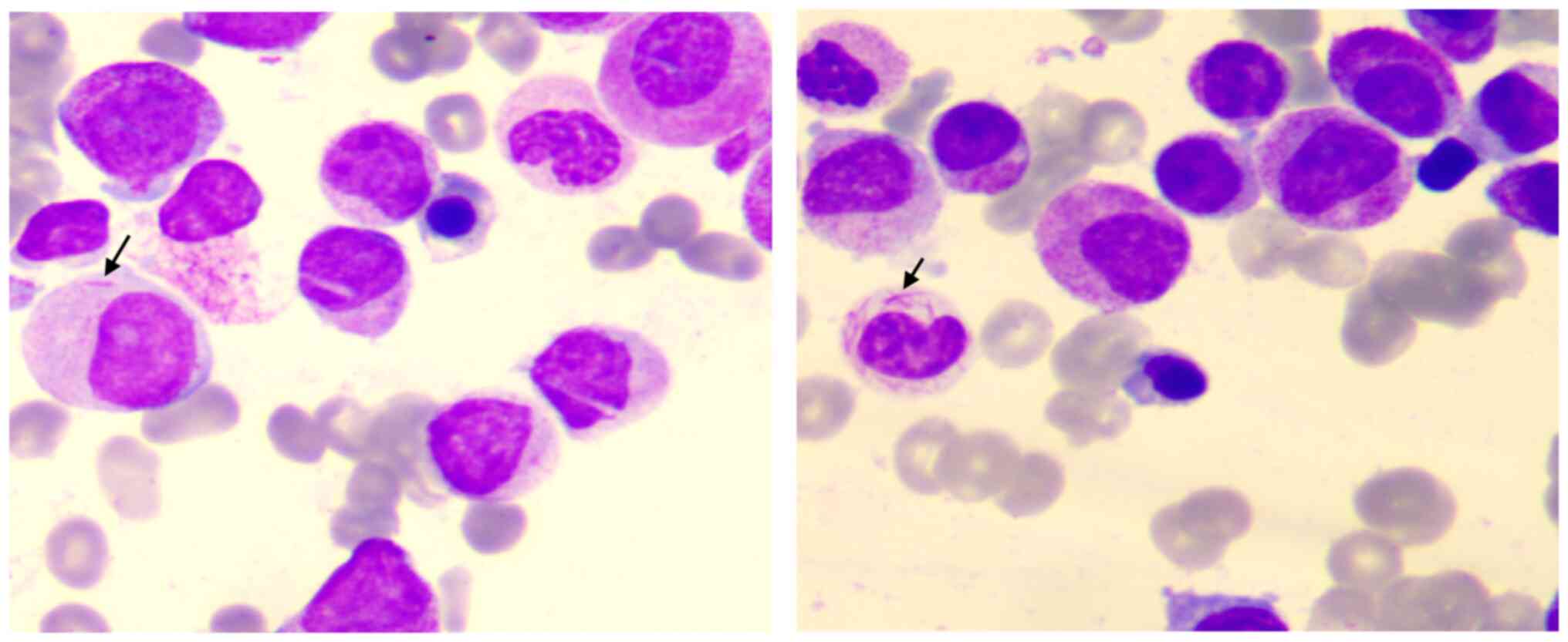
renal and hepatic profiles. The blood film showed abnormal
promyelocytes. Cytomorphological examination of the bone marrow
showed that the proportion of abnormal hypergranular promyelocytes
with Auer rods increased to ~62% (Fig.
2). The results of flow cytometry showed that lymphocytes
accounted for 6.60%, granulocytes for 90.10%, monocytes for 1.70%,
CD45dim cells for 0.80% and CD45- cells for
0.80%. The immunophenotype of abnormal cell population was
CD34-, CD117+, CD33+,
CD13+, HLA-DR-, CD64+,
CD14-, CD56-, CD19-,
CD7-, CD2-, CD5- and
CD20-. Immunophenotyping was consistent with APL
(Fig. S1). Immunophenotyping was
performed on a BD FACSCanto™ Clinical Flow Cytometry (BD
Biosciences) using the FCS Express 3.0 (De Novo Software) flow
cytometry software. The following reagents and antibodies were used
for immunophenotyping: 7-aminoactinomycin D (Beckman Coulter, Inc;
cat. no. A07704); CD15 (BD Biosciences; cat. no. 332778); CD14 (BD
Biosciences; cat. no. 333951); CD8 (BD Biosciences; cat. no.
335822); CD20 (BD Biosciences; cat. no. 335829); CD2 (BD
Biosciences; cat. no. 341024); CD5 (BD Biosciences; cat. no.
341109); CD4 (BD Biosciences; cat. no. 341115); CD10 (BD
Biosciences; cat. no. 341102); CD19 (BD Biosciences; cat. no.
3407224); CD7 (BD Biosciences; cat. no. 340656); CD117 (BD
Biosciences; cat. no. 339206); CD33 (BD Biosciences; cat. no.
340679); cell-surface immunoglobulin (sIg)-λ (BD Biosciences; cat.
no. 555797); sIg-κ (BD Biosciences; cat. no. 555791); CD11b (BD
Biosciences; cat. no. 340936); CD13 (BD Biosciences; cat. no.
347406); CD16 (BD Biosciences; cat. no. 335806); CD56 (BD
Biosciences; cat. no. 555517); human leucocyte antigen (HLA)-DR (BD
Biosciences; cat. no. 642276); CD64 (BD Biosciences; cat. no.
561194); CD3 (BD Biosciences; cat. no. 560176); CD5 (BD
Biosciences; cat. no. 555352); CD71 (BD Biosciences; cat. no.
551374); CD34 (BD Biosciences; cat. no. 555823); CD58 (Beckman
Coulter, Inc; cat. no. IM1218U); CD123 (Beckman Coulter, Inc; cat.
no. B14808); CD9 (BD Biosciences; cat. no. 555372); CD41 (Beckman
Coulter, Inc.; cat. no. 6607117); CD45 (Beckman Coulter, Inc; cat.
no. IM3548U); CD36 (Beckman Coulter, Inc; cat. no. IM0766U); CD38
(Beckman Coulter, Inc; cat. no. IM0775U); CD138 (Beckman Coulter,
Inc; cat. no. A87787); CD200 (BD Biosciences; cat. no. 562126); and
CD61 (Beckman Coulter, Inc; cat. no. IM1758U).
Reverse transcription PCR was positive for the L
subtype of promyelocytic leukemia- retinoic acid receptor α fusion
gene.
A diagnosis of APL was made, and a discussion ensued
with the patient regarding termination, the possible adverse
effects of the chemotherapy on the fetuses and the risks to the
patient in continuing the pregnancy. The patient decided to
continue the pregnancy and received induction chemotherapy with
ATRA-based regimens.
After 3 days from admission, the coagulation
function (PT, 27.60 sec; PT-international normalized ratio, 2.58;
activated partial thromboplastin clotting time, 46.90 sec;
fibrinogen, <0.30g/l; thrombin time, 32.0 sec; D-dimer, 25.83
µg/ml) suggested that the patient was at risk of disseminated
intravascular coagulation (DIC), therefore fresh frozen plasma (4
units) were used for treatment. Significant improvement in
coagulation parameters was observed after 5 days of treatment.
Induction chemotherapy with ATRA [10 mg/m2 ter in
die (tid) day (D)1-D28] + ATO (5 mg/m2 D1; 10
mg/m2 D2-D28) regimen commenced on the 4th day of
admission. On day 17 after the commencement of induction
chemotherapy, laboratory data showed HGB 70 g/l, WBC
6.39x10⁹/l, neutrophils 2.76x10⁹/l, PLT
101x109/l and D-dimer 1.52 µg/ml. A total of two units
of suspended red blood cells were transfused to improve anemia on
D21 and D24 after admission. After the first course of induction
chemotherapy (D34), laboratory data showed that the patient's HGB
remained stable (HGB, 85 g/l), the WBC count was significantly
reduced (3.63x10⁹/l, of which neutrophils were
1.38x10⁹/l) and the PLT count increased
(330x109/l), whereas fibrinogen (3.86 g/l) and D-dimer
(0.83 µg/ml) returned to normal levels (Fig. 3). Moreover, liver and kidney
function were normal. Bone marrow cytomorphology (data not shown)
and flow cytometry (lymphocytes accounted for 19.70%, granulocytes
for 67.90%, monocytes for 4.40%, CD45dim cells for 1.60%
and CD45- cells for 6.40%) showed that the patient
achieved CR (Fig. S2), and no
promyelocyte of abnormal immunophenotype was found. At the same
time, the color Doppler ultrasound (data not shown) showed that the
fetuses were normal, and the rash on the patient's extremities
subsided.
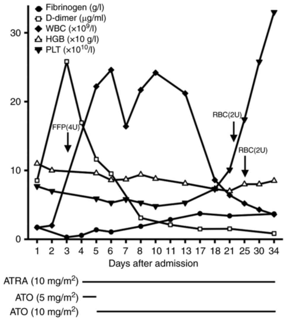 | Figure 3Procedure of first induction
chemotherapy and time course of changes in fibrinogen, D-dimer, WBC
count, HGB and PLT in the patient after admission. Different
parameters were measured at different time points as per the
medical requirements. WBC, white blood cells; HGB, hemoglobin; PLT,
platelets; ATRA, all-trans-retinoic acid; ATO, arsenic trioxide;
FFP, fresh frozen plasma; RBC, red blood cells; U, unit(s). |
The patient then underwent four successive courses
of induction chemotherapy with ATRA + ATO regimen at the hospital
of admission (Table I). During the
third induction chemotherapy, color Doppler ultrasonography of both
lower extremities showed venous thrombosis in the left lower
extremity (data not shown) and anticoagulation therapy with low
molecular weight heparin (200 U/kg) was administered. In addition,
the patient was infected with the influenza B virus and developed
herpes zoster on the left chest wall with hypoalbuminemia during
the fourth induction chemotherapy. The patient was transferred to
the intensive care unit for respiratory isolation, antiviral
therapy with oseltamivir phosphate (75 mg q12h D1-D5) and
intermittent albumin supplementation (5-10 g iv).
 | Table IProcedure for induction
chemotherapy. |
Table I
Procedure for induction
chemotherapy.
| Course no. | Chemotherapy
regimen |
|---|
| 1-5 | ATRA (10
mg/m2 tid D1-D28) + ATO (5 mg/m2 D1, 10
mg/m2 D2-D28) |
| 6 | Cytarabine (100
mg/m2 D1-D5) + Pirarubicin (30 mg/m2
D1-D3) |
| 7 | ATO (10
mg/m2 D1-D15) + Intrathecal chemotherapy (Methotrexate
+ |
| | Cytarabine +
Dexamethasone, D14) + ATRA (10 mg/m2 tid D16-D30) |
| 8 | ATO (10
mg/m2 D1-D15) |
| 9 | ATO (10
mg/m2 D1-D15) + ATRA (10 mg/m2 tid
D16-D30) |
| 10 | ATO (10
mg/m2 D1-D15) |
| 11-13 | ATO (10
mg/m2 D1-D15) + ATRA (10mg/m2 tid
D16-D30) |
| 14-18 | ATO (10
mg/m2 D1-D15) |
At 34 weeks gestational age, the patient developed
edema, increased blood pressure (138-170/85-98 mmHg) and
proteinuria. The obstetrician recommended ending the pregnancy and
two healthy male infants (Apgar score of 10) were successfully
delivered by cesarean section. Subsequently, the patient underwent
consolidation therapy for ~27 months (the therapeutic procedures
are shown in Table I), their
medical condition was stable and they continued to be in CR. During
the ~4-year follow-up period, the two infants were not found to
have any health problems.
Discussion
Cases of APL in pregnancy are rare. The risks of APL
in pregnancy include sequelae of abortion, perinatal mortality,
intrauterine growth retardation, preterm delivery (7), high risk of bleeding, infection,
inflammation and placental abruption (8). Therefore, timely treatment of
maternal leukemia is necessary. The current management of APL in
pregnancy is a challenge, as it cannot be based on evidence from
well-designed trials, but instead relies on data from historical
cases and discussions with individual patients. The present patient
presented to the hospital at 13 weeks gestational age and elected
to continue with the pregnancy and receive induction chemotherapy
for her leukemia after an informed discussion.
A retrospective study by Santolaria et al
(3) showed that among pregnant
patients with APL, most of them were treated with ATRA alone (32%)
or combined with chemotherapy (cytarabine or daunorubicin) (43%),
while the remaining patients received chemotherapy alone and a
small number of patients were treated with ATO-based regimens after
delivery. ATRA is controversial during pregnancy owing to the
teratogenicity and fatal retinoic acid syndrome, especially in the
first 3-5 weeks of gestation (9,10).
Other complications include craniofacial alterations, neural tube
defects, cardiovascular malformations, thymic aplasia and
psychological impairments (9,11).
However, there are currently data showing that ATRA appears to be
reasonably safe and well tolerated if given outside the first
trimester (12). In the present
case, the patient received an ATRA-based induction chemotherapy
regimen at 13 weeks of gestational age and achieved CR status. It
should be noted that clinical bleeding problems were more marked
before the introduction of ATRA, therefore continued vigilance
during induction therapy was required to monitor and prevent fatal
bleeding. Throughout the course of treatment, the occurrence of DIC
was successfully prevented through blood infusion and continuous
monitoring of coagulation factors. Once initial chemotherapy had
been administered and remission had been achieved, the subsequent
course was simpler. The present study showed that CR is likely to
be achieved with maintenance chemotherapy and ATRA.
Over the last two decades, single administration or
combination of ATO with other agents have been successfully used
for the treatment of APL and several other myeloid tumors, such as
non-APL acute myeloid leukemia (13). However, as stated in the European
LeukemiaNet recommendations and other published guidelines for the
treatment of APL, its use in pregnant women should be avoided due
to its teratogenic effects (2,14).
In animal studies, arsenic has been reported to cause anencephaly,
cranial neural tube defects, that affect embryonic growth by
altering the glucocorticoid signaling system during embryonic
development and several different maternal toxicities (15). In the present case, the decision of
treating the patient with ATO was taken due to their strong desire
to protect the fetuses in the womb and the fact that the pregnancy
was in the second trimester. Considering a large amount of
trivalent arsenic excretion in breast milk and the possible fetal
complications caused by ATO (16),
strict feto-maternal surveillance and withheld breastfeeding were
performed. It has been reported that the sequel of arsenic exposure
to the fetus in pregnancy has been based on long-term exposure to
high doses, mostly from environmental sources, such as drinking
water (17,18). To the best of our knowledge,
pregnant women that have received APL and were cured without
affecting delivery in previous cases were relatively rare (2) and very few patient cases were treated
with ATO + ATRA regimen (19-21),
which is what makes the present case special.
In conclusion, the present study reports the case of
successful treatment of APL in a pregnant woman of 13-week
gestational age with an ATRA + ATO-based induction regimen, without
any feto-maternal complications, provided the patient is managed
strictly clinically. In-depth research to verify this observation
is necessary.
Supplementary Material
Images from the flow cytometry
analysis of bone marrow cells before treatment. Markers run: CD5,
CD7, CD56, CD8, CD4, CD3, CD2, CD10, CD19, CD20, CD14, CD13, CD64,
CD16, CD11b, CD15, CD36, CD33, CD34, CD117, CD71, HLA-DR, CD38,
CD138, CD200, CD61, CD45, sIg-κ and sIg-λ. sIg, cell-surface
immunoglobulin; HLA, human leucocyte antigen; granu, granulocyte;
lym, lymphocyte; Mono, monocyte; NEG, negative.
Images from the flow cytometry
analysis of bone marrow cells after CR. No promyelocytes with
abnormal immunophenotype were found. Markers run: CD58, CD10, CD34,
CD19, HLA-DR, CD33, CD117, CD123, CD9, CD71, CD41, and CD45. HLA,
human leucocyte antigen.
Acknowledgements
Not applicable.
Funding
Funding: This research was funded by the Social Welfare and
Basic Research Project Fund of Zhongshan City (grant. no.
2021B1088).
Availability of data and materials
All data generated or analyzed during this study are
included in this published article.
Authors' contributions
WN and KD confirm the authenticity of all the raw
data. WN, KD, YC, LL, JL, WJ and LW collected and analyzed cell
morphology and Doppler ultrasound images, complete blood count and
serum biochemical data, flow cytometry data, and other clinical
information. WN wrote the manuscript. JL, YC and LL revised the
manuscript. All authors have read and approved the final
manuscript.
Ethics approval and consent to
participate
The present study was conducted according to the
guidelines of the Declaration of Helsinki and approved by the
Ethics Committee of Zhongshan Boai Hospital Affiliated to Southern
Medical University (Zhongshan, China). Written informed consent was
obtained from the patient.
Patient consent for publication
Written informed consent was obtained from the
patient for the publication of the data and images in this case
report.
Competing interests
The authors declare that they have no competing
interests.
References
|
1
|
Gong S, Wang H, Zhang H, Liu W, Zhang X
and Zhao C: Real-world data on the dose-related effect of arsenic
trioxide in the relapse of acute promyelocytic leukemia. Mol Clin
Oncol. 213(91)2020.PubMed/NCBI View Article : Google Scholar
|
|
2
|
Sanz MA, Fenaux P, Tallman MS, Estey EH,
Löwenberg B, Naoe T, Lengfelder E, Döhner H, Burnett AK, Chen SJ,
et al: Management of acute promyelocytic leukemia: Updated
recommendations from an expert panel of the European LeukemiaNet.
Blood. 133:1630–1643. 2019.PubMed/NCBI View Article : Google Scholar
|
|
3
|
Santolaria A, Perales A, Montesinos P and
Sanz MA: A cute promyelocytic leukemia during pregnancy: A
systematic review of the literature. Cancers (Basel).
12(968)2020.PubMed/NCBI View Article : Google Scholar
|
|
4
|
Saleh AJ, Alhejazi A, Ahmed SO, Al Mohareb
F, AlSharif F, AlZahrani H, Mohamed SY, Rasheed W, AlDawsari G,
Ibrahim K, et al: Leukemia during pregnancy: Long term follow up of
32 cases from a single institution. Hematol Oncol Stem Cell Ther.
7:63–68. 2014.PubMed/NCBI View Article : Google Scholar
|
|
5
|
Jain N, Hubbard J, Vega F, Vidal G,
Garcia-Manero G and Borthakur G: Spontaneous remission of acute
myeloid leukemia: Report of three cases and review of the
literature. Clin Leuk. 2:64–67. 2008.
|
|
6
|
Pentheroudakis G, Orecchia R, Hoekstra HJ
and Pavlidis N: ESMO Guidelines Working Group. Cancer, fertility
and pregnancy: ESMO clinical practice guidelines for diagnosis,
treatment and follow-up. Ann Oncol. 21 (Suppl 5):v266–v273.
2010.PubMed/NCBI View Article : Google Scholar
|
|
7
|
Chelghoum Y, Vey N, Raffoux E, Huguet F,
Pigneux A, Witz B, Pautas C, de Botton S, Guyotat D, Lioure B, et
al: Acute leukemia during pregnancy: A report on 37 patients and a
review of the literature. Cancer. 104:110–117. 2005.PubMed/NCBI View Article : Google Scholar
|
|
8
|
Rizack T, Mega A, Legare R and Castillo J:
Management of hematological malignancies during pregnancy. Am J
Hematol. 84:830–841. 2009.PubMed/NCBI View Article : Google Scholar
|
|
9
|
Azim HA, Pavlidis N and Peccatori FA:
Treatment of the pregnant mother with cancer: A systematic review
on the use of cytotoxic, endocrine, targeted agents and
immunotherapy during pregnancy. Part II: Hematological tumors.
Cancer Treat Rev. 36:110–121. 2010.PubMed/NCBI View Article : Google Scholar
|
|
10
|
Fenaux P, Chevret S, Guerci A, Fegueux N,
Dombret H, Thomas X, Sanz M, Link H, Maloisel F, Gardin C, et al:
Long-term followup confirms the benefit of all-trans retinoic acid in
acute promyelocytic leukemia. European APL group. Leukemia.
14:1371–1377. 2000.PubMed/NCBI View Article : Google Scholar
|
|
11
|
Valappil S, Kurkar M and Howell R: Outcome
of pregnancy in women treated with all-trans retinoic acid: A case
report and review of literature. Hematology. 12:415–418.
2007.PubMed/NCBI View Article : Google Scholar
|
|
12
|
Giagounidis AA, Beckmann MW, Giagounidis
AS, Aivado M, Emde T, Germing U, Riehs T, Heyll A and Aul C: Acute
promyelocytic leukemia and pregnancy. Eur J Haematol. 64:267–271.
2002.PubMed/NCBI View Article : Google Scholar
|
|
13
|
Hoonjan M, Jadhav V and Bhatt P: Arsenic
trioxide: Insights into its evolution to an anticancer agent. J
Biol Inorg Chem. 23:313–329. 2018.PubMed/NCBI View Article : Google Scholar
|
|
14
|
Pagnano KB, Rego EM, Rohr S, Chauffaille
Mde L, Jacomo RH, Bittencourt R, Firmato AB, Fagundes EM, Melo RA
and Bernardo W: Guidelines on the diagnosis and treatment for acute
promyelocytic leukemia: Associação Brasileira de Hematologia,
Hemoterapia e Terapia Celular guidelines project: Associação Médica
Brasileira-2013. Rev Bras Hematol Hemoter. 36:71–92.
2014.PubMed/NCBI View Article : Google Scholar
|
|
15
|
Caldwell KE, Labrecque MT, Solomon BR, Ali
A and Allan AM: Prenatal arsenic exposure alters the programming of
the glucocorticoid signaling system during embryonic development.
Neurotoxicol Teratol. 47:66–79. 2015.PubMed/NCBI View Article : Google Scholar
|
|
16
|
Samanta G, Das D, Mandal BK, Chowdhury TR,
Chakraborti D, Pal A and Ahamed S: Arsenic in the breast milk of
lactating women in arsenic-affected areas of West Bengal, India and
its effect on infants. J Environ Sci Health A Tox Hazard Subst
Environ Eng. 42:1815–1825. 2015.PubMed/NCBI View Article : Google Scholar
|
|
17
|
Nyanza EC, Dewey D, Manyama M, Martin JW,
Hatfield J and Bernier FP: Maternal exposure to arsenic and mercury
and associated risk of adverse birth outcomes in small-scale gold
mining communities in northern Tanzania. Environ Int.
137(105450)2015.PubMed/NCBI View Article : Google Scholar
|
|
18
|
Vahter M, Skröder H, Rahman SM, Levi M,
Derakhshani Hamadani J and Kippler M: Prenatal and childhood
arsenic exposure through drinking water and food and cognitive
abilities at 10 years of age: A prospective cohort study. Environ
Int. 139(105723)2020.PubMed/NCBI View Article : Google Scholar
|
|
19
|
Naithani R, Dayal N, Chopra A and Sundar
J: Fetal outcome in pregnancy with acute promyelocytic leukemia.
Indian J Paediatr. 83:752–753. 2016.PubMed/NCBI View Article : Google Scholar
|
|
20
|
Fei F, Faye-Petersen OM, Vachhani P, Jamy
O and Reddy VV: Acute promyelocytic leukemia during pregnancy: A
case report and 10-year institutional review of hematologic
malignancies during pregnancy. Pathol Res Pract.
215(152672)2019.PubMed/NCBI View Article : Google Scholar
|
|
21
|
Cochet C, Simonet M, Cattin J, Metz JP,
Berceanu A, Deconinck E, Daguindau E, Schillinger F, Fenaux P,
Mottet N and Desbrosses Y: Arsenic trioxide treatment during
pregnancy for acute promyelocytic leukemia in a 22-year-old woman.
Case Rep Hematol. 2020(3686584)2020.PubMed/NCBI View Article : Google Scholar
|