Angiogenesis is the process that leads to the
formation of new blood vessels, and, if induced by tumors, may also
contribute to the growth of the disorganized vasculature able to
sustain cancer progression over 2–3 mm and metastasis (1). The events that trigger tumor
angiogenesis derive from the interaction between cancer cells and
host microenvironment that includes immune cells, connective tissue
and soluble factors. Vascular endothelial growth factor (VEGF) and
its receptor (VEGFR) are the main contributors to proliferation of
endothelial cells, thus representing suitable targets for
antiangiogenic therapies (2).
Vascular endothelial growth factor is the most
important mediator of angiogenesis. It is overexpressed in various
tumors, stimulating endothelial cell proliferation and migration,
and leading to the formation of new blood vessels from pre-existing
ones (3–8). The VEGF family is composed of five
glycoproteins (VEGF-A, VEGF-B, VEGF-C, VEGF-D and VEGF-E). VEGF-A
is a homodimeric, disulfide-bound glycoprotein, which exists in
several isoforms with different numbers of amino acid residues,
such as VEGF121, VEGF189 and VEGF165. Different VEGF-A isoforms
exhibit varying biological properties, such as the ability to bind
to cell surface heparin sulfate proteoglycans. VEGF121, commonly
existing as a homodimer, is freely diffusible without heparin
binding. The angiogenic actions of VEGF are mediated primarily via
two closely related endothelium-specific receptor tyrosine kinases,
Flt-1 (VEGFR1) and Flk-1/KDR (VEGFR2) (9). Both are largely restricted to
vascular endothelial cells and are overexpressed on the endothelium
of tumor vasculature, yet they are almost undetectable in the
vascular endothelium of adjacent normal tissues (10). All of the VEGF-A isoforms bind to
both VEGFR1 and VEGFR2.
VEGF and its receptors are overexpressed in a
variety of solid tumor biopsy specimens, and overexpression of
VEGFR2 or VEGF-A has been considered as a poor prognostic marker in
various clinical studies (11–13).
Indeed, new vasculature allows tumor cells to grow by supplying
nutrients and oxygen, enabling disposal of metabolic waste products
and providing a route for metastatic spreading. VEGF production by
tumor cells is thought to be regulated by hypoxemia, growth factors
signaling, cytokines, and cell differentiation (8).
Given the role of VEGF and VEGFR in several
oncological and non-oncological diseases, pharmaceutical companies
and researchers are deeply involved in developing agents
potentially useful in the prevention of VEGF-A binding to its
receptors (14), or antibodies
blocking VEGFR2 (11) or small
molecules that inhibit the kinase activity of VEGFR2 (7,15)
and thereby block growth factor signaling. Indeed, VEGF/VEGFR
targeting has already been proved successful in many cancer types
(16).
VEGF-A and its receptors are the best-characterized
signaling pathway in developmental angiogenesis as well as tumor
angiogenesis (10). VEGFR2 appears
to be the most important receptor in VEGF-induced mitogenesis,
angiogenesis, and permeability increase, whereas the role of VEGFR1
in endothelial cell function is less clear (17). During the exponential growth stage,
VEGFR expression is highly upregulated on the newly developed tumor
vasculature. Being the naturally existing VEGFR ligand, VEGF121
offers several advantages over the synthetic small-molecule VEGFR
ligands or anti-VEGFR antibodies. It has much higher binding
affinity to VEGFR (nanomolar range) than reported peptidic VEGFR
inhibitors (submicromolar to micromolar range) (18,19).
If compared to antibody-based radiopharmaceuticals, VEGF121 clears
much faster from the blood pool and the non-targeting organs
because of its smaller size.
Regulation of inflammatory cell recruitment by
VEGFR1 appears to be exerted mainly through placental growth factor
(PGF). Notably, the expression of PGF is very low under
physiological conditions, but it may be strongly upregulated in
various cell types by different pathological stimuli such as
hypoxia, inflammatory cytokines, or oncogenes (20–22).
PGF has recently been regarded as an attractive candidate for
anti-angiogenic therapy. Indeed, it has been shown that PGF plays a
key role in promoting pathological angiogenesis associated with
tumor progression (23) and
overexpression of PGF in a mouse melanoma model resulted in
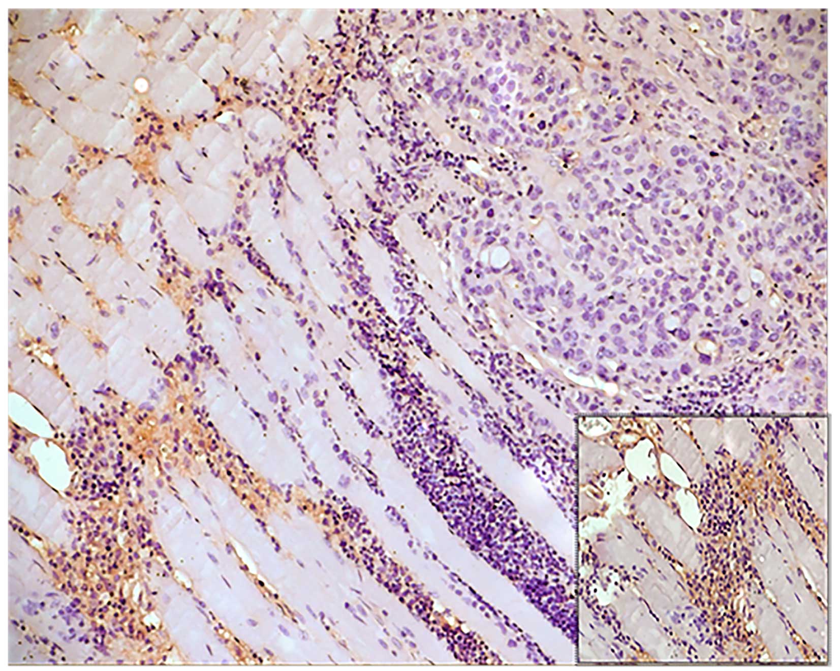
increased tumor growth and metastasis (24). Tumor cells may also express VEGFR2,
although epithelial and mesenchymal tumor cells typically express
VEGFR1 rather than VEGFR2 (25,26)
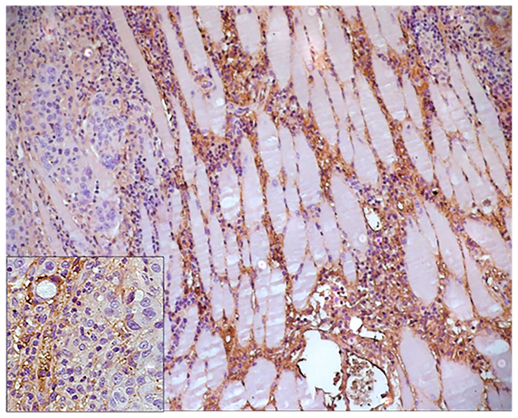
(Fig. 1). Nevertheless, increased
expression of VEGFR2 on tumor cells has been described for melanoma
and hematological malignancies (27). It has been shown that
VEGFR2-mediated signaling allowed survival of cancer cells under
chronic hypoxic conditions and might contribute to a more
aggressive phenotype (28)
(Fig. 2).
Growing evidence supports an important link between
chronic inflammation and tumor development. Induction of VEGFR2
expression in tumor cells, and also in intestinal epithelium during
colitis, is mediated by the pro-inflammatory cytokine
interleukin-6, which is a strong promoter of tumor growth in
experimental colitis-associated colon cancer (29). A soluble form of the VEGFR2
(sVEGFR2) has been also described and may have important biological
roles. sVEGFR2 binds VEGF-C and prevents activation of VEGFR3,
consequently inhibiting lymphatic endothelial cell proliferation
(30). Notably, regulation of
sVEGFR2 in advanced metastatic neuroblastoma may promote
lymphogenic spread of metastases (31). The expression of VEGFR3 in tumor
cells is still controversial (32); however, it has been ascertained
that inhibition of VEGFR3 activity arrests tumor vascularization,
leading to decreased vascular density in several tumor models
(33). The axis VEGFC/VEGFR3 plays
a fundamental role in the tumor microenvironment by promoting the
formation of new lymphatic vessels from pre-existing ones (34). VEGFC, produced by neoplastic cells,
induces lymphatic endothelial destabilization, resulting in
endothelial sprouting as well as leakage and enlargement of the
vessels. These modifications induce entry of tumor cells into the
lymphatics vessels and further dissemination of metastasis to
sentinel lymph nodes (35,36).
High expression of VEGFs and/or VEGFRs in various
tumor biopsy specimens is indicative of poor prognosis for cancer
patients (2,37,38).
Therefore, non-invasive imaging and quantification of VEGFR
expression is of relevant importance in cancer patient management.
Many strategies have been adopted to block the VEGF/VEGFR signaling
pathway for cancer treatment, such as agents that can bind to
VEGF-A to prevent its interaction with VEGFRs (such as bevacizumab,
and VEGF-trap) (39,40) antibodies/antibody fragments that
target VEGFR-2 (ramucirumab, and CDP791) (41,42)
and small molecule inhibitors that interrupt the downstream
signaling of VEGFR-2 (axitinib, sunitinib, and sorafenib) (43,44).
Many of these agents have been approved by the Food and Drug
Administration (FDA) for various medical indications in cancer
therapy (2,45).
VEGFR-2 mediates the majority of VEGF-A signaling in
the tumor microenvironment including microvascular permeability and
endothelial cell proliferation (8,10).
Several agents, including antibodies and soluble receptor
constructs, have been developed to target the VEGF system. The drug
that is currently most widely used in the clinical practice to
modulate VEGF-A is the humanized monoclonal antibody. It blocks
VEGF-induced endothelial cell proliferation, permeability, and
survival, and it inhibits human tumor cell line growth. The likely
mechanism is that bevacizumab binds to VEGF both soluble and bound
to the extracellular matrix and thereby prevents VEGF binding to
its receptors, blocking the biologic pathways induced after VEGF
binding. Bevacizumab is approved both by the United States Food and
Drug Administration (FDA) and the European Medicines Agency (EMA)
for the treatment of metastatic colorectal cancer, non-small cell
lung cancer, breast cancer and glioblastoma multiforme in
combination with chemotherapy (46,47).
One of the greatest challenges in bevacizumab therapy is the lack
of predictive biomarkers and tools that can predict the efficacy of
anti-VEGF therapy (47).
Development of anti-angiogenic therapy including
anti-VEGF antibodies and VEGF-tyrosine kinase receptors has been a
major landmark in cancer therapy leading to improvement in survival
in several cancers. The pharmacologic inhibition of angiogenesis
via the VEGF pathway is an important therapeutic approach that
prevents cancer growth and metastasis formation. In addition to
anti-VEGF antibodies, other strategies have been explored and
include the blocking of its signaling receptor, receptor tyrosine
kinase inhibitors (16,48–50),
and gene therapy approaches, in which the vector produces an
antisense molecule or a soluble receptor that acts in a
dominant-negative manner (51).
Several studies have shown that anti-VEGF treatment,
in association with chemotherapy (52) or radiation therapy (53,54),
results in greater antitumor effects than either treatment alone.
An issue that is now being debated is the mechanism of such
potentiation, and a variety of hypotheses, which are not mutually
exclusive, have been put forward. Klement et al proposed
that chemotherapy, especially when delivered at low dose,
preferentially damages endothelial cells and the blockade of VEGF
blunts a key survival signal for endothelial cells, thereby
amplifying the antitumor-cell effects of chemotherapy (52). Jain suggested that antiangiogenic
therapy ‘normalizes’ the tumor vasculature, leading to pruning of
excessive endothelial cells and perivascular cells, reduction in
vessel tortuosity and drop in interstitial pressure and consequent
improved oxygenation and delivery of chemotherapy to tumor cells
(55). These effects are
accompanied by a reduction in permeability of macromolecules
(56,57). Willett et al have recently
shown that VEGF blockade by bevacizumab decreases tumor perfusion,
vascular volume, microvascular density, interstitial fluid pressure
and the number of viable circulating endothelial and progenitor
cells in colorectal cancer patients (58). Surprisingly, these studies have
also shown that permeability to small molecules actually increases
following VEGF blockade (58).
Bevacizumab was initially approved for the treatment
of metastatic colorectal cancer in combination with intravenous
5-fluorouracil-based chemotherapy (59). Subsequently, bevacizumab was
approved for various indications in non squamous cell lung
carcinoma (NSCLC), metastatic renal cell carcinoma, and
glioblastoma multiforme (38,60–63).
The antitumor activity of bevacizumab is primarily manifested in
combination with chemotherapy, except for renal cell carcinoma,
where it has shown efficacy as a single agent (64). Presently, bevacizumab is being used
in nearly 1,000 clinical trials, and despite promising results, its
effects in many types of cancer are modest or even irrelevant
(65). Furthermore, recent studies
have raised the possibility that treatment with bevacizumab may be
associated with a more aggressive invasive tumor phenotype,
particularly in glioblastoma (66,67),
which is often a greatly vascularized brain tumor. Although the
clinical impact of these results is far from clear, it is obvious
that anti-angiogenic therapy will have to be closely evaluated
depending on disease stage and molecular profile of different
patients and tumors. Preclinical data with anti-VEGFR2 antibodies
have demonstrated a reduction in VEGF-induced signaling as well as
angiogenesis and primary or metastatic growth in a variety of
different tumor models (7,68,69);
therefore, the specific antibody-based blockade of VEGFR2 has also
received special attention in clinical trials.
Ramucirumab (IMC-1121B; Imclone Systems) is
currently being tested in several clinical trials, including breast
cancer, gastric cancer, and HCC (70). Basing on preliminary results, this
antibody has shown activity in patients previously treated with
other antiangiogenic agents, suggesting a more efficient antitumor
response by direct targeting of VEGFR2.
Small molecule inhibitors of VEGFR tyrosine kinase
activity represent another major approach to blocking VEGF-mediated
angiogenesis. Several tyrosine kinase inhibitors have been
developed to selectively inhibit VEGFR2, but they have also
activity on other VEGFRs and tyrosine kinase receptors, including
basic fibroblast growth factor (FGF) receptor, EGFR family members,
PDGFR-a, PDGFR-b, c-kit, and Flt3. Sunitinib was approved in 2006
for its clinical use in imatinib-resistant gastrointestinal stromal
tumors and advanced metastatic renal cell carcinoma (71,72),
whereas sorafenib received FDA approval for the treatment of
metastatic renal cell carcinoma (73) and HCC (74). Sunitinib and sorafenib have shown
clinical efficacy as single agents, possibly due to their ability
to inhibit multiple RTKs and in particular those regulating tumor
angiogenesis. Additional clinical trials aimed to evaluate
combinations of sorafenib and sunitinib with different
chemotherapeutic agents and other antiangiogenic agents are under
evaluation.
There has been a worldwide research program to
develop antiangiogenic agents for the treatment of cancer. Many
families of antiangiogenic drugs now exist, but their clinical
development has been hampered by scarce data concerning the optimal
biologically active dose. In addition, although the classical phase
I study design focuses on toxicity as an endpoint to establish the
maximum tolerated dose, many humanized monoclonal antibodies have
no clinically significant toxicity, which precludes identification
of the maximum tolerated dose. Furthermore, biologic dose-response
relationships may follow a bell-shaped curve (75) and therefore the maximum tolerated
dose may not even be the best dose for clinical applications. To
overcome these issues, biologic pharmacodynamic investigations
(76) have entered phase I
clinical trial design with the goal of establishing the optimum
biologically active dose.
Antiangiogenic therapies are promising approaches
for cancer treatment. However, their systematic application remains
problematic because of poor understanding of mechanisms of action
and occurrence of resistance (77). Indeed, a significant fraction of
patients do not respond to antiangiogenic drugs (78), whereas those who respond have
relatively modest benefits, mostly in progression-free survival
rather than in overall survival. In addition, a number of
significant toxicities have been observed in patients treated with
antiangiogenic agents, emphasizing that a careful assessment of the
risk-benefit ratio needs to be conducted in individual patients.
Despite disease stabilization and increase in the proportion of
patients with progression-free survival, tumors eventually become
resistant to antiangiogenic agents and relapse (79–82).
Antiangiogenic therapy depends on several factors,
including the tumor stage, the nature of the tumor vascular bed and
the origin and genotype of the neoplastic cells. Tumorigenesis
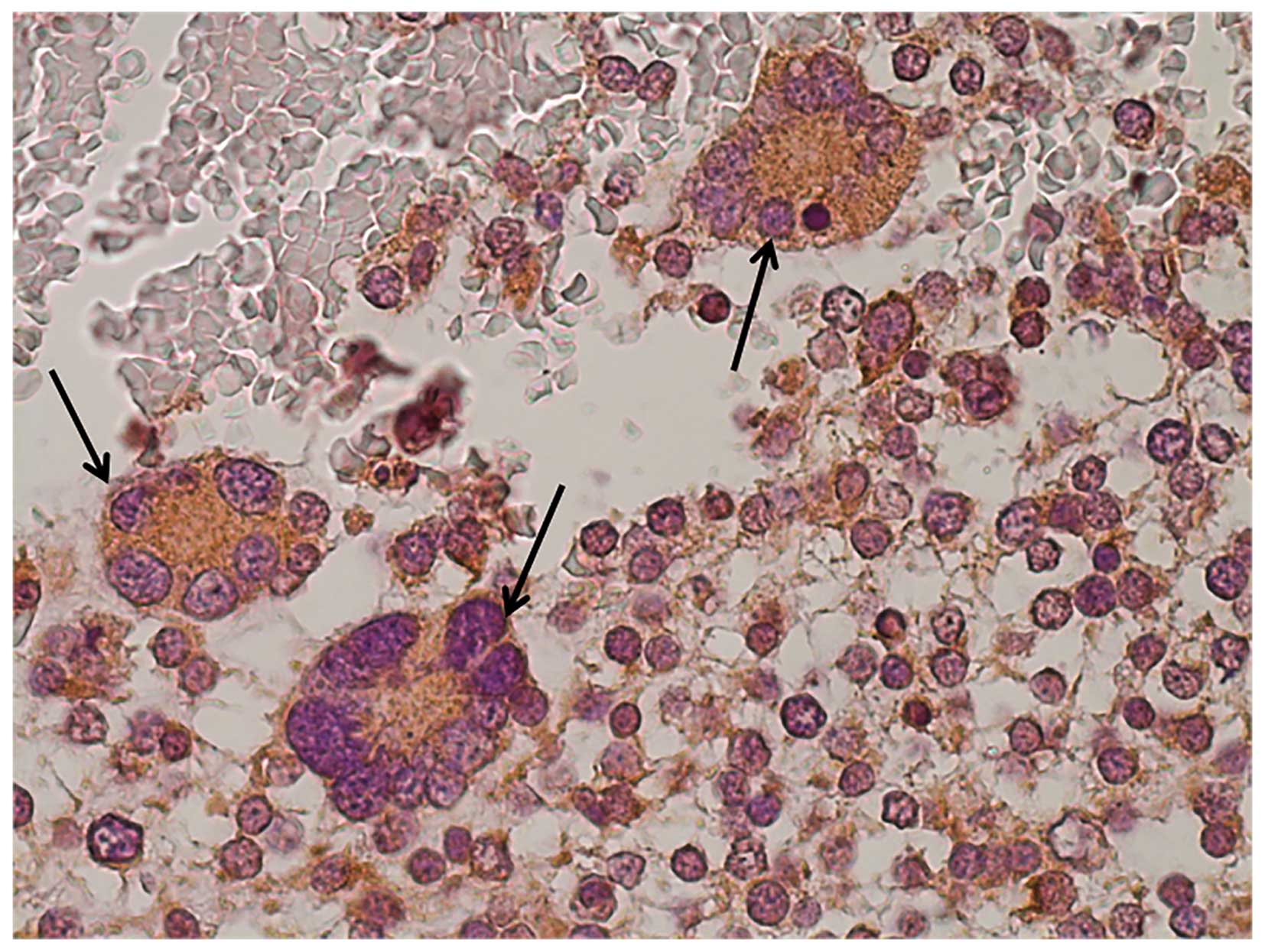
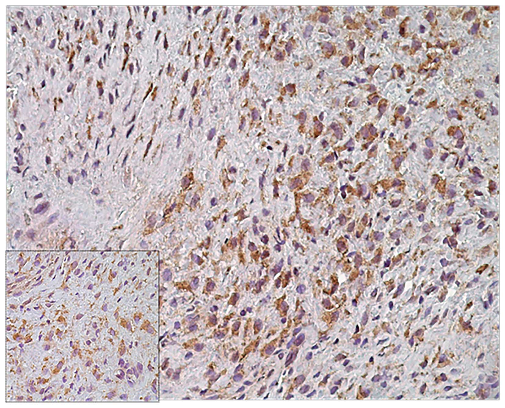
(17), and progression (83) are often associated with a modified
expression of different angiogenic factors (83) (Figs.
3 and 4). Advanced human
breast cancers may express different pro-angiogenic factors,
including VEGF, acidic and basic fibroblast growth factors (aFGF
and bFGF), transforming growth factor β1 (TGFβ1), platelet-derived
growth factor (PDGF), placental growth factor (PGF) and
pleiotrophin (83). The mechanism
of action of certain drugs is also different at various stages of
tumorigenesis. For example, the release of VEGF, following the
remodeling of the extracellular matrix by matrix metalloproteinase
9 (MMP9), is reported to be a component of the RIP1-Tag2 angiogenic
switch (84–88). Inhibition of VEGF is not effective
against established β-cell islet tumors (85,89),
and this finding may lead to hypothesize that the vasculature
matures with increased pericyte coverage, thereby reducing
dependence on VEGF2. The success of targeted therapies, such as
trastuzumab (Genentech), is often dependent on the expression of
the drug target by the tumor (90). Given that bevacizumab is a
monoclonal antibody with a well-defined target, VEGF2, it is
logical that VEGF expression might predict benefit. However, in
retrospective subset analyses, VEGF expression by primary tumors of
metastatic, treatment-refractory breast cancers (91,92)
or metastatic colorectal cancers did not predict benefit from the
addition of bevacizumab (93). The
reasons responsible for this behavior are not entirely clear.
Perhaps VEGF expression by primary tumors is not representative of
metastatic disease, but detailed research indicates that they are
equivalent (44).
CT remains still a fundamental imaging technique in
the diagnosis of neoplastic human pathologies. Positron emission
tomography (PET), very sensitive technique (down to 10–12 molar)
and quantitative with superb tissue penetration, has been widely
used in clinical oncology for tumor staging and treatment
monitoring, where 18F-FDG was used as the tracer for
measuring tumor glucose metabolism (47). High-resolution PET scanners
continue to be developed and made available for imaging small
animals, improving the capacity for in vivo studies in mice,
primates, and humans.
As already discussed, antiangiogenic targeted
therapies are a promising approach for the treatment of cancer.
However, clinical trials showed variable response due to intra- and
inter-tumor heterogeneity and non-invasive tools to monitor
treatment response and drug efficacy are needed. Several methods
have been developed to image tumor angiogenesis, but there is no
general agreement as to which strategy is the most suitable for
monitoring antiangiogenic therapy in single-center and multicenter
trials. There is also evidence that angiogenic imaging data may be
a useful predictor of response to chemo-radiotherapy, the success
of which depends on good perfusion of the tumor. Personalized
medicine allows to identify the suitable patient population for the
appropriate therapy at the right time, as well as to provide
quantitative, non-invasive, and accurate information on the
therapeutic responses in real-time. In this scenario nuclear
medicine offers several radiopharmaceuticals for ‘in vivo’
imaging of angiogenic markers, but to date, none emerged as a gold
standard. As an example, radiolabeled bevacizumab is one of the
most studied radiopharmaceuticals since it is able to bind VEGF
with high affinity. Indeed, development of a bevacizumab-based
imaging agent can play important roles in these aspects, as well as
elucidating the function and modulation of VEGF/VEGFR signaling
during cancer development/intervention.
Being the most important angiogenic effector and
already established therapeutic target, many VEGF-targeting
radio-pharmaceuticals were developed and studied in vitro
and in vivo. In particular, the mAb bevacizumab is one of
the most studied radiolabelled anti-VEGF drugs and, to date, it has
been labeled with a number of PET isotopes such 89Zr (94), 124I (95), 86Y (96), and 64Cu (97). In addition, it has also been
investigated with various other imaging techniques such as single
photon emission computed tomography (SPECT) (98), ultrasound (99), and optical imaging (100). Studies with radiolabeled
bevacizumab for imaging tumor angiogenesis were performed in
preclinical models proposing that its accumulation in the tumor was
due to interactions with the VEGF-A-165 and -189 isoforms,
associated with the tumor cell surface and/or the extracellular
matrix (101,102). However, in a clinical study with
111In-bevacizumab in patients affected by colorectal
cancer liver metastases, there was a lack of correlation between
radiolabeled bevacizumab uptake and VEGF-A expression in the
lesions (103). Authors
speculated that the accumulation of the mAb was due to enhanced
vascular permeability leading to unspecific uptake in the tumor.
This could limit the usefulness of radiolabeled bevacizumab in
imaging tumor angiogenesis. However, this radiopharmaceutical
showed promising results in many other cancers such as breast
cancer. Various studies have reported overexpression of VEGF-A in
the breast cancer microenvironment, compared with normal breast
tissue (104–106). All VEGF-A splice variants are
bound by the clinically used monoclonal antibody bevacizumab. When
labeled with the PET isotope 89Zr, it preserves its VEGF-A-binding
properties. Thus, tracer dosages of radiolabeled bevacizumab can be
used for tumor-specific, whole-body imaging of VEGF-A. In
preclinical studies (94,101) and in a study in renal cell cancer
patients (107), we have already
shown an excellent tumor-to-background ratio with an optimum at 4 d
after tracer injection when using 89Zr-bevacizumab.
89Zr-bevacizumab might be potentially valuable for biologic
characterization of tumors and for prediction and evaluation of the
effect of VEGF-A-targeting therapeutics. VEGF-A is reported in
several studies to be over-expressed in malignant breast tumors and
in ductal carcinoma in situ (106,108), thus covering the full spectrum
from early-stage breast cancer to more advanced stages. More
frequent VEGF-A staining was found to be related to aggressiveness
as assessed by VEGF-A staining in a study with 1,788 breast tumors
(106). 89Zr-bevacizumab PET
proved to be able to detect a broad range of VEGF-A expression
levels. Quantitative tumor analyses showed a >10-fold difference
between individual SUVmax measurements, suggesting large
differences in VEGF-A tumor levels between patients,
89Zr-bevacizumab might be potentially valuable for biologic
characterization of tumors and for prediction and evaluation of the
effect of VEGF-A-targeting therapeutics. Because of better and more
accurate scatter and attenuation corrections associated with PET,
86Y-labeled bevacizumab was developed for imaging VEGF-A tumor
angiogenesis and as a surrogate marker for 90Y-based RIT. The
111In and 89Zr-labeled probes have been proposed as
surrogate imaging markers for 90Y therapy, however, deviations were
observed due to subtle differences in the metalchelate complexes
and metabolism (102,109) highlighting the need for the
development of isotopically matched 86Y-labeled probes for 90Y.
However, 86Y possesses its own set of challenges, in particular,
its high positron energy (Emax 1/4 3.1 MeV) and emission of 1.08
MeV (83% abundance), which can significantly affect the image
quality and recovery coefficients due to spurious coincidences.
When appropriate corrections are performed, the image quality is
greatly improved and is quantifiable (110,111).
PET imaging with 86Y-CHX-A00-DTPA-bevacizumab may
have a useful role in patient selection for bevacizumab-related
therapy as it would indicate accessibility of the antibody to
VEGF-A target sites. However, 86Y-CHX-A00-DTPA-bevacizumab imaging
by itself may not predict the response to therapy as it is only
indicative of how much bevacizumab reaches the tumor and not the
overall tumor microenvironment and the biomolecular
characteristics. The primary use of 86Y-CHX-A00-DTPA-bevacizumab
will be for the selection of patients for
90Y-CHX-A00-DTPA-bevacizumab RIT, monitoring of those patients
during therapy as well as to provide information for dosimetry
calculations (102,112). To achieve the long-term goal of
clinical translation of 86Y-CHX-A00-DTPA-bevacizumab, PET/CT and
MRI studies are currently being performed with mice bearing
orthotopic and disseminated ascites forming colorectal and ovarian
tumors.
In conclusion, the utility of
86Y-CHX-A00-DTPA-bevacizumab for noninvasive PET imaging of VEGF-A
secreting tumors in preclinical models has been demonstrated
(96) 86Y-CHX-A00-DTPA-bevacizumab
may be useful for the assessment of bevacizumab uptake and
localization, which may be important for risk stratification,
patient screening and appropriate dosage selection. Ultimately,
86Y-CHX-A00-DTPA-bevacizumab would serve as a surrogate PET marker
for dosimetry and selection of subjects for
90Y-CHX-A00-DTPA-bevacizumab RIT of VEGF-A-secreting cancers
(96). The limiting factor for
more general application of imaging with radionuclides is the
radiation burden. In a study comparing the risks of
radiation-induced cancer from mammography, molecular breast
imaging, and positron emitting mammography, the cumulative cancer
incidence is 15–30 times higher for positron emission mammography
and molecular breast imaging than for mammography (113). The estimated radiation burden of
89Zr-bevacizumab-PET is 19 mSv per tracer injection, on the basis
of extrapolation from 111In-bevacizumab data and a
dosimetry study on 89Zr-U36, compared with 5.3 mSv for
18F-FDG PET (114–117). Besides bevacizumab, other
radiolabeled anti-VEGF antibodies such as I-labeled VG76e (118) and HuMV833 (119) have been reported. Phase I trials
of the latter revealed that antibody distribution and clearance was
quite heterogeneous, not only between and within patients but also
between and within individual tumors, which underscored the
importance of patient selection to achieve maximum therapeutic
effect.
In addition to VEGF, VEGFR is another important
target for cancer diagnosis and monitoring the therapeutic efficacy
of anti-angiogenic therapies. Over the last decade, imaging of
VEGFR expression has gained enormous interest not only in cancer
but also in many other angiogenesis-related diseases (120). Examination of the tumor in the
same animals or cancer patients with both VEGF- and VEGFR-targeted
radiopharmaceuticals or fluorescent probes may give important
insight into the expression kinetics of VEGF and VEGFRs during
cancer development and cancer therapy. Substantial effort has been
devoted to non-invasive imaging of VEGFR expression in cancer over
the last two decades and various agents have been developed for
SPECT (120–122), PET (121,123,124), optical imaging, magnetic
resonance imaging (MRI) and ultrasound (US). Because of the high
affinity to VEGFRs, VEGF121 has emerged as a particularly desirable
candidate for tracer development in the literature (125). To avoid signifi-cant interference
with VEGFR binding, site-specific labeling of VEGF-based proteins
has been adopted in many literature reports which typically
utilizes a cysteine residue for radio-labeling (121,126). It is important to develop a PET
tracer for the imaging of VEGFR expression using lysine tagged
recombinant human VEGF121 (denoted as K3-VEGF121). The three lysine
residues at the N-terminus, far from the VEGFR binding sites, can
facilitate radiolabeling without affecting the biological activity
and receptor binding. In the design of novel radiotracers, it is
important to minimize the radiation dose to normal organs without
compromising the imaging characteristics.
This study was supported by NOBILE S.p.A. Thanks are
also due to REGIONE LAZIO Prot. FILAS-RU-2014 - 1020 (E.A.).
|
1
|
Folkman J: Tumor angiogenesis: Therapeutic
implications. N Engl J Med. 285:1182–1186. 1971. View Article : Google Scholar
|
|
2
|
Folkman J: Angiogenesis: An organizing
principle for drug discovery? Nat Rev Drug Discov. 6:273–286. 2007.
View Article : Google Scholar : PubMed/NCBI
|
|
3
|
Ellis LM, Liu W and Wilson M:
Down-regulation of vascular endothelial growth factor in human
colon carcinoma cell lines by antisense transfection decreases
endothelial cell proliferation. Surgery. 120:871–878. 1996.
View Article : Google Scholar : PubMed/NCBI
|
|
4
|
Gerber HP, Kowalski J, Sherman D, Eberhard
DA and Ferrara N: Complete inhibition of rhabdomyosarcoma xenograft
growth and neovascularization requires blockade of both tumor and
host vascular endothelial growth factor. Cancer Res. 60:6253–6258.
2000.PubMed/NCBI
|
|
5
|
Kim KJ, Li B, Winer J, Armanini M, Gillett
N, Phillips HS and Ferrara N: Inhibition of vascular endothelial
growth factor-induced angiogenesis suppresses tumour growth ‘in
vivo’. Nature. 362:841–844. 1993. View
Article : Google Scholar : PubMed/NCBI
|
|
6
|
Klohs WD and Hamby JM: Antiangiogenic
agents. Curr Opin Biotechnol. 10:544–549. 1999. View Article : Google Scholar : PubMed/NCBI
|
|
7
|
Prewett M, Huber J, Li Y, Santiago A,
O'Connor W, King K, Overholser J, Hooper A, Pytowski B, Witte L, et
al: Antivascular endothelial growth factor receptor (fetal liver
kinase 1) monoclonal antibody inhibits tumor angiogenesis and
growth of several mouse and human tumors. Cancer Res. 59:5209–5218.
1999.PubMed/NCBI
|
|
8
|
Ferrara N and Davis-Smyth T: The biology
of vascular endothelial growth factor. Endocr Rev. 18:4–25. 1997.
View Article : Google Scholar : PubMed/NCBI
|
|
9
|
Sato Y, Kanno S, Oda N, Abe M, Ito M,
Shitara K and Shibuya M: Properties of two VEGF receptors, Flt-1
and KDR, in signal transduction. Ann NY Acad Sci. 902:201–205.
2000. View Article : Google Scholar : PubMed/NCBI
|
|
10
|
Ferrara N: The role of VEGF in the
regulation of physiological and pathological angiogenesis. EXS.
94:209–231. 2005.
|
|
11
|
Tang RF, Itakura J, Aikawa T, Matsuda K,
Fujii H, Korc M and Matsumoto Y: Overexpression of lymphangiogenic
growth factor VEGF-C in human pancreatic cancer. Pancreas.
22:285–292. 2001. View Article : Google Scholar : PubMed/NCBI
|
|
12
|
Rydén L, Linderholm B, Nielsen NH, Emdin
S, Jönsson PE and Landberg G: Tumor specific VEGF-A and VEGFR2/KDR
protein are co-expressed in breast cancer. Breast Cancer Res Treat.
82:147–154. 2003. View Article : Google Scholar
|
|
13
|
Decaussin M, Sartelet H, Robert C, Moro D,
Claraz C, Brambilla C and Brambilla E: Expression of vascular
endothelial growth factor (VEGF) and its two receptors
(VEGF-R1-Flt1 and VEGF-R2-Flk1/KDR) in non-small cell lung
carcinomas (NSCLCs): Correlation with angiogenesis and survival. J
Pathol. 188:369–377. 1999. View Article : Google Scholar : PubMed/NCBI
|
|
14
|
Sun J, Wang DA, Jain RK, Carie A, Paquette
S, Ennis E, Blaskovich MA, Baldini L, Coppola D, Hamilton AD, et
al: Inhibiting angiogenesis and tumorigenesis by a synthetic
molecule that blocks binding of both VEGF and PDGF to their
receptors. Oncogene. 24:4701–4709. 2005. View Article : Google Scholar : PubMed/NCBI
|
|
15
|
Wood JM, Bold G, Buchdunger E, Cozens R,
Ferrari S, Frei J, Hofmann F, Mestan J, Mett H, O'Reilly T, et al:
PTK787/ZK 222584, a novel and potent inhibitor of vascular
endothelial growth factor receptor tyrosine kinases, impairs
vascular endothelial growth factor-induced responses and tumor
growth after oral administration. Cancer Res. 60:2178–2189.
2000.PubMed/NCBI
|
|
16
|
Wedge SR, Ogilvie DJ, Dukes M, Kendrew J,
Curwen JO, Hennequin LF, Thomas AP, Stokes ES, Curry B, Richmond
GH, et al: ZD4190: An orally active inhibitor of vascular
endothelial growth factor signaling with broad-spectrum antitumor
efficacy. Cancer Res. 60:970–975. 2000.PubMed/NCBI
|
|
17
|
Ferrara N: Vascular endothelial growth
factor: Basic science and clinical progress. Endocr Rev.
25:581–611. 2004. View Article : Google Scholar : PubMed/NCBI
|
|
18
|
El-Mousawi M, Tchistiakova L, Yurchenko L,
Pietrzynski G, Moreno M, Stanimirovic D, Ahmad D and Alakhov V: A
vascular endothelial growth factor high affinity receptor
1-specific peptide with antiangiogenic activity identified using a
phage display peptide library. J Biol Chem. 278:46681–46691. 2003.
View Article : Google Scholar : PubMed/NCBI
|
|
19
|
Gonçalves M, Estieu-Gionnet K, Berthelot
T, Laïn G, Bayle M, Canron X, Betz N, Bikfalvi A and Déléris G:
Design, synthesis, and evaluation of original carriers for
targeting vascular endothelial growth factor receptor interactions.
Pharm Res. 22:1411–1421. 2005. View Article : Google Scholar : PubMed/NCBI
|
|
20
|
Failla CM, Odorisio T, Cianfarani F,
Schietroma C, Puddu P and Zambruno G: Placenta growth factor is
induced in human keratinocytes during wound healing. J Invest
Dermatol. 115:388–395. 2000. View Article : Google Scholar : PubMed/NCBI
|
|
21
|
Green CJ, Lichtlen P, Huynh NT, Yanovsky
M, Laderoute KR, Schaffner W and Murphy BJ: Placenta growth factor
gene expression is induced by hypoxia in fibroblasts: A central
role for metal transcription factor-1. Cancer Res. 61:2696–2703.
2001.PubMed/NCBI
|
|
22
|
Larcher F, Franco M, Bolontrade M,
Rodriguez-Puebla M, Casanova L, Navarro M, Yancopoulos G, Jorcano
JL and Conti CJ: Modulation of the angiogenesis response through
Ha-ras control, placenta growth factor, and angiopoietin expression
in mouse skin carcinogenesis. Mol Carcinog. 37:83–90. 2003.
View Article : Google Scholar : PubMed/NCBI
|
|
23
|
Carmeliet P, De Smet F, Loges S and
Mazzone M: Branching morphogenesis and antiangiogenesis candidates:
Tip cells lead the way. Nat Rev Clin Oncol. 6:315–326. 2009.
View Article : Google Scholar
|
|
24
|
Li B, Sharpe EE, Maupin AB, Teleron AA,
Pyle AL, Carmeliet P and Young PP: VEGF and PlGF promote adult
vasculogenesis by enhancing EPC recruitment and vessel formation at
the site of tumor neovascularization. FASEB J. 20:1495–1497. 2006.
View Article : Google Scholar
|
|
25
|
Hicklin DJ and Ellis LM: Role of the
vascular endothelial growth factor pathway in tumor growth and
angiogenesis. J Clin Oncol. 23:1011–1027. 2005. View Article : Google Scholar
|
|
26
|
Podar K and Anderson KC: The
pathophysiologic role of VEGF in hematologic malignancies:
Therapeutic implications. Blood. 105:1383–1395. 2005. View Article : Google Scholar
|
|
27
|
Youssoufian H, Hicklin DJ and Rowinsky EK:
Review: Monoclonal antibodies to the vascular endothelial growth
factor receptor-2 in cancer therapy. Clin Cancer Res.
13:S5544–S5548. 2007. View Article : Google Scholar
|
|
28
|
Calvani M, Rapisarda A, Uranchimeg B,
Shoemaker RH and Melillo G: Hypoxic induction of an
HIF-1alpha-dependent bFGF autocrine loop drives angiogenesis in
human endothelial cells. Blood. 107:2705–2712. 2006. View Article : Google Scholar
|
|
29
|
Waldner MJ, Wirtz S, Jefremow A, Warntjen
M, Neufert C, Atreya R, Becker C, Weigmann B, Vieth M, Rose-John S,
et al: VEGF receptor signaling links inflammation and tumorigenesis
in colitis-associated cancer. J Exp Med. 207:2855–2868. 2010.
View Article : Google Scholar : PubMed/NCBI
|
|
30
|
Albuquerque RJC, Hayashi T, Cho WG,
Kleinman ME, Dridi S, Takeda A, Baffi JZ, Yamada K, Kaneko H, Green
MG, et al: Alternatively spliced vascular endothelial growth factor
receptor-2 is an essential endogenous inhibitor of lymphatic vessel
growth. Nat Med. 15:1023–1030. 2009. View Article : Google Scholar : PubMed/NCBI
|
|
31
|
Becker J, Pavlakovic H, Ludewig F, Wilting
F, Weich HA, Albuquerque R, Ambati J and Wilting J: Neuroblastoma
progression correlates with downregulation of the lymphangiogenesis
inhibitor sVEGFR-2. Clin Cancer Res. 16:1431–1441. 2010. View Article : Google Scholar : PubMed/NCBI
|
|
32
|
Petrova TV, Bono P, Holnthoner W, Chesnes
J, Pytowski B, Sihto H, Laakkonen P, Heikkilä P, Joensuu H and
Alitalo K: VEGFR-3 expression is restricted to blood and lymphatic
vessels in solid tumors. Cancer Cell. 13:554–556. 2008. View Article : Google Scholar : PubMed/NCBI
|
|
33
|
Laakkonen P, Waltari M, Holopainen T,
Takahashi T, Pytowski B, Steiner P, Hicklin D, Persaud K, Tonra JR,
Witte L, et al: Vascular endothelial growth factor receptor 3 is
involved in tumor angiogenesis and growth. Cancer Res. 67:593–599.
2007. View Article : Google Scholar : PubMed/NCBI
|
|
34
|
He Y, Rajantie I, Ilmonen M, Makinen T,
Karkkainen MJ, Haiko P, Salven P and Alitalo K: Preexisting
lymphatic endothelium but not endothelial progenitor cells are
essential for tumor lymphangiogenesis and lymphatic metastasis.
Cancer Res. 64:3737–3740. 2004. View Article : Google Scholar : PubMed/NCBI
|
|
35
|
Achen MG and Stacker SA: Targeting tumor
stroma. Curr Cancer Drug Targets. 8:4462008. View Article : Google Scholar : PubMed/NCBI
|
|
36
|
He Y, Rajantie I, Pajusola K, Jeltsch M,
Holopainen T, Yla-Herttuala S, Harding T, Jooss K, Takahashi T and
Alitalo K: Vascular endothelial cell growth factor receptor
3-mediated activation of lymphatic endothelium is crucial for tumor
cell entry and spread via lymphatic vessels. Cancer Res.
65:4739–4746. 2005. View Article : Google Scholar : PubMed/NCBI
|
|
37
|
Gerber HP and Ferrara N: Pharmacology and
pharmacodynamics of bevacizumab as monotherapy or in combination
with cytotoxic therapy in preclinical studies. Cancer Res.
65:671–680. 2005.PubMed/NCBI
|
|
38
|
Sandler A, Gray R, Perry MC, Brahmer J,
Schiller JH, Dowlati A, Lilenbaum R and Johnson DH:
Paclitaxel-carboplatin alone or with bevacizumab for non-small-cell
lung cancer. N Engl J Med. 355:2542–2550. 2006. View Article : Google Scholar : PubMed/NCBI
|
|
39
|
Miles DW, Chan A, Dirix LY, Cortés J,
Pivot X, Tomczak P, Delozier T, Sohn JH, Provencher L, Puglisi F,
et al: Phase III study of bevacizumab plus docetaxel compared with
placebo plus docetaxel for the first-line treatment of human
epidermal growth factor receptor 2-negative metastatic breast
cancer. J Clin Oncol. 28:3239–3247. 2010. View Article : Google Scholar : PubMed/NCBI
|
|
40
|
Robert NJ, Diéras V, Glaspy J, Brufsky AM,
Bondarenko I, Lipatov ON, Perez EA, Yardley DA, Chan SY, Zhou X, et
al: RIBBON-1: Randomized, double-blind, placebo-controlled, phase
III trial of chemotherapy with or without bevacizumab for
first-line treatment of human epidermal growth factor receptor
2-negative, locally recurrent or metastatic breast cancer. J Clin
Oncol. 29:1252–1260. 2011. View Article : Google Scholar : PubMed/NCBI
|
|
41
|
Miller K, Wang M, Gralow J, Dickler M,
Cobleigh M, Perez EA, Shenkier T, Cella D and Davidson NE:
Paclitaxel plus bevacizumab versus paclitaxel alone for metastatic
breast cancer. N Engl J Med. 357:2666–2676. 2007. View Article : Google Scholar : PubMed/NCBI
|
|
42
|
Valachis A, Polyzos NP, Patsopoulos NA,
Georgoulias V, Mavroudis D and Mauri D: Bevacizumab in metastatic
breast cancer: A meta-analysis of randomized controlled trials.
Breast Cancer Res Treat. 122:1–7. 2010. View Article : Google Scholar : PubMed/NCBI
|
|
43
|
Pivot X, Schneeweiss A, Verma S, Thomssen
C, Passos-Coelho JL, Benedetti G, Ciruelos E, von Moos R, Chang HT,
Duenne AA, et al: Efficacy and safety of bevacizumab in combination
with docetaxel for the first-line treatment of elderly patients
with locally recurrent or metastatic breast cancer: Results from
AVADO. Eur J Cancer. 47:2387–2395. 2011. View Article : Google Scholar
|
|
44
|
Vach W, Høilund-Carlsen PF, Fischer BM,
Gerke O and Weber W: How to study optimal timing of PET/CT for
monitoring of cancer treatment. Am J Nucl Med Mol Imaging. 1:54–62.
2011.PubMed/NCBI
|
|
45
|
Rakheja R, Ciarallo A, Alabed YZ and
Hickeson M: Intravenous administration of diazepam significantly
reduces brown fat activity on 18F-FDG PET/CT. Am J Nucl Med Mol
Imaging. 1:29–35. 2011.
|
|
46
|
Eary JF, Hawkins DS, Rodler ET and Conrad
EUI III: (18)F-FDG PET in sarcoma treatment response imaging. Am J
Nucl Med Mol Imaging. 1:47–53. 2011.PubMed/NCBI
|
|
47
|
Iagaru A: 18F-FDG PET/CT: Timing for
evaluation of response to therapy remains a clinical challenge. Am
J Nucl Med Mol Imaging. 1:63–64. 2011.
|
|
48
|
Mendel DB, Schreck RE, West DC, Li G,
Strawn LM, Tanciongco SS, Vasile S, Shawver LK and Cherrington JM:
The angiogenesis inhibitor SU5416 has long-lasting effects on
vascular endothelial growth factor receptor phosphorylation and
function. Clin Cancer Res. 6:4848–4858. 2000.
|
|
49
|
Laird AD, Vajkoczy P, Shawver LK, Thurnher
A, Liang C, Mohammadi M, Schlessinger J, Ullrich A, Hubbard SR,
Blake RA, et al: SU6668 is a potent antiangiogenic and antitumor
agent that induces regression of established tumors. Cancer Res.
60:4152–4160. 2000.PubMed/NCBI
|
|
50
|
Drevs J, Hofmann I, Hugenschmidt H, Wittig
C, Madjar H, Müller M, Wood J, Martiny-Baron G, Unger C and Marmé
D: Effects of PTK787/ZK 222584, a specific inhibitor of vascular
endothelial growth factor receptor tyrosine kinases, on primary
tumor, metastasis, vessel density, and blood flow in a murine renal
cell carcinoma model. Cancer Res. 60:4819–4824. 2000.PubMed/NCBI
|
|
51
|
Davidoff AM, Leary MA, Ng CY and Vanin EF:
Gene therapy-mediated expression by tumor cells of the angiogenesis
inhibitor flk-1 results in inhibition of neuroblastoma growth in
vivo. J Pediatr Surg. 36:30–36. 2001. View Article : Google Scholar : PubMed/NCBI
|
|
52
|
Klement G, Baruchel S, Rak J, Man S, Clark
K, Hicklin DJ, Bohlen P and Kerbel RS: Continuous low-dose therapy
with vinblastine and VEGF receptor-2 antibody induces sustained
tumor regression without overt toxicity. J Clin Invest.
105:R15–R24. 2000. View Article : Google Scholar : PubMed/NCBI
|
|
53
|
Lee CG, Heijn M, di Tomaso E,
Griffon-Etienne G, Ancukiewicz M, Koike C, Park KR, Ferrara N, Jain
RK, Suit HD, et al: Anti-Vascular endothelial growth factor
treatment augments tumor radiation response under normoxic or
hypoxic conditions. Cancer Res. 60:5565–5570. 2000.
|
|
54
|
Kozin SV, Boucher Y, Hicklin DJ, Bohlen P,
Jain RK and Suit HD: Vascular endothelial growth factor
receptor-2-blocking antibody potentiates radiation-induced
long-term control of human tumor xenografts. Cancer Res. 61:39–44.
2001.PubMed/NCBI
|
|
55
|
Jain RK: Normalizing tumor vasculature
with anti-angiogenic therapy: A new paradigm for combination
therapy. Nat Med. 7:987–989. 2001. View Article : Google Scholar : PubMed/NCBI
|
|
56
|
Yuan F, Chen Y, Dellian M, Safabakhsh N,
Ferrara N and Jain RK: Time-dependent vascular regression and
permeability changes in established human tumor xenografts induced
by an anti-vascular endothelial growth factor/vascular permeability
factor antibody. Proc Natl Acad Sci USA. 93:14765–14770. 1996.
View Article : Google Scholar : PubMed/NCBI
|
|
57
|
Pham CD, Roberts TP, van Bruggen N, Melnyk
O, Mann J, Ferrara N, Cohen RL and Brasch RC: Magnetic resonance
imaging detects suppression of tumor vascular permeability after
administration of antibody to vascular endothelial growth factor.
Cancer Invest. 16:225–230. 1998. View Article : Google Scholar : PubMed/NCBI
|
|
58
|
Willett CG, Boucher Y, di Tomaso E, Duda
DG, Munn LL, Tong RT, Chung DC, Sahani DV, Kalva SP, Kozin SV, et
al: Direct evidence that the VEGF-specific antibody bevacizumab has
antivascular effects in human rectal cancer. Nat Med. 10:145–147.
2004. View Article : Google Scholar
|
|
59
|
Hurwitz H, Fehrenbacher L, Novotny W,
Cartwright T, Hainsworth J, Heim W, Berlin J, Baron A, Griffing S,
Holmgren E, et al: Bevacizumab plus irinotecan, fluorouracil, and
leucovorin for metastatic colorectal cancer. N Engl J Med.
350:2335–2342. 2004. View Article : Google Scholar : PubMed/NCBI
|
|
60
|
Escudier B, Bellmunt J, Négrier S, Bajetta
E, Melichar B, Bracarda S, Ravaud A, Golding S, Jethwa S and
Sneller V: Phase III trial of bevacizumab plus interferon alfa-2a
in patients with metastatic renal cell carcinoma (AVOREN): Final
analysis of overall survival. J Clin Oncol. 28:2144–2150. 2010.
View Article : Google Scholar : PubMed/NCBI
|
|
61
|
Friedman HS, Prados MD, Wen PY, Mikkelsen
T, Schiff D, Abrey LE, Yung WKA, Paleologos N, Nicholas MK, Jensen
R, et al: Bevacizumab alone and in combination with irinotecan in
recurrent glioblastoma. J Clin Oncol. 27:4733–4740. 2009.
View Article : Google Scholar
|
|
62
|
Kreisl TN, Kim L, Moore K, Duic P, Royce
C, Stroud I, Garren N, Mackey M, Butman JA, Camphausen K, et al:
Phase II trial of single-agent bevacizumab followed by bevacizumab
plus irinotecan at tumor progression in recurrent glioblastoma. J
Clin Oncol. 27:740–745. 2009. View Article : Google Scholar :
|
|
63
|
Rini BI, Halabi S, Rosenberg JE, Stadler
WM, Vaena DA, Ou SS, Archer L, Atkins JN, Picus J, Czaykowski P, et
al: Bevacizumab plus interferon alfa compared with interferon alfa
monotherapy in patients with metastatic renal cell carcinoma: CALGB
90206. J Clin Oncol. 26:5422–5428. 2008. View Article : Google Scholar
|
|
64
|
Yang JC, Haworth L, Sherry RM, Hwu P,
Schwartzentruber DJ, Topalian SL, Steinberg SM, Chen HX and
Rosenberg SA: A randomized trial of bevacizumab, an anti-vascular
endothelial growth factor antibody, for metastatic renal cancer. N
Engl J Med. 349:427–434. 2003. View Article : Google Scholar : PubMed/NCBI
|
|
65
|
Van Meter ME and Kim ES: Bevacizumab:
Current updates in treatment. Curr Opin Oncol. 22:586–591. 2010.
View Article : Google Scholar
|
|
66
|
Keunen O, Johansson M, Oudin A, Sanzey M,
Rahim SA, Fack F, Thorsen F, Taxt T, Bartos M, Jirik R, et al:
Anti-VEGF treatment reduces blood supply and increases tumor cell
invasion in glioblastoma. Proc Natl Acad Sci USA. 108:3749–3754.
2011. View Article : Google Scholar : PubMed/NCBI
|
|
67
|
Artico M, Cervoni L, Celli P, Salvati M
and Palma L: Supratentorial glioblastoma in children: A series of
27 surgically treated cases. Childs Nerv Syst. 9:7–9. 1993.
View Article : Google Scholar : PubMed/NCBI
|
|
68
|
Bruns CJ, Shrader M, Harbison MT, Portera
C, Solorzano CC, Jauch KW, Hicklin DJ, Radinsky R and Ellis LM:
Effect of the vascular endothelial growth factor receptor-2
antibody DC101 plus gemcitabine on growth, metastasis and
angiogenesis of human pancreatic cancer growing orthotopically in
nude mice. Int J Cancer. 102:101–108. 2002. View Article : Google Scholar : PubMed/NCBI
|
|
69
|
Shaheen RM, Tseng WW, Vellagas R, Liu W,
Ahmad SA, Jung YD, Reinmuth N, Drazan KE, Bucana CD, Hicklin DJ, et
al: Effects of an antibody to vascular endothelial growth factor
receptor-2 on survival, tumor vascularity, and apoptosis in a
murine model of colon carcinomatosis. Int J Oncol. 18:221–226.
2001.
|
|
70
|
Spratlin J: Ramucirumab (IMC-1121B):
Monoclonal antibody inhibition of vascular endothelial growth
factor receptor-2. Curr Oncol Rep. 13:97–102. 2011. View Article : Google Scholar : PubMed/NCBI
|
|
71
|
Demetri GD, van Oosterom AT, Garrett CR,
Blackstein ME, Shah MH, Verweij J, McArthur G, Judson IR, Heinrich
MC, Morgan JA, et al: Efficacy and safety of sunitinib in patients
with advanced gastrointestinal stromal tumour after failure of
imatinib: A randomised controlled trial. Lancet. 368:1329–1338.
2006. View Article : Google Scholar : PubMed/NCBI
|
|
72
|
Motzer RJ, Michaelson MD, Rosenberg J,
Bukowski RM, Curti BD, George DJ, Hudes GR, Redman BG, Margolin KA
and Wilding G: Sunitinib efficacy against advanced renal cell
carcinoma. J Urol. 178:1883–1887. 2007. View Article : Google Scholar : PubMed/NCBI
|
|
73
|
Escudier B, Eisen T, Stadler WM, Szczylik
C, Oudard S, Siebels M, Negrier S, Chevreau C, Solska E, Desai AA,
et al; TARGET Study Group. Sorafenib in advanced clear-cell
renal-cell carcinoma. N Engl J Med. 356:125–134. 2007. View Article : Google Scholar : PubMed/NCBI
|
|
74
|
Llovet JM, Ricci S, Mazzaferro V, Hilgard
P, Gane E, Blanc JF, de Oliveira AC, Santoro A, Raoul JL, Forner A,
et al; SHARP Investigators Study Group. Sorafenib in advanced
hepatocellular carcinoma. N Engl J Med. 359:378–390. 2008.
View Article : Google Scholar : PubMed/NCBI
|
|
75
|
Gruber BL, Marchese MJ and Kew R:
Angiogenic factors stimulate mast-cell migration. Blood.
86:2488–2493. 1995.PubMed/NCBI
|
|
76
|
Thomas AL, Morgan B, Drevs J, Jivan A,
Buchert M, Horsfield M, et al: Pharmacodynamic results using
dynamic contrast enhanced magnetic resonance imaging of 2 Phase 1
studies of the VEGF inhibitor PTK787/ZK 222584 in patients with
liver metastases from colorectal cancer. Proc ASCO. 20:2792001.
|
|
77
|
Jain RK, Duda DG, Clark JW and Loeffler
JS: Lessons from phase III clinical trials on anti-VEGF therapy for
cancer. Nat Clin Pract Oncol. 3:24–40. 2006. View Article : Google Scholar : PubMed/NCBI
|
|
78
|
Burris H III and Rocha-Lima C: New
therapeutic directions for advanced pancreatic cancer: Targeting
the epidermal growth factor and vascular endothelial growth factor
pathways. Oncologist. 13:289–298. 2008. View Article : Google Scholar : PubMed/NCBI
|
|
79
|
Bergers G and Hanahan D: Modes of
resistance to anti-angiogenic therapy. Nat Rev Cancer. 8:592–603.
2008. View Article : Google Scholar
|
|
80
|
Ellis LM and Hicklin DJ: Pathways
mediating resistance to vascular endothelial growth factor-targeted
therapy. Clin Cancer Res. 14:6371–6375. 2008. View Article : Google Scholar : PubMed/NCBI
|
|
81
|
Kerbel RS: Tumor angiogenesis. N Engl J
Med. 358:2039–2049. 2008. View Article : Google Scholar : PubMed/NCBI
|
|
82
|
Shojaei F and Ferrara N: Role of the
microenvironment in tumor growth and in refractoriness/resistance
to anti-angiogenic therapies. Drug Resist Updat. 11:219–230. 2008.
View Article : Google Scholar : PubMed/NCBI
|
|
83
|
Relf M, LeJeune S, Scott PA, Fox S, Smith
K, Leek R, Moghaddam A, Whitehouse R, Bicknell R and Harris AL:
Expression of the angiogenic factors vascular endothelial cell
growth factor, acidic and basic fibroblast growth factor, tumor
growth factor β-1, platelet-derived endothelial cell growth factor,
placenta growth factor, and pleiotrophin in human primary breast
cancer and its relation to angiogenesis. Cancer Res. 57:963–969.
1997.PubMed/NCBI
|
|
84
|
Christofori G, Naik P and Hanahan D:
Vascular endothelial growth factor and its receptors, flt-1 and
flk-1, are expressed in normal pancreatic islets and throughout
islet cell tumorigenesis. Mol Endocrinol. 9:1760–1770.
1995.PubMed/NCBI
|
|
85
|
Inoue M, Hager JH, Ferrara N, Gerber HP
and Hanahan D: VEGF-A has a critical, nonredundant role in
angiogenic switching and pancreatic β cell carcinogenesis. Cancer
Cell. 1:193–202. 2002. View Article : Google Scholar : PubMed/NCBI
|
|
86
|
Joyce JA, Laakkonen P, Bernasconi M,
Bergers G, Ruoslahti E and Hanahan D: Stage-specific vascular
markers revealed by phage display in a mouse model of pancreatic
islet tumorigenesis. Cancer Cell. 4:393–403. 2003. View Article : Google Scholar : PubMed/NCBI
|
|
87
|
Bergers G, Javaherian K, Lo KM, Folkman J
and Hanahan D: Effects of angiogenesis inhibitors on multistage
carcinogenesis in mice. Science. 284:808–812. 1999. View Article : Google Scholar : PubMed/NCBI
|
|
88
|
Bergers G, Brekken R, McMahon G, Vu TH,
Itoh T, Tamaki K, Tanzawa K, Thorpe P, Itohara S, Werb Z, et al:
Matrix metalloproteinase-9 triggers the angiogenic switch during
carcinogenesis. Nat Cell Biol. 2:737–744. 2000. View Article : Google Scholar : PubMed/NCBI
|
|
89
|
Bergers G, Song S, Meyer-Morse N,
Bergsland E and Hanahan D: Benefits of targeting both pericytes and
endothelial cells in the tumor vasculature with kinase inhibitors.
J Clin Invest. 111:1287–1295. 2003. View Article : Google Scholar :
|
|
90
|
Slamon DJ, Leyland-Jones B, Shak S, Fuchs
H, Paton V, Bajamonde A, Fleming T, Eiermann W, Wolter J, Pegram M,
et al: Use of chemotherapy plus a monoclonal antibody against HER2
for metastatic breast cancer that overexpresses HER2. N Engl J Med.
344:783–792. 2001. View Article : Google Scholar : PubMed/NCBI
|
|
91
|
Hillan KJ: The role of VEGF expression in
response to bevacizumab plus capcitabine in metastatic breast
cancer (MBC). J Clin Oncol. 21:S2842003.
|
|
92
|
Gobbi G, Mirandola P, Micheloni C,
Solenghi E, Sponzilli I, Artico M, Soda G, Zanelli G, Pelusi G,
Fiorini T, et al: Expression of HLA class I antigen and proteasome
subunits LMP-2 and LMP-10 in primary vs. metastatic breast
carcinoma lesions. Int J Oncol. 25:1625–1629. 2004.PubMed/NCBI
|
|
93
|
Jubb AM, Hurwitz HI, Bai W, Holmgren EB,
Tobin P, Guerrero AS, Kabbinavar F, Holden SN, Novotny WF, Frantz
GD, et al: Impact of vascular endothelial growth factor-A
expression, thrombospondin-2 expression, and microvessel density on
the treatment effect of bevacizumab in metastatic colorectal
cancer. J Clin Oncol. 24:217–227. 2006. View Article : Google Scholar
|
|
94
|
Nagengast WB, de Korte MA, Oude Munnink
TH, Timmer-Bosscha H, den Dunnen WF, Hollema H, de Jong JR, Jensen
MR, Quadt C, Garcia-Echeverria C, et al: 89Zr-bevacizumab PET of
early antiangiogenic tumor response to treatment with HSP90
inhibitor NVP-AUY922. J Nucl Med. 51:761–767. 2010. View Article : Google Scholar : PubMed/NCBI
|
|
95
|
Christoforidis JB, Carlton MM, Knopp MV
and Hinkle GH: PET/CT imaging of I-124-radiolabeled bevacizumab and
ranibizumab after intravitreal injection in a rabbit model. Invest
Ophthalmol Vis Sci. 52:5899–5903. 2011. View Article : Google Scholar : PubMed/NCBI
|
|
96
|
Nayak TK, Garmestani K, Baidoo KE, Milenic
DE and Brechbiel MW: PET imaging of tumor angiogenesis in mice with
VEGF-A-targeted (86)Y-CHX-A″-DTPA-bevacizumab. Int J Cancer.
128:920–926. 2011. View Article : Google Scholar
|
|
97
|
Paudyal B, Paudyal P, Oriuchi N, Hanaoka
H, Tominaga H and Endo K: Positron emission tomography imaging and
biodistribution of vascular endothelial growth factor with
64Cu-labeled bevacizumab in colorectal cancer xenografts. Cancer
Sci. 102:117–121. 2011. View Article : Google Scholar
|
|
98
|
Nagengast WB, Hooge MN, van Straten EM,
Kruijff S, Brouwers AH, den Dunnen WF, de Jong JR, Hollema H,
Dierckx RA, Mulder NH, et al: VEGF-SPECT with
111In-bevacizumab in stage III/IV melanoma patients. Eur
J Cancer. 47:1595–1602. 2011. View Article : Google Scholar : PubMed/NCBI
|
|
99
|
Zhang L, Xu JS, Sanders VM, Letson AD,
Roberts CJ and Xu RX: Multifunctional microbubbles for image-guided
anti-vascular endothelial growth factor therapy. J Biomed Opt1.
5:0305152010. View Article : Google Scholar
|
|
100
|
Terwisscha van Scheltinga AG, van Dam GM,
Nagengast WB, Ntziachristos V, Hollema H, Herek JL, Schröder CP,
Kosterink JG, Lub-de Hoog MN and de Vries EG: Intraoperative
near-infrared fluorescence tumor imaging with vascular endothelial
growth factor and human epidermal growth factor receptor 2
targeting antibodies. J Nucl Med. 52:1778–1785. 2011. View Article : Google Scholar : PubMed/NCBI
|
|
101
|
Nagengast WB, de Vries EG, Hospers GA,
Mulder NH, de Jong JR, Hollema H, Brouwers AH, van Dongen GA, Perk
LR and Lub-de Hooge MN: In vivo VEGF imaging with radiolabeled
bevacizumab in a human ovarian tumor xenograft. J Nucl Med.
48:1313–1319. 2007. View Article : Google Scholar
|
|
102
|
Helisch A, Förster GJ, Reber H, Buchholz
HG, Arnold R, Göke B, Weber MM, Wiedenmann B, Pauwels S, Haus U, et
al: Pre-therapeutic dosimetry and biodistribution of
86Y-DOTA-Phe1-Tyr3-octreotide versus 111In-pentetreotide in
patients with advanced neuroendocrine tumours. Eur J Nucl Med Mol
Imaging. 31:1386–1392. 2004. View Article : Google Scholar : PubMed/NCBI
|
|
103
|
Scheer MG, Stollman TH, Boerman OC,
Verrijp K, Sweep FC, Leenders WP, Ruers TJ and Oyen WJ: Imaging
liver metastases of colorectal cancer patients with radiolabelled
bevacizumab: Lack of correlation with VEGF-A expression. Eur J
Cancer. 44:1835–1840. 2008. View Article : Google Scholar : PubMed/NCBI
|
|
104
|
Vogl G, Bartel H, Dietze O and
Hauser-Kronberger C: HER2 is unlikely to be involved in directly
regulating angiogenesis in human breast cancer. Appl
Immunohistochem Mol Morphol. 14:138–145. 2006. View Article : Google Scholar : PubMed/NCBI
|
|
105
|
Kostopoulos I, Arapantoni-Dadioti P, Gogas
H, Papadopoulos S, Malamou-Mitsi V, Scopa CD, Markaki S, Karagianni
E, Kyriakou V, Margariti A, et al: Evaluation of the prognostic
value of HER-2 and VEGF in breast cancer patients participating in
a randomized study with dose-dense sequential adjuvant
chemotherapy. Breast Cancer Res Treat. 96:251–261. 2006. View Article : Google Scholar : PubMed/NCBI
|
|
106
|
Liu Y, Tamimi RM, Collins LC, Schnitt SJ,
Gilmore HL, Connolly JL and Colditz GA: The association between
vascular endothelial growth factor expression in invasive breast
cancer and survival varies with intrinsic subtypes and use of
adjuvant systemic therapy: Results from the Nurses' Health Study.
Breast Cancer Res Treat. 129:175–184. 2011. View Article : Google Scholar : PubMed/NCBI
|
|
107
|
Oosting SF, Brouwers AH, Van Es SC,
Nagengast WB, Oude Munnink TH, Lub-de Hooge MN, Hollema H, de Jong
JR, de Jong IJ, de Haas S, et al: 89Zr-bevacizumab PET imaging in
metastatic renal cell carcinoma patients before and during
antiangiogenic treatment. J Clin Oncol. 30(Suppl): 105812012.
|
|
108
|
Bluff JE, Menakuru SR, Cross SS, Higham
SE, Balasubramanian SP, Brown NJ, Reed MW and Staton CA:
Angiogenesis is associated with the onset of hyperplasia in human
ductal breast disease. Br J Cancer. 101:666–672. 2009. View Article : Google Scholar : PubMed/NCBI
|
|
109
|
Perk LR, Visser OJ, Stigter-van Walsum M,
Vosjan MJ, Visser GW, Zijlstra JM, Huijgens PC and van Dongen GA:
Preparation and evaluation of (89)Zr-Zevalin for monitoring of
(90)Y-Zevalin biodistribution with positron emission tomography.
Eur J Nucl Med Mol Imaging. 33:1337–1345. 2006. View Article : Google Scholar : PubMed/NCBI
|
|
110
|
Herzog H, Tellmann L, Scholten B, Coenen
HH and Qaim SM: PET imaging problems with the non-standard positron
emitters Yttrium-86 and Iodine-124. Q J Nucl Med Mol Imaging.
52:159–165. 2008.
|
|
111
|
Buchholz HG, Herzog H, Förster GJ, Reber
H, Nickel O, Rösch F and Bartenstein P: PET imaging with
yttrium-86: Comparison of phantom measurements acquired with
different PET scanners before and after applying background
subtraction. Eur J Nucl Med Mol Imaging. 30:716–720. 2003.
View Article : Google Scholar : PubMed/NCBI
|
|
112
|
Palm S, Enmon RM Jr, Matei C, Kolbert KS,
Xu S, Zanzonico PB, Finn RL, Koutcher JA, Larson SM and Sgouros G:
Pharmacokinetics and biodistribution of (86)Y-Trastuzumab for (90)Y
dosimetry in an ovarian carcinoma model: Correlative MicroPET and
MRI. J Nucl Med. 44:1148–1155. 2003.PubMed/NCBI
|
|
113
|
O'Connor MK, Li H, Rhodes DJ, Hruska CB,
Clancy CB and Vetter RJ: Comparison of radiation exposure and
associated radiation-induced cancer risks from mammography and
molecular imaging of the breast. Med Phys. 37:6187–6198. 2010.
View Article : Google Scholar
|
|
114
|
De Jong JR, Warnders FJ, Nagengast WB,
Dierckx RAJO, Hospers GAP, Brouwers AH, De Vries EGE and De Hooge
MN: Radiation dosimetry of 111In-bevacizumab for
VEGF-SPECT in melanoma patients. Eur J Nucl Med Mol Imaging.
37(Suppl): S477. 2010.
|
|
115
|
Börjesson PK, Jauw YW, de Bree R, Roos JC,
Castelijns JA, Leemans CR, van Dongen GA and Boellaard R: Radiation
dosimetry of 89Zr-labeled chimeric monoclonal antibody U36 as used
for immuno-PET in head and neck cancer patients. J Nucl Med.
50:1828–1836. 2009. View Article : Google Scholar : PubMed/NCBI
|
|
116
|
Murano T, Minamimoto R, Senda M, Uno K,
Jinnouchi S, Fukuda H, Iinuma T, Tsukamoto E, Terauchi T, Yoshida
T, et al: Radiation exposure and risk-benefit analysis in cancer
screening using FDG-PET: Results of a Japanese nationwide survey.
Ann Nucl Med. 25:657–666. 2011. View Article : Google Scholar : PubMed/NCBI
|
|
117
|
Gaykema SB, Brouwers AH, Lub-de Hooge MN,
Pleijhuis RG, Timmer-Bosscha H, Pot L, van Dam GM, van der Meulen
SB, de Jong JR, Bart J, et al: 89Zr-bevacizumab PET imaging in
primary breast cancer. J Nucl Med. 54:1014–1018. 2013. View Article : Google Scholar : PubMed/NCBI
|
|
118
|
Collingridge DR, Carroll VA, Glaser M,
Aboagye EO, Osman S, Hutchinson OC, Barthel H, Luthra SK, Brady F,
Bicknell R, et al: The development of [(124)I]iodinated-VG76e: A
novel tracer for imaging vascular endothelial growth factor in vivo
using positron emission tomography. Cancer Res. 62:5912–5919.
2002.PubMed/NCBI
|
|
119
|
Jayson GC, Zweit J, Jackson A, Mulatero C,
Julyan P, Ranson M, Broughton L, Wagstaff J, Hakannson L,
Groenewegen G, et al; European Organisation for Research and
Treatment of Cancer Biological Therapeutic Development Group.
Molecular imaging and biological evaluation of HuMV833 anti-VEGF
antibody: Implications for trial design of antiangiogenic
antibodies. J Natl Cancer Inst. 94:1484–1493. 2002. View Article : Google Scholar
|
|
120
|
Chan C, Sandhu J, Guha A, Scollard DA,
Wang J, Chen P, Bai K, Lee L and Reilly RM: A human
transferrin-vascular endothelial growth factor (hnTf-VEGF) fusion
protein containing an integrated binding site for (111)In for
imaging tumor angiogenesis. J Nucl Med. 46:1745–1752.
2005.PubMed/NCBI
|
|
121
|
Backer MV, Levashova Z, Patel V, Jehning
BT, Claffey K, Blankenberg FG and Backer JM: Molecular imaging of
VEGF receptors in angiogenic vasculature with single-chain
VEGF-based probes. Nat Med. 13:504–509. 2007. View Article : Google Scholar
|
|
122
|
Blankenberg FG, Backer MV, Levashova Z,
Patel V and Backer JM: In vivo tumor angiogenesis imaging with
site-specific labeled (99m)Tc-HYNIC-VEGF. Eur J Nucl Med Mol
Imaging. 33:841–848. 2006. View Article : Google Scholar : PubMed/NCBI
|
|
123
|
Wang H, Cai W, Chen K, Li ZB, Kashefi A,
He L and Chen X: A new PET tracer specific for vascular endothelial
growth factor receptor 2. Eur J Nucl Med Mol Imaging. 34:2001–2010.
2007. View Article : Google Scholar : PubMed/NCBI
|
|
124
|
Hsu AR, Cai W, Veeravagu A, Mohamedali KA,
Chen K, Kim S, Vogel H, Hou LC, Tse V, Rosenblum MG, et al:
Multimodality molecular imaging of glioblastoma growth inhibition
with vasculature-targeting fusion toxin VEGF121/rGel. J Nucl Med.
48:445–454. 2007.PubMed/NCBI
|
|
125
|
Cai W and Chen X: Multimodality imaging of
vascular endothelial growth factor and vascular endothelial growth
factor receptor expression. Front Biosci. 12:4267–4279. 2007.
View Article : Google Scholar : PubMed/NCBI
|
|
126
|
Backer MV and Backer JM: Imaging key
biomarkers of tumor angiogenesis. Theranostics. 2:502–515. 2012.
View Article : Google Scholar : PubMed/NCBI
|