Introduction
Esophageal cancer (EC) is one of the most common
gastrointestinal cancers in the world and there is a clear regional
and ethnic difference in terms of its incidence rate and etiology
factors (1). The incidence of EC
has been as high as 68.88/100,000 among the Kazakhs living in the
Xinjiang Uyghur Autonomous Region (Northwest region of China). In
China, the histopathological type of EC is different from that
found in Europe and America and majority of cases are squamous cell
carcinomas. EC is related to the activation of multiple genes and
is a multi-step process. Oncogene activation and tumor suppressor
gene inactivation are the basis for the development of molecular
genetics. Currently, the p53 gene is the most important tumor
suppressor gene and is located on human chromosome 17p13.1, which
regulates the cell cycle and induces cell apoptosis (2,3).
Fluorescence in situ hybridization (FISH) is
a cytogenetic technique that has gradually developed in recent
years, involving the use of fluorescent labeled DNA probes to
detect cell changes within the chromosome (4). Several studies have demonstrated that
p53 gene deletion is important in the development and progression
of EC (5–7). However, reports on the p53 gene
deletion associated with Xinjiang Kazakh EC patients are
absent.
In this study, we used FISH to detect TP53 gene
deletion in 40 esophageal carcinoma cases in Kazakh patients, in
order to analyze its clinical significance in evaluating patients’
prognosis based on molecular pathology.
Patients and methods
From October 2010 to December 2011, 40 Kazakh EC
patients were admitted to the Department of Thoracic Surgery, First
Affiliated Hospital of Xinjiang Medical University, and underwent
surgical resection. There were 32 male and 8 female patients, with
an average age of 56 years (ranging from 31 to 82). None of the
patients had received any preoperative radiotherapy, chemotherapy
or other special treatment.
Pathological diagnosis confirmed esophageal squamous
cell carcinoma in 40 cases post-operatively, including
well-differentiated tumors in 14 cases, moderately differentiated
tumors in 12 cases and poorly differentiated tumors in 14 cases
(Table I). Ten samples of normal
esophageal tissues (>5 cm from the tumor) were collected as
normal controls. Informed consent was obtained from all
patients.
 | Table IResults of p53 and CEP17 by FISH. |
Table I
Results of p53 and CEP17 by FISH.
| | | Copy no. | |
|---|
| | |
| |
|---|
| Case | Age (years) | Gender | 0
p53/CEP17 | 1
p53/CEP17 | 2
p53/CEP17 | 3
p53/CEP17 | 4
p53/CEP17 | FISH |
|---|
| 1 | 53 | M | 3/0 | 35/0 | 60/26 | 2/34 | 0/40 | Deletion |
| 2 | 65 | M | 4/0 | 33/0 | 40/30 | 15/45 | 8/25 | Deletion |
| 3 | 45 | M | 5/0 | 23/0 | 58/54 | 13/28 | 1/18 | Normal |
| 4 | 54 | F | 1/0 | 21/0 | 51/62 | 21/30 | 6/8 | Normal |
| 5 | 50 | M | 3/0 | 35/0 | 56/79 | 4/11 | 2/10 | Deletion |
| 6 | 51 | F | 0/0 | 25/0 | 74/77 | 1/12 | 0/11 | Normal |
| 7 | 41 | M | 4/0 | 21/0 | 69/72 | 5/15 | 1/13 | Normal |
| 8 | 63 | M | 5/0 | 11/0 | 84/68 | 0/18 | 0/14 | Normal |
| 9 | 46 | M | 0/0 | 18/0 | 76/71 | 6/21 | 0/8 | Normal |
| 10 | 56 | M | 9/0 | 36/0 | 53/46 | 2/39 | 0/15 | Deletion |
| 11 | 68 | M | 10/0 | 40/0 | 49/67 | 1/19 | 0/14 | Deletion |
| 12 | 63 | M | 14/0 | 44/0 | 33/51 | 9/41 | 0/8 | Deletion |
| 13 | 66 | M | 4/0 | 10/0 | 63/48 | 16/21 | 7/8 | Normal |
| 14 | 71 | M | 5/0 | 42/1 | 50/48 | 3/36 | 0/15 | Deletion |
| 15 | 44 | M | 3/0 | 30/0 | 60/69 | 4/29 | 3/2 | Deletion |
| 16 | 63 | M | 5/0 | 40/0 | 49/51 | 6/28 | 0/21 | Deletion |
| 17 | 71 | M | 7/0 | 44/0 | 48/68 | 1/20 | 0/12 | Deletion |
| 18 | 52 | M | 10/0 | 31/0 | 41/52 | 13/15 | 5/33 | Deletion |
| 19 | 45 | M | 11/0 | 45/0 | 39/61 | 5/30 | 0/9 | Deletion |
| 20 | 70 | M | 0/0 | 19/0 | 58/66 | 14/31 | 9/3 | Normal |
| 21 | 52 | F | 3/0 | 25/0 | 45/59 | 20/28 | 7/13 | Normal |
| 22 | 37 | F | 2/0 | 24/0 | 69/60 | 4/24 | 1/16 | Normal |
| 23 | 56 | M | 5/0 | 36/0 | 41/50 | 16/34 | 2/16 | Deletion |
| 24 | 56 | M | 2/0 | 39/0 | 54/60 | 3/21 | 2/19 | Deletion |
| 25 | 60 | F | 2/0 | 38/0 | 35/46 | 25/39 | 0/15 | Deletion |
| 26 | 64 | F | 3/0 | 40/0 | 42/41 | 15/53 | 0/6 | Deletion |
| 27 | 54 | F | 4/0 | 41/0 | 33/52 | 22/20 | 0/28 | Deletion |
| 28 | 66 | M | 4/0 | 35/0 | 41/56 | 20/30 | 0/14 | Deletion |
| 29 | 51 | M | 3/0 | 30/0 | 50/67 | 10/23 | 7/10 | Deletion |
| 30 | 45 | M | 5/0 | 23/0 | 58/49 | 13/30 | 1/21 | Normal |
| 31 | 58 | M | 3/0 | 44/1 | 50/45 | 2/41 | 1/13 | Deletion |
| 32 | 70 | M | 0/0 | 40/0 | 55/42 | 5/36 | 0/22 | Deletion |
| 33 | 64 | F | 4/0 | 20/0 | 63/49 | 12/32 | 1/19 | Normal |
| 34 | 50 | M | 1/0 | 28/0 | 47/50 | 22/34 | 2/16 | Normal |
| 35 | 62 | M | 0/0 | 25/0 | 68/70 | 6/17 | 1/13 | Normal |
| 36 | 52 | M | 3/0 | 33/0 | 58/62 | 6/36 | 0/2 | Deletion |
| 37 | 50 | M | 3/0 | 21/0 | 70/70 | 6/22 | 0/8 | Normal |
| 38 | 48 | M | 4/0 | 19/0 | 58/51 | 15/39 | 4/10 | Normal |
| 39 | 60 | M | 5/0 | 21/0 | 74/60 | 0/26 | 0/14 | Normal |
| 40 | 60 | M | 3/0 | 20/0 | 57/50 | 20/37 | 0/13 | Normal |
Touch preparations of cells were made on glass
slides from fresh specimens and air-dried overnight at room
temperature and then stored at −80°C ready for FISH. The same
specimens were stained with hematoxylin and eosin (H&E) for
pathological evaluation. FISH detection was followed by Nakamura’s
improved Vysis protocol (8).
Direct fluorochrome-labeled centromeric probes were used for
enumeration of different chromosomes. Spectrum orange-labeled and
spectrum green-labeled probes for the TP53 and centromere of
chromosome 17 (CEP17) were purchased from the manufacturer (Vysis,
Inc., Downers Grove, IL, USA).
Cells were denatured with 70% formamide and then
washed twice in standard saline citrate (SSC) at 74°C and at room
temperature, respectively, for 2 min in a water bath. The slides
were then dehydrated through a graded ethanol series (70, 85 and
100%, each for 2 min). We then applied 10 μl of hybridization
solution containing 1 μl of each of the DNA probes, 7 μl of
hybridization buffer and 1 μl of double distilled water. This was
covered with a cover slip and sealed with rubber cement. Following
incubation for 16 h at 42°C in a humidity-controlled chamber, the
slides were washed with an SSC solution for 5 min at 74°C and at
room temperature for 2 min. Then 5 μl diamidinophenylindole (DAPI,
II) was applied to each spot and covered with a cover slip. The
slides were observed under a fluorescence microscope that was
connected to a cooled charge-coupled device camera. According to
the kit instructions, under a fluorescence microscope, a special
image acquisition and analysis system (Leica Microsystems, Ltd.,
Germany) was used for the signal count. In total, 100 nuclei were
observed to obtain the number of p53 gene signals, such as 1 or 0;
when nucleus fluorescence was >30%, we determined p53 gene
deletion.
Microsoft Social Sciences 15.0 software (SPSS
software, Chicago, IL, USA) was used for statistical analysis. The
corrected χ2 test or the Scheffe method were used for
univariate analysis of data from each group. P<0.05 was
considered to indicate a statistically significant difference.
Results
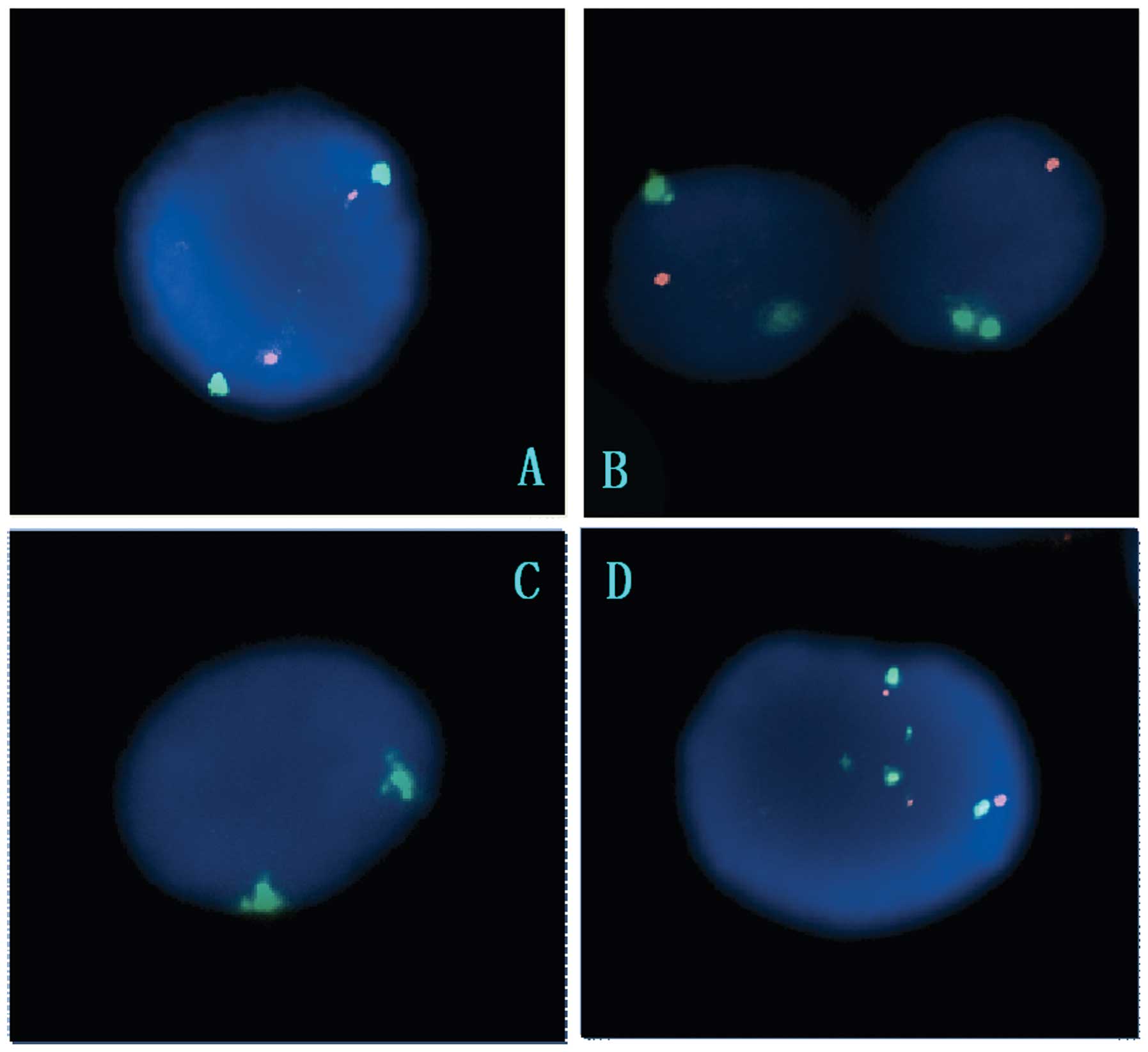
Orange (TP53) and green (CEP17) fluorescence signals
were detected in all EC specimens. CEP17 hybridization signals were
larger and brighter than TP53 gene signals. The representative FISH
models included: normal, 2 intranuclear orange signals and 2
intranuclear green signals; partial deletion, ≥2 intranuclear green
signals and 1 intranuclear orange signal; complete deletion, ≥2
intranuclear green signals and 0 orange signals (Fig. 1). Table I shows the TP53 gene deletion in
the 40 esophageal carcinoma cases.
Table II shows the
correlation between clinicopathological factors and TP53 deletion.
Statistical analysis indicated that there were no significant
correlations among TP53 gene deletion rate, age and gender
(P>0.05). TP53 gene deletion rates were 28% (4/14) and 78%
(11/14) in well-differentiated and poorly-differentiated EC,
respectively (P=0.028). TP53 gene deletion rates were 38% (7/18)
and 72% (16/22) in the lymph node metastasis and no lymph node
metastasis groups, respectively (P=0.0313). A high frequency of
CEP17 hyperdisomy was detected.
| Table IICorrelation between p53 gene deletion
and clinicopathological factors. |
Table II
Correlation between p53 gene deletion
and clinicopathological factors.
| Clinicopathological
parameters | Case | p53 deletion | P-value |
|---|
| Age (years) |
| <50 | 11 | 3 | 0.0695 |
| ≥50 | 29 | 19 | |
| Gender |
| Male | 32 | 19 | 0.4745 |
| Female | 8 | 3 | |
|
Differentiation |
|
Well-differentiated | 14 | 4 | 0.028 |
| Moderately
differentiated | 12 | 7 | |
| Poorly
differentiated | 14 | 11 | |
| Lymph node
metastasis |
| No metastasis | 18 | 7 | 0.0313 |
| Metastasis | 22 | 16 | |
Discussion
The causes of EC are complicated. At present, EC is
considered as a multi-factor, polygenic variant, multi-staged
disease. The variation of correlated proto-oncogenes and tumor
suppressor genes is the key to tumorigenesis and development.
Deletion of the tumor suppressor gene is the major mode of
variation (9).
The p53 gene is currently thought to be the most
important tumor suppressor gene and more than 50% of tumors are
associated with its abnormality. p53 genetic mutation is important
in the development of EC. Studies have indicated that patients with
p53 genetic mutation had poorer prognosis and greater malignancy
(10,11). Multiple studies have proven that
p53 genetic mutation is an independent and reliable biological
parameter for evaluating the disease prognosis (12,13).
Due to p53 gene deletion or abnormality, cells with injured DNA
enter into S stage, resulting in changes to hereditary
characteristics, chromosomal aberration and, ultimately,
carcinomatous change.
FISH is a technique using fluorescently labeled
single-chain nucleotide DNA probes, based on the base
complementarity principle, to form double-stranded nucleic acid
with complementary single chain nucleotides by specific binding,
and to detect cell chromosome changes and definite gene copy number
changes in order to diagnose malignant cells. FISH is capable of
detecting all types of cytogenetic changes, including aneusomy,
amplification and deletion. Recently, FISH has been used in the
diagnosis of hematologic malignancy, lung, breast and renal cancer,
and it is a technique with high sensitivity and specificity
(4,14–17).
In the past, there were limitations in the detection
of mutant p53 protein by immunohistochemical methods since viral
infection and stress may also produce p53 protein aggregation, and
therefore samples may have contained both mutant and wild-type
variants. Several studies found that the p53 protein positive rate
was lower than that observed using PCR-SSCP detection, which
indicates that p53 mutation or abnormality in the malignant tumor
is much higher than that shown by immunohistochemisty detection in
real situations (18). The
sensitivity of FISH is similar to isotope hybridization in
situ, but the space resolution and gene mapping accuracy is
higher; therefore, FISH plays a significant role in tumor studies
(8,18,19).
In this study, FISH was applied using double-color
DNA probes (fluorescent orange-labeled LSI TP53 probe and
fluorescent green-labeled CEP17 probe) to detect TP53 gene deletion
in Kazakh esophageal squamous cell cancer patients (7,20).
In the normal controls, in over 90% of nuclei, CEP17
and TP53 genes displayed 2 signals, indicating that FISH was
successful in our study (data not shown). Among 40 EC cases, the
mean TP53 gene deletion rate was 55% (22/40), and 28% (4/14), 58%
(7/12) and 78% (11/14) in well-differentiated, moderately
differentiated and poorly differentiated cases, respectively. This
indicates that the TP53 gene deletion rate is correlated with the
level of differentiation; the higher the differentiation, the
higher the TP53 deletion rate. Moreover, the TP53 gene deletion
rate also correlated with lymph node metastasis (P=0.0313). Similar
studies support our results (21,22).
Our study used FISH to detect the TP53 gene in
Kazakh patients with EC, and the results show for the first time
that the TP53 gene deletion rate is correlated with the level of
differentiation and lymph node metastasis in ECs, and may be an
important molecular biological marker for evaluating the condition
of ECs in Kazakh patients. Further study on the post-operative life
span of these patients is required to confirm the correlation of
TP53 gene deletion and EC prognosis.
Acknowledgements
This study was funded by the Xinjiang Uyghur
Autonomous Region Key Discipline and Specialties Foundation
(2010-09), and the First Affiliated Hospital of Xinjiang Medical
Univeristy Research Award Foundation (2012-YFY-27). Special thanks
to the Department of Hematology of the First Affiliated Hospital of
Xinjiang Medical University for technical support.
Abbreviations:
|
FISH
|
fluorescence in situ
hybridization
|
|
EC
|
esophageal cancer
|
|
CEP17
|
centromere of chromosome 17
|
|
SSC
|
standard saline citrate
|
References
|
1
|
Lu XM, Zhang YM, Lin RY, Arzi G, Wang X,
Zhang YL, Zhang Y, Wang Y and Wen H: Relationship between genetic
polymorphisms of metabolizing enzymes CYP2E1, GSTM1 and Kazakh’s
esophageal squamous cell cancer in Xinjiang. World J Gastroenterol.
11:3651–3654. 2005.PubMed/NCBI
|
|
2
|
Schmale H and Bamberger C: A novel protein
with strong homology to the tumor suppressor p53. Oncogene.
15:1363–1367. 1997. View Article : Google Scholar : PubMed/NCBI
|
|
3
|
Graesslin O, Chantot-Bastaraud S,
Lorenzato M, Birembaut P, Quéreux C and Daraï E: Fluorescence in
situ hybridization and immunohistochemical analysis of p53
expression in endometrial cancer: prognostic value and relation to
ploidy. Ann Surg Oncol. 15:484–492. 2008. View Article : Google Scholar : PubMed/NCBI
|
|
4
|
Halling KC and Kipp BR: Fluorescence in
situ hybridization in diagnostic cytology. Hum Pathol.
38:1137–1144. 2007. View Article : Google Scholar : PubMed/NCBI
|
|
5
|
Li WJ and Duan JX: Detecting of p53 gene
heterozygosity loss in esophageal carcinoma tissues. JZU Med Sci.
39:499–501. 2004.
|
|
6
|
Hollstein MC, Metcalf RA, Welsh JA,
Montesano R and Harris CC: Frequent mutation of the p53 gene in
human esophageal cancer. Proc Natl Acad Sci USA. 87:9958–9961.
1990. View Article : Google Scholar : PubMed/NCBI
|
|
7
|
Audrézet MP, Robaszkiewicz M, Mercier B,
et al: TP53 gene mutation profile in esophageal squamous cell
carcinomas. Cancer Res. 53:5745–5749. 1993.PubMed/NCBI
|
|
8
|
Nakamura H, Saji H, Idiris A, et al:
Chromosomal instability detected by fluorescence in situ
hybridizationin surgical specimens of non-small cell lung cancer is
associated with poor survival. Clin Cancer Res. 9:2294–2299.
2003.
|
|
9
|
Cheng ZQ, Wang XM, Shan J, et al:
Detection of p53 gene deletion in primary lung cancer by dual-FISH
technique and its clinical significance. Clin Med Chin. 26:368–371.
2010.
|
|
10
|
Uchino S, Saito T, Inomata M, Osawa N,
Chikuba K, Etoh K and Kobayashi M: Prognostic significance of the
p53 mutation in esophageal cancer. Jpn J Clin Oncol. 26:287–292.
1996. View Article : Google Scholar : PubMed/NCBI
|
|
11
|
da Manoel-Caetano FS, Borim AA, Caetano A,
Cury PM and Silva AE: Cytogenetic alterations in chagasic achalasia
compared to esophageal carcinoma. Cancer Genet Cytogenet.
149:17–22. 2004.PubMed/NCBI
|
|
12
|
Curtis C, Harris MD and Hollstein MC:
Clinical implications of the p53 tumor suppessor gene. N Eng J Med.
329:1318–1325. 1993. View Article : Google Scholar : PubMed/NCBI
|
|
13
|
Smith DR, Ji CY and Coh HS: Prognostic
significance of p53 over expression and mutation in colorectal
adrecarcinomas. Br J Cancer. 74:216–223. 1996. View Article : Google Scholar : PubMed/NCBI
|
|
14
|
Fritcher EG, Brankley SM, Kipp BR, et al:
A comparison of conventional cytology, DNA ploidy analysis, and
fluorescence in situ hybridization for the detection of
dysplasia and adenocarcinoma in patients with Barrett’s esophagus.
Hum Pathol. 39:1128–1135. 2008. View Article : Google Scholar : PubMed/NCBI
|
|
15
|
Kawai T, Hiroi S, Nakanishi K, Sakurai Y
and Torikata C: Abnormalities in chromosome 17 and p53 in lung
carcinoma cells detected by fluorescence in situ hybridization.
Pathol Int. 54:413–419. 2004. View Article : Google Scholar : PubMed/NCBI
|
|
16
|
Varshney D, Zhou YY, Geller SA and Alsabeh
R: Determination of HER-2 status and chromosome 17 polysomy in
breast carcinomas comparing Hercep Test and PathVysion FISH assay.
Am J Clin Pathol. 121:70–77. 2004. View Article : Google Scholar : PubMed/NCBI
|
|
17
|
Fujiwara T, Cai DW, Georges R, et al:
Therapeutic effect of a retroviral wild-type p53 expression vector
in an orthotopic lung cancer model. J Natl Cancer Inst.
86:1458–1462. 1994. View Article : Google Scholar : PubMed/NCBI
|
|
18
|
Lu SM, Cheng CL and Sheng BY:
Immunohistochemical detection of p53 protein altered expression in
1364 patients with maligant tumors and its clinicopathological
significance. Chin J Cancer. 20:620–623. 2001.
|
|
19
|
Li JD, Li H and Shen ZY: Esophageal
spontaneous apoptosis and nuclear value-added antigen, relationship
with p53. Chin J Oncol. 20:415–417. 1998.
|
|
20
|
Idiris A, Madiniyet N, Hadeti B, Zhang Z,
Ilyar S and Wen H: Molecular pathological diagnosis for early
esophageal cancer in Kazakh patients. Oncol Lett. 13:549–553.
2012.
|
|
21
|
Abedi-Ardekani B, Kamangar F, Sotoudeh M,
et al: Extremely high Tp53 mutation load in esophageal squamous
cell carcinoma in Golestan Province, Iran. PLoS One. 6:e294882011.
View Article : Google Scholar : PubMed/NCBI
|
|
22
|
King SI, Purdie CA, Bray SE, Quinlan PR,
Jordan LB, Thompson AM and Meek DW: Immunohistochemical detection
of Polo-like Kinase-1 (PLK1) in primary breast cancer is associated
with TP53 mutation and poor clinical outcome. Breast Cancer Res.
14:402012. View
Article : Google Scholar : PubMed/NCBI
|