Introduction
Chronic myeloid leukemia (CML) is a malignant
hematological disease affecting the hematopoietic stem/progenitor
cells. CML is characterized by the presence of a constitutively
active tyrosine kinase, known as the BCR/ABL oncoprotein, which is
produced as a result of a reciprocal translocation between
chromosomes 9 and 22 (1). In the
United States, 5,000 patients are diagnosed with CML each year
(2). However, CML remains one of
the most difficult malignant hematological diseases to treat.
Chemotherapy is a common therapeutic, however, certain natural
anti-tumor medicines, including camptothecin, vinblastine and
paclitaxel often cause problems, including adverse reactions and
drug resistance. Therefore, the development of an effective natural
antitumor drug is required (3).
Securinega alkaloids are a group of natural
compounds, which are isolated from the Euphorbiaceae family of
plants. Securinine, a major alkaloid found in the leaves of
Securinega suffruticosa, was initially isolated in 1956 and
the structure was determined in 1963 (4). Although securinine, the first
alkaloid from this class, was found to be a specific γ-aminobutyric
acid receptor antagonist with significant in vivo central
nervous system activity (5),
further examination of this class of compounds led to the
identification of molecules exhibiting potent biological
properties, including antimalarial (6) and antibiotic properties (7). There are two optical isomers,
l-securinine and virosecurinine. Previous pharmacological studies
have demonstrated that virosecurinine also possesses antitumor
properties (8), improves bone
marrow function (9) and induces
apoptosis (10). Therefore, there
is potential for the clinical use of virosecurinine in the
treatment of cancer. Apoptosis is a physiological mechanism for the
elimination of cells, which occurs during embryonic development,
hormone-induced atrophy and normal cellular homeostasis (11). Since the definition of apoptosis by
Kerr (23) in l972, it has been
observed that apoptosis is also involved in certain cases of
drug-induced tumor cell death (12–13).
Numerous currently used anticancer drugs kill particular types of
tumor cells via apoptosis.
In the present study, the effect of virosecurinine
on the apoptosis of human leukemic K562 cells was investigated to
further elucidate the underlying mechanism. In addition, the
present study aimed to investigate the efficacy of natural
anti-tumor drugs.
Materials and methods
Chemicals
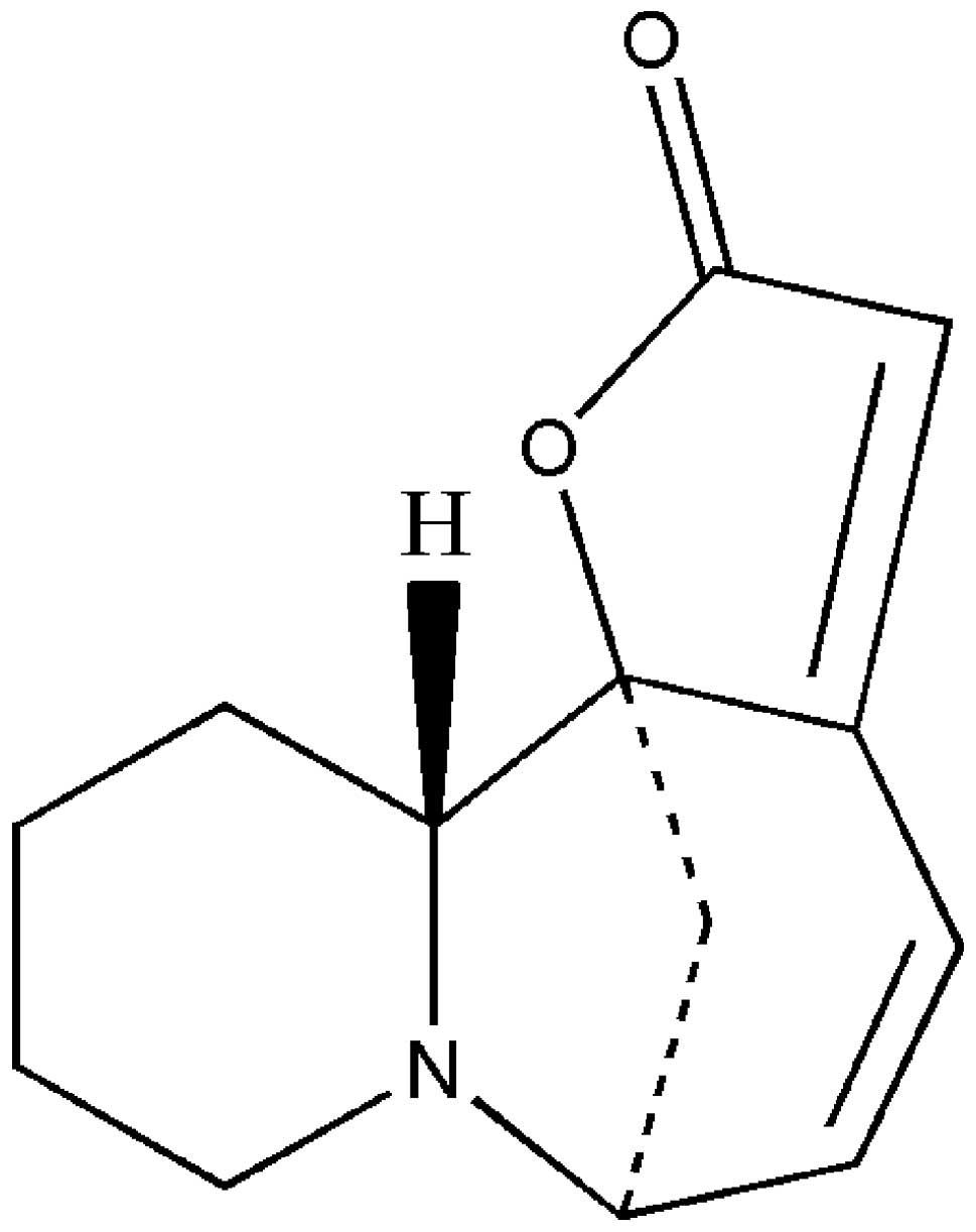
Isolation and extraction of pure virosecurinine
(Fig. 1) from Securinega
suffrutico (Pall.) Rehd was achieved using the ion exchange
resin method. The pure sample of virosecurinine was provided
by the Institute of Traditional Chinese Medicine and Natural
Products, Jinan University (Guangzhou, China). The Cell Counting
kit-8 (CCK-8) was purchased from KeyGen Biotech Co., Ltd. (Nanjing,
China; cat no. KGA317). The cell culture media and solutions were
obtained from Gibco-BRL (Carlsbad, CA, USA; cat no. 31800-105).
Cell culture
The human leukemic K562 cell line was purchased from
KeyGen Biotech Co., Ltd. The K562 cells were cultured in RPMI-1640
(Gibco-BRL; cat no. 31800-105) containing 10% fetal bovine serum
(FBS; Hangzhou Sijiqing Biological Engineering Materials Co., Ltd.,
Hangzhou, China; cat no. 120316) and 100 U/ml each of penicillin
and streptomycin (KeyGen Biotech Co. Ltd). Cells were grown and
maintained at 37°C in a 5% CO2 humidified
atmosphere.
Analysis of cell viability
CCK-8 was used to measure cell viability.
Exponentially growing K562 cells (100 μl, 5×10⁴
cells/ml) were seeded into 96-well plates (Corning Incorporated,
Corning, NY, USA; cat no. 3599). After 24 h, the K562 cells were
cultured in RPMI-1640 medium containing 10% FBS and then treated
with virosecurinine at concentrations ranging between 6.25 and 200
μmol/l. This was repeated six times at each concentration. The
plates were then incubated in a humidified incubator (Sanyo XD-101;
Sanyo, Osaka, Japan) at 37°C and 5% CO2 for 24, 48 and
72 h. Subsequently, 3 h prior to measuring the absorbance, 10 μl
CCK-8 solution was added to each well. The optical density was
measured at an absorbance of 450 nm using a microplate reader
(ELx800; BioTek Instruments, Inc., Winooski, VT, USA). The
inhibitory rate of cellular proliferation was calculated using the
following formula: Cellular proliferation inhibitory rate = (1 -
mean A450 of experimental group / mean A450 of control group) ×
100%.
Electron microscopy
The K562 cell suspension was seeded at a density of
5×10⁵ cells/well in 6-well plates and was treated with
or without virosecurinine at a concentration of 25 μmol/l for 48 h
at 37°C. Following incubation, the cells were collected and fixed
for 2 h at 4°C in 2.5% ice-cold glutaraldehyde (KeyGen Biotech Co.,
Ltd.) and then washed three times in 0.1 mol/l phosphate-buffered
saline (PBS). The cells were then post-fixed at 4°C in 1% osmium
tetroxide (KeyGen Biotech Co., Ltd.) for 2 h and dehydrated through
serial dilutions of ethanol (50, 70, 90 and 100% each for 15 min
and three times at 100%) prior to being embedded in epoxy resin.
The embedded cells were then cut into ultrathin sections (50–60 nm)
and stained using uranyl acetate and lead citrate (KeyGen Biotech
Co., Ltd.). The sections were viewed using a transmission electron
microscope (TEM-1011; Jeol, Tokyo, Japan).
Analysis of cell apoptosis
To analyze apoptosis, the K562 cells were cultured
in 6-well plates (cat no. 3516; Corning Incorporated) with at least
3.0×10⁵ cells/well in medium containing RPMI-1640
(Gibco-BRL, Carslbad, CA, USA), 10% FBS (Sijiqing Biological
Engineering Materials, Hangzhou, China) and 100 U/ml penicillin and
streptomycin (KeyGen Biotech Co., Ltd.) for 24 h. The cells were
then treated with different concentrations of virosecurinine (6.25,
25 and 50 μmol/l) for 48 h. The cells were washed twice with cold
PBS (cat no. KGB500; KeyGen Biotech Co., Ltd.), followed by the
addition of 5 μl annexin V-fluorescein isothiocyanate (FITC; cat
no. KGA105; KeyGen Biotech Co., Ltd.) and propidium iodide (PI; cat
no. KGA511; KeyGen Biotech Co., Ltd). After 15 min incubation at
room temperature in the dark, the cells were analyzed using flow
cytometry. Fluorescence was measured using a FACScan flow cytometer
(Becton-Dickinson, Franklin Lakes, NJ, USA) equipped with an argon
laser (488 nm). The cell apoptotic rate was calculated using the
internal software system of the FACScan (Becton-Dickinson).
Cell cycle analysis
For cell cycle analysis, the K562 cells were
cultured in 6-well plates (at least 3.0×10⁵ cells/well)
with medium containing RPMI-1640, 10% FBS and 100 U/ml penicillin
and streptomycin for 24 h and then treated with different
concentrations of virosecurinine (6.25, 25 and 50 μmol/l) for 48 h.
The cells were washed, collected, fixed using 70% ethanol and
stored at 4°C overnight. Following that, the cells were treated
with Tris-HCl buffer (pH 7.4) containing 1% RNase A (cat. no
KGA511; KeyGen Biotech Co., Ltd) and stained using PI (5 mg/ml).
Flow cytometry (FACSCalibur; Becton-Dickinson) was used to
determine the distribution of cells with different DNA contents.
The data were analyzed using multicycle DNA content and cell cycle
analysis software (FlowJo, version 7.6.5; KeyGen Biotech Co.,
Ltd.).
Reverse transcription quantitative
real-time polymerase chain reaction (RT-qPCR)
RT-qPCR was performed to analyze the mRNA levels of
mammalian target of rapamycin (mTOR), phosphatase and tensin
homologue (PTEN), breakpoint cluster region (BCR)/Abelson (ABL) and
SH2 domain-containing inositol-5′-phosphatase 2 (SHIP2) in the K562
cells treated with or without virosecurinine. Total RNA was
isolated from the K562 cells using TRIzol reagent (cat no.
15596-026; Invitrogen Life Technologies, Carlsbad, CA, USA)
according to the manufacturer’s instructions. First strand cDNA
synthesis was performed using the ProSTARt First Strand RT-PCR kit
(cat no. PC0002; Fermentas, Vilnius, Lithuania) according to
the manufacturer’s instructions. Following reverse transcription,
20 μl of the reaction mixture (cat no. EP0702; Fermentas) was used
in a qPCR program (cat no. DA7600; Zhongshan Bio-Tech Co., Ltd.,
Zhongshan, China) comprising 40 cycles consisting of denaturation
(15 sec at 95°C), annealing (20 sec at 60°C) and extension (40 sec
at 72°C). The 20 μl reaction mixture contained 10 μM each primer, 2
μl 2X QuantiTect SYBR green RT-PCR master mix, 10 μl QuantiTect
reverse transcriptase mix and nuclease-free water (cat no.
KGDN4500; KeyGen Biotech Co., Ltd) up to 8 μl. Data were analyzed
using the 2−ΔΔCt method. The experiment was repeated
three times and the efficiency of cDNA synthesis from each sample
was estimated using GAPDH-specific primers. The primers used were
as follows: mTOR, forward 5′-ATTTGATCAGGTGTGCCAGT-3′ and reverse
5′-GCTTAGGACATGGTTCATGG-3′; GAPDH, forward
5′-TGTTGCCATCAATGACCCCTT-3′ and reverse 5′-CTCCACGACGTACTCAGCG-3′;
PTEN, forward 5′-CAAGATGATGTTTGAAACTATTCCAATG-3′ and reverse
5′-CCTTTAGCTGGCAGACCACAA-3′; BCR/ABL, forward
5′-CTCCAGACTGTCCACAGCATTCCG-3′ and reverse
5′-CAGACCCTGAGGCTCAAAGTCAGA-3′ and SHIP2, forward
5′-GAGCACGAGAACCGTATCAGC-3′ and reverse
5′-CCAAATGAGGTGCCATTAAACA-3′.
Statistical analysis
All data are expressed as the mean ± standard
deviation. SPSS software version 18.0 (SPSS, Inc., Chicago, IL,
USA) was used for statistical analyses. Differences between the
groups were evaluated using the Student-Newman-Keuls test.
P<0.05 was considered to indicate a statistically significant
difference.
Results
Effect of virosecurinine on the
proliferation of K562 cell lines
In order to understand the mechanism underlying
virosecurinine-induced apoptosis in K562 cells, a CCK-8 assay was
used to determine the effects of virosecurinine on the
proliferation of K562 cells. The K562 cells were treated with
virosecurinine at concentrations ranging between 6.25 and 200
μmol/l or with dimethyl sulfoxide (DMSO; KeyGen Biotech Co., Ltd.)
alone for 24, 48 and 72 h and the number of viable cells were
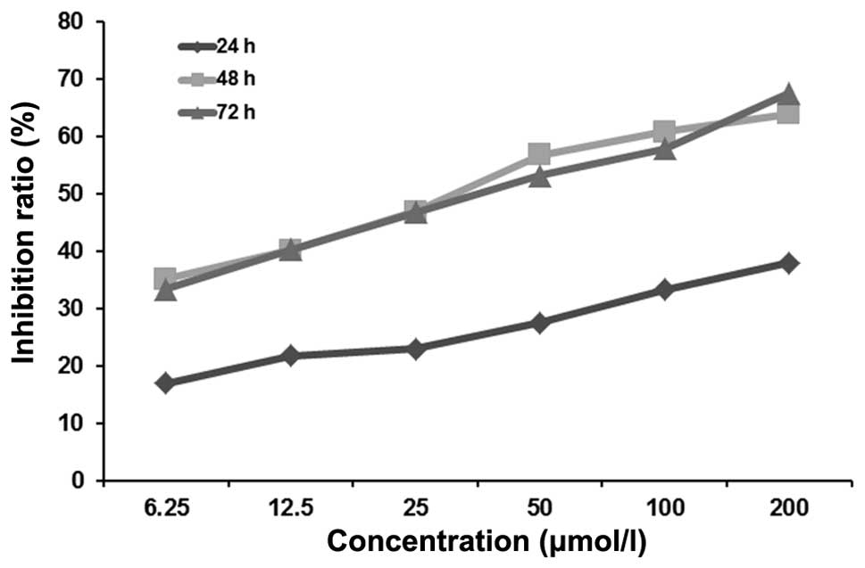
determined. The CCK-8 assay revealed that virosecurinine markedly
inhibited the proliferation of K562 cells in a dose- and
time-dependent manner (Fig. 2).
The inhibitory rate at 48 h was significantly higher compared with
at 24 or 72 h and the IC50 was 32.984 μM/l at 48 h.
K562 cell ultrastructure
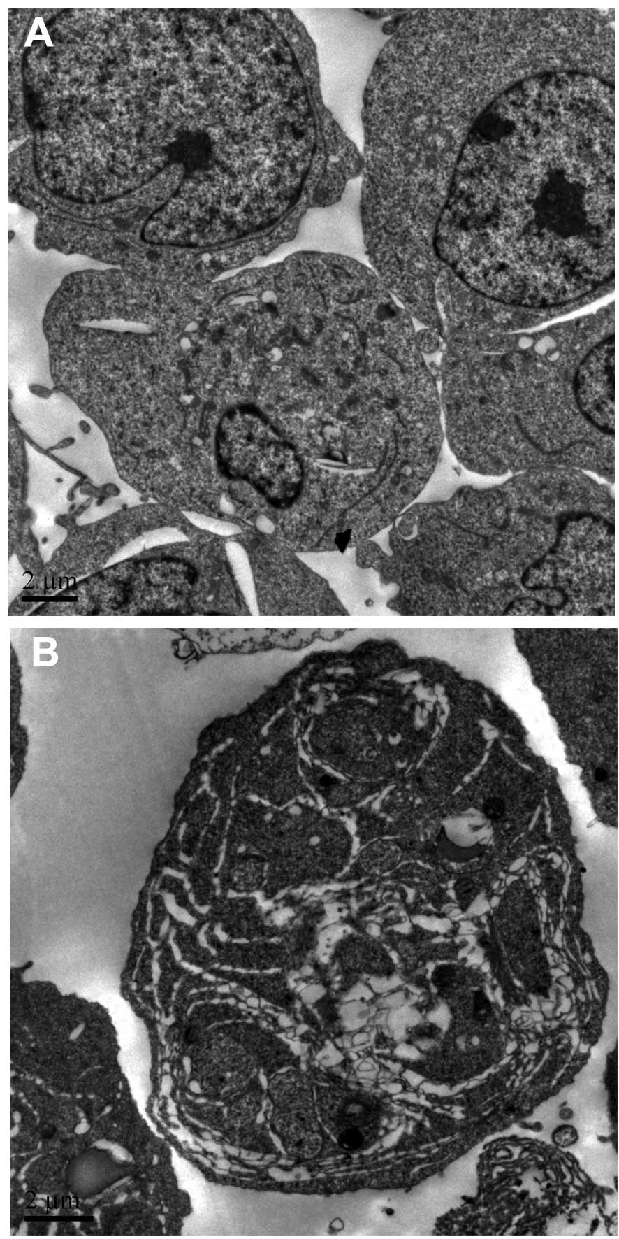
Furthermore, ultra-structural analysis by electron
microscopy further confirmed that apoptotic cells were not observed
in the DMSO group (Fig. 3A).
However, apoptotic bodies were observed in the K562 cells treated
with 25 μmol/l virosecurinine for 48 h (Fig. 3B). The assay demonstrated that K562
cells treated with 25 μM/l virosecurinine for 48 h were undergoing
apoptosis.
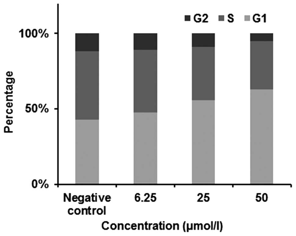
Flow cytometric analysis of the cell
cycle
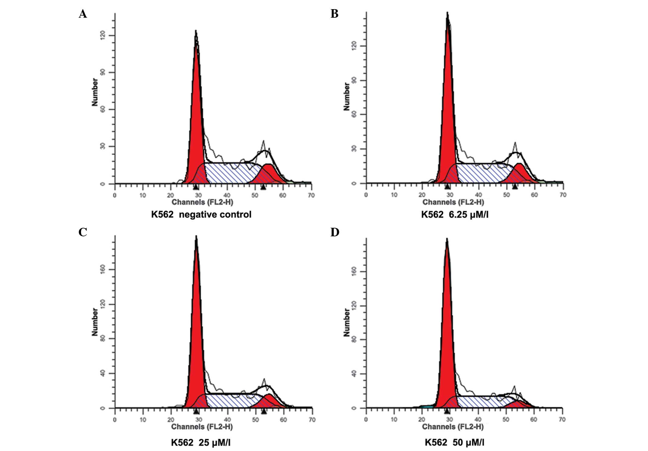
To understand the mechanisms of
virosecurinine-induced K562 cell apoptosis, the present study
analyzed the cell cycle phase distribution. K562 cells were treated
with 6.25, 25 and 50 μmol/l of virosecurinine for 48 h and the cell
cycle was analyzed using flow cytometry according to the DNA
content. The results indicated that virosecurinine arrested the
cells at the G1 phase of the cell cycle (Figs. 4 and 5).
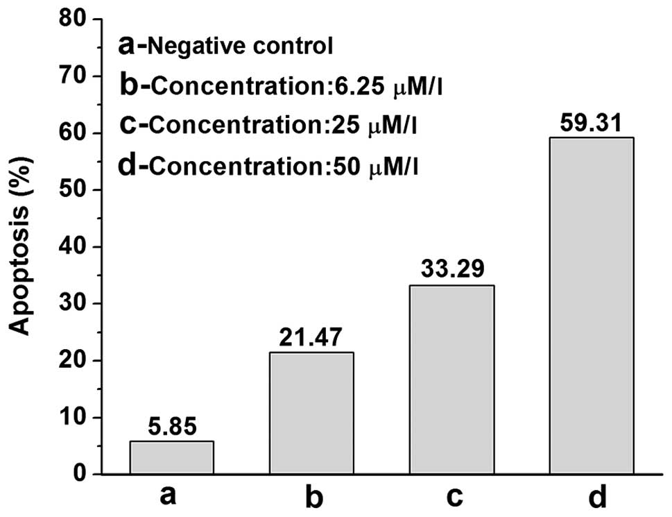
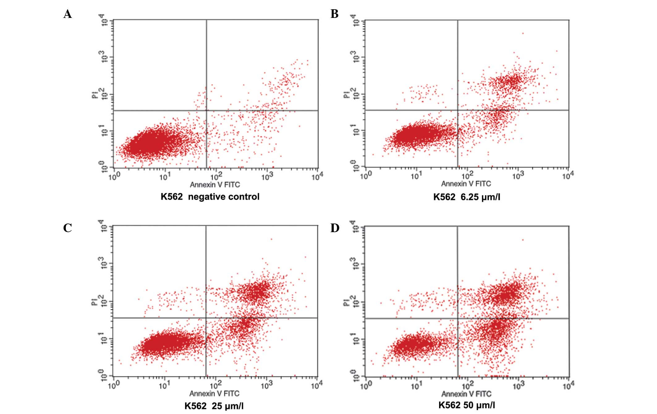
Apoptotic statistical flow cytometric
assay
To further determine whether virosecurinine-treated
K562 cells underwent apoptosis, the cells were stained using
double-staining with Annexin V and PI. The K562 cells were treated
with virosecurinine (6.25, 25 and 50 μmol/l) or without
virosecurinine for 48 h. The results demonstrated that
virosecurinine increased the percentage of K562 cells undergoing
apoptosis in a dose-dependent manner (Fig. 6). The percentage of apoptotic cells
following treatment with 6.25, 25 and 50 μmol/l virosecurinine for
48 h was 21.47, 33.29 and 59.31%, respectively (Figs. 6 and 7).
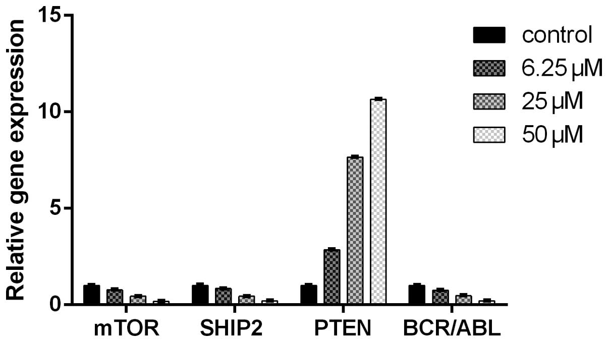
RT-qPCR analysis of the mRNA expression
of mTOR, PTEN, BCR/ABL and SHIP2
In order to further understand the molecular
mechanism underlying virosecurinine-induced apoptosis, the mRNA
levels of mTOR, PTEN, BCR/ABL and SHIP2 mRNA were measured in
virosecurinine-deprived and control K562 cells using RT-qPCR
analysis. The results indicated that virosecurinine effectively
downregulated the expression level of mTOR, SHIP2 and BCR/ABL and
upregulated the expression of PTEN (P<0.05; Fig. 8).
Discussion
Although securinine, a major alkaloid of the plant
Securinega suffruticosa, has been found to inhibit the
growth of MCF-7, SW480 and HL-60 tumor cells, the underlying
mechanism remains to be elucidated (4,12).
The present study investigated the efficacy of virosecurinine
against the human CML cell line K562 and demonstrated that
virosecurinine significantly inhibited the proliferation of K562
cells at low concentrations. The IC50 of virosecurinine to K562
cells 48 h after treatment was 32.984 μM/l. This result indicated
that virosecurinine markedly inhibited the proliferation of K562
cells in a dose- and time-dependent manner. In addition, the
present study demonstrated that virosecurinine induced cell
apoptosis, with the observation of apoptotic bodies and the
appearance of a typical sub-G1 peak in the K562 cells treated with
25 μmol/l virosecurinine for 48 h. Apoptosis is important in
development and the prevention of apoptosis is a hallmark of
cancer. The investigation of apoptosis may be important for the
development of novel anticancer therapies.
Previous studies from different groups have used
mathematical models of apoptosis and applied them to cancer cells
(14–16). The data from these studies
demonstrate that the inhibitory effect of virosecurinine on the
growth of the human CML cell line K562 may be partly due to the
induction of apoptosis. mTOR is a serine/threonine protein kinase,
a downstream partner in the phosphoinositide 3-kinase (PI3K)/Akt
pathway, which regulates protein translation, cell growth and
apoptosis (17). The PTEN gene is
a novel tumor suppressor gene and the PTEN protein has protein
phosphatase and lipid phosphatase dual activity, with activity
against phosphatidylinositol 3,4,5-trisphosphate, the major
bioactive product of PI3K (18).
It has been identified as a candidate tumor suppressor gene based
on the presence of gene deletion or inactivating mutations in human
brain, breast and prostate cancers as well as tumor cell lines
(19–22). BCR-ABL fusion proteins result from
the chromosomal translocation t(9;22), which produces the
Philadelphia chromosome and ultimately leads to CML (23). The activity of BCR-ABL has several
effects on cells, causing an increase in proliferation and a
reduction in apoptosis, leading to the malignant growth of groups
of hematopoietic stem cells. Inhibition of ABL by imatinib, its
tyrosine kinase inhibitor, has markedly improved the prognosis of
patients with CML (24). It has
been suggested that SHIP2 is involved in type 2 diabetes and in
obesity (25), as well as cancer
and atherosclerosis (26).
Therefore, there has been an interest in developing compounds that
selectively target SHIP2. A previous study described the inhibition
of the catalytic activity of SHIP2 by specific molecular compounds
(27). In addition, cell permeable
pan-SHIP1/2 inhibitors have been identified, which have been
reported to cause cell death in multiple myeloma (28). The identification of SHIP2 specific
compounds suggests that SHIP2 may be a potential target with which
to treat a range of diseases and also enables a deeper
understanding of the role of SHIP2 in the immune system.
To further investigate the molecular mechanism of
virosecurinine, the present study measured the expression levels of
four genes linked to apoptosis, mTOR, PTEN, BCR/ABL and SHIP2 in
virosecurinine-treated K562 cells. The results demonstrated that
virosecurinine upregulated the gene expression of PTEN and
downregulated the expression of mTOR, BCR/ABL and SHIP2 in the K562
cells. Therefore, the present study demonstrated for the first
time, to the best of our knowledge, that virosecurinine induced
apoptosis in K562 cells by altering the expression of mTOR, PTEN,
BCR/ABL and SHIP2. These results demonstrated that virosecurinine
effectively suppressed the proliferation of the human CML cell line
K562 and suggested that growth inhibition may be in part due to
virosecurinine-induced apoptosis through the downregulation of
mTOR, BCR/ABL and SHIP2 and the upregulation of PTEN.
The results from the present study, suggest that
virosecurinine may serve as a potential lead for future drug
development in the prevention and treatment of CML.
Acknowledgements
This study was supported by grants from the National
Natural Science Foundation of China (no. 81241102). The authors
would like to thank the Institute of Traditional Chinese Medicine
and Natural Products, Jinan University for providing a pure sample
of virosecurinine.
References
|
1
|
Deininger MW, Goldman JM and Melo JV: The
molecular biology of chronic myeloid leukemia. Blood. 96:3343–3356.
2000.PubMed/NCBI
|
|
2
|
Jemal A, Bray F, Center MM, Ferlay J, Ward
E and Forman D: Cancer statistics, 2009. CA Cancer J Clin.
59:225–249. 2009. View Article : Google Scholar
|
|
3
|
Lin H and Chen ZL: Progress of researching
in anti-tumor drugs. Chin J Hosp Pharm. 8:226–228. 1998.(In
Chinese).
|
|
4
|
Saito S, Kotera K, Shigematsu N, Ide A,
Sugimoto N, Horii Z, et al: Structure of securinine. Tetrahedron.
19:2085–2099. 1963. View Article : Google Scholar : PubMed/NCBI
|
|
5
|
Beutler JA, Karbon EW, Brubaker AN, Malik
R, Curtis DR and Enna SJ: Securinine alkaloids: a new class of GABA
receptor antagonist. Brain Res. 330:135–140. 1985. View Article : Google Scholar : PubMed/NCBI
|
|
6
|
Weenen H, Nkunya MH, Bray DH, Mwasumbi LB,
Kinabo LS, Kilimali VA, et al: Antimalarial compounds containing an
alpha, beta-unsaturated carbonyl moiety from Tanzanian medicinal
plants. Planta Med. 56:371–373. 1990. View Article : Google Scholar : PubMed/NCBI
|
|
7
|
Mensah JL, Lagarde I, Ceschin C, Michel G,
Gleye J and Fouraste I: Antibacterial activity of the leaves of
Phyllanthus discoideus. J Ethnopharmacol. 28:129–133. 1990.
View Article : Google Scholar : PubMed/NCBI
|
|
8
|
Dong NZ and Gu ZL: Study on the antitumor
effect of securinine and its mechanism. Chin Tradit Pat Med.
21:193–195. 1999.(In Chinese).
|
|
9
|
Jiang XY: The effect of securinine and
aminophylline on hemopoietic stem cells and granulopoietic
progenitors in mice. Tianjin Med J. 10:99–100. 1982.(In
Chinese).
|
|
10
|
Liu WJ, Gu ZL and Zhou WX: Securinine
induced apoptosis in K562 cells. Chin Pharmacol Bull. 115:135–158.
1999.
|
|
11
|
Pérez JM, Quiroga AG, Montero EI, Alonso C
and Navarro-Ranninger C: A cycloplatinated compound of
p-isopropylbenzaldehyde thiosemicarbazone and its chloro-bridged
derivative induce apoptosis in cis-DDP resistant cells which
overexpress the H-ras oncogene. J Inorg Biochem. 73:235–243.
1999.
|
|
12
|
Kerr JF, Wyllie AH and Currie AR:
Apoptosis: a basic biological phenomenon with wide ranging
implications in tissue kinetics. Br J Cancer. 26:239–257. 1972.
View Article : Google Scholar : PubMed/NCBI
|
|
13
|
Steller H: Mechanisms and genes of
cellular suicide. Science. 267:1445–1449. 1995. View Article : Google Scholar : PubMed/NCBI
|
|
14
|
Hector S, Rehm M, Schmid J, Kehoe J,
McCawley N, Dicker P, et al: Clinical application of a systems
model of apoptosis execution for the prediction of colorectal
cancer therapy responses and personalisation of therapy. Gut.
11:725–733. 2012. View Article : Google Scholar : PubMed/NCBI
|
|
15
|
Huber HJ, Dussmann H, Kilbride SM, Rehm M
and Prehn JH: Glucose metabolism determines resistance of cancer
cells to bioenergetic crisis after cytochrome-c release. Mol Syst
Biol. 7:4702011. View Article : Google Scholar
|
|
16
|
Lee MJ, Ye AS, Gardino AK, Heijink AM,
Sorger PK, MacBeath G and Yaffe MB: Sequential application of
anticancer drugs enhances cell death by rewiring apoptotic
signaling networks. Cell. 149:780–794. 2012. View Article : Google Scholar : PubMed/NCBI
|
|
17
|
Hay N and Sonenberg N: Upstream and
downstream of mTOR. Genes Dev. 18:1926–1945. 2001. View Article : Google Scholar
|
|
18
|
Montiel-Duarte C, Cordeu L, Agirre X,
Román-Gómez J, Jiménez-Velasco A, José-Eneriz ES, et al: Resistance
to Imatinib Mesylate-induced apoptosis in acute lymphoblastic
leukemia is associated with PTEN down-regulation due to promoter
hypermethylation. Leuk Res. 32:709–716. 2008. View Article : Google Scholar
|
|
19
|
Wiencke JK, Zheng S, Jelluma N, Tihan T,
Vandenberg S, Tamgüney T, et al: Methylation of the PTEN promoter
defines low-grade gliomas and secondary glioblastoma. Neuro Oncol.
9:271–279. 2007. View Article : Google Scholar : PubMed/NCBI
|
|
20
|
Mirmohammadsadegh A, Marini A, Nambiar S,
Hassan M, Tannapfel A, Ruzicka T, et al: Epigenetic silencing of
the PTEN gene in melanoma. Cancer Res. 66:6546–6552. 2006.
View Article : Google Scholar : PubMed/NCBI
|
|
21
|
Noro R, Gemma A, Miyanaga A, Kosaihira S,
Minegishi Y, Nara M, et al: PTEN inactivation in lung cancer cells
and the effect of its recovery on treatment with epidermal growth
factor receptor tyrosine kinase inhibitors. Int J Oncol.
31:1157–1163. 2007.PubMed/NCBI
|
|
22
|
Athanassiadou P, Athanassiades P, Grapsa
D, Gonidi M, Athanassiadou AM, Stamati PN and Patsouris E: The
prognostic value of PTEN, p53, and beta-catenin in endometrial
carcinoma: a prospective immunocytochemical study. Int J Gynecol
Cancer. 17:697–704. 2007. View Article : Google Scholar : PubMed/NCBI
|
|
23
|
Goldman JM and Melo JV: BCR-ABL in chronic
myelogenous leukemia-how does it work? Acta Haematol. 13:212–217.
2008. View Article : Google Scholar : PubMed/NCBI
|
|
24
|
Kantarjian H, Sawyers C, Hochhaus A,
Guilhot F, Schiffer C, Gambacorti-Passerini C, et al: Hematologic
and cytogenetic responses to imatinib mesylate in chronic
myelogenous leukemia. N Engl J Med. 346:645–652. 2002. View Article : Google Scholar : PubMed/NCBI
|
|
25
|
Ooms LM, Horan KA, Rahman P, Seaton G,
Gurung R, Kethesparan DS and Mitchell CA: The role of the inositol
polyphosphate 5-phosphatases in cellular function and human
disease. Biochem J. 419:29–49. 2009. View Article : Google Scholar : PubMed/NCBI
|
|
26
|
Suwa A, Kurama T and Shimokawa T: HIP2 and
its involvement in various diseases. Expert Opin Ther Targets.
14:727–737. 2010. View Article : Google Scholar
|
|
27
|
Suwa A, Yamamoto T, Sawada A, Minoura K,
Hosogai N, Tahara A, et al: Discovery and functional
characterization of a novel small molecule inhibitor of the
intracellular phosphatase, SHIP2. J Pharmacol. 158:879–887.
2009.PubMed/NCBI
|
|
28
|
Fuhler GM, Brooks R, Toms B, Iyer S, Gengo
EA, Park MY, et al: Therapeutic potential of SH2 domain-containing
inositol-5′-phosphatase 1 (SHIP1) and SHIP2 inhibition in cancer.
Mol Med. 18:65–75. 2012.
|