Introduction
Prostate cancer is one of the most common
malignancies and the second leading cause of mortality among male
cancer patients in developed countries (1). Due to the limited treatments
available for this disease, although chemotherapy and radiotherapy
are currently used, and poor prognosis and therapy failure in
prostate cancers usually occur (2,3). One
of the main reasons is that the growth and survival of prostate
cancer cells depend on androgens, which are also associated with
tumor regression (4). At present,
no effective treatments prostate cancers are available, and
therefore, it is important to identify novel chemotherapeutic drugs
and to develop effective treatment strategies for prostate
cancer.
Apoptosis has an important role in keeping the
balance between cell proliferation and death in normal tissues
(5). B-cell lymphoma 2 (Bcl-2)
family proteins regulate apoptosis in cancer progression, and they
have been divided into pro- and anti-apoptotic groups, including
Bcl-2, Bcl-extra large (xL), myeloid leukemia cell differentiation
protein (Mcl-1), and Bcl-2 family proteins 1, 2 and 10 (6,7). It
is noteworthy that the anti-apoptotic Bcl-2 family proteins are
actively involved in various cancer types in humans, making them
attractive targets for developing novel anti-cancer drugs (8,9).
Additionally, structural studies reveal that the anti-apoptotic
Bcl-2 protein has a hydrophobic groove (10,11),
and it could form a binding pocket for the pro-apoptotic members
with Bcl-2 homology domain 3 (BH3) domains, interfering with their
pro-apoptotic functions (12).
Thus, targeting the binding pocket of anti-apoptotic Bcl-2 proteins
is a new and promising strategy for cancer therapy and drug
discovery (13).
Recently, several non-peptide small molecule
inhibitors of Bcl-2 family proteins have been synthesized and
extracted (14). Gossypol, a
polyphenolic compound isolated from cotton seeds and roots, is one
of these effective anti-tumor drugs undergoing evaluation in
pre-clinical trials, which has been reported to have
anti-proliferation activities against several types of cancer in
vitro and anti-tumor effects in vivo (15,16).
This natural product is also a potent and effective male
contraceptive drug in an early clinical stage (17–19).
However, the two aldehyde groups in gossypol are associated with
toxicity and potential non-specific activities (20). Therefore, it is imperative to
develop novel gossypol derivatives with a higher binding affinity
to Bcl‑2 proteins as well as good selectivity between normal and
cancer cells with varying levels of Bcl-2 proteins (21). Researchers are synthesizing novel
gossypol derivatives in order to optimize its chemical structure
and improve its anti-cancer effect by removing aldehyde groups, to
achieve superior anti-proliferation activity with less toxicity in
nasopharyngeal carcinoma, prostate cancer, human leukemic monocyte
lymphoma, diffuse large-cell lymphoma, follicular lymphoma,
pancreatic cancer cells and human hepatocellular carcinoma
(22,23). The derivative apogossypolone has
been synthesized and its anti-cancer effects have been
investigated. The results revealed that apogossypolone effectively
inhibited the growth and proliferation of gastric and prostate
cancer cell lines in vitro and in vivo (24,25).
In addition, our group and others have designed and synthesized
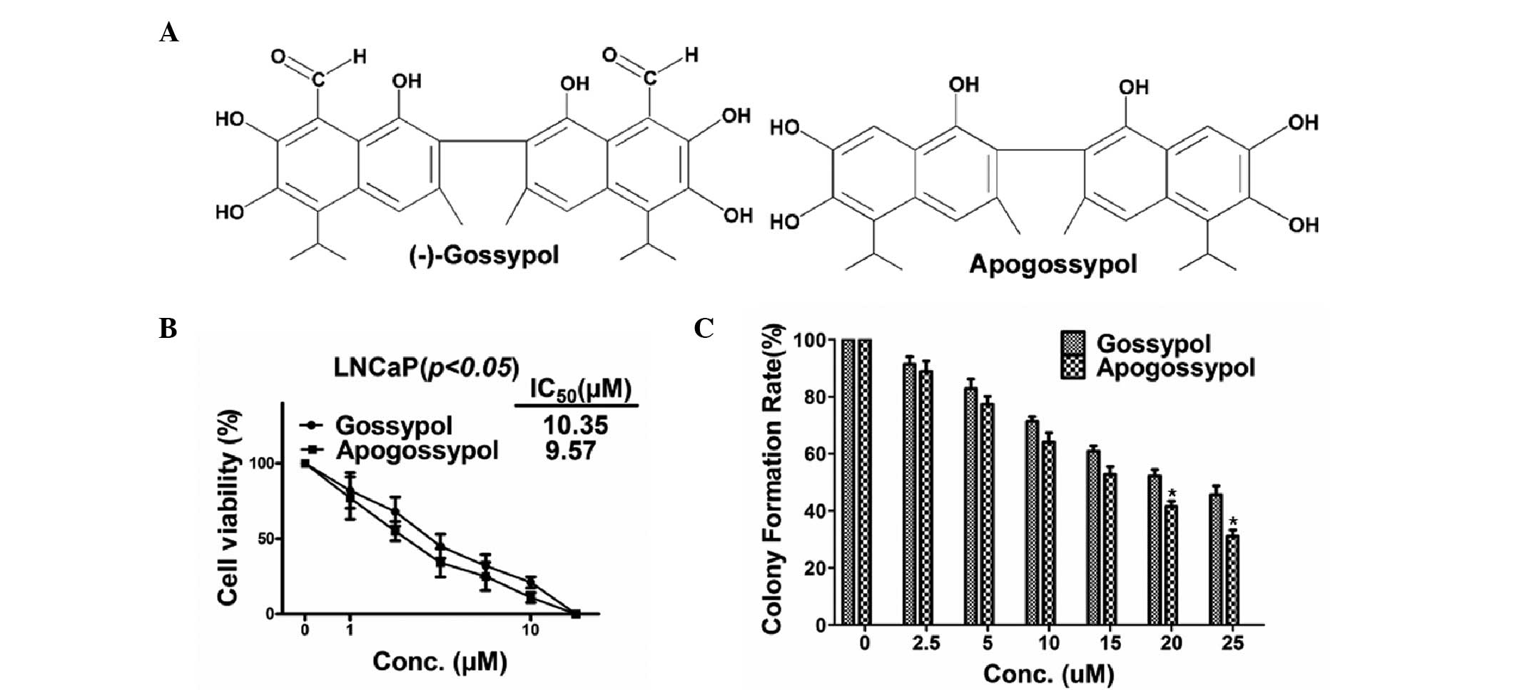
apogossypol (Fig. 1A), a novel
gossypol derivative lacking two aldehyde groups, which retains the
activity against the anti-apoptotic Bcl-2 family proteins in
vitro (26). Based on the
chemical design, apogossypol was expected to exert significantly
lower toxicity while maintaining a similar anti-cancer activity to
that of gossypol. However, whether or not apogossypol could
actually inhibit the growth and proliferation of prostate cancer
cells has yet to be established. In the present study, the
inhibitory effects of apogossypol on human prostate cancers were
investigated in order to demonstrate and compare the anti‑cancer
efficiencies between apogossypol and gossypol on prostate cancers
in vitro and in vivo.
Materials and methods
Cell lines and reagents
The LNCaP human prostate cancer cell line was
purchased from the American Type Culture Collection (Manassas, VA,
USA). The cells were cultured in RPMI1640 medium (Gibco-BRL, Grand
Island, NY, USA) supplemented with 10% fetal bovine serum (FBS;
Gibco-BRL) and 1% penicillin/streptomycin in a humidified incubator
at 37°C with 5% CO2. Apogossypol and gossypol were
synthesized and extracted in our laboratory (25), dissolved in dimethyl sulfoxide
(DMSO) and stored at ‑20°C. Working solutions were prepared by
diluting the stock solution with culture medium before use. MTT was
purchased from Sigma-Aldrich (St. Louis, MO, USA). The anti-Bcl-2,
anti-caspase-3, and anti-caspase-8 antibodies were purchased from
Maixin Biotechnology (Fuzhou, China), Zhongshan Golden Bridge
Biotechnology (Beijing, China) and Boster Biological Engineering
(Wuhan, China), respectively. Monkey anti-mouse immunoglobulin
(Ig)G labeled with fluorescein isothiocyanate (FITC) and goat
anti-rabbit IgG labeled with rhodamine were purchased from Santa
Cruz Biotechnology (Santa Cruz, CA, USA).
MTT assay
The cytotoxic effect of apogossypol and gossypol on
prostate cancer cell lines was measured by the MTT assay. LNCaP
cells were seeded onto sterile 96‑well flat‑bottomed plates and
incubated overnight. Then diluted apogossypol and gossypol were
added into each well with gradient concentrations (2–20
μmol/l). For the cell viability test, tumor cells were
suspended in a mixed solution of 200 μl complete medium and
0.2 μl DMSO, and wells with 200 μl complete medium
were used as blank controls. The plates were incubated at 37°C with
5% CO2 for 72 h. The medium was then removed, and 0.5
μmol/l MTT was added into the wells. After another 4 h, 150
μl DMSO was added into each well. The absorbance was read at
570 nm on a microplate reader (SpectraMax® M2, Molecular
Devices, Sunnyvale, CA, USA). The drug concentration yielding 50%
cell inhibition (IC50) was determined. All experiments
were performed in triplicate.
Colony formation assay
The colony formation assay was conducted on LNCaP
cells. The cells were seeded in six-well plates at a density of
200/well. Apogossypol and gossypol were added 24 h later at
appropriate doses. Following five days of incubation, 0.5 ml serum
was added into each well. The colonies were stained with crystal
violet on day 14 and the colonies consisting of >50 cells were
counted.
Terminal deoxynucleotidyl transferase
dUTP nick end labeling (TUNEL) assay
To assess apoptosis in the tumors, the TUNEL assay
was carried out using an In Situ Cell Death Detection Kit
(Boehringer-Mannheim, Mannheim, Germany). Briefly,
paraffin‑embedded tissue sections were treated with proteinase K
(20 μg/ml) in 10 mmol/l Tris-HCl (pH 7.5) for 30 min at room
temperature and afterwards they were dewaxed and rehydrated. The
slides were rinsed with phosphate-buffered saline (PBS) twice, for
5 min each time. The sections were then incubated with 50 μl
TUNEL reaction mixture at 37°C for 1 h in a humidified chamber.
Following incubation, the slides were rinsed with PBS three times,
for 5 min each time, and the apoptotic cells were visualized with
an Olympus FV1000 laser scanning confocal microscope (Olympus,
Tokyo, Japan). A positive control was prepared by treating the
samples with DNase I prior to TUNEL staining.
Animal experiments
Female athymic nude (nu/nu) mice (4–6 weeks of age,
weighing 20–25 g) were purchased from the animal center of the
Fourth Military Medical University. All animal experiments were
performed according to the protocol approved by the Fourth Military
Medical University Guidelines for the Use and Care of Animals.
LNCaP cells (2×10⁶) were injected subcutaneously into
each mouse. The tumor volume was measured every two days using a
caliper and calculated according to the following formula: Tumor
volume=L×W2, where L and W were the length and width,
respectively (24). When
subcutaneous tumor sizes reached 150–200 mm3, these mice
were randomly divided into three groups, each group consisting of
10 mice. Next, they were treated with apogossypol and gossypol,
respectively, at 20 mg/kg intraperitoneally, q.d. every 7 d for 28
d. The vehicle control group received the same amount of DMSO as in
the treatment groups. The tumor volume was detected every day. The
tumor tissues were fixed in 10% formalin solution. The tissues were
embedded with paraffin, and the sections were prepared. Samples
were stained with hematoxylin and eosin (H&E) and
microscopically examined (Olympus IX81; Olympus, Tokyo, Japan).
Immunofluorescence
The tumor tissues were dissected, fixed with
formaldehyde (40 μg/ml), embedded in paraffin and
deparaffinized with xylene. Following washing with water, the
sections were blocked with 250 μl/ml goat serum for 30 min.
Next, mouse anti-human Bcl-2 monoclonal antibody (1:50 dilution),
rabbit anti-caspase-3 polyclonal antibody (1:100 dilution) or
rabbit anti-caspase-8 polyclonal antibody (1:100 dilution) was
separately used for incubation at 4°C overnight in a humidified
chamber. Following washing with PBS for three times, monkey
anti-mouse IgG labeled with FITC or goat anti-rabbit IgG labeled
with rhodamine (1:100 dilution) was added and the samples were
incubated at 37°C for another 1 h. After washing with PBS for three
times, the samples were mounted with glycerol buffer and examined
under a microscope (Olympus IX81). In total, 10 high-power fields
per section were observed.
Statistical analysis
Data were presented as the mean ± standard
deviation. All the statistical analysis was performed with SPSS
16.0 software (Chicago, IL, USA). Student’s t-test was used for
statistical comparisons between groups. P<0.05 was used to
indicate a statistically significant difference.
Results
Apogossypol inhibits the survival of
LNCaP cells
To investigate the inhibitory effects of apogossypol
and gossypol on LNCaP cell survival, the MTT assay was performed.
The results demonstrated that apogossypol inhibited the
proliferation of LNCaP cells in a time- and dose-dependent manner,
in a similar way with gossypol (Fig.
1B). The concentration for 50% inhibition (IC50) on
LNCaP cells within ~72 h was 9.57 μmol/l, while the
IC50 of gossypol on LNCaP cells was 10.35 μmol/l.
The inhibitory activities of apogossypol and gossypol at the same
drug concentration were not significantly different from each
other, suggesting that the removal of two aldehyde groups had
little effect on the anti-tumor effect of gossypol.
Apogossypol inhibits the proliferation of
prostate cancer cells
To further evaluate the anti-tumor effects of
apogossypol and gossypol, the colony formation assay was performed.
The treatment groups were treated with different drug
concentrations of apogossypol or gossypol, and the control group
was treated with DMSO. After 14 days of treatment, the percentage
of colony formation was calculated. Apogossypol potently inhibited
the colony formation of LNCaP cells (Fig. 1C). Compared with the vehicle
control, 15 μmol/l apogossypol inhibited >56% of the
colony formation, while 25 μmol/l gossypol inhibited >45%
of the colony formation. These data indicated that apogossypol and
gossypol had strong anti-tumor activities. Furthermore, the results
also indicated that the anti-tumor activity of apogossypol was
slightly stronger compared to that of gossypol, which was in
accordance with the result of the MTT assay.
Apogossypol induces apoptosis in LNCaP
cells
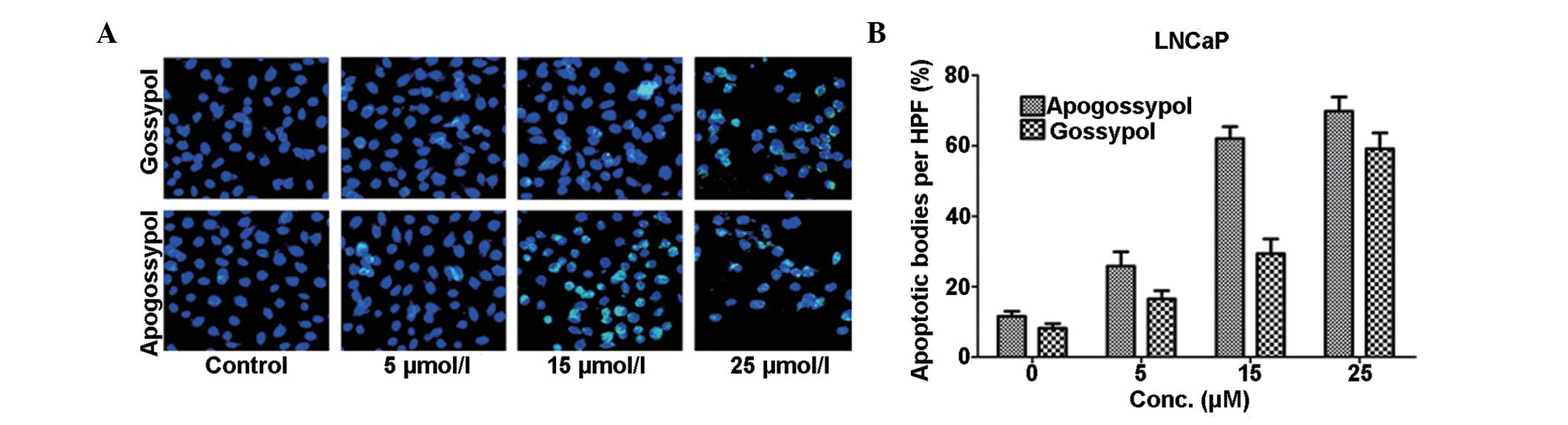
Hoechst 33258 staining was performed next to
evaluate the levels of apoptosis in different drug concentration
groups. The cells with changed nuclear morphologies indicated by
Hoechst 33258 staining (cyan) were TUNEL-positive cells, while the
nuclei in normal cells were stained with DAPI (blue). The cells
treated with high drug concentrations exhibited evident apoptotic
characteristics, including shrinkage and nuclear fragmentation
(Fig. 2A). Compared with the
gossypol group, it was notable that there was an evident increase
in the number of TUNEL-positive LNCaP cells following treatment
with apogossypol at a concentration of 15 μmol/l. The
apoptotic rate following gossypol treatment was 24%, which was
significantly lower than 62% following apogossypol treatment
(P<0.01; Fig. 2B). The results
indicated that inducing cell apoptosis may be a possible mechanism
underlying the anti-tumor activity of apogossypol in prostate
cancer.
Apogossypol alters the expression levels
of Bcl‑2, caspase‑3 and ‑8 in prostate tumors
To investigate the possible molecular mechanism(s)
through which apogossypol triggered apoptosis, immunofluorescence
was performed to observe the changes of the protein expression
levels of the Bcl-2 family members in LNCaP xenograft cells treated
with apogossypol. An evidently high percentage of tumor cells
expressed Bcl-2 in the control group, while apogossypol treatment
caused a significant decrease in Bcl-2 expression levels in tumor
tissues, indicating that apogossypol induced apoptosis in the LNCaP
xenograft tumor (Fig. 3A). In
addition, the expression levels of caspase-3 and -8 were weak in
the control group, while they were strong in the groups treated
with apogossypol. Compared with the control group, it was notable
that there was a clear increase in the protein expression levels in
LNCaP cells treated with 25 μmol/l apogossypol. The relative
protein expression levels of Bcl-2 were only 37% in cells treated
with apogossypol, compared with 84% in the control group
(P<0.01; Fig. 3B). However, the
relative expression levels of caspase-3 and caspase-8 in the
apogossypol group were 67 and 81%, respectively, which were both
significantly higher compared to those in the control group
(P<0.01). These findings indicated that apogossypol induced
tumor cell apoptosis by downregulating the Bcl-2 protein expression
levels and upregulating caspase-3 and -8 expression levels.
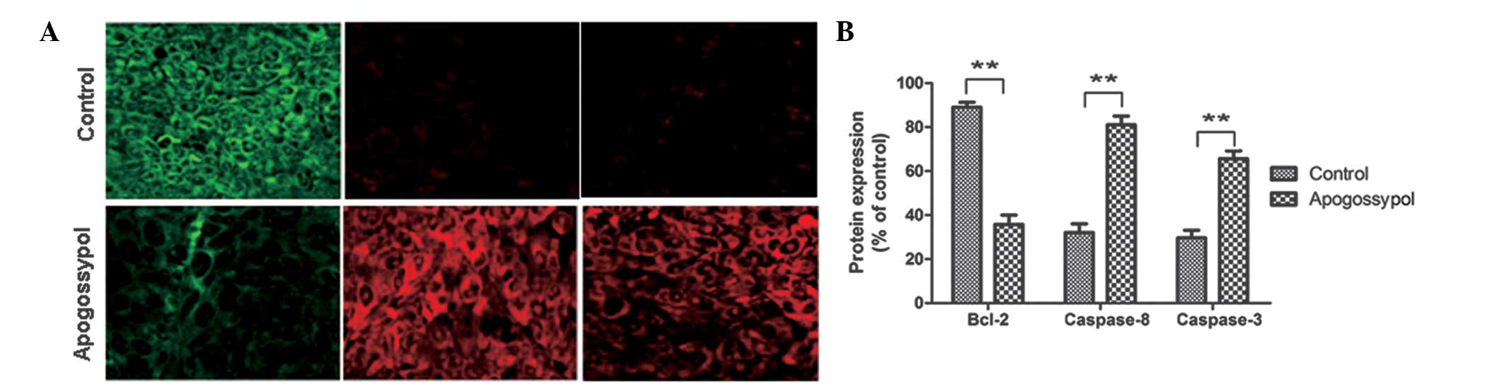 | Figure 3Apogossypol alters the expression
levels of Bcl‑2, caspase‑3, and caspase‑8 in prostate tumors. (A)
Immunofluorescence was performed to detect the expression levels of
Bcl‑2, caspase‑3, and caspase‑8 in LNCaP xenografts treated with
apogossypol. Control group was treated with DMSO (magnification,
×400). (B) Statistical analysis of protein expression levels in
LNCaP cells. The histogram represents the relative expression
levels of Bcl-2, caspase-3, and caspase-8, compared with
non-treated cells. Values expressed as mean ± standard deviation
from three independent experiments. **P<0.01 compared
with the control group. DMSO, dimethyl sulfoxide; Bcl-2, B-cell
lymphoma 2. |
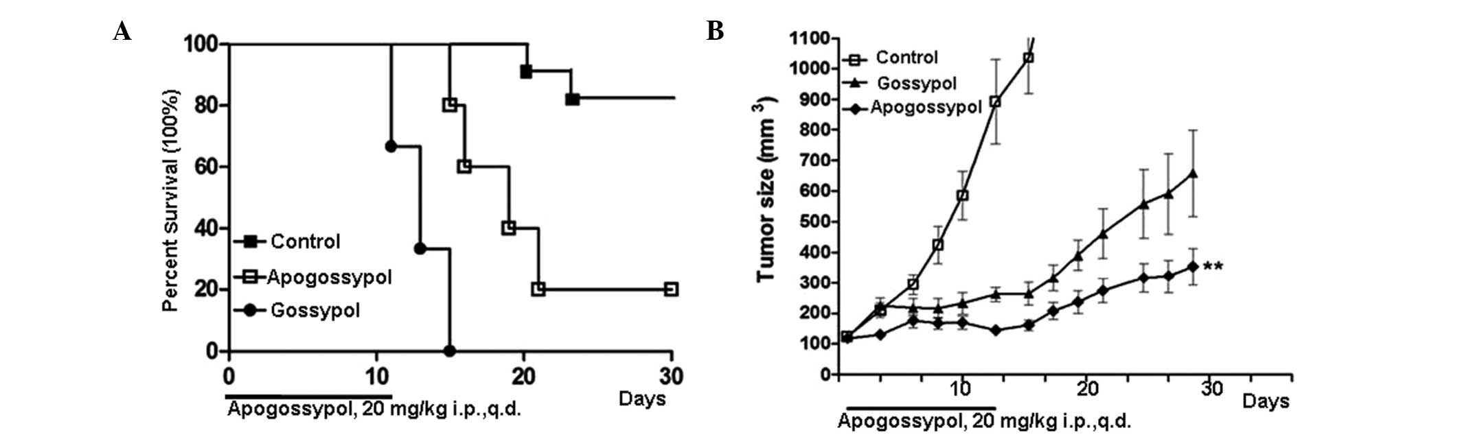
Apogossypol reduces tumor growth of LNCaP
xenografts in vivo
Due to its modified structure, apogossypol was
expected to exhibit lower toxicity while maintaining the
significant anti-growth and anti-tumor activities in vitro,
similar to those of gossypol. Therefore, the present study further
evaluated the anti-cancer effect of apogossypol in mice bearing
subcutaneous LNCaP cell xenografts. The tumor growth was monitored
and measured by a caliper and balance. The survival rate of the
mice was notably improved by apogossypol (Fig. 4). Of note, the tumor sizes were
also markedly decreased by apogossypol treatment (P<0.01). These
results indicated that apogossypol had significant anti-cancer
activity in prostate cancers in vivo, compared with gossypol
at the same drug concentration (20 mg kg−1). Finally,
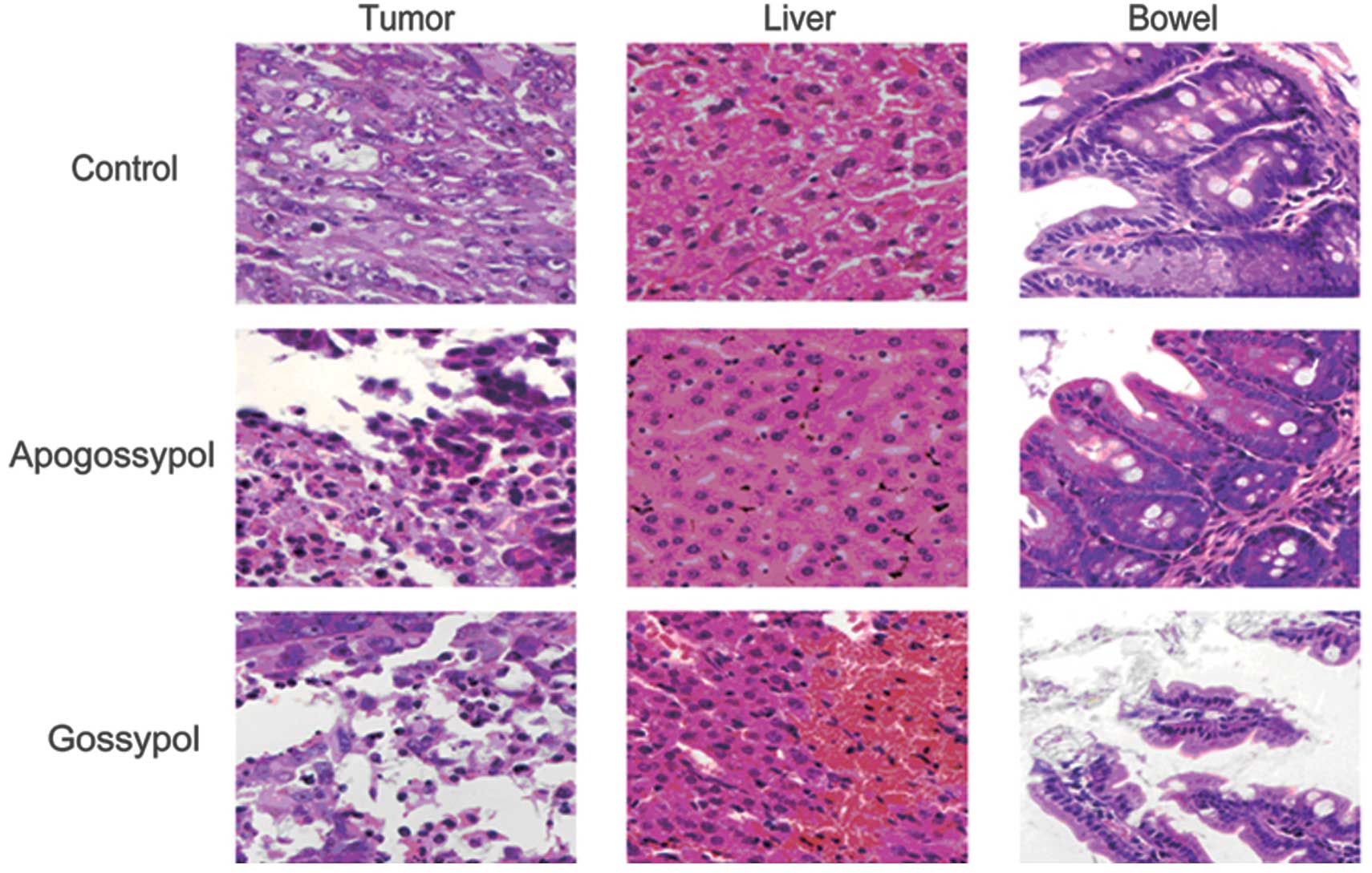
the pathological examination was carried out by H&E staining.
The cells in tumor tissues of the apogossypol treatment group
exhibited necrotic or pyknotic nuclei (Fig. 5). However, there were no evident
lesions in other normal tissues. The results demonstrated that
apogossypol produced an excellent anti-cancer therapeutic response,
while having low toxicity to normal tissues.
Discussion
Although chemotherapy and radiotherapy are currently
common treatments for prostate cancers, limited therapeutic methods
are available for the disease (27). Thus, identifying novel
chemotherapeutic drugs or developing effective treatment strategies
is important for disease management (28). Recently, the strategy of blocking
anti-apoptotic protein activities has gained increasing attention.
Several non-peptide small molecular inhibitors of Bcl-2 family
proteins have been synthesized and used in studies for therapies
against various types of cancers (29,30).
However, the effects of apogossypol in prostate cancer therapy have
never been established. To the best of our knowledge, proliferation
and apoptosis are extensively used biomarkers for diagnosis and
measurement of tumor aggressiveness, which thereby contribute to
evaluating the tumor responses to novel anti-cancer drugs (31,32).
Thus, the anti-proliferation and apoptosis-inducing effects of
apogossypol in prostate cancers were evaluated in vitro. The
MTT and colony formation assays revealed that apogossypol
effectively inhibited cell growth and induced evident apoptosis in
prostate cancer cells. The anti-growth effects and
apoptosis-inducing abilities between apogossypol and gossypol were
further compared. Based on these results, it is reasonable to
postulate that the removal of two aldehyde groups may have no
effect on the in vitro growth inhibition ability of
gossypol. In addition, several previous studies have reported that
gossypol has a synergistic effect in enhancing anti-cancer
therapies (33,34). Therefore, it is hypothesized that
apogossypol may be used as a safe and effective agent in
combination with other targeting or conventional drugs for therapy
of prostate cancers, which is now actively underway in our
laboratory.
To facilitate the translation of apogossypol from
research into clinical practice for prostate cancer therapy, the
in vivo response to drug therapy must be addressed. The two
aldehyde groups in the chemical structure of gossypol are
associated with toxicity (35,36).
Thus, apogossypol was synthesized by removing the two aldehyde
groups and has been found to maintain the anti-cancer effects for
several types of cancers, while exhibiting reduced toxicity
(37,38). In the present study, the toxicities
and tumor-inhibiting activities between apogossypol and gossypol
were compared in nude mouse xenografts. The results showed that
apogossypol exhibited significantly lower toxicity and caused more
significant reduction in tumor size compared to gossypol, which is
consistent with previous reports. Therefore, the in vivo
data further verified the fact that the removal of the two aldehyde
groups did not affect the BH3, which creates a hydrophobic surface
pocket that may be a binding groove for anti-tumor drugs (39). These results indicated that
apogossypol may be a novel and useful anti-cancer agent for
prostate cancer therapy.
The death receptor pathway, the mitochondrial
pathway and endoplasmic reticulum stress-induced apoptosis are
three common ways to induce apoptosis (40,41).
Bcl-2 family proteins are regarded as the central regulators of the
apoptotic process, which has been divided into two groups,
pro-apoptotic proteins, including Bcl-2 homologous antagonist
killer, Bcl-2-associated death promoter, Bcl-2-interacting killer
and Bcl-2-like protein 11, as well as anti-apoptotic proteins,
including Bcl-2, Bcl-xL and Mcl-1 (42,43).
All of them have become hot spots to develop novel anti-cancer
drugs (44,45). As a result, there are numerous
newly designed and synthesized chemotherapeutic agents targeting
the BH-3 domains of anti-apoptotic Bcl-2 members to induce
apoptosis and inhibit the function of Bcl-2/Bcl-xL proteins, which
may be useful in the management and treatment of cancers (46,47).
In fact, the ratio between Bcl-2, caspase-3 and -8 may be used to
determine whether cancer cells are undergoing apoptosis or not
(14,48). Thus, the expression levels of these
proteins were examined in the xenografts treated with apogossypol
through immunofluorescence. Our results demonstrated that
apogossypol could alter the expression levels of Bcl-2 family
proteins, downregulate Bcl-2 expression levels and lead to the
activation of apoptosis proteins, including caspase-3 and -8.
Therefore, in prostate cancers, apogossypol could activate the
mitochondrial signaling pathway to promote cell death.
In conclusion, it was demonstrated that a novel
small-molecule inhibitor of the anti-apoptotic Bcl-2 family
proteins, apogossypol, had significant anti‑tumor activity in
vitro and in vivo in prostate cancers. Apogossypol may
bind to Bcl-2 family proteins and prevent the binding of
pro-apoptotic proteins with BH-3 domains, unleashing the
pro-apoptotic proteins to induce the apoptotic response. The
present study indicated that apogossypol may be a promising novel
agent for prostate cancer therapy.
Acknowledgments
The present study was supported by the National
Nature Science Foundation of China (no. 81101100), the Natural
Science Research Plan in Ningxia Province of China (no. NZ14147)
and the Fundamental Research Funds Central Universities (nos.
K50510100002 and K50510100004).
References
|
1
|
Parkin DM: International variation.
Oncogene. 23:6329–6340. 2004. View Article : Google Scholar : PubMed/NCBI
|
|
2
|
Jemal A, Bray F, Center MM, et al: Global
cancer statistics. CA Cancer J Clin. 61:69–90. 2011. View Article : Google Scholar : PubMed/NCBI
|
|
3
|
Morse MA and Stoner GD: Cancer
chemoprevention: principles and prospects. Carcinogenesis.
14:1737–1746. 1993. View Article : Google Scholar : PubMed/NCBI
|
|
4
|
Debatin KM: Apoptosis pathways in cancer
and cancer therapy. Cancer Immunol Immunother. 53:153–159. 2004.
View Article : Google Scholar : PubMed/NCBI
|
|
5
|
Oltersdorf T, Elmore SW, Shoemaker AR, et
al: An inhibitor of Bcl-2 family proteins induces regression of
solid tumors. Nature. 435:677–681. 2005. View Article : Google Scholar : PubMed/NCBI
|
|
6
|
Krajewska M, Fenoglio-Preiser CM,
Krajewski S, et al: Immunohistochemical analysis of Bcl-2 family
proteins in adenocarcinomas of the stomach. Am J Pathol.
149:1449–1457. 1996.PubMed/NCBI
|
|
7
|
Reed JC, Zha H, Aime-Sempe C, Takayama S
and Wang HG: Structure-function analysis of Bcl-2 family proteins:
Regulators of programmed cell death. Adv Exp Med Biol. 406:99–112.
1996. View Article : Google Scholar
|
|
8
|
Petros AM, Olejniczak ET and Fesik SW:
Structural biology of the Bcl-2 family of proteins. Biochim Biophys
Acta. 1644:83–94. 2004. View Article : Google Scholar : PubMed/NCBI
|
|
9
|
Marzo I and Naval J: Bcl-2 family members
as molecular targets in cancer therapy. Biochem Pharmacol.
76:939–946. 2008. View Article : Google Scholar : PubMed/NCBI
|
|
10
|
Huang Z: Small molecule inhibitors of
Bcl-2 function: modulators of apoptosis and promising anticancer
agents. Curr Opin Drug Discov Devel. 3:565–574. 2000.PubMed/NCBI
|
|
11
|
Wang S, Yang D and Lippman ME: Targeting
Bcl-2 and Bcl-XL with nonpeptidic small-molecule antagonists. Semin
Oncol. 30(5 Suppl 16): 133–142. 2003. View Article : Google Scholar : PubMed/NCBI
|
|
12
|
Al-Katib AM, Sun Y, Goustin AS, et al: SMI
of Bcl-2 TW-37 is active across a spectrum of B-cell tumors
irrespective of their proliferative and differentiation status. J
Hematol Oncol. 2:82009. View Article : Google Scholar : PubMed/NCBI
|
|
13
|
Azmi AS and Mohammad RM: Non-peptidic
small molecule inhibitors against Bcl-2 for cancer therapy. J Cell
Physiol. 218:13–21. 2009. View Article : Google Scholar :
|
|
14
|
Wei MC, Zong WX, Cheng EH, et al:
Proapoptotic BAX and BAK: a requisite gateway to mitochondrial
dysfunction and death. Science. 292:727–730. 2001. View Article : Google Scholar : PubMed/NCBI
|
|
15
|
Coutinho EM: Gossypol: a contraceptive for
men. Contraception. 65:259–263. 2002. View Article : Google Scholar : PubMed/NCBI
|
|
16
|
Naik H, Petrylak D, Yagoda A, et al:
Preclinical studies of gossypol in prostate carcinoma. Int J Oncol.
6:209–213. 1995.PubMed/NCBI
|
|
17
|
Gilbert NE, O’Reilly JE, Chang CJ, Lin YC
and Brueggemeier RW: Antiproliferative activity of gossypol and
gossypolone on human breast cancer cells. Life Sci. 57:61–67. 1995.
View Article : Google Scholar : PubMed/NCBI
|
|
18
|
Bushunow P, Reidenberg MM, Wasenko J, et
al: Gossypol treatment of recurrent adult malignant gliomas. J
Neurooncol. 43:79–86. 1999. View Article : Google Scholar : PubMed/NCBI
|
|
19
|
Balakrishnan K, Wierda WG, Keating MJ and
Gandhi V: Gossypol, a BH3 mimetic, induces apoptosis in chronic
lymphocytic leukemia cells. Blood. 112:1971–1980. 2008. View Article : Google Scholar : PubMed/NCBI
|
|
20
|
Gunassekaran GR, Priya DK, Gayathri R and
Sakthisekaran D: In vitro and in vivo studies on antitumor effects
of gossypol on human stomach adenocarcinoma (AGS) cell line and
MNNG induced experimental gastric cancer. Biochem Biophys Res
Commun. 411:661–666. 2011. View Article : Google Scholar : PubMed/NCBI
|
|
21
|
Jiang J, Slivova V, Jedinak A and Sliva D:
Gossypol inhibits growth, invasiveness, and angiogenesis in human
prostate cancer cells by modulating NF-κB/AP-1 dependent- and
independent-signaling. Clin Exp Metastasis. 29:165–178. 2012.
View Article : Google Scholar
|
|
22
|
Baggstrom MQ, Qi Y, Koczywas M, et al: A
phase II study of AT-101 (Gossypol) in chemotherapy-sensitive
recurrent extensive-stage small cell lung cancer. J Thorac Oncol.
6:1757–1760. 2011. View Article : Google Scholar : PubMed/NCBI
|
|
23
|
Wei J, Kitada S, Stebbins JL, et al:
Synthesis and biological evaluation of Apogossypolone derivatives
as pan-active inhibitors of antiapoptotic B-cell
lymphoma/leukemia-2 (Bcl-2) family proteins. J Med Chem.
53:8000–8011. 2010. View Article : Google Scholar : PubMed/NCBI
|
|
24
|
Zhang XQ, Huang XF, Hu XB, et al:
Apogossypolone, a novel inhibitor of antiapoptotic Bcl-2 family
proteins, induces autophagy of PC-3 and LNCaP prostate cancer cells
in vitro. Asian J Androl. 12:697–708. 2010. View Article : Google Scholar : PubMed/NCBI
|
|
25
|
Zhan Y, Jia G, Wu D, Xu Y and Xu L: Design
and synthesis of a gossypol derivative with improved antitumor
activities. Arch Pharm (Weinheim). 342:223–229. 2009. View Article : Google Scholar
|
|
26
|
Wei J, Rega MF, Kitada S, et al: Synthesis
and evaluation of Apogossypol atropisomers as potential Bcl-xL
antagonists. Cancer Lett. 273:107–113. 2009. View Article : Google Scholar :
|
|
27
|
Jiang J, Sugimoto Y, Liu S, et al: The
inhibitory effects of gossypol on human prostate cancer cells-PC3
are associated with transforming growth factor beta1 (TGFbeta1)
signal transduction pathway. Anticancer Res. 24:91–100.
2004.PubMed/NCBI
|
|
28
|
Karaca B, Kucukzeybek Y, Gorumlu G, et al:
Profiling of angiogenic cytokines produced by hormone- and
drug-refractory prostate cancer cell lines, PC-3 and DU-145 before
and after treatment with gossypol. Eur Cytokine Netw. 19:176–184.
2008.PubMed/NCBI
|
|
29
|
Zhang M, Liu H, Tian Z, et al: Gossypol
induces apoptosis in human PC-3 prostate cancer cells by modulating
caspase-dependent and caspase-independent cell death pathways. Life
Sci. 80:767–774. 2007. View Article : Google Scholar
|
|
30
|
Reed JC: Apoptosis-based therapies. Nat
Rev Drug Discov. 1:111–121. 2002. View
Article : Google Scholar : PubMed/NCBI
|
|
31
|
Adams JM and Cory S: The Bcl-2 protein
family: arbiters of cell survival. Science. 281:1322–1326. 1998.
View Article : Google Scholar : PubMed/NCBI
|
|
32
|
Gross A, McDonnell JM and Korsmeyer SJ:
BCL-2 family members and the mitochondria in apoptosis. Genes Dev.
13:1899–1911. 1999. View Article : Google Scholar : PubMed/NCBI
|
|
33
|
Wang JL, Liu D, Zhang ZJ, et al:
Structure-based discovery of an organic compound that binds Bcl-2
protein and induces apoptosis of tumor cells. Proc Natl Acad Sci
USA. 97:7124–7129. 2000. View Article : Google Scholar : PubMed/NCBI
|
|
34
|
Degterev A, Lugovskoy A, Cardone M, et al:
Identification of small-molecule inhibitors of interaction between
the BH3 domain and Bcl-xL. Nat Cell Biol. 3:173–182. 2001.
View Article : Google Scholar : PubMed/NCBI
|
|
35
|
Reed JC: Bcl-2 family proteins. Oncogene.
17:3225–3236. 1998. View Article : Google Scholar
|
|
36
|
Reed JC: Bcl-2 family proteins: strategies
for overcoming chemoresistance in cancer. Adv Pharmacol.
41:501–532. 1997. View Article : Google Scholar : PubMed/NCBI
|
|
37
|
Kitada S, Leone M, Sareth S, et al:
Discovery, characterization, and structure-activity relationships
studies of proapoptotic poly-phenols targeting B-cell
lymphocyte/leukemia-2 proteins. J Med Chem. 46:4259–4264. 2003.
View Article : Google Scholar : PubMed/NCBI
|
|
38
|
Zhang M, Liu H, Guo R, et al: Molecular
mechanism of gossypol-induced cell growth inhibition and cell death
of HT-29 human colon carcinoma cells. Biochem Pharmacol. 66:93–103.
2003. View Article : Google Scholar : PubMed/NCBI
|
|
39
|
Wang G, Nikolovska-Coleska Z, Yang CY, et
al: Structure-based design of potent small-molecule inhibitors of
anti-apoptotic Bcl-2 proteins. J Med Chem. 49:6139–6142. 2006.
View Article : Google Scholar : PubMed/NCBI
|
|
40
|
Scopa CD, Vagianos C, Kardamakis D,
Kourelis TG, Kalofonos HP and Tsamandas AC: bcl-2/bax ratio as a
predictive marker for therapeutic response to radiotherapy in
patients with rectal cancer. Appl Immunohistochem Mol Morphol.
9:329–334. 2001. View Article : Google Scholar
|
|
41
|
Shelley MD, Hartley L, Groundwater PW and
Fish RG: Structure-activity studies on gossypol in tumor cell
lines. Anticancer Drugs. 11:209–216. 2000. View Article : Google Scholar : PubMed/NCBI
|
|
42
|
Becattini B, Kitada S, Leone M, et al:
Rational design and real time, in-cell detection of the
proapoptotic activity of a novel compound targeting Bcl-X(L). Chem
Biol. 11:389–395. 2004. View Article : Google Scholar : PubMed/NCBI
|
|
43
|
Kitada S, Kress CL, Krajewska M, Jia L,
Pellecchia M and Reed JC: Bcl-2 antagonist apogossypol (NSC736630)
displays single-agent activity in Bcl-2-transgenic mice and has
superior efficacy with less toxicity compared with gossypol
(NSC19048). Blood. 111:3211–3219. 2008. View Article : Google Scholar : PubMed/NCBI
|
|
44
|
Coward L, Gorman G, Noker P, et al:
Quantitative determination of apogossypol, a proapoptotic analog of
gossypol, in mouse plasma using LC/MS/MS. J Pharm Biomed Anal.
42:581–586. 2006. View Article : Google Scholar : PubMed/NCBI
|
|
45
|
Sattler M, Liang H, Nettesheim D, et al:
Structure of Bcl-xL-Bak peptide complex: recognition between
regulators of apoptosis. Science. 275:983–986. 1997. View Article : Google Scholar : PubMed/NCBI
|
|
46
|
Eldridge MD, Murray CW, Auton TR, Paolini
GV and Mee RP: Empirical scoring functions: I. The development of a
fast empirical scoring function to estimate the binding affinity of
ligands in receptor complexes. J Comput Aided Mol Des. 11:425–445.
1997. View Article : Google Scholar : PubMed/NCBI
|
|
47
|
Ramjaun AR, Tomlinson S, Eddaoudi A and
Downward J: Upregulation of two BH3-only proteins, Bmf and Bim,
during TGF beta-induced apoptosis. Oncogene. 26:970–981. 2007.
View Article : Google Scholar
|
|
48
|
Katsumata M, Siegel RM, Louie DC, et al:
Differential effects of Bcl-2 on T and B cells in transgenic mice.
Proc Natl Acad Sci USA. 89:11376–11380. 1992. View Article : Google Scholar : PubMed/NCBI
|