Introduction
Polycystic ovary syndrome (PCOS) is a complex
syndrome and is the most common endocrine disorder in females of
reproductive age globally (1). In
a previous study, 4–4.8% of white females and 3.5% of African
females exhibited PCOS (2).
Similarly, a previous study demonstrated that the prevalence of
PCOS was 4.9% among female college students in Korea (3). According to the American Society for
Reproductive Medicine (ASRM) and the European Society of Human
Reproduction and Embryology (ESHRE), PCOS is diagnosed when the
phenotype of patients satisfy two of the following three criteria:
Oligomenorrhea or amenorrhea, biochemical hyperandrogenism or
polycystic ovaries (4,5). Generally, females with PCOS also
exhibit symptoms of an increased risk of infertility, obesity,
hyperlipidemia, insulin resistance, type 2 diabetes (T2D), diabetic
nephropathy (DN) and possibly cardiovascular disease (6–8).
The pathogenesis of PCOS is initiated by various
signaling pathways: Metabolism, insulin-signaling, inflammation and
angiogenesis (9). Among these
pathways, insulin resistance or hyperinsulinemia triggers changes
in the hypothalamo-pituitary-ovarian axis, causing excessive
production of androgen, leading to aberrant follicular development.
Hyperinsulinemia in PCOS inhibits the hepatic sex hormone binding
globulin from increasing the levels of free testosterone (T) in the
body. This promotes the secretion of luteinizing hormone (LH) with
relatively low follicle-stimulating hormone (FSH) (10). For this reason, it is recognized
that females with PCOS are susceptible to insulin resistance and
T2D (11). A case-control genetic
association investigation regarding the pathogenesis of PCOS
focused on the single nucleotide polymorphisms (SNPs) affecting the
inflammatory processes and the activity of transforming growth
factor-β1 (TGF-β1) (12).
In previous years, genetic studies have linked PCOS to a
dinucleotide repeat marker, D19S884, in the fibrillin 3
gene. Fibrillins are important molecules, which assemble into
microfibrils in the extracellular matrix (ECM) to modulate the
TGF-β1 signaling pathway (13,14).
Therefore, variations in fibrillin 3 and the subsequent
dysregulation of TGF-β may contribute to the pathogenesis of PCOS
(15).
TGF-β is a multifunctional cytokine synthesized in a
wide variety of tissue types and it is secreted from various cell
types (16). The TGF-β superfamily
consists of three isoforms: TGF-β1, TGF-β2 and
TGF-β3 (17). The members
of the human TGF-β superfamily are critical modulators in apoptosis
and cell survival (18). The TGF-β
signaling pathway is initiated when a TGF-β superfamily ligand
binds to a high-affinity transmembrane receptor complex, composed
of the activin-like kinase 5/TGF-β type 1 receptor and the TGF-β
type 2 receptor. Each class of ligand binds to a specific type 2
receptor, which has a serine/threonine kinase domain (19). It subsequently recruits and
phosphorylates a specific type 1 receptor. The type 1 receptor
phosphorylates receptor-regulated Smads, which subsequently bind to
the coSmad, Smad4. R-Smad/coSmad complexes activated by
phosphorylation translocate into the nucleus to bind to gene
promoters and activate the expression of the target genes involved
in cell proliferation and differentiation (20,21).
This TGF-β superfamily signaling demonstrates its potential role in
embryonic development, cellular differentiation, hormone secretion
and immune system functions (22,23).
In addition to this Smad-mediated gene transcription, TGF-β
signaling is involved in activating Smad-independent pathways,
including the nuclear factor-κB pathway (24), the mitogen-activated protein
kinase/ERK pathway (25) and the
phosphatidylinositol-3-kinase/Akt signaling pathway (26). Therefore, Smad-independent pathways
in the TGF-β family signaling pathways have significant effects on
the different biological functions of TGF-β, including cell cycle
inhibition, immune suppression and neuroprotective effects
(27,28).
The TGF-β signaling pathway has a vital role in the
development of multiple tissues or cells, including
folliculogenesis, which is the process of developing ovarian
follicles (29). Members of the
TGF-β superfamily are expressed in mammalian oocytes and thereby,
the subsequent dysregulation of TGF-β has been implicated in the
pathogenesis of abnormal follicle development and hyperandrogenism
in patients with PCOS (30,31).
A previous study indicated that the ovaries of females with PCOS
exhibited all the markers of increased TGF-β activity (32).
The TGF-β1 gene has been suggested as a
genetic factor due to the clinical symptoms of PCOS, including an
increased risk of T2D. TGF-β1 promotes the production of the
ECM in response to high levels of glucose. Therefore, TGF-β1
is considered to be central in the pathogenesis of DN (33).
The human TGF-β1 gene is located on
chromosome 19q13.1-13.3 and has six known SNPs: C-988A (rs1800820),
G-800A (rs1800468), C509T (rs1800469), T869C (rs1800470;
Leu10/P.ro10; T29->C), G915C (rs1800471) and C11929T (THr263Ile;
rs1800472) (34,35). In a previous study, a statistically
significant difference was detected between the control group and
Egyptian patients with T2D in the frequencies of the TGF-β1
codon 10 (T869C) (36). The
present study, therefore, focused on the T869C polymorphism located
on exon 1, which may be one of the candidate genes associated with
an increased risk of DN. The present study aimed to determine
whether the T869C polymorphism of TGF-β1 was associated with
PCOS.
Patients and methods
Study subjects
All individuals were Korean females (n=414) of which
129 were healthy controls and 285 were patients with PCOS,
recruited from the Fertility Center at CHA General Hospital (Seoul,
Korea) between 2008 and 2011. The diagnosis of PCOS was based on
the criteria proposed by the 2003 ASRM/ESHRE Rotterdam consensus
(4,5). The present study was approved by the
Gangnam CHA Fertility Center (Gyeonggi-Do, Korea). Written informed
consent was provided by the patient
Phenotypic characterization of all
subjects
Basal blood samples were obtained from patients with
PCOS and the controls to measure the levels of the following:
Plasma FSH, LH, estrogen (E2), prolactin (PRL), thyroid stimulating
hormone (TSH), dehydroepiandrosteronesulphate (DHEAS), T, fasting
glucose and insulin.
DNA extraction and genetic analysis
Blood samples were collected in tubes containing
EDTA as an anti-clotting factor and stored at 4°C. The genomic DNA
was extracted from the blood of patients with PCOS and the
controls. Restriction fragment length polymorphism (RFLP) analysis
was performed to determine the genotypes for the T869C polymorphism
in exon 1 of the TGF-β1 gene. The T869C polymorphism was
amplified by polymerase chain reaction (PCR) using the following
primers: Forward, 5′-GTA CCA GAT CGC GCC CATCT-3′ and reverse,
5′-TAG CCA CAG CAT CGG TAG CAG-3′, in a total volume of 30 µl. In
the reaction mixture (Solgent, Seoul, Korea), 100 ng genomic DNA
was used as a template. The cycling parameters were as follows:
Denaturation at 95°C for 5 min, 30 cycles at 95°C for 40 sec, 65°C
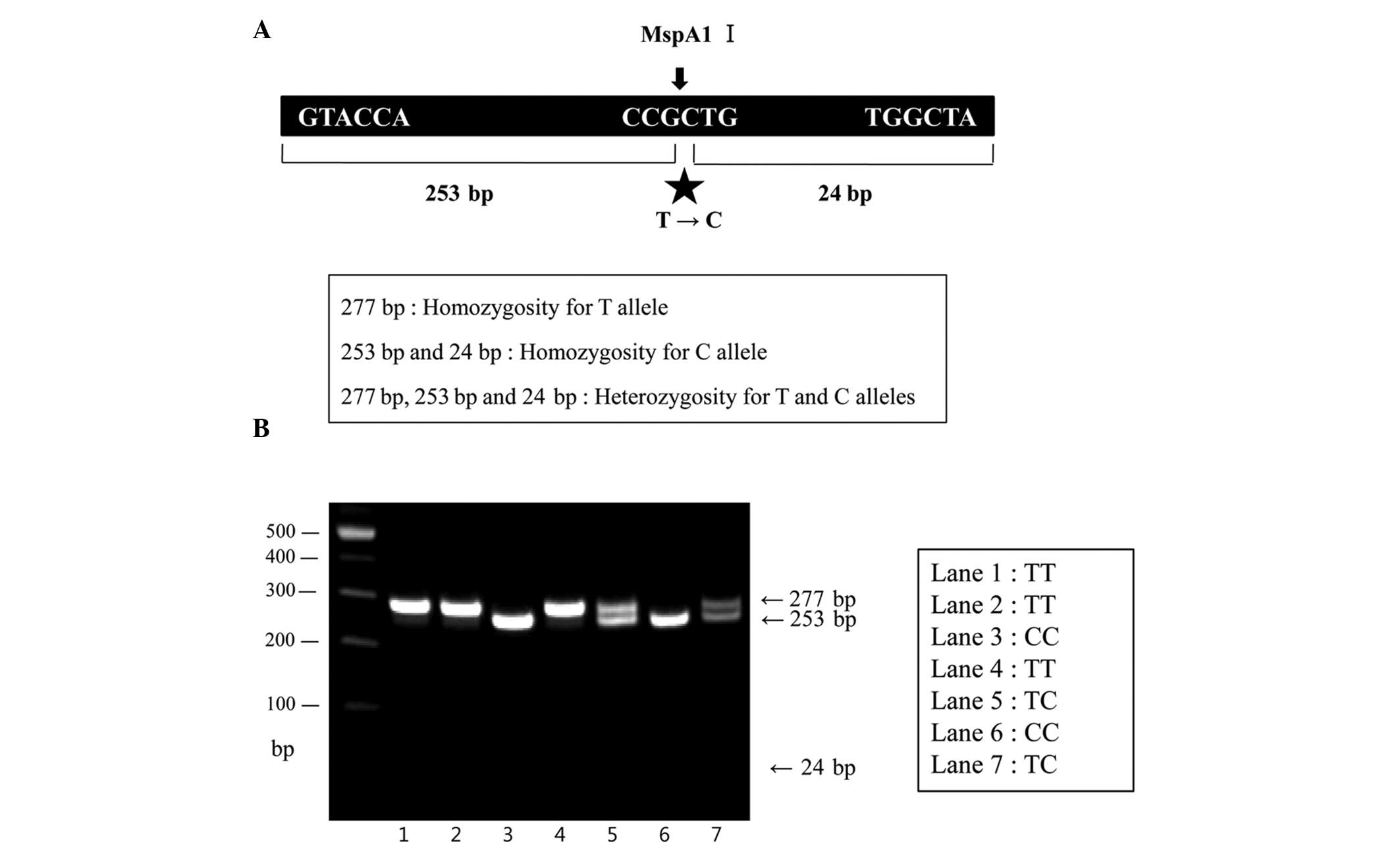
for 40 sec and 72°C for 40 sec, followed by 72°C for 7 min.
Following PCR (C1000 Thermal cycler; Bio-Rad Laboratories, Inc.,
Hercules, CA, USA), the PCR products of 277 bp were digested with
MspA1 Ι (Enzynomics, Daejeon, Korea) for 2 h at 37°C
(Fig. 1A). The restricted DNA
fragments were electrophoresed on a 2% agarose gel (Invitrogen Life
Technologies, Carlsbad, CA, USA), containing ethidium bromide
(Sigma-Aldrich, St. Louis, MO, USA) and visualized on a DNA Image
Visualizer (SeouLin Bioscience Co., Ltd, Seoul, Korea).
A total of three genotypes were observed in the
restricted DNA fragments: A single 277 bp band, indicating
homozygosity for the T allele; the presence of two fragments, 253
bp and 24 bp, indicating homozygosity for the C allele; the
presence of three fragments, 277 bp, 253 bp and 24-bp bands,
indicating heterozygosity for the T allele and the C allele,
respectively (Fig. 1B).
Statistics analysis
Statistical analysis for comparing the genotype
frequencies of the control group and the patient group was
performed using Hap analysis (HapAnalyzer Ver.1 0.1, NGRI, Seoul,
Korea) and the χ2 test. P<0.05 was considered to
inidcate a statistically significant difference.
Results
Patient characteristics
The 2003 ASRM/ESHRE Rotterdam Consensus was followed
to obtain the diagnostic criteria for PCOS. In accordance with
these criteria, 285 patients with PCOS were diagnosed when they
exhibited at least two of the following three symptoms:
Oligomenorrhea or amenorrhea, clinical or biochemical
hyperandrogenism and ultrasonographic polycystic ovarian
morphology. In the present study, the control group had regular
menstrual cycles and no characteristics based on the criteria
proposed by the 2003 ASRM/ESHRE Rotterdam consensus. Conversely,
the PCOS patient group revealed that 50 patients (17.54%) had
hyperandrogenism and oligomenorrhea or amenorrhea, 48 patients
(16.84%) had hyperandrogenism and polycystic ovaries, 143 patients
(50.18%) had oligomenorrhea or amenorrhea and polycystic ovaries
and 44 patients (15.44%) had hyperandrogenism, oligomenorrhea or
amenorrhea and polycystic ovaries (Table I).
 | Table IComparison of disorders and symptoms
between the normal controls and patients with PCOS. |
Table I
Comparison of disorders and symptoms
between the normal controls and patients with PCOS.
| Characteristic | Controls,
n=129 | PCOS patients,
n=285 (%) |
|---|
| Hyperandrogenism
and oligo- or amenorrhea | 0 | 50 (17.54) |
| Hyperandrogenism
and polycystic ovaries | 0 | 48 (16.84) |
| Oligo- or
amenorrhea and polycystic ovaries | 0 | 143 (50.18) |
| Hyperandrogenism,
oligo- or amenorrhea and polycystic ovaries | 0 | 44 (15.44) |
Clinical and biochemical features
The clinical and biochemical features of the
patients with PCOS and the control group are described in Table II, which demonstrated the body
mass index (BMI), waist/hip ratio, obesity and hormone levels,
including FSH, LH, E2, PRL, TSH, DHEAS and T. No particular
differences were observed between the clinical and biochemical
characteristics of the normal controls and the patients with PCOS.
However, high levels of LH and T were observed in the PCOS
patients, and the DHEA-S level was also slightly higher compared
with the control group (Table
II).
| Table IIClinical and biochemical
characteristics of normal controls and patients with PCOS. |
Table II
Clinical and biochemical
characteristics of normal controls and patients with PCOS.
| Characteristic | Controls
(n=129) | PCOS patients
(n=285) | P-value |
|---|
| BMI
(kg/m2) | 20.74±2.42
(16.39–32.56) | 22.53±3.45
(16.67–28.02) | NS |
| Waist/hip
ratio | 0.78±0.05
(0.70–0.98) | 0.80±0.06
(0.68–1.09) | NS |
| FSH levels
(mIU/ml) | 7.35±2.02
(3.05–20.67) | 6.42±1.89
(2.64–18.86) | NS |
| LH levels
(mIU/ml) | 3.30±1.64
(0.82–7.03) | 6.99±5.44
(1.20–20.08) | <0.001 |
| E2 levels
(pg/ml) | 32.38±15.02
(5.06–63.38) | 41.37±17.89
(8.01–86.36) | NS |
| Prolactin levels
(ng/ml) | 12.24±6.47
(4.04–46.29) | 13.15±9.36
(2.30–71.54) | NS |
| TSH levels
(µIU/ml) | 1.82±0.83
(0.04–4.05) | 2.30±1.25
(0.42–11.20) | NS |
| DHEAS levels
(µg/dl) | 148.98±54.85
(65.84–252.45) | 178.92±67.45
(48.33–380.1) | 0.01 |
| Testosterone
(ng/ml) | 0.22±0.14
(0.02–0.53) | 0.43±0.24
(0.07–0.85) | <0.001 |
T869C polymorphism and PCOS
The homozygosity of the T allele of the T869C
polymorphism in the TGF-β1 gene was confirmed by cutting
with the MspA1 Ι restriction enzyme (CCGTTG), resulting in
the presence of a single fragment of 277-bp. The result of the RFLP
analysis demonstrated that the frequencies of three genotypes were
present in the T869C polymorphism of the TGF-β1 gene. As
shown in Table III, the
frequency of the T/T, T/C and C/C genotypes in the control and PCOS
patient groups demonstrated similar proportions: The rate of the
T/T genotype was observed in 41 controls (31.78%) and 78 patients
with PCOS (27.36%), the rate of the T/C genotype was observed in 60
controls (46.51%) and 148 patients with PCOS (51.92%), and the rate
of the C/C genotype was observed in 28 controls (21.71%) and 59
patients with PCOS (20.7%; Table
III). These results revealed no significant association between
the T869C polymorphism in the TGF-β1 gene and the patients
with PCOS.
| Table IIIGenotypes of the T869C polymorphism
of transforming growth factor-β1 gene in control group and patients
with PCOS. |
Table III
Genotypes of the T869C polymorphism
of transforming growth factor-β1 gene in control group and patients
with PCOS.
| Genotype | Control group n
(%) | PCOS patients group
n (%) | P-value |
|---|
| TT | 41 (31.78) | 78 (27.36) | |
| TC | 60 (46.51) | 148 (51.92) | |
| CC | 28 (21.71) | 59 (20.7) | 0.789 |
| Total | 129 | 285 | |
Discussion
TGF-β is a multifunctional cytokine, which exerts
its biological function by regulating several cellular processes,
including proliferation, differentiation, embryonic development,
ECM formation, angiogenesis and immunity (37). Altered expression of TGF-β1
due to polymorphisms exerts an affect on numerous normal cellular
and disease processes, including T-cell activation and
proliferation, tumor development, and asthma (38). Among the TGF-β1
polymorphisms, the polymorphism at codon 10 (T869C) may be
associated with higher or lower TGF-β1 synthesis in
vitro and may affect a variety of autoimmune-associated
diseases, including rheumatoid arthritis, asthma, systemic lupus
erythematous and infectious diseases (39). In addition, TGF-β is known as an
important mediator in ECM molecule production, including
fibronectins, collagens and proteoglycans (40). Its overexpression is one of the
most continuous molecular characteristics of pathological tissue
fibrosis, which leads to multiple organ failure, including the
skin, liver, lung and kidney (41). The SNP in codon 10 of TGF-β1
changes the amino acid sequence and affects the levels of
TGF-β1. For this reason, the increased and thickened ovarian
stroma of patients with PCOS, which is caused by increasing fibrous
tissue and collagen deposition, are signs of the dysregulation of
the local TGF-β superfamily members and its signaling pathway
(42).
Previous studies have suggested the direct effects
of TGF-β dysregulation on females with PCOS (43,44).
Ovarian folliculogenesis is regulated by a balance between extra-
and intra-ovarian factors. An imbalance between extra- and
intra-ovarian factors results in aberrant folliculogenesis and
oogenesis disorder. Intra-ovarian factors include epidermal growth
factor, fibroblast growth factors, the insulin-like growth factor
family, the neurotrophin growth factor family, the TGF-β family,
the vascular endothelial growth factor family, the cytokine family
and other microenvironmental factors (6). The TGF-β superfamily members
expressed in the ovary lead to the pathogenesis of anovulation,
hyperandrogenism and abnormal follicle development in females with
PCOS (30). Furthermore,
folliculogenesis and follicle maturation are a series of
complicated processes in which mature follicles are differentiated
from primordial follicles. This developmental process can be
interfered with by aberrant extra-ovarian factors, resulting in
ovarian malfunction. These abnormal extra-ovarian endocrine
disorders, including FSH deficiency, LH hypersecretion,
hyperandrogenism and hyperinsulinemia with insulin resistance, are
involved in the pathogenesis of PCOS (45). It has been reported that the
majority of patients with PCOS have susceptibility to obesity and
T2D. These diseases are caused by the abnormal expression of target
genes in patients with PCOS. Of the target genes, the aberrant
expression of TGF-β1, a significant protein in the insulin
signaling pathway, results in T2D, which has symptoms, including
glucose tolerance and insulin resistance. In addition, increased
levels of TGF-β1 in the serum are associated with increased
IL-1Ra, an anti-inflammatory cytokine. Additionally, increased
concentrations of IL-1Ra develop the metabolic regulation of
patients with T2D.
It has been reported that an increase in the TC and
CC genotype frequency in TGF-β1 codon 10-gene polymorphisms
was statistically significant in patients with T2D, and an
increased frequency of the TT genotype was significant in the
controls. However, several studies investigated the association
between the TGF-β1 codon 10 gene polymorphism and T2D in the
Polish and Chinese populations (46,47).
The results of these previous studies revealed different
frequencies of genotypes and alleles compared with those of
Egyptians (36). This difference
between the two previous studies of different ethnic groups may be
derived from variations in the allele frequency of the different
populations.
The development of low-grade chronic inflammation
and the innate immune system, which regulates the effects of genes,
fetal programming and metabolic syndrome are significantly involved
in the pathogenesis of T2D. Since TGF-β1 is a central
mediator of the immune system through its primary immunosuppressive
effect, it is a critical anti-inflammatory immune regulator
(48). TGF-β1 also affects
T cells by inhibiting the activation of macrophages. Although the
role of the TGF-β family in the pathogenesis of PCOS remains to be
fully elucidated, reproductive abnormalities appear in knockout
mice, which lose function at all levels of the TGF-β signaling
pathway.
This is the first study, to the best of our
knowledge, on the association between the SNP of the TGF-β1
gene and patients with PCOS. A previous study reported that the
T869C polymorphism of TGF-β1 is associated with T2D and
obesity (35), however the authors
did not analyze its association with PCOS. In the present study,
the results revealed no significant correlation between the T869C
polymorphism in the TGF-β1 gene and females with PCOS.
Therefore, this present genetic association study indicated no
evidence of the involvement of the T869C polymorphism in the
TGF-β1 gene in PCOS. However, further genotypic association
investigations into other SNPs of the TGF-β1 gene and PCOS
are required. In addition, investigations regarding the
TGF-β1 gene and patients of different ethnic groups with
PCOS are required.
Acknowledgments
The authors would like to thank the members of the
Fertility Center and Stem Cell Institute at CHA University and CHA
General Hospital. This study was supported by a grant from the
Brain Korea 21 (BK21) PLUS project in Korea.
References
|
1
|
Zadeh-Vakili A, Ramezani Tehrani F,
Daneshpour MS, Zarkesh M, Saadat N and Azizi F: Genetic
polymorphism of vitamin D receptor gene affects the phenotype of
PCOS. Gene. 515:193–196. 2013. View Article : Google Scholar
|
|
2
|
Carmina E and Lobo RA: Polycystic ovary
syndrome (PCOS): arguably the most common endocrinopathy is
associated with significant morbidity in women. J Clin Endocrinol
Metab. 84:1897–1899. 1999. View Article : Google Scholar : PubMed/NCBI
|
|
3
|
Futterweit W: Polycystic ovary syndrome:
Clinical perspectives and management. Obstet Gynecol Surv.
54:403–413. 1999. View Article : Google Scholar : PubMed/NCBI
|
|
4
|
Rotterdam ESHRE/ASRM-Sponsored PCOS
Consensus Workshop Group: Revised 2003 consensus on diagnostic
criteria and long-term health risks related to polycystic ovary
syndrome. Fertil Steril. 81:19–25. 2004. View Article : Google Scholar
|
|
5
|
Rotterdam ESHRE/ASRM-Sponsored PCOS
Consensus Workshop Group: Revised 2003 consensus on diagnostic
criteria and long-term health risks related to polycystic ovary
syndrome (PCOS). Hum Reprod. 19:41–47. 2004. View Article : Google Scholar
|
|
6
|
Qiao J and Feng HL: Extra- and
intra-ovarian factors in polycystic ovary syndrome: impact on
oocyte maturation and embryo developmental competence. Hum Reprod
Update. 17:17–33. 2011. View Article : Google Scholar
|
|
7
|
Tal R, Seifer DB, Shohat-Tal A, Grazi RV
and Malter HE: Transforming growth factor-beta1 and its receptor
soluble endoglin are altered in polycystic ovary syndrome during
controlled ovarian stimulation. Fertil Steril. 100:538–543. 2013.
View Article : Google Scholar : PubMed/NCBI
|
|
8
|
Diamanti-Kandarakis E, Kandarakis H and
Legro RS: The role of genes and environment in the etiology of
PCOS. Endocrine. 30:19–26. 2006. View Article : Google Scholar : PubMed/NCBI
|
|
9
|
Dantas WS, Gualano B, Rocha MP, Barcellos
CR, dos Reis Vieira Yance V and Marcondes JA: Metabolic disturbance
in PCOS: clinical and molecular effects on skeletal muscle tissue.
Scientific World J. 2013:1783642013.
|
|
10
|
Doi SA, Towers PA, Scott CJ and Al-Shoumer
KA: PCOS: an ovarian disorder that leads to dysregulation in the
hypothalamic-pituitary-adrenal axis? Eur J Obstet Gynecol Reprod
Biol. 118:4–16. 2005. View Article : Google Scholar
|
|
11
|
Ehrmann DA: Metabolic dysfunction in pcos:
Relationship to obstructive sleep apnea. Steroids. 77:290–294.
2012. View Article : Google Scholar
|
|
12
|
Luque-Ramírez M, San Millán JL and
Escobar-Morreale HF: Genomic variants in polycystic ovary syndrome.
Clin Chim Acta. 366:14–26. 2006. View Article : Google Scholar
|
|
13
|
Urbanek M, Woodroffe A, Ewens KG, et al:
Candidate gene region for polycystic ovary syndrome on chromosome
19p13.2. J Clin Endocrinol Metab. 90:6623–6629. 2005. View Article : Google Scholar : PubMed/NCBI
|
|
14
|
Stewart DR, Dombroski BA, Urbanek M, et
al: Fine mapping of genetic susceptibility to polycystic ovary
syndrome on chromosome 19p13.2 and tests for regulatory activity. J
Clin Endocrinol Metab. 91:4112–4117. 2006. View Article : Google Scholar : PubMed/NCBI
|
|
15
|
Jordan CD, Bohling SD, Charbonneau NL and
Sakai LY: Fibrillins in adult human ovary and polycystic ovary
syndrome: is fibrillin-3 affected in PCOS? J Histochem Cytochem.
58:903–915. 2010. View Article : Google Scholar : PubMed/NCBI
|
|
16
|
Letterio JJ and Roberts AB: Regulation of
immune responses by TGF-beta. Annu Rev Immunol. 16:137–161. 1998.
View Article : Google Scholar : PubMed/NCBI
|
|
17
|
Govinden R and Bhoola KD: Genealogy,
expression and cellular function of transforming growth
factor-beta. Pharmacol Ther. 98:257–265. 2003. View Article : Google Scholar : PubMed/NCBI
|
|
18
|
Taipale J, Saharinen J and Keski-Oja J:
Extracellular matrix-associated transforming growth factor-beta:
role in cancer cell growth and invasion. Adv Cancer Res. 75:87–134.
1998. View Article : Google Scholar : PubMed/NCBI
|
|
19
|
Ten Dijke P and Hill CS: New insights into
TGF-beta-Smad signalling. Trends Biochem Sci. 29:265–273. 2004.
View Article : Google Scholar : PubMed/NCBI
|
|
20
|
Bachman KE and Park BH: Duel nature of
TGF-beta signaling: tumor suppressor vs. tumor promoter. Curr Opin
Oncol. 17:49–54. 2005. View Article : Google Scholar
|
|
21
|
Moustakas A, Souchelnytskyi S and Heldin
CH: Smad regulation in TGF-beta signal transduction. J Cell Sci.
114:4359–4369. 2001.
|
|
22
|
Massagué J: How cells read TGF-beta
signals. Nat Rev Mol Cell Biol. 1:169–178. 2000. View Article : Google Scholar
|
|
23
|
Shi Y and Massague J: Mechanisms of
TGF-beta signaling from cell membrane to the nucleus. Cell.
113:685–700. 2003. View Article : Google Scholar : PubMed/NCBI
|
|
24
|
König HG, Kögel D, Rami A and Prehn JH:
TGF-{beta}1 activates two distinct type I receptors in neurons:
implications for neuronal NF-{kappa}B signaling. J Cell Biol.
168:1077–1086. 2005. View Article : Google Scholar : PubMed/NCBI
|
|
25
|
Derynck R and Zhang YE: Smad-dependent and
Smad-independent pathways in TGF-beta family signalling. Nature.
425:577–584. 2003. View Article : Google Scholar : PubMed/NCBI
|
|
26
|
Caraci F, Battaglia G, Busceti C, et al:
TGF-beta 1 protects against Abeta-neurotoxicity via the
phosphatidylinositol-3-kinase pathway. Neurobiol Dis. 30:234–242.
2008. View Article : Google Scholar : PubMed/NCBI
|
|
27
|
Zhu Y, Culmsee C, Klumpp S and Krieglstein
J: Neuroprotection by transforming growth factor-beta1 involves
activation of nuclear factor-kappaB through
phosphatidylinositol-3-OH kinase/Akt and mitogen-activated protein
kinase-extracellular-signal regulated kinase1, 2 signaling
pathways. Neuroscience. 123:897–906. 2004. View Article : Google Scholar
|
|
28
|
Bosco P, Ferri R, Salluzzo MG, et al: Role
of the transformin g-growth-factor-beta1 gene in late-onset
Alzheimer's disease: implications for the treatment. Curr Genomics.
14:147–156. 2013. View Article : Google Scholar : PubMed/NCBI
|
|
29
|
Sproul K, Jones MR, Mathur R, Azziz R and
Goodarzi MO: Association study of four key folliculogenesis genes
in polycystic ovary syndrome. BJOG. 117:756–760. 2010. View Article : Google Scholar : PubMed/NCBI
|
|
30
|
Welt CK, Taylor AE, Fox J, Messerlian GM,
Adams JM and Schneyer AL: Follicular arrest in polycystic ovary
syndrome is associated with deficient inhibin A and B biosynthesis.
J Clin Endocrinol Metab. 90:5582–5587. 2005. View Article : Google Scholar : PubMed/NCBI
|
|
31
|
Eldar-Geva T, Spitz IM, Groome NP,
Margalioth EJ and Homburg R: Follistatin and activin A serum
concentrations in obese and non-obese patients with polycystic
ovary syndrome. Hum Reprod. 16:2552–2556. 2001. View Article : Google Scholar : PubMed/NCBI
|
|
32
|
Hatzirodos N, Bayne RA, Irving-Rodgers HF,
et al: Linkage of regulators of TGF-beta activity in the fetal
ovary to polycystic ovary syndrome. FASEB J. 25:2256–2265. 2011.
View Article : Google Scholar : PubMed/NCBI
|
|
33
|
Akai Y, Sato H, Ozaki H, Iwano M, Dohi Y
and Kanauchi M: Association of transforming growth factor-beta1
T29C polymorphism with the progression of diabetic nephropathy. Am
J Kidney Dis. 38(4 Suppl 1): 182–185. 2001. View Article : Google Scholar
|
|
34
|
Peng Z, Zhan L, Chen S and Xu E:
Association of transforming growth factor-beta1 gene C-509T and
T869C polymorphisms with atherosclerotic cerebral infarction in the
Chinese: a case-control study. Lipids Health Dis. 10:1002011.
View Article : Google Scholar
|
|
35
|
Jia H, Yu L, Gao B and Ji Q: Association
between the T869C polymorphism of transforming growth factor-beta 1
and diabetic nephropathy: a meta-analysis. Endocrine. 40:372–378.
2011. View Article : Google Scholar : PubMed/NCBI
|
|
36
|
El-Sherbini SM, Shahen SM, Mosaad YM,
Abdelgawad MS and Talaat RM: Gene polymorphism of transforming
growth factor-beta1 in Egyptian patients with type 2 diabetes and
diabetic nephropathy. Acta Biochim Biophys Sin (Shanghai).
45:330–338. 2013. View Article : Google Scholar
|
|
37
|
Hou YL, Chen H, Dong ZH, et al: Clinical
significance of serum transforming growth factor-beta1 in lung
cancer. Cancer Epidemiol. 37:750–753. 2013. View Article : Google Scholar : PubMed/NCBI
|
|
38
|
Shah R, Rahaman B, Hurley CK and Posch PE:
Allelic diversity in the TGFB1 regulatory region: characterization
of novel functional single nucleotide polymorphisms. Hum Genet.
119:61–74. 2006. View Article : Google Scholar
|
|
39
|
Awad MR, El-Gamel A, Hasleton P, Turner
DM, Sinnott PJ and Hutchinson IV: Genotypic variation in the
transforming growth factor-beta1 gene: association with
transforming growth factor-beta1 production, fibrotic lung disease
and graft fibrosis after lung transplantation. Transplantation.
66:1014–1020. 1998. View Article : Google Scholar : PubMed/NCBI
|
|
40
|
Sharma K and Ziyadeh FN: The emerging role
of transforming growth factor-beta in kidney diseases. Am J
Physiol. 266:F829–F842. 1994.PubMed/NCBI
|
|
41
|
Border WA and Noble NA: Transforming
growth factor beta in tissue fibrosis. N Engl J Med. 331:1286–1292.
1994. View Article : Google Scholar : PubMed/NCBI
|
|
42
|
Trombly DJ, Woodruff TK and Mayo KE: Roles
for transforming growth factor beta superfamily proteins in early
folliculogenesis. Semin Reprod Med. 27:14–23. 2009. View Article : Google Scholar : PubMed/NCBI
|
|
43
|
Raja-Khan N, Kunselman AR, Demers LM,
Ewens KG, Spielman RS and Legro RS: A variant in the fibrillin-3
gene is associated with TGF-beta and inhibin B levels in women with
polycystic ovary syndrome. Fertil Steril. 94:2916–2919. 2010.
View Article : Google Scholar : PubMed/NCBI
|
|
44
|
Raja-Khan N, Urbanek M, Rodgers RJ and
Legro RS: The role of TGF-beta in polycystic ovary syndrome. Reprod
Sci. 21:20–31. 2014. View Article : Google Scholar
|
|
45
|
Dumesic DA and Abbott DH: Implications of
polycystic ovary syndrome on oocyte development. Semin Reprod Med.
26:53–61. 2008. View Article : Google Scholar : PubMed/NCBI
|
|
46
|
Buraczynska M, Baranowicz-Gaszczyk I,
Borowicz E and Ksiazek A: TGF-beta1 and TSC-22 gene polymorphisms
and susceptibility to microvascular complications in type 2
diabetes. Nephron Physiol. 106:p69–p75. 2007. View Article : Google Scholar : PubMed/NCBI
|
|
47
|
Wong TY, Poon P, Chow KM, Szeto CC, Cheung
MK and Li PK: Association of transforming growth factor-beta
(TGF-beta) T869C (Leu 10Pro) gene polymorphisms with type 2
diabetic nephropathy in Chinese. Kidney Int. 63:1831–1835. 2003.
View Article : Google Scholar : PubMed/NCBI
|
|
48
|
Li MO, Wan YY, Sanjabi S, Robertson AK and
Flavell RA: Transforming growth factor-beta regulation of immune
responses. Annu Rev Immunol. 24:99–146. 2006. View Article : Google Scholar : PubMed/NCBI
|