Introduction
To date, doxorubicin (DOX) remains one of the most
widely administered anticancer therapeutic agents, due to its
potent therapeutic affects on a variety of cancer types, including
leukemia, lymphoma and breast cancer (1). However, its clinical use is limited
by severe toxic side-effects on the heart, which may lead to
dilated cardiomyopathy and congestive heart failure (2). Numerous studies have shown that
reactive oxygen species (ROS) generation has been implicated in
DOX's cardiotoxicity, which ultimately leads to cardiomyocyte
apoptosis (3). The signal
transduction pathway that links DOX-induced oxidative stress and
cardiac injuries is currently a topic of particular interest.
Calreticulin (CRT), a Ca2+-binding
molecular chaperone in the endoplasmic reticulum (ER), is vital in
cardiac physiology and pathology (4,5).
Recently, CRT was identified as a novel embryonic cardiac gene,
which is highly expressed in embryonic hearts (6), however, its expression is suppressed
after birth (7). Postnatally,
elevated levels of CRT expression lead to impaired development of
the cardiac conductive system and may be responsible for complete
heart block (8). In a study of
transgenic mice overexpressing CRT in the heart, the mice developed
bradycardia, associated with sinus node dysfunction, complete
cardiac block and succumbed due to intractable heart failure
(9). Furthermore, overexpression
of CRT enhanced apoptosis in myocardial H9c2 cells under conditions
of retinoic acid-induced differentiation (10) or oxidative stress (11). These findings indicate that CRT
overexpression is a key factor determining cellular susceptibility
to oxidative stress-induced apoptosis. Recent studies on H9c2 cells
indicate that overexpression of CRT in cardiomyocytes affects the
Akt signaling pathway and promotes apoptosis (10,12).
However, the biological significance of CRT expression levels in
DOX-induced cardiotoxicity currently remains unknown.
Hydrogen sulfide (H2S), a well-known
toxic gas, has been specified as the third gasotransmitter along
with nitric oxide and carbon monoxide (13). Accumulating evidence has shown that
H2S exerts important physiologic and pathophysiological
action in the regulation of cardiovascular function (14). Our previous study revealed that
increased endogenous H2S generation in the early
reperfusion phase is significant in ischemia preconditioning
(IPC)-elicited protection in isolated hearts (14). Based on these previous studies, the
present study investigates the role of CRT in the protective
effects of H2S against DOX-induced cardiomyocyte
injuries.
H9c2 cells were treated with 5 μM DOX to
establish a chemotherapy-induced cardiotoxicity model (15) and the aim of the present study was
to establish whether DOX induces expression of CRT in H9c2 cells.
Furthermore, the role of CRT in the protective effect of
H2S against DOX-induced injury in H9c2 cells was
elucidated.
Materials and methods
Materials
Methyl thiazolyl-tetrazolium (MTT), Hoechst 33258,
DOX, sodium hydrosulfide (NaHS), and N-acetyl-L-cysteine (NAC) were
purchased from Sigma-Aldrich (St. Louis, MO, USA). All cell culture
medium components were purchased from Thermo Fisher Scientific,
Inc. (Waltham, MA, USA) unless otherwise stated. The H9c2 cardiac
myocytes were obtained from the Type Culture Collection of the
Chinese Academy of Sciences (Shanghai, China) (originally from the
American Type Culture Collection, Manassas, VA, USA).
Cell culture
H9c2 cardiac myocytes were cultured in Dulbecco's
modified Eagle's medium (DMEM) supplemented with 10% fetal bovine
serum (FBS), 100 μg/ml streptomycin and 100 U/ml penicillin
streptomycin (both Gibco Life Technologies, Carlsbad, CA, USA) in a
humidified 5% CO2 atmosphere at 37°C. The H9c2 cardiac
myocytes were passaged every two days. H9c2 cardiac myocytes were
seeded at a density of 2×10⁶ cells/dish in 100-mm dishes
with 10% fetal calf serum and incubated for 24 h, the culture
medium was subsequently changed to 0.5% FBS DMEM for 24-h
starvation.
MTT assay
The MTT assay is a standard method used to assess
cell viability. Prior to each experiment, H9c2 cardiac myocytes
(5,000 cells/well) were seeded in 96-well microtiter plates.
Following incubation with NaHS for 30 min, the cells were treated
with 5 μM DOX for a further 24 h. Subsequently, 10 μl
MTT solution was added to each well and the plates were incubated
for 4 h at 37°C in a 5% CO2. The absorbance was measured
with the SpectraMax 190 Spectrophotometer (Molecular Devices LLC,
Sunnyvale, CA, USA) at 470 nm and used to calculate the relative
ratio of cell viability (optical density of treatment group/optical
density of control group ×100%). Three independent experiments were
performed for each experimental condition.
Assessment of cardiomyocyte cell
apoptosis
Apoptosis was analyzed by fluorescence microscopy
with the chromatin dye, Hoechst 33258. Following various
treatments, the cells were fixed in ice-cold 4% paraformaldehyde
dissolved in phosphate-buffered saline (PBS) at room temperature
for 20 min. Non-specific binding was blocked using 5% normal goat
serum in 0.01 M PBS containing 0.3% Triton X-100. Cells were washed
twice with PBS and incubated with 10 μg/ml Hoechst 33258 for
15 min at room temperature in the dark. The cells were visualized
under a fluorescence microscope (BX50-FLA; Olympus Corporation,
Tokyo, Japan). Apoptotic cells exhibited condensed, fractured or
distorted nuclei, whereas viable cells displayed normal nuclear
size and uniform fluorescence.
Western blot analysis
Cells were homogenized directly into cell lysis
buffer (Cell Signaling Technology, Inc., Danvers, MA, USA) and
phosphatase inhibitor cocktail (Sigma-Aldrich), and lysates were
centrifuged at 12,000 rpm for 10 min at 4°C. The protein
concentration was determined with the use of a bicinchoninic acid
protein assay kit according to the manufacturer's instruction. The
extracted proteins were mixed with 5% sodium dodecyl sulfate
(SDS)-PAGE sample buffer, then boiled at 100°C for 7 min and
separated by electrophoresis on a 10% SDS-polyacrylamide gel.
Subsequent to electrophoresis, proteins were transferred to
polyvinylidene difluoride membranes. The membranes were blocked in
Tris-buffered saline with 0.1% Tween-20 (TBS-T) containing 5%
non-fat dry milk for 2 h at room temperature with rotation (20 rpm
for 2 h). After blocking, the membranes were incubated with the
following antibodies: Rabbit anti-cystathionine γ-lyase (CSE)
polyclonal antibody (1:1,000; Cell Signaling Technology, Inc.) and
rabbit CRT polyclonal antibody (1:200; Abcam, Cambridge, UK). Then,
membranes were incubated in bovine serum albumin overnight at 4°C.
The primary antibody was removed by washing the membranes three
times in TBS-T and incubated for 2 h with the appropriate
horseradish peroxidase-conjugated secondary antibodies. Following
three washes in TBS-T, the antigen-antibody bands were detected
using an Enhanced Chemiluminescence Reagent kit (Beyotime Institute
of Biotechnology, Shanghai, China) and quantified using the
Quantity One Software Package (Bio-Rad Laboratories, Ltd., Hemel
Hempstead, UK).
Statistical analysis
Results are presented as the mean ± standard error
of the mean. Statistical analysis was performed using Student's
t-test or analysis of variance with SPSS 13.0 (SPSS Inc., Chicago,
IL, USA) and P<0.05 was considered to indicate a statistically
significant difference.
Results
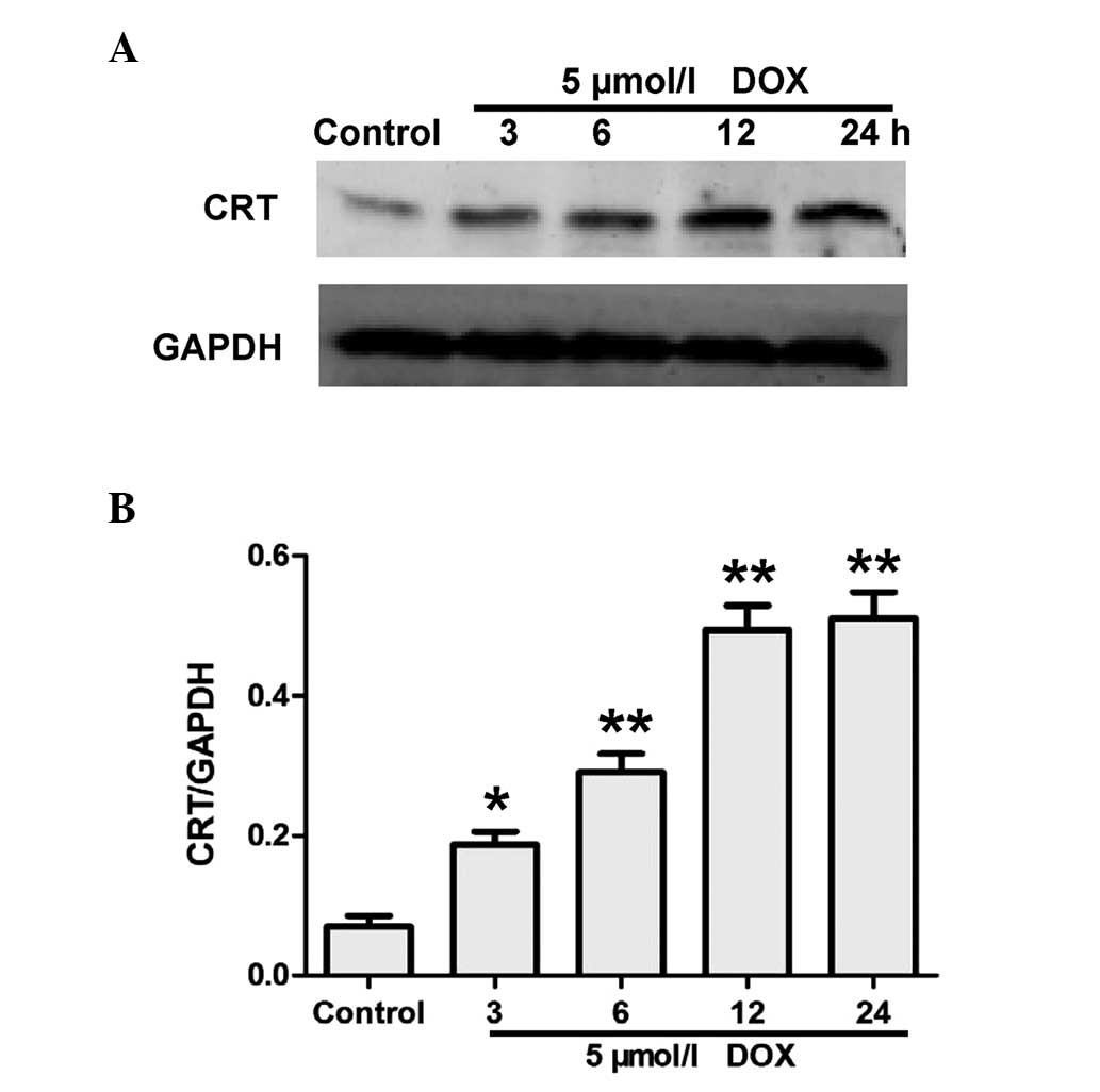
Effects of DOX on the expression of CRT
in H9c2 cells
To investigate the effect of DOX on the expression
of CRT, H9c2 cells were treated with 5 μM DOX for 0 (at the
time of treatment), 3, 6, 12 and 24 h. Western blot analysis
demonstrated that DOX treatment enhanced the CRT expression levels
in a time-dependent manner (Fig.
1).
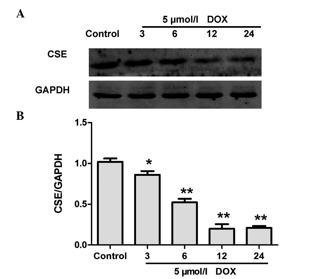
DOX inhibited the expression of CSE in
H9c2 cells
CSE is the major enzyme responsible for endogenous
H2S generation in H9c2 cells (16). Western blot analysis was performed
to evaluate whether DOX decreases endogenous H2S
production by inhibiting the expression of CSE. Treatment with 5
μM DOX for the indicated time periods (0, 3, 6, 12 and 24 h)
caused a significant downregulation of CSE expression in H9c2 cells
(Fig. 2). These data indicate that
DOX induced the inhibition of CSE expression in H9c2 and
contributed to the DOX-elicited decrease in endogenous
H2S production.
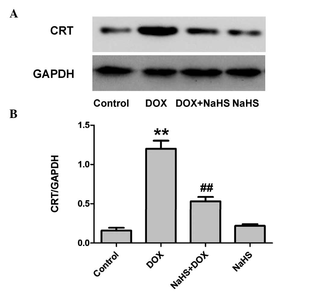
Exogenous H2S inhibits
DOX-induced expression of CRT in H9c2 cells
The effect of NaHS on the expressions of CRT induced
by DOX was detected to assess whether the cyto-protective effect of
H2S against DOX-induced toxicity was associated with the
inhibition of CRT in H9c2 cells. The results demonstrated that
pretreatment of H9c2 cells with 100 μM NaHS (a donor of
H2S) for 30 min prior to exposure to 5 μmol/l DOX
for 24 h significantly inhibited the DOX-induced overexpression of
CRT (Fig. 3). These data indicate
that the cardioprotection of H2S is associated with its
inhibitory effect on DOX-induced CRT expression.
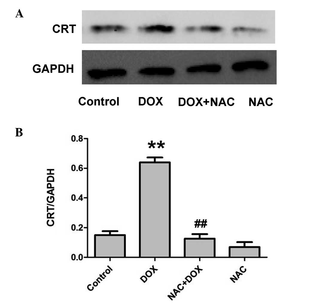
NAC suppresses the DOX-induced expression
of CRT in H9c2 cells
To identify whether the inhibitory effect of NaHS on
the DOX-induced increase in expression of CRT is associated with
its antioxidation, H9c2 cells were pretreated with 1,000 μM
NAC (an ROS scavenger) for 60 min prior to exposure to 5 μM
DOX for 24 h. As shown in Fig. 4,
similar to the inhibitory effect of NaHS pretreatment, the
pretreatment of cells with NAC for 60 min markedly depressed the
expression of CRT. The results revealed that the antioxidant
effect, resulting from NAC administration, contributed to the
inhibitory effect of H2S on the DOX-induced expression
of CRT.
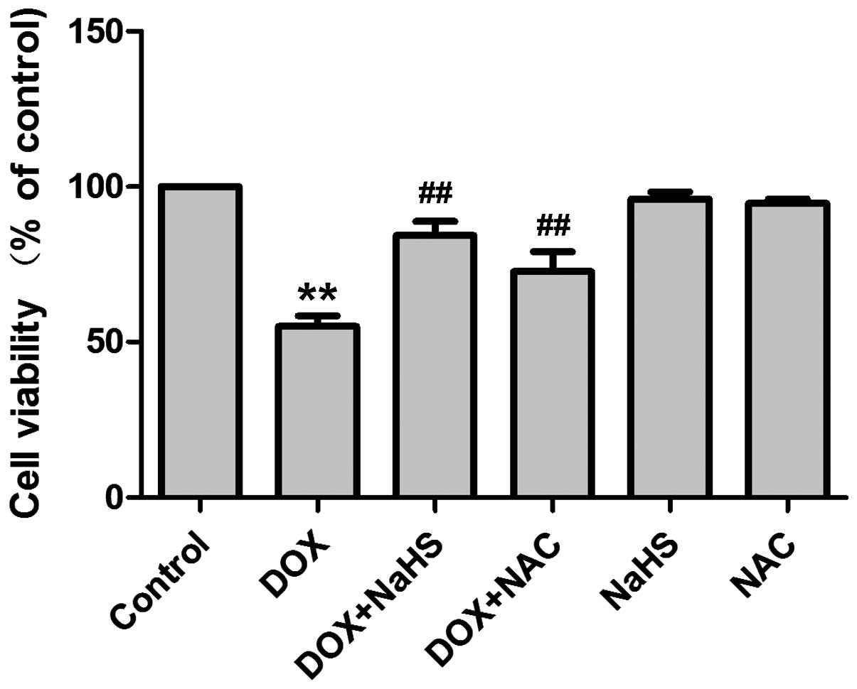
Effect of H2S and NAC on
DOX-induced cytotoxicity
As presented in Fig.
5, exposure of H9c2 cells to DOX at a dose of 5 μM for
24 h induced marked cytotoxicity, leading to a decrease in cell
viability. However, pretreatment of cells with 100 μM NaHS
for 30 min prior to exposure to DOX significantly ameliorated the
DOX-induced cytotoxicity, as evidenced by an increase in cell
viability (P<0.01 compared with the DOX-treated group). Similar
to the effect of NaHS, pretreatment with NAC for 60 min
significantly attenuated the DOX-induced decrease in cell viability
(P<0.01 compared with the DOX-treated group). Neither NaHS nor
NAC alone altered cell viability in the H9c2 cells.
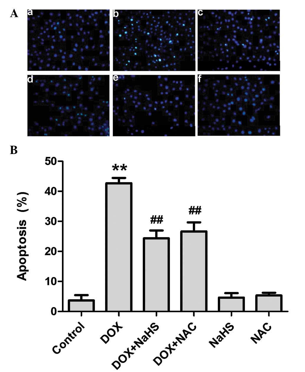
Effect of H2S and NAC on
DOX-induced apoptosis
The effects of NaHS and NAC treatment on DOX-induced
apoptosis was also observed. As shown in Fig. 6, H9c2 cells treated with 5
μM DOX for 24 h exhibited typical characteristics of
apoptosis, including condensation of chromatin, shrinkage of nuclei
and apoptotic bodies. However, pretreatment of cells with 100
μM NaHS for 30 min prior to DOX exposure markedly decreased
the DOX-induced increase in cell number, as well as decreasing the
nuclear condensation and fragmentation. In addition, H9c2 cells
were preconditioned with a common ROS scavenger, NAC (1,000
μM) prior to DOX treatment. The results indicated that
pretreatment of cells with NAC significantly attenuated DOX-induced
H9c2 cell apoptosis. NaHS or NAC alone did not markedly alter the
cell morphology or the percentage of apoptotic H9c2 cells. These
findings suggest that an antioxidant effect participates in the
inhibitory effect of H2S on the DOX-induced apoptosis of
H9c2 cells.
Discussion
Numerous studies have revealed that the major
molecular mechanism involved in DOX-induced cardiac toxicity is
free radical-induced oxidative stress and cardiomyocyte death, as a
result of apoptosis and necrosis. Consistent with previous studies
(17,18), the present study observed that
exposure of H9c2 cells to DOX markedly induced cellular injury,
including decreased cell viability, as well as increased cell
apoptosis and expression of CRT.
Previously, the cardioprotective effects of
H2S have been demonstrated in animal models of disease
(19). H2S infusion
significantly reduces myocardial infract size and improves regional
left ventricular function, as well as endothelium-dependent and
-independent microvascular reactivity in a porcine model of
myocardial ischemia-reperfusion (20). Furthermore, H2S
attenuates myocardial necrosis and apoptosis (21). In addition, endogenous
H2S is associated with the cardioprotection that results
from metabolic inhibition preconditioning in rat ventricular
myocytes (22). Inhibition of
endogenous H2S generation, by its synthesis inhibitor
(DL-propargylglycine), has been shown to block the protective
effect of IPC in isolated hearts, as well as in isolated cardiac
myocytes (23). In the present
study, H9c2 cells were used to elucidate the effect of DOX on
endogenous H2S generation. Exposure of H9c2 cells to DOX
resulted in a significant decrease in H2S
generation.
CRT is a major ER protein that is significant in
cardiac development and pathology (24). Various reviews have revealed that
CRT is highly expressed in embryonic hearts, but not in mature
hearts, and may be an early cardiac gene product (25). Mice with a targeted disruption of
the CRT gene succumb in utero exhibiting decreased
ventricular cell mass due to increased apoptosis of cardiac
myocytes (26). In addition,
studies with CRT-deficient cells suggest that CRT participates in
apoptosis (27). In the CRT
transgenic heart, CRT-dependent cardiac block involves damage to
L-type Ca2+ channels, and gap junction connexin-40 and
-43, due to defective regulation of Ca2+ homeostasis
(8). Overexpression of CRT
suppresses Akt signaling and causes differentiation-induced
apoptosis in H9c2 cells (10). In
the present study, the results showed that the expression of CRT
was increased following DOX treatment, and exogenous H2S
preconditioning was demonstrated to suppress CRT expression while
markedly attenuating DOX-induced apoptosis.
Increasing evidence indicates that ROS are
significant in the pathogenesis of cardiac failure (28). Furthermore, antioxidants have been
shown to exert protective and beneficial effects against heart
failure (29). Oxidative stress is
a primary mechanism by which DOX induces cardiomyocyte injury.
Notably, the present study demonstrated that oxidative stress was
involved in DOX-induced cell injury and established whether DOX
activation of CRT is due to the induction of ROS. Pretreatment of
H9c2 cells with NAC (a ROS scavenger) was shown to significantly
attenuate DOX-induced expression of CRT. Thus, the results of the
present study support the hypothesis that DOX induction of ROS
activates CRT, which mediates DOX-induced injury in H9c2 cells.
In conclusion, the current study identified that
H2S inhibits DOX-induced apoptosis in H9c2 cells, which
may involve inhibition of ROS-mediated CRT expression. Therefore,
the present study has elucidated the mechanisms of
H2S-mediated anti-apoptosis in cardiomyocytes and
provided evidence for identifying H2S as a candidate for
application in the treatment of cardiovascular diseases.
Acknowledgments
The present study was supported by grants from the
Medical Scientific Research Funds of Guangdong province (grant no.
A2014810) and the Graduate Student Research Innovation Project of
Hunan province (grant no. CX2013B397).
References
|
1
|
Menna P, Recalcati S, Cairo G and Minotti
G: An introduction to the metabolic determinants of anthracycline
cardiotoxicity. Cardiovasc Toxicol. 7:80–85. 2007. View Article : Google Scholar : PubMed/NCBI
|
|
2
|
Lipshultz SE, Karnik R, Sambatakos P,
Franco VI, Ross SW and Miller TL: Anthracycline-related
cardiotoxicity in childhood cancer survivors. Curr Opin Cardiol.
29:103–112. 2014. View Article : Google Scholar
|
|
3
|
Spallarossa P, Garibaldi S, Altieri P,
Fabbi P, Manca V, Nasti S, Rossettin P, Ghigliotti G, Ballestrero
A, Patrone F, et al: Carvedilol prevents doxorubicin-induced free
radical release and apoptosis in cardiomyocytes in vitro. J Mol
Cell Cardiol. 37:837–846. 2004. View Article : Google Scholar : PubMed/NCBI
|
|
4
|
Raturi A, Ortiz-Sandoval C and Simmen T:
Redox dependence of endoplasmic reticulum (ER) Ca2+
signaling. Histol Histopathol. 29:543–552. 2014.
|
|
5
|
Ma J and Pan Z: Retrograde activation of
store-operated calcium channel. Cell Calcium. 33:375–384. 2003.
View Article : Google Scholar : PubMed/NCBI
|
|
6
|
Mesaeli N, Nakamura K, Zvaritch E, Dickie
P, Dziak E, Krause KH, Opas M, MacLennan DH and Michalak M:
Calreticulin is essential for cardiac development. J Cell Biol.
144:857–868. 1999. View Article : Google Scholar : PubMed/NCBI
|
|
7
|
Milan D, Griffith J, Su M, Price ER and
McKeon F: The latch region of calcineurin B is involved in both
immunosuppressant-immunophilin complex docking and phosphatase
activation. Cell. 79:437–447. 1994. View Article : Google Scholar : PubMed/NCBI
|
|
8
|
Nakamura K, Robertson M, Liu G, Dickie P,
Guo JQ, Duff HJ, Opas M, Kavanagh K and Michalak M: Complete heart
block and sudden death in mice overexpressing calreticulin. J Clin
Invest. 107:1245–1253. 2001. View
Article : Google Scholar : PubMed/NCBI
|
|
9
|
Lynch JM, Chilibeck K, Qui Y and Michalak
M: Assembling pieces of the cardiac puzzle; calreticulin and
calcium-dependent pathways in cardiac development, health and
disease. Trends Cardiovasc Med. 16:65–69. 2006. View Article : Google Scholar : PubMed/NCBI
|
|
10
|
Kageyama K, Ihara Y, Goto S, Urata Y, Toda
G, Yano K and Kondo T: Overexpression of calreticulin modulates
protein kinase B/Akt signaling to promote apoptosis during cardiac
differentiation of cardiomyoblast H9c2 cells. J Biol Chem.
277:19255–19264. 2002. View Article : Google Scholar : PubMed/NCBI
|
|
11
|
Ihara Y, Urata Y, Goto S and Kondo T: Role
of calreticulin in the sensitivity of myocardiac H9c2 cells to
oxidative stress caused by hydrogen peroxide. Am J Physiol Cell
Physiol. 290:C208–C221. 2006. View Article : Google Scholar
|
|
12
|
Konishi M, Haraguchi G, Ohigashi H,
Ishihara T, Saito K, Nakano Y and Isobe M: Adiponectin protects
against doxorubicin-induced cardiomyopathy by anti-apoptotic
effects through AMPK up-regulation. Cardiovasc Res. 89:309–319.
2011. View Article : Google Scholar
|
|
13
|
Zhang Y, Tang ZH, Ren Z, Qu SL, Liu MH,
Liu LS and Jiang ZS: Hydrogen sulfide, the next potent preventive
and therapeutic agent in aging and age-associated diseases. Mol
Cell Biol. 33:1104–1113. 2013. View Article : Google Scholar : PubMed/NCBI
|
|
14
|
Huang YE, Tang ZH, Xie W, Shen XT, Liu MH,
Peng XP, Zhao ZZ, Nie DB, Liu LS and Jiang ZS: Endogenous hydrogen
sulfide mediates the cardioprotection induced by ischemic
post-conditioning in the early reperfusion phase. Exp Ther Med.
4:1117–1123. 2012.PubMed/NCBI
|
|
15
|
Guo R, Lin J, Xu W, Shen N, Mo L, Zhang C
and Feng J: Hydrogen sulfide attenuates doxorubicin-induced
cardiotoxicity by inhibition of the p38 MAPK pathway in H9c2 cells.
Int J Mol Med. 31:644–650. 2013.PubMed/NCBI
|
|
16
|
Kimura H: Hydrogen sulfide: Its
production, release and functions. Amino Acids. 41:113–121. 2011.
View Article : Google Scholar
|
|
17
|
Wang X, Wang XL, Chen HL, Wu D, Chen JX,
Wang XX, Li RL, He JH, Mo L, Cen X, et al: Ghrelin inhibits
doxorubicin cardiotoxicity by inhibiting excessive autophagy
through AMPK and p38-MAPK. Biochem Pharmacol. 88:334–350. 2014.
View Article : Google Scholar : PubMed/NCBI
|
|
18
|
Guo R, Wu K, Chen J, Mo L, Hua X, Zheng D,
Chen P, Chen G, Xu W and Feng J: Exogenous hydrogen sulfide
protects against doxorubicin-induced inflammation and cytotoxicity
by inhibiting p38MAPK/NFκB pathway in H9c2 cardiac cells. Cell
Physiol Biochem. 32:1668–1680. 2013.
|
|
19
|
Ji Y, Pang QF, Xu G, Wang L, Wang JK and
Zeng YM: Exogenous hydrogen sulfide postconditioning protects
isolated rat hearts against ischemia-reperfusion injury. Eur J
Pharmacol. 587:1–7. 2008. View Article : Google Scholar : PubMed/NCBI
|
|
20
|
Osipov RM, Robich MP, Feng J, Liu Y,
Clements RT, Glazer HP, Sodha NR, Szabo C, Bianchi C and Sellke FW:
Effect of hydrogen sulfide in a porcine model of myocardial
ischemia-reperfusion: Comparison of different administration
regimens and characterization of the cellular mechanisms of
protection. J Cardiovasc Pharmacol. 54:287–297. 2009. View Article : Google Scholar : PubMed/NCBI
|
|
21
|
Sodha NR, Clements RT, Feng J, Liu Y,
Bianchi C, Horvath EM, Szabo C and Sellke FW: The effects of
therapeutic sulfide on myocardial apoptosis in response to
ischemia-reperfusion injury. Eur J Cardiothorac Surg. 33:906–913.
2008. View Article : Google Scholar : PubMed/NCBI
|
|
22
|
Pan TT, Feng ZN, Lee SW, Moore PK and Bian
JS: Endogenous hydrogen sulfide contributes to the cardioprotection
by metabolic inhibition preconditioning in the rat ventricular
myocytes. J Mol Cell Cardiol. 40:119–130. 2006. View Article : Google Scholar
|
|
23
|
Bian JS, Yong QC, Pan TT, Feng ZN, Ali MY,
Zhou S and Moore PK: Role of hydrogen sulfide in the
cardioprotection caused by ischemic preconditioning in the rat
heart and cardiac myocytes. J Pharmacol Exp Ther. 316:670–678.
2006. View Article : Google Scholar
|
|
24
|
Michalak M, Lynch J, Groenendyk J, Guo L,
Robert Parker JM and Opas M: Calreticulin in cardiac development
and pathology. Biochim Biophys Acta. 1600:32–37. 2002. View Article : Google Scholar : PubMed/NCBI
|
|
25
|
Coe H and Michalak M: Calcium binding
chaperones of the endoplasmic reticulum. Gen Physiol Biophys.
28:F96–F103. 2009.
|
|
26
|
Rauch F, Prud'homme J, Arabian A, Dedhar S
and St-Arnaud R: Heart, brain and body wall defects in mice lacking
calreticulin. Exp Cell Res. 256:105–111. 2000. View Article : Google Scholar : PubMed/NCBI
|
|
27
|
Nakamura K, Bossy-Wetzel E, Burns K, Fadel
MP, Lozyk M, Goping IS, Opas M, Bleackley RC, Green DR and Michalak
M: Changes in endoplasmic reticulum luminal environment affect cell
sensitivity to apoptosis. J Cell Biol. 150:731–740. 2000.
View Article : Google Scholar : PubMed/NCBI
|
|
28
|
Schwarzer M, Osterholt M, Lunkenbein A,
Schrepper A, Amorim P and Doenst T: Mitochondrial reactive oxygen
species production and respiratory complex activity in rats with
pressure overload-induced heart failure. J Physiol. 592:3767–3782.
2014. View Article : Google Scholar : PubMed/NCBI
|
|
29
|
Matsushima S, Ide T, Yamato M, Matsusaka
H, Hattori F, Ikeuchi M, Kubota T, Sunagawa K, Hasegawa Y, Kurihara
T, et al: Overexpression of mitochondrial peroxiredoxin-3 prevents
left ventricular remodeling and failure after myocardial infarction
in mice. Circulation. 113:1779–1786. 2006. View Article : Google Scholar : PubMed/NCBI
|