Introduction
Osteosarcoma is the most common type of primary bone
cancer in children and is associated with a high rate of
metastasis, relapse and mortality. Approximately 80% of cases occur
in the appendicular skeleton and the most common site of metastasis
is the lung. The overall five-year survival rate of patients with
the disease is 20–70% (1–3). Treatment strategies including
chemotherapy, radical resection and irradiation followed by
extensive rehabilitation can be effective in certain patients,
particularly those with low-grade neoplasms; however, up to a third
of patients who initially respond to treatment relapse (4,5).
Novel treatments are therefore urgently required (6).
A number of chemotherapeutic agents are derived from
plants, such as paclitaxel (from Taxus brevifolia L.),
vincristine (from Catharanthus roseus G. Don),
podophyllotoxin (from Podophyllum peltatum L.) and
camptothecin (from Camptotheca acuminata) (7). While they are effective treatments
for different types of cancer, a number of these drugs are
associated with serious side effects. Foods high in polyphenolic
compounds may represent an alternative or adjunctive therapy for
the prevention or treatment of cancer. Polyphenols, namely phenolic
acids and flavonoids, are naturally occurring compounds found
predominantly in fruits, vegetables and beverages (8), and a number of these have been shown
to offer protection against various diseases, including cancer
(9).
Gallic acid, an intermediate component of plant
metabolism, and its ester derivative gallate have been shown to be
associated with a wide variety of biological actions, including
antioxidant, antifungal, antibacterial, antimalarial and
antiherpetic activities (10–13).
Dodecyl gallate (DG) has been shown to protect human cells from
oxidative damage and to inhibit tumor growth while exhibiting low
toxicity to normal cells (14,15).
In addition, DG has also been reported to induce apoptosis and
inhibit the proliferation of leukemic cells, lymphocytes and
melanoma cells derived from rats (16–18).
It has also been demonstrated that DG is able to disrupt
mitochondrial membrane permeability, promote cytochrome c
release into the cytosol resulting in caspase activation, and
induce the degradation of DNA into oligonucleosomal fragments in
the murine Wehi 231 B-cell lymphoma line (19). However, the mechanism of apoptosis
induction by DG in tumor cells is not clearly understood. Thus, the
aim of this study was to investigate the mechanism by which DG
induces antiproliferative and apoptotic effects in MG-63 human
osteosarcoma cells.
Materials and methods
Cell culture and reagents
The MG-63 human osteosarcoma cell line was obtained
from the Bioresource Collection and Research Center (Hsinchu,
Taiwan). Cells were cultured in Minimum Essential Medium (MEM;
Gibco-BRL, Thermo Fisher Scientific, Waltham, MA, USA) supplemented
with 10% fetal bovine serum (Gibco), 1% penicillin/streptomycin
(Gibco), 0.1 mM non-essential amino acids (Gibco), and 1.0 mM
sodium pyruvate (Gibco) in 75-cm2 tissue culture flasks.
Cells were incubated at 37°C in a humidified atmosphere of 5%
CO2 in air. Medium was changed twice weekly and cells
were subcultured once they reached 80–90% confluence. A stock
solution (20 mM) of DG (Sigma-Aldrich, St. Louis, MO, USA) was
prepared by dissolving 3.384 mg of the compound in 0.5 ml dimethyl
sulfoxide (Sigma-Aldrich).
Cell viability assay
Cell viability was evaluated by the
3-(4,5-dimethylthiazol-2-y1)-2,5-diphenyltetrazolium bromide (MTT)
assay as previously described (20). MTT was obtained from Amresco (St.
Louis, MO, USA). Briefly, MG-63 cells were seeded in 96-well plates
overnight and then treated with various concentrations of DG for
24, 48 or 72 h. The optical density of the MTT formazan product was
measured at a wavelength of 590 nm with a microplate reader (Thermo
Multiskan SPECTRUM Thermo Fisher Scientific). Results are expressed
as a percentage of the untreated controls. Data were calculated as
the percentage of proliferation using the following formula:
Proliferation (%) = (ODtest−ODblank) × 100, where ODtest and
ODblank are the optical density (OD) of the test substances and the
blank controls, respectively.
Cell cycle analysis
MG-63 cells were plated on 12-well plates at a
density of 1×10⁴ cells/well, treated with different
concentrations of DG, and then incubated for 24 h. Adherent cells
were harvested with trypsin (Gibco), washed in phosphate-buffered
saline (PBS), collected by centrifugation at 400 × g for 10 min,
and then fixed with 75% ethanol at 4°C for 24 h. Cell pellets were
then collected by centrifugation at 400 × g for 10 min and
incubated with propidium iodine (PI; Sigma-Aldrich) solution (0.1%
Triton X-100, 0.2 µg/ml Ribonuclease A and 40 µg/ml
PI) for 30 min. Cell cycle phase was determined by flow cytometry
using a Cytomics FC500 cytometer (Beckman Coulter, Brea, CA, USA)
and CXP Analysis Software version 2.1 (Beckman Coulter). Other
materials and reagents not specified were obtained from
Sigma-Aldrich or Merck Millipore (Billerica, MA, USA).
Annexin V/PI staining to detect
apoptosis
MG-63 cells were plated on 12-well plates at a
density of 1×10⁴ cells/well for 24 h. Cells were then
treated with different concentrations of DG and incubated for 24 h.
Following incubation, cells were harvested with trypsin, washed
with PBS and collected by centrifugation at 400 × g for 5 min.
After removal of the supernatant, cells were resuspended in 500
µl of 1X Binding Buffer [10X Binding Buffer-100 mM HEPES (pH
7.4); 18 mM CaCl2; 50 mM KCl; 1.5 M NaCl; and 10 mM
MgCl2 which had been diluted to 1X prior to use], to
which 1 µl Annexin V-fluorescein isothiocyanate (FITC;
BioVision, Milpitas, CA, USA) and 10 µl PI (Sigma-Aldrich)
were added. Cells were then incubated at room temperature for 15
min in the dark. Qualitative and quantitative assessments of
apoptosis were conducted with a Muse Cell analyzer (Merck
Millipore). Other materials and reagents not specified were
obtained from Sigma-Aldrich or Merck.
Mitochondrial membrane potential
assay
MG-63 cells were seeded on 12-well plates for 24 h
and then treated with different concentrations of DG for 1 h. Cells
were then harvested with trypsin, washed with PBS and collected by
centrifugation at 400 × g for 5 min. After centrifugation, the
supernatant was removed and the cell pellets were stained with the
Muse MitoPotential kit (Merck Millipore) for 25 min at 37°C and
data was analyzed by the Muse Cell Analyzer assays (Merck
Millipore).
Western blot assay
MG-63 cells were lysed on ice for 30 min with
radioimmunoprecipitation assay buffer (Merck Millipore) containing
protease inhibitors (BioVision). Cells were then scraped off the
dishes and centrifuged at 15,900 × g at 4°C for 10 min, and the
supernatant was collected. Equal amounts of protein (30 µg)
were loaded onto 8–12% polyacrylamide gels (Bio-Rad Laboratories,
Hercules, CA, USA). Proteins were then transferred to
polyvinylidene difluoride membranes (Millipore, Bedford, MA), which
were blocked for 1 h with 5% non-fat dried milk in 0.1% PBST (500
ml 1X PBS with 0.5 ml Tween-20) at room temperature. Membranes were
then incubated with primary antibodies against cleaved caspase-3
(cat. no. 9661; 1:1,000 dilution; Cell Signaling Technology Inc.,
Beverly, MA, USA), caspase-9 (cat. no. 9508; 1:1,000 dilution; Cell
Signaling Technology Inc.), bcl-2 (cat. no. 2872; 1:1,000 dilution;
Cell Signaling Technology Inc.), X-linked inhibitor of apoptosis
protein (XIAP; cat. no. 2042; 1:1,000 dilution; Cell Signaling
Technology Inc.), survivin (cat. no. 2808; 1:1,000 dilution; Cell
Signaling Technology Inc.), glyceraldehyde 3-phosphate
dehydrogenase (GAPDH) (cat. no. 2118; 1:1,000 dilution; Cell
Signaling Technology Inc.), poly (ADPribose) polymerase (PARP; cat.
no. 9542; 1:1,000 dilution; Cell Signaling Technology Inc.), bax
(sc-493; 1:1,000 dilution; Santa Cruz Biotechnology Inc., Santa
Cruz, CA, USA) and caspase-8 (NB100-56116; 1:1,000 dilution; Novus
Biologicals, Littleton, CO, USA) overnight at 4°C. Later, membranes
were washed three times with PBST and incubated with horseradish
peroxidase-conjugated goat anti-rabbit (cat. no. 7074; 1:10,000
dilution; Cell Signaling Technology Inc.) and horse anti-mouse
(cat. no. 7076; 1:10,000 dilution; Cell Signaling Technology Inc.)
secondary antibodies for 1 h at room temperature. After washing
with PBS three times, the protein signals were detected by an
enhanced chemiluminescence detection system (Merck-Millipore). The
band densities were quantified using Image J software 1.42q
(National Institutes of Health, Bethesda, MD, USA).
Statistical analysis
All data were obtained from at least three separate
experiments and are expressed as the mean ± standard deviation.
Statistical comparisons of differences between groups were
conducted using Student's t-test. P<0.05 was considered to
indicate a statistically significant difference. All statistical
analyses were performed using the software package GraphPad Prism
(Version 4.0, GraphPad Software; San Diego, CA, USA).
Results
Cytotoxic effect of DG on the MG-63 human
osteosarcoma cell line
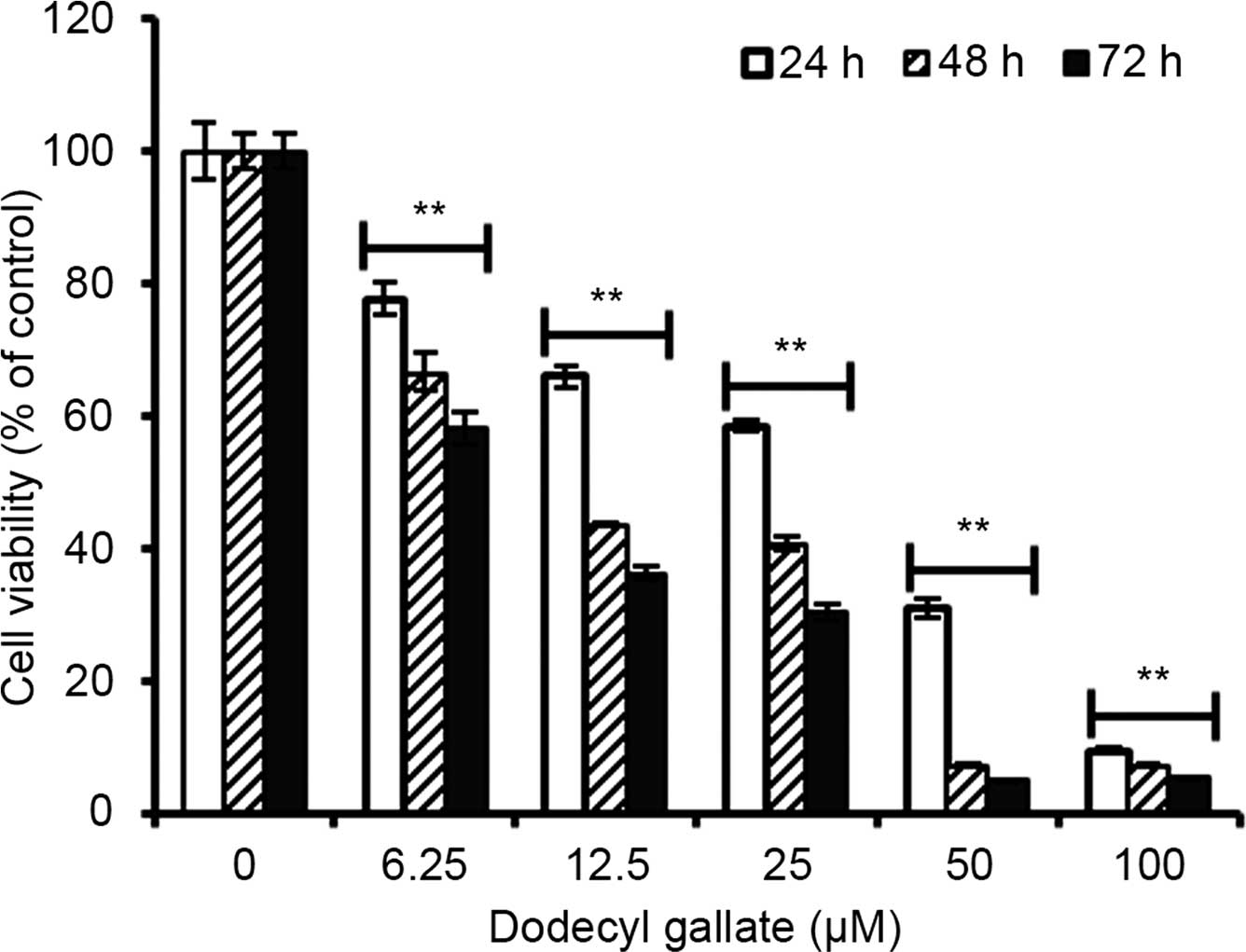
MG-63 cells were exposed to various concentrations
(6.25–100 µM) of DG for 24, 48 or 72 h, and cell viability
was determined by the MTT assay. As shown in Fig. 1, the estimated IC50
value of DG in MG-63 cells was 31.15 µM at 24 h, 10.66
µM at 48 h, and 9.06 µM at 72 h. The viability of
MG-63 cells decreased markedly in a dose- and time-dependent
manner.
Effects of DG on cell cycle distribution
in MG-63 cells
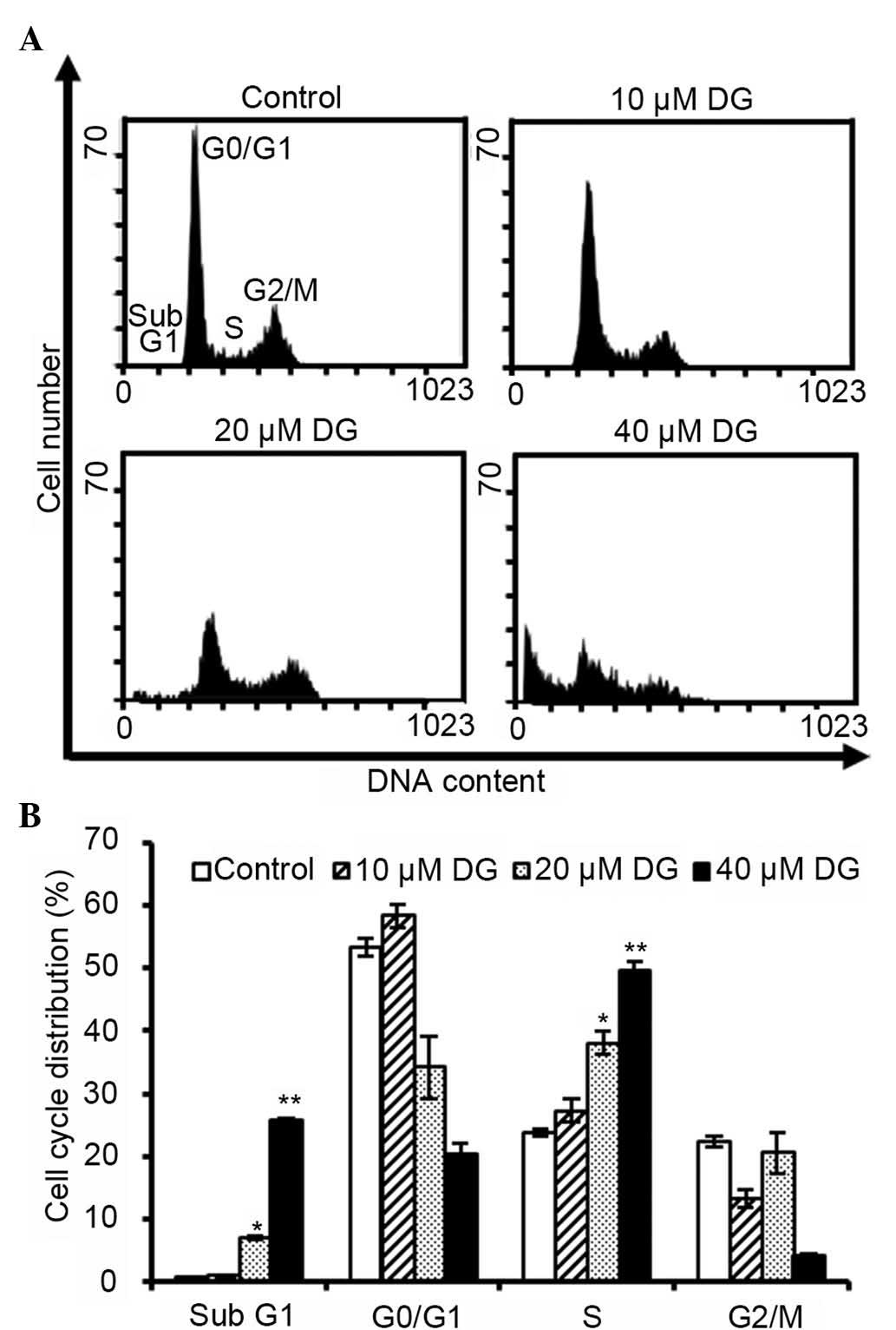
Flow cytometry was used to determine whether DG
causes cell growth inhibition by inducing cell cycle arrest. MG-63
cells were exposed to a series of concentrations of DG for 24 h. As
shown in Fig. 2, exposure to DG at
concentrations of 20 and 40 µM for 24 h resulted in a
significantly increased number of cells arrested in the S phase.
Additionally, the sub-G1 population, which is an
indication of cell death, increased significantly in the presence
of 20 and 40 µM DG.
Flow cytometric analysis of DG-induced
apoptosis in MG-63 cells
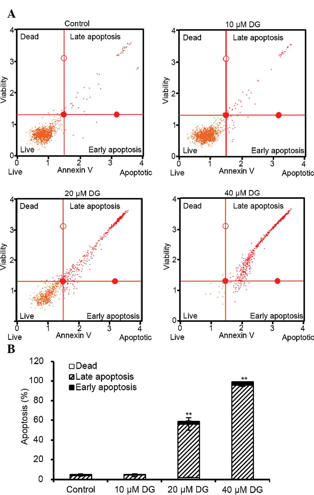
To further investigate whether the growth inhibition
induced by DG in human osteosarcoma cells was caused by apoptosis,
MG-63 cells were treated with various concentrations of DG for 24 h
and the percentage of apoptotic cells was measured by flow
cytometry. As shown in Fig. 3, the
percentages of cells demonstrating early stages of apoptosis
(Annexin V+/PI− and lower right quadrant) and
late stages of apoptosis and necrotic death (Annexin
V+/PI+ and upper right quadrant) increased in
a dose-dependent manner.
DG induces the loss of mitochondrial
membrane potential
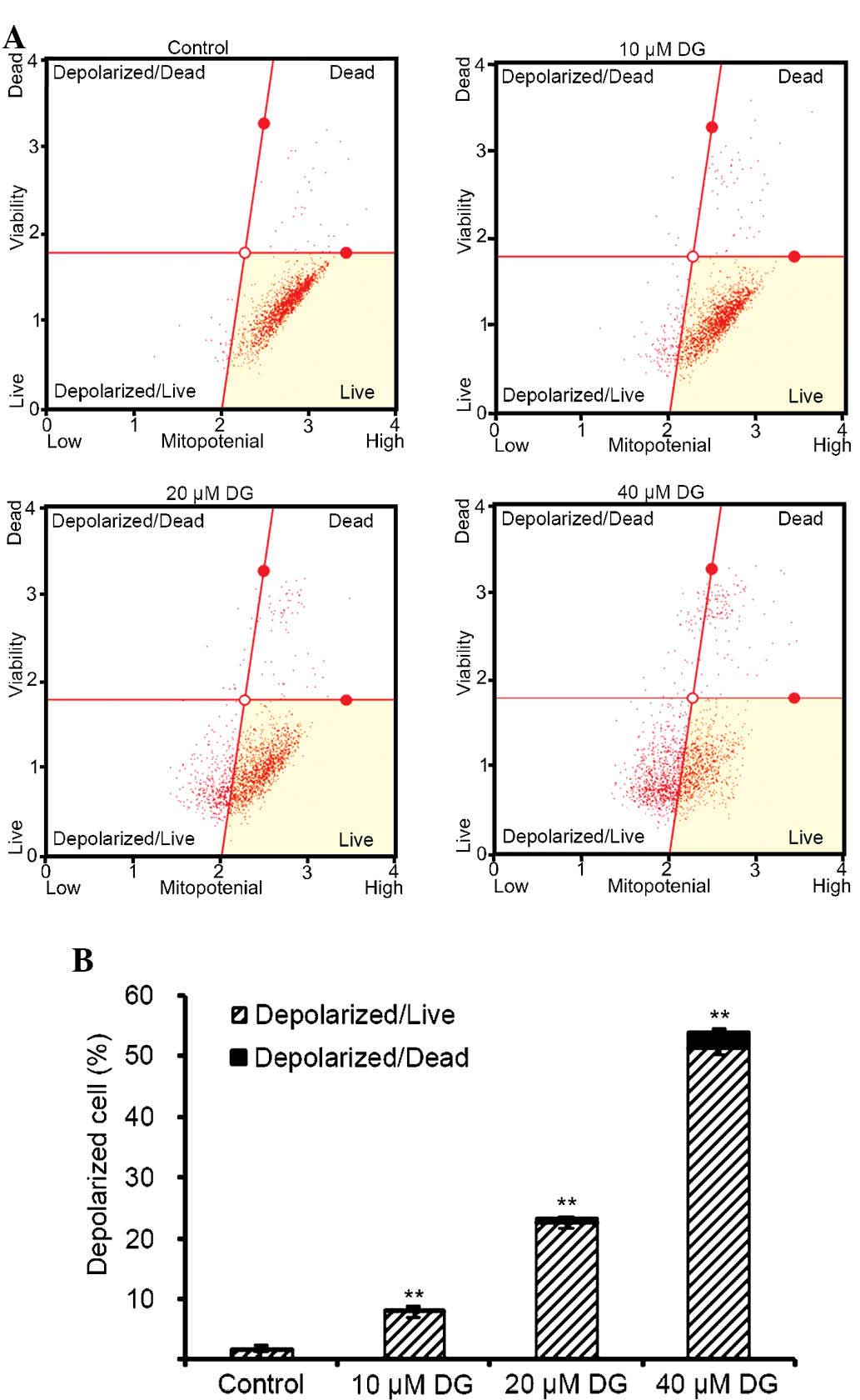
To determine whether DG-induced apoptosis involves
the mitochondria-mediated apoptotic pathway, DG-treated cells were
stained with the Muse MitoPotential kit and Muse Cell Analyzer
assays were conducted. As shown in Fig. 4, treatment with 10–40 µM DG
resulted in an increase depolarized/live percentage, indicating
loss of mitochondria membrane potential. DG induced dose-dependent
collapse of mitochondrial membrane potential. The results indicate
that DG-induced apoptosis is mediated by the mitochondrial
pathway.
Effects of DG on caspase activation in
MG-63 cells
To further clarify the type of apoptotic pathway
induced by DG, western blot analysis was used to detect the
expression levels of cleaved forms of caspase-8, caspase 9,
caspase-3 and PARP. As shown in Fig.
5, levels of cleaved forms of caspase-3, caspase-8 and PARP
were higher in cells exposed to 20 µM DG compared with those
in the control group.
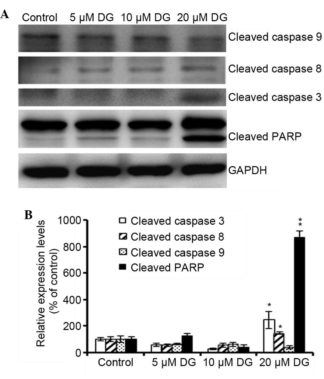 | Figure 5Effects of DG on caspase activation in
MG-63 cells. (A) The cells were treated with various concentrations
of DG (0, 5, 10 or 20 µM) for 24 h. Total cell lysates were
prepared, resolved by SDS-PAGE, and immunoblotted with the
indicated antibodies to detect the cleaved forms of caspase-8,
caspase-9, caspase-3 and PARP. GAPDH served as a loading control.
(B) Results of cleaved caspase-8, caspase-9, caspase-3 and PARP
protein levels after being normalized to the levels of GAPDH. Data
are representative of three independent experiments with similar
results. *P<0.05 and **P<0.001 as
compared with the control group. DG, dodecyl gallate; PARP, poly
(ADPribose) polymerase; GAPDH, glyceraldehyde 3-phosphate
dehydrogenase. |
DG decreases Bcl-2 and XIAP protein
expression in MG-63 cells
To confirm that the mitochondrial pathway is
involved in DG-induced apoptosis, the protein expression of Bax, a
pro-apoptotic member of the Bcl-2 family of apoptotic apoptotic
regulators, and Bcl-2 protein, an anti-apoptotic member of the
Bcl-2 family was examined. As shown in Fig. 5, treatment of MG-63 cells with DG
for 24 h resulted in increased levels of Bax/Bcl-2 protein. In
addition to measuring expression levels of members of the Bcl-2
family, the changes in expression of IAP family proteins were also
investigated. The results showed that expression of two IAP family
proteins, namely survivin and XIAP, were reduced following
treatment with higher doses of DG, as shown in Fig. 6.
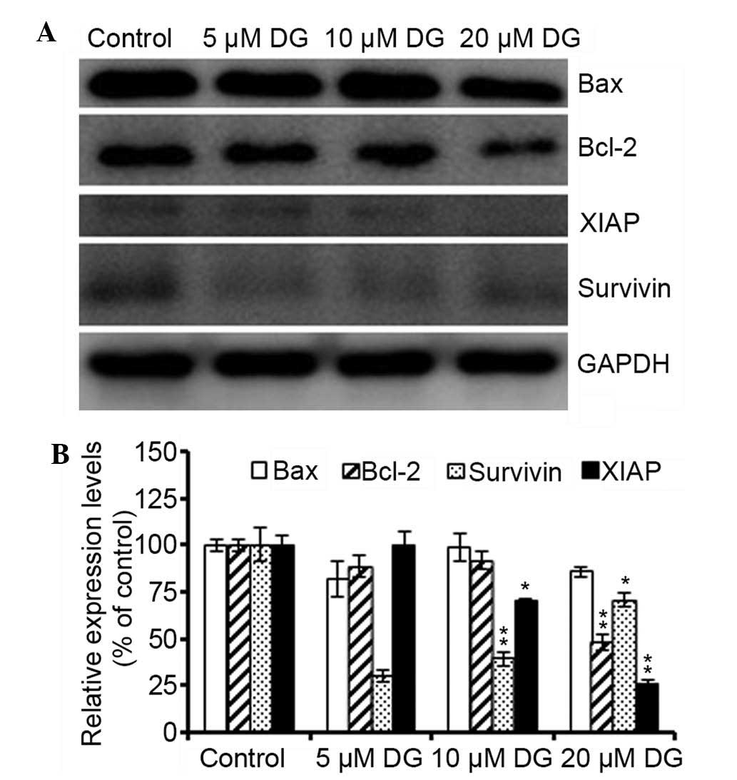 | Figure 6Decreased Bcl-2 and XIAP protein
expression in MG-63 cells. (A) The cells were treated with various
concentrations of DG (0, 5, 10 or 20 µM) for 24 h. Total
cell lysates were prepared, resolved by SDS-PAGE, and immunoblotted
with the indicated antibodies to detect the cleaved forms of Bax,
Bc1-2, XIAP and survivin. GAPDH served as a loading control. (B)
Results of Bax, Bcl-2, XIAP and survivin protein levels after being
normalized to the levels of GAPDH. Data are representative of three
independent experiments with similar results. *P<0.05
and **P<0.001 as compared with the control group. DG,
dodecyl gallate; XIAP, X-linked inhibitor of apoptosis protein;
GAPDH, glyceraldehyde 3-phosphate dehydrogenase. |
Discussion
Gallic acid is a naturally abundant plant phenolic
compound in the human diet (21).
Gallic acid derivatives, such as hydrogenated farnesyl gallate, DG,
gallic acid laurylamide and cholesteryl gallate, have been shown to
induce apoptosis in U937 and L1210 human monoblastic leukemia cells
(22,23). The molecular mechanism governing
the movement of gallate into cells and its interaction with lipid
membranes remains unclear. It has been reported that the lipophilic
group on the dodecyl ester allows for its uptake into cells and
organelles via lipophilic membranes. In addition, binding of the
hydrophilic pyrogallol group of gallate to the hydrophilic portion
of membrane surfaces results in disruption of membrane fluidity at
the membrane lipid bilayer (17).
DG has been shown to exhibit antiproliferative
effects in the B16F10 mouse melanoma cell line (24). The present results demonstrate that
DG inhibits tumor growth (Fig. 1)
with IC50 values of 31.15 µM at 24 h, 10.66
µM at 48 h and 9.06 µM at 72 h. These values are
similar to those reported by de Cordova et al (17), who showed that DG induced apoptosis
in the B16F10 melanoma cell line.
Cell cycle arrest is a common phenomenon of cell
growth inhibition (25).
Calcabrini et al (15)
showed that treatment with DG inhibited cell growth by arresting
MCF-7 breast cancer cells in the G1 phase. By contrast, it was
demonstrated that DG induced S-phase arrest in MG-63 cells. The
difference in the type of cell cycle arrest caused by DG may be due
to differences in cell type or species.
Apoptosis is governed by two predominant pathways,
namely the extrinsic pathway, which is caused by the stimulation of
pro-apoptotic cell surface receptor signal pathways, and the
intrinsic pathway, which includes a cascade of reactions resulting
in the activation of caspases. Apoptosis is defined by several
biochemical criteria such as disruption of mitochondrial membrane
potential, activation of caspase signaling, induction of DNA
fragmentation of oligonucleosomal DNA, and the release of inter
membrane mitochondrial proteins into the cytosol (25). DG has been shown to induce
caspase-dependent apoptosis in human melanoma cells (17). In the present study, Annexin
V-FITC/PI double staining was used to understand the mechanisms
governing the anticancer actions of DG. It was demonstrated that DG
(10–40 µM) induced apoptosis in a dose-dependent manner and
activated caspase-8, caspase-3 and PARP cleavage. The intrinsic
apoptotic pathway is regulated by members of the Bcl-2 family.
Bcl-2 family proteins include anti-apoptotic proteins, such as
Bcl-2, Bcl-XL, and Mcl-1, which inhibit apoptosis by preventing
cytochrome c release from the mitochondria, and
pro-apoptotic proteins, such as Bad, Bax, and Bid, which facilitate
apoptosis by promoting cytochrome c release from
mitochondria. Bax and Bcl-2 combine to form of a Bax/Bcl-2
heterodimer or Bax/Bax and Bcl-2/Bcl-2 homodimers thus maintaining
the balance between expression levels of anti- and pro-apoptotic
Bcl-2 family proteins in the cell (17,25).
Bcl-2 protein is located in the outer mitochondrial membrane, and
its control of cell death effect major related with mitochondrial
membrane structure and function. The Bcl-2 protein inhibits the
permeability of mitochondrial transition pores and prevents the
collapse of mitochondrial membrane potential and release of
pro-apoptotic factors into the cytosol (15,26).
Moreover, inhibitors of apoptosis can act directly by binding to
activated caspases, such as caspase-3 and caspase-7 (27). It was demonstrated that 24 h
treatment with DG resulted in an increase in the Bax/Bcl-2 ratio
and a decrease in protein expression of anti-apoptotic members of
the Bcl-2 family, namely Bcl-2, XIAP and survivin (Fig. 6).
In conclusion, this study demonstrated that DG
induces apoptosis by upregulating the caspase-dependent apoptotic
pathway and inhibiting the expression of anti-apoptotic Bcl-2
proteins in human osteosarcoma cells. These findings provide useful
information for the development of anticancer agents.
Acknowledgments
This study was supported by a grant from the
Changhua Christian Hospital, Changhua, Taiwan (grant no.
1Y_103_0263).
References
|
1
|
Liang W, Li X, Li C, Liao L, Gao B, Gan H,
Yang Z, Liao L and Chen X: Quercetin-mediated apoptosis via
activation of the mitochondrial-dependent pathway in MG-63
osteosarcoma cells. Mol Med Rep. 4:1017–1023. 2011.PubMed/NCBI
|
|
2
|
Ma JF, Liu L, Yang WJ, Zang LN and Xi YM:
RNAi-mediated knockdown of relaxin decreases in vitro proliferation
and invasiveness of osteosarcoma MG-63 cells by inhibition of
MMP-9. Eur Rev Med Pharmacol Sci. 17:1102–1109. 2013.PubMed/NCBI
|
|
3
|
Wang XF and Wang J: Icaritin suppresses
the proliferation of human osteosarcoma cells in vitro by
increasing apoptosis and decreasing MMP expression. Acta Pharmacol
Sin. 35:531–539. 2014. View Article : Google Scholar : PubMed/NCBI
|
|
4
|
Honicke AS, Ender SA and Radons J:
Combined administration of EGCG and IL-1 receptor antagonist
efficiently downregulates IL-1-induced tumorigenic factors in U-2
OS human osteosarcoma cells. Int J Oncol. 41:753–758.
2012.PubMed/NCBI
|
|
5
|
Crompton BD, Goldsby RE, Weinberg VK,
Feren R, O'Donnell RJ and Ablin AR: Survival after recurrence of
osteosarcoma: A 20-year experience at a single institution. Pediatr
Blood Cancer. 47:255–259. 2006. View Article : Google Scholar
|
|
6
|
Siclari VA and Qin L: Targeting the
osteosarcoma cancer stem cell. J Orthop Surg Res. 5:782010.
View Article : Google Scholar : PubMed/NCBI
|
|
7
|
Ji SJ, Han DH and Kim JH: Inhibition of
proliferation and induction of apoptosis by EGCG in human
osteogenic sarcoma (HOS) cells. Arch Pharm Res. 29:363–368. 2006.
View Article : Google Scholar : PubMed/NCBI
|
|
8
|
Naasani I, Oh-Hashi F, Oh-Hara T, Feng WY,
Johnston J, Chan K and Tsuruo T: Blocking telomerase by dietary
polyphenols is a major mechanism for limiting the growth of human
cancer cells in vitro and in vivo. Cancer Res. 63:824–830.
2003.PubMed/NCBI
|
|
9
|
Saint-Cricq De Gaulejac N, Provost C and
Vivas N: Comparative study of polyphenol scavenging activities
assessed by different methods. J Agric Food Chem. 47:425–431. 1999.
View Article : Google Scholar : PubMed/NCBI
|
|
10
|
Manna SK, Kuo MT and Aggarwal BB:
Overexpression of gamma-glutamylcysteine synthetase suppresses
tumor necrosis factor-induced apoptosis and activation of nuclear
transcription factor-kappaB and activator protein-1. Oncogene.
18:4371–4382. 1999. View Article : Google Scholar : PubMed/NCBI
|
|
11
|
Fiuza SM, Gomes C, Teixeira LJ, Girão da
Cruz MT, Cordeiro MN, Milhazes N, Borges F and Marques MP: Phenolic
acid derivatives with potential anticancer properties-a
structure-activity relationship study. Part 1: methyl, propyl and
octyl esters of caffeic and gallic acids. Bioorg Med Chem.
12:3581–3589. 2004. View Article : Google Scholar : PubMed/NCBI
|
|
12
|
Grundhöfer P, Niemetz R, Schilling G and
Gross GG: Biosynthesis and subcellular distribution of hydrolyzable
tannins. Phytochemistry. 57:915–927. 2001. View Article : Google Scholar : PubMed/NCBI
|
|
13
|
Klein E and Weber N: In vitro test for the
effectiveness of antioxidants as inhibitors of thiyl
radical-induced reactions with unsaturated fatty acids. J Agric
Food Chem. 49:1224–1227. 2001. View Article : Google Scholar : PubMed/NCBI
|
|
14
|
Jagan S, Ramakrishnan G, Anandakumar P,
Kamaraj S and Devaki T: Antiproliferative potential of gallic acid
against diethylnitrosamine-induced rat hepatocellular carcinoma.
Mol Cell Biochem. 319:51–59. 2008. View Article : Google Scholar : PubMed/NCBI
|
|
15
|
Calcabrini A, García-Martínez JM, González
L, Tendero MJ, Ortuño MT, Crateri P, Lopez-Rivas A, Arancia G,
González-Porqué P and Martín-Pérez J: Inhibition of proliferation
and induction of apoptosis in human breast cancer cells by lauryl
gallate. Carcinogenesis. 27:1699–1712. 2006. View Article : Google Scholar : PubMed/NCBI
|
|
16
|
Inoue M, Suzuki R, Koide T, Sakaguchi N,
Ogihara Y and Yabu Y: Antioxidant, gallic acid, induces apoptosis
in HL-60RG cells. Biochem Biophys Res Commun. 204:898–904. 1994.
View Article : Google Scholar : PubMed/NCBI
|
|
17
|
de Cordova CA, Locatelli C, Assuncão LS,
Mattei B, Mascarello A, Winter E, Nunes RJ, Yunes RA and
Creczynski-Pasa TB: Octyl and dodecyl gallates induce oxidative
stress and apoptosis in a melanoma cell line. Toxicol In Vitro.
25:2025–2034. 2011. View Article : Google Scholar : PubMed/NCBI
|
|
18
|
Nakagawa Y, Moldéus P and Moore G: Propyl
gallate-induced DNA fragmentation in isolated rat hepatocytes. Arch
Toxicol. 72:33–37. 1997. View Article : Google Scholar : PubMed/NCBI
|
|
19
|
Roy G, Lombardía M, Palacios C, Serrano A,
Cespón C, Ortega E, Eiras P, Lujan S, Revilla Y and Gonzalez-Porqué
P: Mechanistic aspects of the induction of apoptosis by lauryl
gallate in the murine B-cell lymphoma line Wehi 231. Arch Biochem
Biophys. 383:206–214. 2000. View Article : Google Scholar
|
|
20
|
Yan MY, Chien SY, Kuo SJ, Chen DR and Su
CC: Tanshinone IIA inhibits BT-20 human breast cancer cell
proliferation through increasing caspase 12, GADD153 and
phospho-p38 protein expression. Int J Mol Med. 29:855–863.
2012.PubMed/NCBI
|
|
21
|
Hsu CL, Lo WH and Yen GC: Gallic acid
induces apoptosis in 3T3-L1 pre-adipocytes via a Fas- and
mitochondrial-mediated pathway. J Agric Food Chem. 55:7359–7365.
2007. View Article : Google Scholar : PubMed/NCBI
|
|
22
|
Saeki K, Yuo A, Isemura M, Abe I, Seki T
and Noguchi H: Apoptosis-inducing activity of lipid derivatives of
gallic acid. Biol Pharm Bull. 23:1391–1394. 2000. View Article : Google Scholar : PubMed/NCBI
|
|
23
|
Locatelli C, Rosso R, Santos-Silva MC, de
Souza CA, Licínio MA, Leal P, Bazzo ML, Yunes RA and
Creczynski-Pasa TB: Ester derivatives of gallic acid with potential
toxicity toward L1210 leukemia cells. Bioorg Med Chem.
16:3791–3799. 2008. View Article : Google Scholar : PubMed/NCBI
|
|
24
|
Locatelli C, Leal PC, Yunes RA, Nunes RJ
and Creczynski-Pasa TB: Gallic acid ester derivatives induce
apoptosis and cell adhesion inhibition in melanoma cells: The
relationship between free radical generation, glutathione depletion
and cell death. Chem Biol Interact. 181:175–184. 2009. View Article : Google Scholar : PubMed/NCBI
|
|
25
|
Wu CS, Chen YJ, Chen JJ, Shieh JJ, Huang
CH, Lin PS, Chang GC, Chang JT and Lin CC: Terpinen-4-ol induces
apoptosis in human nonsmall cell lung cancer in vitro and in vivo.
Evid Based Complement Alternat Med. 2012:8182612012. View Article : Google Scholar
|
|
26
|
Kowaltowski AJ, Cosso RG, Campos CB and
Fiskum G: Effect of Bcl-2 overexpression on mitochondrial structure
and function. J Biol Chem. 277:42802–42807. 2002. View Article : Google Scholar : PubMed/NCBI
|
|
27
|
Yang YL and Li XM: The IAP family:
Endogenous caspase inhibitors with multiple biological activities.
Cell Res. 10:169–177. 2000. View Article : Google Scholar : PubMed/NCBI
|