Introduction
According to data from the National Center for
Health Statistics, primary hepatocellular carcinoma (HCC) is one of
the most common types of cancer in the world. α-fetoprotein (AFP)
is a glycoprotein that is produced in the endodermal cells of the
yolk sac and fetal liver. The synthesis of AFP decreases markedly
following birth. However, expression of the AFP gene is reactivated
in adults during hepatocarcinogenesis (1). Human T cell repertoires recognize
AFP-derived peptide epitopes in the context of major human
leukocyte antigen (HLA) class I molecules and induce AFP-specific
protection (1,2). Thus, AFP, as a tumor-associated
antigen, may be a suitable target for dendritic cell (DC)-based
cytotoxic T lymphocyte (CTL)-mediated antigen-targeted
immunotherapy. CTLs are the predominant effector cells against
cancers (3). DCs are the most
effective antigen-presenting cells, and stimulate naive T
lymphocytes to initiate an antigen-specific, HLA class I-restricted
CTL response (4). However, in a
number of cases, HCC cells express few or no HLA class I molecules
due to genetic, transcriptional and post-transcriptional
regulation, which results in escape of recognition of tumor cells
by CTLs (5–8).
Various protocols for generating DCs in vitro
from peripheral blood mononuclear cells have been developed
(9). Recombinant adeno-associated
virus (rAAV) is one of the safest virus vectors in gene therapy
(10,11). AAV type 2 vectors have been
demonstrated to be effective vectors for delivery of antigen genes
into human DCs, and to generate a marked CTL response against the
antigen-positive target cells (10,11).
It has been reported that interferon-γ (IFN-γ) upregulates the
expression of HLA class I molecules (12,13).
The present study demonstrated that rAAV carrying the
cytomegalovirus promoter (CMVp) and AFP gene
(rAAV/CMVp-AFP)-transduced DCs elicited an AFP-targeted,
HLA-A2-restricted CTL response against Hep3B cells. In order to
improve the activity of CTLs against HLA-A2-deficient Hep3B cells,
HCC-specific rAAV carrying human α-fetoprotein promoter (AFPp) and
the IFN-γ gene (rAAV/AFPp-IFN-γ) were used to transduce Hep3B cells
in order to recover the expression of HLA-A2.
Materials and methods
Cells
Hep3B (hepatocellular carcinoma), NCI-H2126 (lung
cancer), HEK293 (embryonic kidney cells), HeLa (cervical cancer),
Hs578T (breast cancer), LNCaP-FGC (prostate cancer), LoVo
(colorectal adenocarcinoma), PANC-1 (pancreatic cancer), SNU-1
(gastric carcinoma), THP-1 (monocytic leukemia) and K562 (myeloid
leukemia) cell lines were obtained from the American Type Culture
Collection (Manassas, VA, USA). All of these cells were cultured in
complete Dulbecco's modified Eagle's medium (Invitrogen; Thermo
Fisher Scientific, Inc., Waltham, MA, USA) or RPMI-1640 medium
(Invitrogen; Thermo Fisher Scientific, Inc.) supplemented with 5 or
10% fetal bovine serum (Invitrogen; Thermo Fisher Scientific,
Inc.). The primary human hepatocytes, hNHeps, were obtained from
Lonza Group AG (Basel, Switzerland) and cultured in HCM hepatocyte
culture medium (Lonza Group AG). Peripheral blood mononuclear cells
(PBMCs) from healthy donors were separated using a routine Ficoll
(Sigma-Aldrich, St. Louis, MO, USA) gradient method and cultured in
AIM-V medium (Invitrogen; Thermo Fisher Scientific, Inc.). All
blood donors were enrolled by the Provincial Hospital Affiliated to
Shandong University (Jinan, China) and provided written informed
consent. The study protocol received a priori approval by
the Human Research Internal Review Board of the Provincial Hospital
Affiliated to Shandong University. The HLA haplotype of all donors
was HLA-A2.
Construction of rAAV vectors
The wild-type AAV type 2 genome, pSM620, was
digested in order to delete the internal AAV sequences from map
units 3–97, including the p5 promoter, and a specially designed
polylinker was ligated in place, resulting in the AAV vector
plasmid, dl3–97 (14). The
cytomegalovirus enhancer and the SV40 early mRNA polyadenylation
signal DNA were derived from the pEGFP-N1 plasmid (Clontech
Laboratories, Inc., Mountain View, CA, USA) and inserted into the
dl3–97 vector (provided by Professor Yong Liu, Gene and Biotherapy
Center, Cancer Institute, University of Arkansas for Medical
Sciences, Little Rock, AR, USA). The plasmid, pDRIVE-hAFP
(Invivogen, San Diego, CA, USA) was digested with PacI and
NcoI to obtain 275 bp AFP promoter DNA. Subsequently, the
DNA was inserted into the downstream of CMV enhancer gene
(dl3–97/AFPp). CMV immediate early promoter was inserted into the
dl3–97 vector using the identical method (dl3–97/CMVp), which was
derived from the pEGFP-N1 plasmid.
Human AFP and IFN-γ cDNA were amplified by reverse
transcription-polymerase chain reaction (RT-PCR). The total mRNA
was isolated from the Hep3B cells and
phytohemag-glutinin-stimulated human T lymphocytes using an
Oligotex mRNA isolation Mini kit (Qiagen, Inc., Valencia, CA, USA).
The RNA was reverse transcribed using the SuperScript™ first-strand
synthesis system, according to the manufacturer's protocols
(Invitrogen; Thermo Fisher Scientific, Inc.). Subsequent to the
generation of the first-strand of cDNA, PCR amplification was
conducted using the following primer pairs: Sense, 5′-CTT CCA CCA
CTG CCA ATAAC-3′ and antisense, 5′-TTG TCT TCT CTT CCC CTG-3′ for
AFP, which amplifies the sequence from nucleotides 12 to 1,902
(15), and sense, 5′-TTT CTC TCG
GAA ACG ATG-3′ and antisense, 5′-GGC AGG ACA ACC ATTAC-3′ for
IFN-γ, which amplifies the sequence from nucleotides 95 to 622
(16). The PCR conditions for AFP
were 30 cycles of denaturation at 94°C for 50 sec, annealing at
58°C for 55 sec and elongation at 72°C for 50 sec. The PCR
conditions for IFN-γ were 30 cycles of denaturation at 94°C for 50
sec, annealing at 58°C for 55 sec and elongation at 72°C for 40
sec. According to the routine method of molecular cloning, AFP and
IFN-γ cDNA were inserted into the dl3–97 vectors, respectively. The
dl3–97 vectors and AFP and IFN-γ cDNA were digested with a
restriction enzyme (New England BioLabs, Inc., Ipswich, MA, USA).
The cDNA was inserted downstream of the promoter using ligase (New
England BioLabs, Inc.). The AAV vectors were generated, including
rAAV/CMVp-AFP and rAAV/AFPp-IFN-γ. The enhanced green fluorescent
protein (eGFP) was derived from pEGFP-N1, and also inserted into
the AAV vectors to generate rAAV/AFPp-eGFP.
Generation of rAAV virus
The pSH3 plasmid expresses the AAV type 2 rep
and cap genes and adenovirus type 5 E2A, VA1 and E4 genes,
to allow rAAV DNA replication and packaging into viral particles
without contaminating the wild-type AAV and adenovirus (17). The rAAV vectors were colipofected
into HEK293 cells with the pSH3 plasmid using Lipofectamine 2000
(Invitrogen; Thermo Fisher Scientific, Inc.), and the rAAVs were
harvested after 4 days. A one-step column purification technique,
which used gravity flow based on its affinity to heparin, without
ultracentrifugation, was performed in order to generate the
purified rAAV (18). The rAAVs
were titered as described previously by dot blot hybridization
using a DIG DNA Labeling and Detection kit (Roche Diagnostics,
Basel, Switzerland) (10).
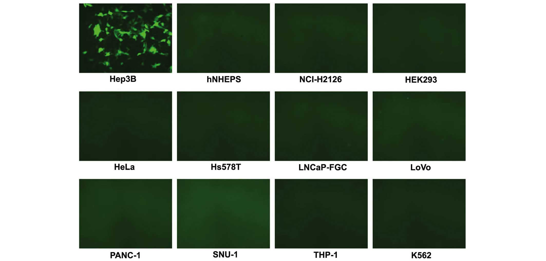
Analysis of cell-specific expression of
rAAV with AFP promoter
To observe cell-specific expression of the rAAV with
an AFP promoter, Hep3B and a series of AFP-negative control cells
were infected with rAAV/AFPp-eGFP virus. After 5 days, the cells
were observed under a BX61 inverted fluorescence microscope
(Olympus Corporation, Tokyo, Japan).
Analysis of IFN-γ expression in the
rAAV/AFPp-IFN-γ-transduced Hep3B cells
At day 5 of transduction, the Hep3B cells were
harvested. Intracellular staining was performed to analyze the
expression of IFN-γ in rAAV/AFPp-IFN-γ-transduced Hep3B cells. The
cells were fixed with 4% parafomaldehyde (Sigma-Aldrich) at 4°C for
30 min and permeabilized with 1 ml 0.5% saponin (Sigma-Aldrich) for
20 min at room temperature. The cells were subsequently incubated
with phycoerythrin (PE)-conjugated mouse anti-human IFN-γ
monoclonal antibody (1:100; cat. no. 559326; BD Pharmingen, San
Diego, CA, USA). A FACSCalibur flow cytometer (BD Biosciences, San
Jose, CA, USA) was used for data acquisitions, and 10,000 cells
were counted for each sample. rAAV/AFPp-eGFP-transduced Hep3B cells
served as a control.
The mRNA expression of IFN-γ was analyzed by RT-PCR
under conditions of 30 cycles of denaturation at 94°C for 50 sec,
annealing at 58°C for 55 sec and elongation at 72°C for 40 sec. At
day 5 of transduction, the total mRNA was isolated from
rAAV/AFPp-IFN-γ-transduced Hep3B cells using an Oligotex mRNA
isolation Mini kit. Subsequent to the generation of the first
strand of the cDNA, PCR amplification was conducted using the
primer pairs, 5′-TTT CTC TCG GAA ACG ATG-3′ and 5′-GGC AGG ACA ACC
ATTAC-3′, which amplify the sequence from nucleotides 95 to 622
(16). Transcription factor IIB
was also amplified by PCR as a control, the resulting products were
analyzed by 2% gel electrophoresis.
The expression of IFN-γ was also measured in the
Hep3B cell medium by enzyme-linked immunosorbent assay (ELISA)
using the Human IFN-γ Direct ELISA kit (Invitrogen; Thermo Fisher
Scientific, Inc.). From day 6 to day 60, the secretion of IFN-γ was
measured in duplicate, according to the manufacturer's
protocol.
Observation of HLA-A2 expression in
rAAV/AFPp-IFN-γ-transduced Hep3B cells
At day 5 of rAAV/AFPp-IFN-γ transduction, the Hep3B
cells were harvested. To observe HLA-A2 expression, the cells were
fixed with 4% paraformaldehyde at 4°C for 30 min and stained with
PE-conjugated mouse anti-human HLA-A2 monoclonal antibody (1:100;
cat. no. 558570; BD Pharmingen). rAAV/AFPp-eGFP-transduced Hep3B
cells served as a control.
The mRNA expression of HLA-A2 was also analyzed by
RT-PCR. At day 5 of transduction, the total mRNA was isolated from
rAAV/AFPp-IFN-γ-transduced Hep3B cells using an Oligotex mRNA
isolation Mini kit. Subsequent to the generation of the first
strand of the cDNA, PCR amplification was conducted using the
primer pairs, 5′-ATG GCC GTC ATG GCG CCC CGAAC-3′ and 5′-GGC AGC
TGT CTC ACA CTT-3′. The PCR conditions were 30 cycles of
denaturation at 94°C for 50 sec, followed by annealing at 58°C for
55 sec, and elongation at 72°C for 60 sec. The resulting PCR
products were analyzed by 2% gel electrophoresis.
Generation of AFP-expressing DCs
PBMCs were obtained from the peripheral blood of
HLA-A2-positive healthy volunteers, separated by Ficoll
density-gradient centrifugation at 400 x g for 20 min, and
incubated in six-well culture plates at 37°C for 2 h in AIM-V
medium. Following incubation, the non-adherent cells were removed,
and the adherent PBMCs were cultured in AIM-V medium with 800
units/ml human granulocyte macrophage colony-stimulating factor
(GM-CSF; R&D Systems, Inc., Minneapolis, MN, USA) and infected
with 1010 encapsidated genomes (eg)/ml of rAAV/CMVp-AFP.
Uninfected PBMCs served as controls. After 8 h, the medium/virus
solution was removed, and replaced with fresh AIM-V medium
containing 800 units/ml GM-CSF and 1,000 IU/ml human interleukin-4
(IL-4; R&D Systems, Inc.). Every 2 days, the culture was
replaced with the fresh medium, including the cytokines. At day 4,
50 IU/ml human tumor necrosis factor-α (R&D Systems, Inc.) was
added into the medium. At day 6, the DCs were harvested. To analyze
AFP expression, the intracellular staining was conducted as
described above. The DCs were stained with PE-conjugated mouse
anti-human AFP monoclonal antibody (1:100; cat. no. 563002; BD
Pharmingen) and analyzed by fluorescence-activated cell sorting
(FACS).
The mRNA expression of AFP was also analyzed by
RT-PCR. At day 6 of transduction, the total mRNA was isolated from
rAAV/CMVp-AFP-transduced DCs using an Oligotex mRNA isolation Mini
kit. Subsequent to the generation of the first strand of the cDNA,
PCR amplification was conducted using the primer pairs, 5′-AGT TTG
AGG AGA ATA TTTG-3′ and 5′-GGT TGC TAG TTA TTT TGTT-3′. The PCR
conditions were 30 cycles of denaturation at 94°C for 50 sec,
annealing 58°C for 55 sec and elongation at 72°C for 50 sec. The
resulting PCR products were analyzed by 2% gel electrophoresis.
Characterization of DCs
Following 6 days of culture, the transduced and
control DCs were harvested. A panel of fluorescein isothiocyanate
(FITC)-conjugated mouse anti-human monoclonal antibodies was used,
including HLA-death receptor (DR; cat. no. 555811), cluster of
differentiation (CD)1a (cat. no. 555806), CD40 (cat. no. 555588),
CD80 (cat. no. 555683), CD83 (cat. no. 555910) and CD86 (cat. no.
555657). They were all diluted at 1:100 and obtained from BD
Pharmingen. For FACS analysis, all the samples were stained with
the antibodies.
Analysis of the level of expressed
cytokines in AFP-expressed DCs
Following 6 days of culture, the transduced and
control DCs were harvested. The intracellular staining assay was
performed to analyze the expression of IL-10 and IL-12 using
PE-conjugated rat anti-human IL-10 (1:100; cat. no. 559330; BD
Pharmingen) and FITC-conjugated mouse anti-human IL-12 (1:100; cat.
no. 554574; BD Pharmingen) monoclonal antibodies.
Analysis of activated T cell
populations
The rAAV/CMVp-AFP-transduced DCs were mixed with
CD3+ T cells subsequent to the DCs being harvested at
day 6 (ratio, T:DC, 20:1). CD3+ T cells were isolated
from the PBMCs using a Pan T Cell Isolation kit II, according to
the manufacturer's protocol (Miltenyi Biotec, Inc., Auburn, CA,
USA). The DC-T cell mixtures were cultured in AIM-V medium in the
presence of 20 units/ml recombinant human IL-2 (R&D Systems,
Inc.) and 20 ng/ml recombinant human IL-7 (R&D Systems, Inc.).
The medium in the DC-T cell culture was replaced with the fresh
medium, including the cytokines, every 2 days. At day 8, the primed
T cell populations were analyzed for their surface markers with
immunofluorescence staining by FACS. A panel of FITC- or
PE-conjugated mouse anti-human monoclonal antibodies recognizing
the following antigens was used: CD4 (cat. no. 555347), CD8 (cat.
no. 555634), CD25 (cat. no. 555431) and CD69 (cat. no. 557050).
They were all diluted 1:100 and obtained from BD Pharmingen.
Analysis of the expression levels of
cytokines in primed T cells
At day 8 post-priming, the T cells were harvested.
The intracellular staining assay was performed to analyze the
expression of IFN-γ and IL-4. The T cells were incubated at 37°C
for 6 h in AIM-V medium containing 50 ng/ml phorbol 12-myristate
13-acetate (Sigma-Aldrich) and 500 ng/ml ionomicin (Sigma-Aldrich).
Brefeldin A (10 ng/ml; Sigma-Aldrich) was added for the final 5 h
of the incubation to completely inhibit cytokine secretion. The
cells were washed and fixed with 4% paraformaldehyde and
permeabilized with phosphate-buffered saline/0.5% saponin
(Sigma-Aldrich) for 10 min. Following incubation, the cells were
labeled with FITC-conjugated mouse anti-human IFN-γ (1:100; cat.
no. 552882; BD Pharmingen) and PE-conjugated mouse anti-human IL-4
(1:100; cat. no. 559333; BD Pharmingen) monoclonal antibodies for
30 min on ice, prior to flow cytometric analysis.
Cytotoxicity assays
After 8 days of the DC-T cell culture, 6-hour
chromium-51 (51Cr) release assays were used to analyze
the activity of the CTLs elicited by the rAAV/CMVp-AFP-transduced
and control DCs against the target cells (11). The target cells included untreated,
rAAV/AFPp-IFN-γ- and rAAV/AFPp-eGFP-transduced Hep3B cells. The
51Cr-labeled target cells were mixed with the CTLs
(1:20) and incubated for 6 h at 37°C with 5% CO2. To
determine the structures on the target cells, the anti-human HLA-A2
monoclonal antibody was used to completely inhibit the
cytotoxicity. The 51Cr-labeled targets were preincubated
with mouse anti-human HLA-A2 antibody (1:100; cat. no. 551230; BD
Pharmingen) for 2 h prior to the 51Cr release assay
being performed. To demonstrate the AFP-specific activity of the
CTLs, a series of AFP-negative cells were also assessed. K562 cells
were used as targets to observe natural killer (NK) cell
activity.
Statistics
All data are expressed as the mean ± standard
deviation and differences between groups were analyzed using the
Student's t-test with SPSS, version 15.0 software (SPSS, Inc.,
Chicago, IL, USA). P<0.05 was considered to indicate a
statistically significant difference.
Results
A series of rAAV vectors were
constructed
To construct the rAAV vectors for the present study,
the AFP promoter and CMV immediate early promoter were successfully
inserted into the dl3–97 vectors. AFP and IFN-γ mRNA was isolated
from the human cells and amplified by RT-PCR (data not shown). This
cDNA and eGFP cDNA were also successfully cloned into the rAAV
vectors. All the cDNA was sequenced and determined to be identical
to the published sequence (15,16).
The virus stocks of rAAV were generated and titered (data not
shown). The viral titers were from 1011 to
1012 eg/ml.
rAAV with an AFP promoter was
specifically expressed in AFP-positive Hep3B cells
To verify that rAAV with the AFP promoter only
expressed protein in AFP-positive cells, the AFP-positive Hep3B,
and a series of AFP-negative, cells were transduced using
rAAV/AFPp-eGFP. After 5 days, only the transduced Hep3B cells
expressed eGFP protein (Fig.
1).
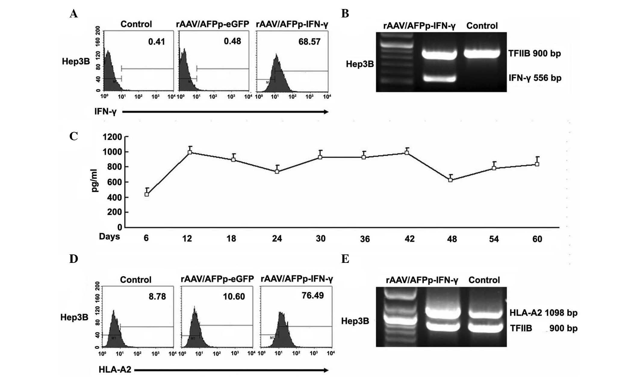
IFN-γ was expressed in
rAAV/AFPp-IFN-γ-transduced Hep3B cells
To observe whether IFN-γ was expressed in the
rAAV/AFPp-IFN-γ-transduced Hep3B cells, intracellular staining was
performed. As shown in Fig. 2A, at
day 5 post-transduction, the percentage of the IFN-γ-positive cells
present was 68.57%. The rAAV/AFPp-eGFP-transduced and untransduced
Hep3B cells were IFN-γ-negative. The mRNA expression of IFN-γ
analyzed by RT-PCR is presented in Fig. 2B. Furthermore, the secretion of
IFN-γ was also observed at various times post-transduction using
ELISA. As presented in Fig. 2C,
the secretion remained comparably stable for at least 60 days.
HLA-A2 expression was elevated in
rAAV/AFPp-IFN-γ-transduced Hep3B cells
To observe whether the expression of HLA-A2 was
elevated in rAAV/AFPp-IFN-γ-transduced Hep3B cells, the level of
expression of HLA-A2 was observed at day 5 post-transduction. As
presented in Fig. 2D, the
expressed IFN-γ effectively promoted the expression of HLA-A2. The
percentage of HLA-A2-positive cells was higher compared with that
of the untransduced cells (P<0.05). In addition, rAAV/AFPp-eGFP
transduction did not result in the elevation of level of HLA-A2
expression (P>0.05). The upregulated mRNA expression of HLA-A2
is presented in Fig. 2E.
AFP-pulsed monocyte-derived DCs were
generated
To generate AFP-pulsed DCs, the PBMCs were
transduced by rAAV/CMVp-AFP at day 0, and cultured in AIM-V medium
containing the cytokines. At day 6, the monocyte-derived DCs were
harvested and analyzed for AFP protein expression by FACS. The
percentage of AFP-positive DCs at day 6 following rAAV/CMVp-AFP
transduction was 74.33%, as presented in Fig. 3A. The mRNA expression of AFP
analyzed by RT-PCR is presented in Fig. 3B.
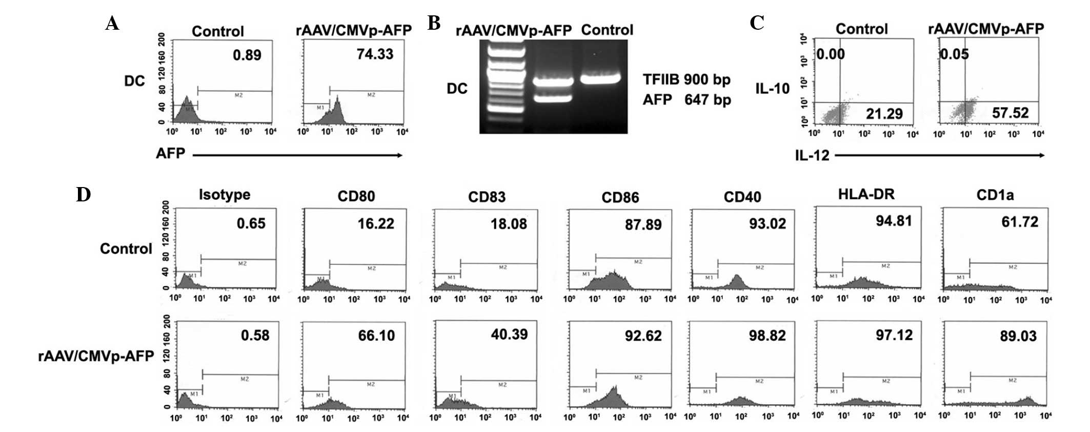 | Figure 3Characterization of DCs by FACS. (A)
Percentage of AFP-positive DCs was 74.33% at day 6 following
rAAV/CMVp-AFP transduction. (B) mRNA expression of AFP was assessed
in rAAV/CMVp-AFP-transduced DCs by reverse transcription-polymerase
chain reaction. (C) Higher levels of IL-12 production were detected
in rAAV/CMVp-AFP-transduced DCs than in untreated DCs. Almost no
IL-10 was detected in the two groups of the DCs. (D) Phenotype of
the rAAV/CMVp-AFP-transduced DCs was determined by FACS analysis.
The expression levels of CD1a, CD80 and CD83 of the
rAAV/CMVp-AFP-transduced DCs were higher. P<0.05 vs. untreated
DCs. CD, cluster of differentiation; rAAV, recombinant
adeno-associated virus; DCs, dendritic cells; AFP, α-fetoprotein,
FACS, fluorescence-activated cell sorting; CMVp, cytomegalovirus
promoter; TFIIB, transcription factor II B; IL-10, interleukin-10;
IL-12, interleukin-12. |
DCs were activated by rAAv/CMVp-AFP
The intracellular staining assay was performed to
analyze the expression of IL-10 and IL-12 in
rAAV/CMVp-AFP-transduced DCs after 6 days of culture. A notably
higher level of IL-12 production was detected in
rAAV/CMVp-AFP-transduced DCs than in untreated DCs (P<0.05).
Almost no IL-10 was detected in the rAAV/CMVp-AFP-transduced DCs
and untreated DCs in the present study (Fig. 3C).
The phenotype of the rAAV/CMVp-AFP-transduced DCs
was determined by FACS analysis to observe whether significant
differences were present between untreated and
rAAV/CMVp-AFP-transduced DCs. The results demonstrated that the
expression levels of HLA-DR, CD40 and CD86 were not significantly
different in the two groups of DCs (P>0.05). However, the
expression levels of CD1a, CD80 and CD83 of the
rAAV/CMVp-AFP-transduced DCs were higher (P<0.05), as presented
in Fig. 3D.
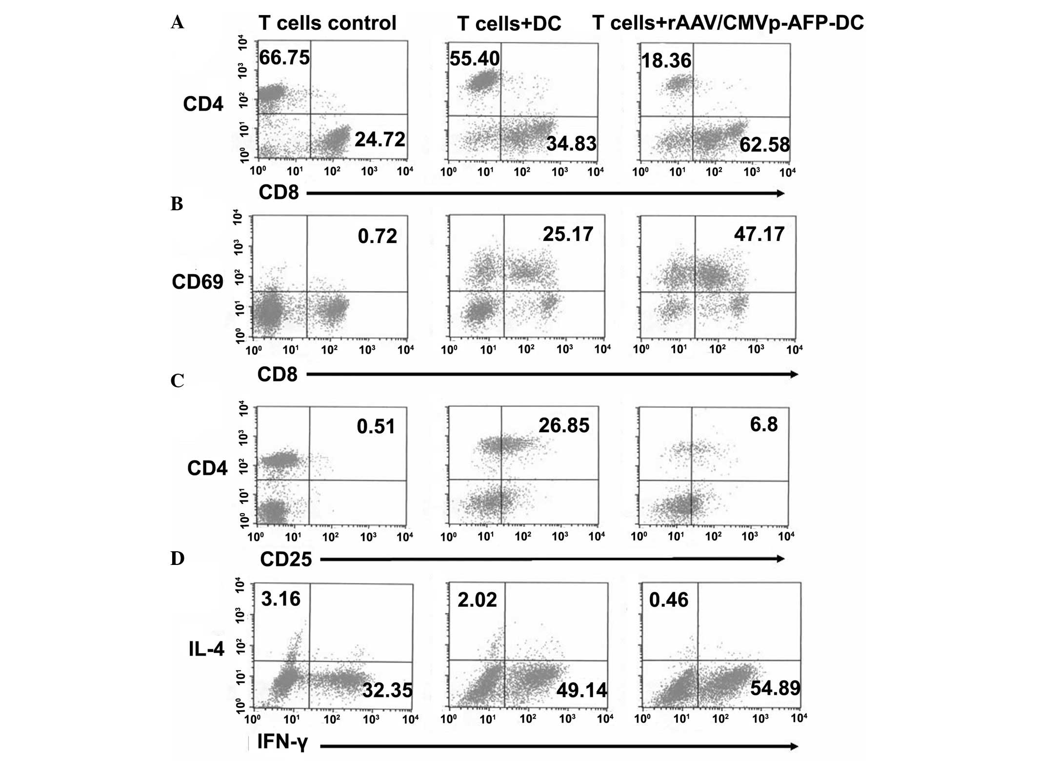
T cells primed by
rAAV/CMVp-AFP-transduced DCs were activated
The DC-T cell mixtures were cultured for 8 days. The
primed T cell populations were analyzed for their surface CD
markers by FACS analysis. As presented in Fig. 4A, the T cells primed by
rAAV/CMVp-AFP-transduced DCs had a greater ratio of CD8/CD4 (3.4:1)
than the T cells stimulated by untreated DCs (0.63:1; P<0.05).
Furthermore, the expression level of the CD69 molecules, an early
activation marker of T cells, was observed in the primed
CD8+ T cells (19). The
percentage of the CD69+ T cells elicited by the
rAAV/CMVp-AFP-transduced DCs was higher than that primed by the
untreated DCs (47.17 vs. 25.17%; P<0.05; Fig. 4B). CD25+/CD4+
T regulator (Treg) cells are critical, as they are involved in the
suppression of the Th1 response (20,21).
As presented in Fig. 4C, the
percentages of CD25+/CD4+ T cells in the T
cells primed by the untreated and rAAV/CMVp-AFP-transduced DCs were
26.85 and 6.8%, respectively (P<0.05).
IFN-γ expression was higher in the T cells primed
by rAAV/CMVp-AFP-transduced DCs
The intracellular staining assay was performed to
analyze the expression of IFN-γ and IL-4 in the T cells on day 8
post-priming. The ratio of the expressed IFN-γ/IL-4 molecules in
the T cells elicited by rAAV/CMVp-AFP-transduced DCs (119.3:1) was
higher compared with those primed by untreated DCs (24.3:1;
P<0.05; Fig. 4D).
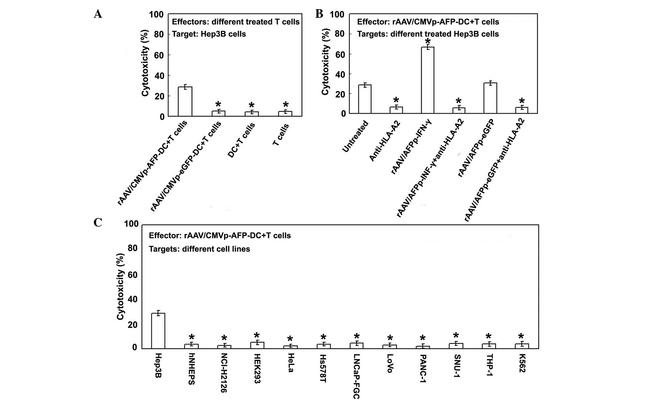
rAAV/CMVp-AFP-transduced DCs elicited an
AFP-specific and HLA class I-restricted CTL response
51Cr release assays were conducted over 6
h to analyze the activity of the CTLs elicited by
rAAV/CMVp-AFP-transduced DCs against a series of the cells. The
activity of CTLs primed by rAAV/CMVp-AFP-transduced DCs observed
against Hep3B cells was 28.6±2.3%. The CTLs primed by the untreated
or rAAV/CMVp-eGFP-transduced DCs did not lyse or induce cell death
in Hep3B cells (Fig. 5A). As
presented in Fig. 5B, the level of
CTLs against the rAAV/AFPp-IFN-γ-transduced Hep3B cells (66.7±2.8%)
was markedly higher than untreated (28.6±2.3%) and
rAAV/AFPp-eGFP-transduced cells (30.6±2.4%; P<0.05). To
determine whether the CTL response was HLA class I
(HLA-A2)-restricted, the target cells were completely inhibited
with an anti-HLA-A2 antibody prior to performing the
51Cr release assays. As presented in Fig. 5B, the activity was significantly
decreased (P<0.05). To verify that the activity was
AFP-specific, a series of AFP-negative cancer cells and primary
hepatic cells were also assessed. The results of the
51Cr release assays demonstrated that the CTLs primed by
the rAAV/CMVp-AFP-transduced DCs did not function against these
cells (Fig. 5C). In addition, NK
cell activity was observed using K562 cells as the target. As
presented in Fig. 5C, in K562
cells cell death did not occur at notable levels (3.86±1.9%).
Discussion
A number of previous studies have reported that AFP
serves as a target for immunotherapy (1,2). The
CTLs are activated by antigen-presenting DCs, and HLA class I genes
are key in cellular immune responses mediated by CTLs. To elicit a
CTL immune response, the tumor antigenic peptides must form
complexes with HLA class I heavy chain and β2-microglobulin
(22). However, in certain cases
of HCC, few or no HLA class I molecules are expressed by the tumor
cells (6–8). A deficiency of HLA class I results in
failure of the CTL response.
In the present study, transduction of rAAV/CMVp-AFP
resulted in a total of 74.33% of AFP-positive DCs, and high levels
of expression of HLA-DR, CD1a, CD40, CD80, CD83 and CD86. In
addition, in the rAAV/CMVp-AFP-transduced DCs, the expression level
of IL-10 was very low, and the expression level of IL-12 was
markedly increased. Mature DCs release large quantities of IL-12,
which stimulate a Th1 immune response. The release of IL-10,
however, completely inhibits the DC maturation process by
interfering with the upregulation of co-stimulatory molecules and
production of IL-12, subsequently limiting the ability of the DCs
to initiate a Th1 response (23,24).
The data from the present study indicated that mature AFP-pulsed
DCs were generated. After 8 days of DC-CD3+ T cell
culture, activation of the CTLs was achieved by
rAAV/CMVp-AFP-transduced DCs. The ratio of the
CD8+/CD4+ T cells (3.4:1) was notably higher
compared with the T cells stimulated by untreated DCs (0.63;
P<0.05). These data suggest that the predominant T cell
population was the CTLs (62.58%); 73.7% of the CD8+ T
cells were CD69-positive, which is an early activation marker of T
cells. Thus, large quantities of activated CD8+ T cells
were generated. Furthermore, the Treg cells
(CD25+/CD4+) were present only at as level of
6.8% in the T cell population. In addition, in the primed T cells,
the expression level of IL-4 was particularly low, and the
expression level of IFN-γ was markedly increased. IFN-γ is a
representative Th1 cytokine, and IL-4 is a representative Th2
response cytokine. These data suggest that the use of
rAAV/CMVp-AFP-transfected DCs was effective in generating a marked
Th1 response. However, the cytotoxicity assays demonstrated that
the level of the CTL activity against the AFP-positive Hep3B cells
was 28.6±2.3% due to a low level of HLA-A2 expression in the Hep3B
cells (8.78%). This suggests that a deficiency of the HLA-A2
expression in the Hep3B cells completely inhibits the CTL
response.
HLA class I genes may be induced by type 1
interferon (IFN-α/β) and IFN-γ (12,13).
To recover the expression of HLA-A2 in the Hep3B cells, in the
present study the human IFN-γ gene was transduced into Hep3B cells
by rAAV/AFPp. It is well known that AAV infects a variety of human
cells, and rAAVs have a similar ability to infect the cells
(25). To provide valuable data
for anti-HCC immunotherapy, the transduced genes were expressed in
the AFP-positive target cells, but not in the other cells. The AAV
main promoter, p5, was replaced with the human AFP promoter in an
AAV type 2 vector. All the cells were infected by rAAV/AFPp-eGFP.
As presented in Fig. 1, the eGFP
was expressed in the AFP-positive cells, Hep3B, but not in
AFP-negative cells, including hNHeps. These results indicate that
the expression of rAAV with the AFP promoter is AFP-positive and
target-specific.
Subsequently, the present study investigated the
ability of human IFN-γ gene transduction by rAAV to promote the
AFP-specific, HLA-A2-restricted CTL response against the Hep3B
cells. IFN-γ proteins were continuously expressed in the
rAAV/AFPp-IFN-γ-transfected Hep3B cells for at least 60 days. The
expression of HLA class I genes was upregulated at the
transcriptional level by IFN-γ. It has been demonstrated that IFN-γ
resulted in an increased expression of HLA class I molecules in
normal cells (26,27). In the current study, the expression
levels of HLA-A2 were upregu-lated by IFN-γ subsequently to the
HLA-A2-deficient HCC cells, Hep3B, being transduced with
rAAV/AFPp-IFN-γ. This may be one mechanism underlying the higher
percentages of induced cell death in rAAV/AFPp-IFN-γ-transduced
Hep3B cells compared with untreated and rAAV/AFPp-eGFP transduced
cells.
IFN-γ is the characteristic cytokine of Th1 cells,
and is produced by NK cells and CTLs. The level of IFN-γ has been
shown to be consistent with the CTL activity (27). The IFN-γ concentration increased
when the cytokine was secreted from the rAAV/AFPp-IFN-γ-transduced
Hep3B cells into the culture medium of the CTL and Hep3B cell
mixtures, thus, the anti-AFP CTL activity was further enhanced.
This may be another mechanism that underlies the advantageous
transduction of the IFN-γ gene into Hep3B cells.
It was observed that blocking HLA-A2 inhibited
cytotoxity, consistent with HLA-A2 restriction. The anti-AFP CTLs
were only able to induce cell death in the AFP-expressed Hep3B
cells, and exerted little toxicity on the AFP-negative primary
hepatocytes and other tumor cells, all of which were AFP-negative.
It was demonstrated that the CTLs are HLA class I
(HLA-A2)-restricted and AFP antigen-specific.
In conclusion, the data from the present study
indicated that rAAV/CMVp-AFP-transduced DCs elicit an AFP-specific
and HLA-class I-restricted CTL response against Hep3B cells. The
transduction of Hep3B cells with rAAV/AFPp-IFN-γ upregulated the
expression of HLA-A2 and improved the sensitivity to the CTL
response. The present study also provides a foundation for future
studies. There are various possibilities for improving this basic
protocol. First, the HLA-A2 gene may be transduced directly into
Hep3B cells using rAAV/AFPp in order to compare its ability to
improve the CTL response with rAAV/AFPp-IFN-γ. Secondly, the DC
vaccine may be further pretreated with additional important
cytokines (for example, IL-12) prior to mixing with T cells to
enhance DC phenotype and function. Thirdly, a burgeoning body of
evidence has demonstrated that rAAV6 is a more efficient serotype
for transducing human DCs (28,29),
and rAAV3 serotype may specifically and efficiently transduce human
HCC cells in vitro and in vivo (30–32).
These approaches may markedly improve the transduction techniques.
Furthermore, naturally occurring chemical compounds may also be
used to facilitate rAAV vector-mediated transgene expression
(33–36).
Acknowledgments
The present study was funded by grants from the
China Scholarship Council and the National Natural Science
Foundation of China (grant no. 81173615) and the Research Award
Fund for Outstanding Young Scientists of Shandong Province (grant
no. BS2013YY062).
References
|
1
|
Behboudi S and Pereira SP:
Alpha-fetoprotein specific CD4 and CD8 T cell responses in patients
with hepatocellular carcinoma. World J Hepatol. 2:256–260. 2010.
View Article : Google Scholar : PubMed/NCBI
|
|
2
|
Thimme R, Neagu M, Boettler T,
Neumann-Haefelin C, Kersting N, Geissler M, Makowiec F, Obermaier
R, Hopt UT, Blum HE and Spangenberg HC: Comprehensive analysis of
the alpha-fetoprotein-specific CD8+ T cell responses in patients
with hepatocellular carcinoma. Hepatology. 48:1821–1833. 2008.
View Article : Google Scholar : PubMed/NCBI
|
|
3
|
Oelkrug C and Ramage JM: Enhancement of T
cell recruitment and infiltration into tumours. Clin Exp Immunol.
178:1–8. 2014. View Article : Google Scholar : PubMed/NCBI
|
|
4
|
Anguille S, Smits EL, Lion E, van Tendeloo
VF and Berneman ZN: Clinical use of dendritic cells for cancer
therapy. Lancet Oncol. 15:e257–e267. 2014. View Article : Google Scholar : PubMed/NCBI
|
|
5
|
Bodmer WF, Browning MJ, Krausa P, Rowan A,
Bicknell DC and Bodmer JG: Tumor escape from immune response by
variation in HLA expression and other mechanisms. Ann NY Acad Sci.
690:42–49. 1993. View Article : Google Scholar : PubMed/NCBI
|
|
6
|
Kurokohchi K, Carrington M, Mann DL,
Simonis TB, Alexander-Miller MA, Feinstone SM, Akatsuka T and
Berzofsky JA: Expression of HLA class I molecules and the
transporter associated with antigen processing in hepatocellular
carcinoma. Hepatology. 23:1181–1188. 1996. View Article : Google Scholar : PubMed/NCBI
|
|
7
|
Wadee AA, Paterson A, Coplan KA and Reddy
SG: HLA expression in hepatocellular carcinoma cell lines. Clin Exp
Immunol. 97:328–333. 1994. View Article : Google Scholar : PubMed/NCBI
|
|
8
|
Fujiwara K, Higashi T, Nouso K,
Nakatsukasa H, Kobayashi Y, Uemura M, Nakamura S, Sato S, Hanafusa
T, Yumoto Y, et al: Decreased expression of B7 costimulatory
molecules and major histocompatibility complex class-I in human
hepato-cellular carcinoma. J Gastroenterol Hepatol. 19:1121–1127.
2004. View Article : Google Scholar : PubMed/NCBI
|
|
9
|
Salem ML: The use of dendritic cells for
peptide-based vaccination in cancer immunotherapy. Methods Mol
Biol. 1139:479–503. 2014. View Article : Google Scholar : PubMed/NCBI
|
|
10
|
Chiriva-Internati M, Liu Y, Salati E, Zhou
W, Wang Z, Grizzi F, Roman JJ, Lim SH and Hermonat PL: Efficient
generation of cytotoxic T lymphocytes against cervical cancer cells
by adeno-associated virus/human papillomavirus type 16 E7 antigen
gene transduction into dendritic cells. Eur J Immunol. 32:30–38.
2002. View Article : Google Scholar
|
|
11
|
Mahadevan M, Liu Y, You C, Luo R, You H,
Mehta JL and Hermonat PL: Generation of robust cytotoxic T
lymphocytes against prostate specific antigen by transduction of
dendritic cells using protein and recombinant adeno-associated
virus. Cancer Immunol Immunother. 56:1615–1624. 2007. View Article : Google Scholar : PubMed/NCBI
|
|
12
|
Nilsen EM, Johansen FE, Kvale D, Krajci P
and Brandtzaeg P: Different regulatory pathways employed in
cytokine-enhanced expression of secretory component and epithelial
HLA class I genes. Eur J Immunol. 29:168–179. 1999. View Article : Google Scholar : PubMed/NCBI
|
|
13
|
Brouwer RE, van der Heiden P, Schreuder
GM, Mulder A, Datema G, Anholts JD, Willemze R, Claas FH and
Falkenburg JH: Loss or downregulation of HLA class I expression at
the allelic level in acute leukemia is infrequent but functionally
relevant and can be restored by interferon. Hum Immunol.
63:200–210. 2002. View Article : Google Scholar : PubMed/NCBI
|
|
14
|
Samulski RJ, Berns KI, Tan M and Muzyczka
N: Cloning of adeno-associated virus into pBR322: Rescue of intact
virus from the recombinant plasmid in human cells. Proc Natl Acad
Sci USA. 79:2077–2081. 1982. View Article : Google Scholar : PubMed/NCBI
|
|
15
|
Morinaga T, Sakai M, Wegmann TG and
Tamaoki T: Primary structures of human alpha-fetoprotein and its
mRNA. Proc Natl Acad Sci USA. 80:4604–4608. 1983. View Article : Google Scholar : PubMed/NCBI
|
|
16
|
Devos R, Cheroutre H, Taya Y, Degrave W,
Van Heuverswyn H and Fiers W: Molecular cloning of human immune
interferon cDNA and its expression in eukaryotic cells. Nucleic
Acids Res. 10:2487–2501. 1982. View Article : Google Scholar : PubMed/NCBI
|
|
17
|
Collaco RF, Cao X and Trempe JP: A helper
virus-free packaging system for recombinant adeno-associated virus
vectors. Gene. 238:397–405. 1999. View Article : Google Scholar : PubMed/NCBI
|
|
18
|
Auricchio A, Hildinger M, O'Connor E, Gao
GP and Wilson JM: Isolation of highly infectious and pure
adeno-associated virus type 2 vectors with a single-step
gravity-flow column. Hum Gene Ther. 12:71–76. 2001. View Article : Google Scholar : PubMed/NCBI
|
|
19
|
de la Fuente H, Cruz-Adalia A, Martinez
Del Hoyo G, Cibrián-Vera D, Bonay P, Pérez-Hernández D, Vázquez J,
Navarro P, Gutierrez-Gallego R, Ramirez-Huesca M, et al: The
leukocyte activation receptor CD69 controls T cell differentiation
through its interaction with galectin-1. Mol Cell Biol.
34:2479–2487. 2014. View Article : Google Scholar : PubMed/NCBI
|
|
20
|
Zheng W, Wang QH, Feng H, Liu J, Meng HR
and Cao YM: CD4+CD25+Foxp3+ regulatory T cells prevent the
development of Th1 immune response by inhibition of dendritic cell
function during the early stage of Plasmodium yoelii infection in
susceptible BALB/c mice. Folia Parasitol (Praha). 56:242–250. 2009.
View Article : Google Scholar
|
|
21
|
Xu L, Xu W, Jiang Z, Zhang F, Chu Y and
Xiong S: Depletion of CD4 (+) CD25 (high) regulatory T cells from
tumor infiltrating lymphocytes predominantly induces Th1 type
immune response in vivo which inhibits tumor growth in adoptive
immunotherapy. Cancer Biol Ther. 8:66–72. 2009. View Article : Google Scholar
|
|
22
|
Chang CC, Ogino T, Mullins DW, Oliver JL,
Yamshchikov GV, Bandoh N, Slingluff CL Jr and Ferrone S: Defective
human leukocyte antigen class I-associated antigen presentation
caused by a novel beta2-microglobulin loss-of-function in melanoma
cells. J Biol Chem. 281:18763–18773. 2006. View Article : Google Scholar : PubMed/NCBI
|
|
23
|
Ruffell B, Chang-Strachan D, Chan V,
Rosenbusch A, Ho CM, Pryer N, Daniel D, Hwang ES, Rugo HS and
Coussens LM: Macrophage IL-10 blocks CD8+ T cell-dependent
responses to chemotherapy by suppressing IL-12 expression in
intratumoral dendritic cells. Cancer Cell. 26:623–637. 2014.
View Article : Google Scholar : PubMed/NCBI
|
|
24
|
Chattopadhyay G and Shevach EM:
Antigen-specific induced T regulatory cells impair dendritic cell
function via an IL-10/MARCH1-dependent mechanism. J Immunol.
191:5875–5884. 2013. View Article : Google Scholar : PubMed/NCBI
|
|
25
|
Muzyczka N: Use of adeno-associated virus
as a general transduction vector for mammalian cells. Curr Top
Microbiol Immunol. 158:97–129. 1992.PubMed/NCBI
|
|
26
|
Schoenborn JR and Wilson CB: Regulation of
interferon-gamma during innate and adaptive immune responses. Adv
Immunol. 96:41–101. 2007. View Article : Google Scholar : PubMed/NCBI
|
|
27
|
Schroder K, Hertzog PJ, Ravasi T and Hume
DA: Interferon-gamma: An overview of signals, mechanisms and
functions. J Leukoc Biol. 75:163–189. 2004. View Article : Google Scholar
|
|
28
|
Ussher JE and Taylor JA: Optimized
transduction of human monocyte-derived dendritic cells by
recombinant adeno-associated virus serotype 6. Hum Gene Ther.
21:1675–1686. 2010. View Article : Google Scholar : PubMed/NCBI
|
|
29
|
Pandya J, Ortiz L, Ling C, Rivers AE and
Aslanidi G: Rationally designed capsid and transgene cassette of
AAV6 vectors for dendritic cell-based cancer immunotherapy. Immunol
Cell Biol. 92:116–123. 2014. View Article : Google Scholar
|
|
30
|
Glushakova LG, Lisankie MJ, Eruslanov EB,
Ojano-Dirain C, Zolotukhin I, Liu C, Srivastava A and Stacpoole PW:
AAV3-mediated transfer and expression of the pyruvate
dehy-drogenase E1 alpha subunit gene causes metabolic remodeling
and apoptosis of human liver cancer cells. Mol Genet Metab.
98:289–299. 2009. View Article : Google Scholar : PubMed/NCBI
|
|
31
|
Cheng B, Ling C, Dai Y, Lu Y, Glushakova
LG, Gee SW, McGoogan KE, Aslanidi GV, Park M, Stacpoole PW, et al:
Development of optimized AAV3 serotype vectors: Mechanism of
high-efficiency transduction of human liver cancer cells. Gene
Ther. 19:375–384. 2012. View Article : Google Scholar :
|
|
32
|
Ling C, Lu Y, Kalsi JK, Jayandharan GR, Li
B, Ma W, Cheng B, Gee SW, McGoogan KE, Govindasamy L, et al: Human
hepatocyte growth factor receptor is a cellular coreceptor for
adeno-associated virus serotype 3. Hum Gene Ther. 21:1741–1747.
2010. View Article : Google Scholar : PubMed/NCBI
|
|
33
|
Zhang FL, Jia SQ, Zheng SP and Ding W:
Celastrol enhances AAV1-mediated gene expression in mice adipose
tissues. Gene Ther. 18:128–134. 2011. View Article : Google Scholar
|
|
34
|
Mitchell AM, Li C and Samulski RJ: Arsenic
trioxide stabilizes accumulations of adeno-associated virus virions
at the perinuclear region, increasing transduction in vitro and in
vivo. J Virol. 87:4571–4583. 2013. View Article : Google Scholar : PubMed/NCBI
|
|
35
|
Wang LN, Wang Y, Lu Y, Yin ZF, Zhang YH,
Aslanidi GV, Srivastava A, Ling CQ and Ling C: Pristimerin enhances
recombinant adeno-associated virus vector-mediated transgene
expression in human cell lines in vitro and murine hepatocytes in
vivo. J Integr Med. 12:20–34. 2014. View Article : Google Scholar : PubMed/NCBI
|
|
36
|
Ling CQ, Wang LN, Wang Y, Zhang YH, Yin
ZF, Wang M and Ling C: The roles of traditional Chinese medicine in
gene therapy. J Integr Med. 12:67–75. 2014. View Article : Google Scholar : PubMed/NCBI
|