Introduction
Lower back pain (LBP) is a common disorder with
quick recovery observed in the majority of patients. However, the
rate of LBP recurrence is high and adversely affects the quality of
life of the patients (1). Studies
have indicated that LBP can arise following lumbar facet joint
osteoarthritis (LFJOA), which accounts for 15–45% of non-specific
LBP cases (2). Facet joints are
paired zygophyseal joints between two consecutive vertebrae. LFJOA
is intimately linked to the distinct but functionally associated
condition of degenerative disc disease, which affects structures in
the anterior aspect of the vertebral column. The prevalence of
facet-mediated pain in clinical populations increases with
increasing age, suggesting that LFJOA may have a particularly
important role in older adults with spinal pain.
Over the past two decades conceptualization of knee
OA has shifted away from a predominant focus on cartilage
degeneration towards a view of OA as a heterogeneous and dynamic
process of whole-joint failure resulting from an imbalance between
the breakdown and repair of joint tissues. However, clinical
treatment of LFJOA is currently restricted to conservative
management, including medicine administration (such as nonsteroidal
anti-inflammatory drugs) and physiotherapy. Surgical intervention
can be introduced when the disorder is complicated by lumbar
degenerative disease. Direct resection of the zygapophyseal joint
and bone graft fusion is common; however, this approach cannot
fully cure LFJOA or restore function. An optimal LFJOA treatment
should not only eliminate pain and remove the cause of pain, but
also restore and maintain the normal structure and physiological
function of the lumbar zygapophyseal joint (LZJ).
The application of stem cells in cartilage tissue
repair is developing rapidly, providing a potential new therapeutic
approach for LFJOA (3,4). However, the presence of stem cells
within the lumbar zygapophyseal articular cartilage (LZAC) has yet
to be confirmed. The repair and regeneration of LFJOA cartilage may
be achieved through a single injection of lab-grown LZAC to provide
a cell source with multilineage differentiation potential combined
with a cell scaffold biomaterial and/or cartilage
differentiation-inducing factors.
The present study aimed to investigate mesenchymal
stem cell (MSC)-like cells isolated from human LZAC and to compare
their differentiation potentials in differentially degenerated
articular cartilage tissues. The findings are expected to further
the current understanding on stem cell restoration and LFJOA
therapy.
Materials and methods
Patient data and inclusion
criteria
The present study was approved by the Ethics
Committee of Yijishan Hospital, The First Affiliated Hospital of
Wannan Medical College (Wuhu, China). A total of 36 LZJ tissue
samples were obtained during surgery performed on patients with
lumbar degenerative disease between March 2017 and August 2017. The
patient age ranged between 27 and 71 years, and the most commonly
affected surgical segments were L4-S1. The inclusion criteria were
as follows: i) Patients who were clinically diagnosed with lumbar
spinal stenosis, spondylolisthesis and degenerative lumbar
scoliosis, exhibited little or no curative effects following
conservative management and volunteered to undergo surgery
(5); ii) patients without symptoms
of rachiterata or spinal cord tumors; iii) the surgery included
posterior lumbar decompression, bone fusion and vertebral pedicle
internal fixation. Written informed consent was obtained from all
enrolled patients. All the subsequent research analyses were
carried out in accordance with the approved guidelines.
Pathological staining of the LZJ
The collected LZJ samples were fixed in 10% formalin
for 48 h, cut into 2-mm slices along the coronal plane, decalcified
for 1 week, embedded in paraffin and sectioned. The 5 mm sections
were then dewaxed, dehydrated and air-dried prior to staining with
hematoxylin/eosin (HE) and Oil Red O (Gibco; Thermo Fisher
Scientific, Inc., Waltham, MA, USA). Tissue morphology was observed
using an inverted microscope.
The samples were classified according to the
Osteoarthritis Cartilage Histopathology Assessment System of the
Osteoarthritis Research Society International and the classifying
methods detailed by Kim et al (6). Samples with an intact articular
cartilage surface and tissue structures were assigned to the normal
group (G0). Samples with discontinuities or fissures on the
articular cartilage surface, mild loss of mesochondrium and/or
chondrocyte responsive hypertrophy were included in the mildly
degenerated group (G1-G2). Finally, samples with defects or
deformations of the articular cartilage surface, severe loss of
mesochondrium and/or a significantly decreased cartilage cell
number were assigned to the severely degenerated group (G3-G4).
Cultivation of primary chondrocytes
and isolation of stem cells
LZJ articular cartilage was isolated from the joint
surface under sterile conditions, cut into 1-mm3
sections using ophthalmic scissors and digested for 4–5 h at 37°C
using collagenase type-II (0.25%; Gibco; Thermo Fisher Scientific,
Inc.). Following digestion, cell suspensions were filtered twice
through a 200-mesh stainless steel filter and centrifuged at 1,000
× g for 5 min at 37°C to obtain the cell pellet. Subsequent to the
removal of the supernatant, Dulbecco's modified Eagle's
medium/Ham's F12 (DMEM/F12; Gibco; Thermo Fisher Scientific, Inc.)
supplemented with 15% fetal bovine serum (FBS; Gibco; Thermo Fisher
Scientific, Inc.) was used to disperse the pellet by pipetting, and
then cells were seeded into a 10-cm culture plate at a density of
100 cells/cm2. Cells were incubated at 5% CO2
and 37°C, and the medium was changed every 3 days until monoclonal
cells were observed. Subsequent experiments demonstrate that the
monoclonal cells have various properties of stem cells.
Induced differentiation and cell
staining
Primary stem cells were seeded into 24-well plates
at a density of 2×10⁴ cells/well in 1 ml DMEM/F12
supplemented with 10% FBS. When the cells reached 80% confluence,
the medium was replaced with osteogenic, adipocytic or chondrogenic
differentiation medium (Gibco; Thermo Fisher Scientific, Inc.). The
differentiation media were changed every 3 days. After induction
for 2–3 weeks, cells were fixed in 4% paraformaldehyde solution at
25°C for 40 min and washed with PBS. Cells were then stained with
500 µl alizarin red S (0.4%), Oil Red O or safranin O (Gibco;
Thermo Fisher Scientific, Inc.) to identify the osteogenic,
adipogenic or chondrogenic differentiation, respectively. Following
incubation at 25°C for 1 h, the staining solutions were removed,
and the samples were washed with PBS, prior to observation and
imaging using an inverted phase-contrast microscope.
Flow cytometry
Isolated primary stem cells were washed with PBS,
digested in trypsin (0.25%; Gibco; Thermo Fisher Scientific, Inc.)
and placed into Eppendorf tubes (2 ml).
Phycoerythrin-conjugated mouse anti-human monoclonal
antibodies against CD14 (eBioscience; Thermo Fisher Scientific,
Inc.; 11-0149-41; 1:50), CD90 (eBioscience; Thermo Fisher
Scientific, Inc.; 11-0909-41; 1:50), CD105 (eBioscience; Thermo
Fisher Scientific, Inc.; 12-1057-41; 1:50), CD73 (eBioscience;
Thermo Fisher Scientific, Inc.; 11-0739-41; 1:50), CD45
(eBioscience; Thermo Fisher Scientific, Inc.; 11-9459-41; 1:50),
CD34 (eBioscience; Thermo Fisher Scientific, Inc.; 11-0349-41;
1:50), STRO-1 (eBioscience; Thermo Fisher Scientific, Inc.;
14-6688-82; 1:50) and HLA-DR (eBioscience; Thermo Fisher
Scientific, Inc. 11-9956-42; 1:50) were then added. IgG (mouse IgG1
κ isotype control-FITC, eBioscience; Thermo Fisher Scientific,
Inc.; 11-4714-81; 1:20; mouse IgG1 κ isotype control-PE;
eBioscience; Thermo Fisher Scientific, Inc.; 12-4714-41, 1:100;
mouse IgG2b κ isotype control-PerCP-Cyanine5.5; eBioscience; Thermo
Fisher Scientific, Inc.; 45-4732-80; 1:100) were used as isotype
control. Subsequent to mixing, the samples were incubated for 30
min in the dark 25°C, washed with PBS, re-suspended in 500 µl PBS
and analyzed using flow cytometry (BD Biosciences, San Diego, CA,
USA). Threshold values were set using negative cell lines in
accordance with the fluorescence intensities of isotype controls.
Positive expression rates and fluorescence intensities against each
of the aforementioned monoclonal antibodies were detected using
duplicate samples.
Cytoskeleton staining
Primary stem cells were seeded into 6-well plates at
a density of 2×10⁵ cells/well. The cells were fixed in
4% paraformaldehyde at 25°C for 15 min and permeabilized using 0.3%
Triton X-100 (2 ml/well) for 10 min at 37°C. The cells were then
washed with PBS and 100 µl/well phalloidin (Gibco; Thermo Fisher
Scientific, Inc.) was added prior to incubation for 10 min in the
dark 25°C. DAPI (100 µl) was subsequently added and the cells were
incubated for 5 min in the dark 25°C. Following washing in PBS,
cytoskeletal F-actin was observed and imaged using laser-scanning
confocal microscopy (Zeiss LSM 510-META laser scanning microscope;
Carl Zeiss AG, Jena, Germany).
Reverse transcription-quantitative
polymerase chain reaction (RT-qPCR)
RNA was extracted from the induced cells using
TRIzol® reagent (Invitrogen; Thermo Fisher Scientific,
Inc.). The quantity and quality of isolated RNA were evaluated
using absorbance at wavelengths of 260 and 280 nm.
Reverse-transcribed into cDNA using a RevertAid™ First
Strand cDNA Synthesis kit (Thermo Fisher Scientific, Inc.)
according to the manufacturer's protocol of the kit. Next, qPCR was
performed using a QuantiTect SYBR Green PCR kit (Thermo Fisher
Scientific, Inc.), according to the manufacturer's protocol, and
the mRNA levels of aggrecan (ACAN), collagen type-II (COL-II) and
SRY-related high-mobility-group box 9 (SOX9) were measured in each
group. The conditions of real-time PCR were as follows:
Denaturation at 95°C for 10 sec, 40 cycles at 95°C for 10 sec and
60°C for 30 sec. Dissociation curves revealed no nonspecific
amplification. The primers used for qPCR assay are listed in
Table I. GAPDH was used as an
internal reference, and the results were analyzed using the
2−ΔΔCq method (7).
 | Table I.Primer sequences for quantitative
polymerase chain reaction. |
Table I.
Primer sequences for quantitative
polymerase chain reaction.
| Gene | Primer sequence
(5′-3′) |
|---|
| ACAN | F:
CATTCACCAGTGAGGACCTCGT |
|
| R:
TCACACTGCTCATAGCCTGCTTC |
| COL-II | F:
TGAGGGCGCGGTAGAGACCC |
|
| R:
TGCACACAGCTGCCAGCCTC |
| SOX9 | F:
ATCTGAAGAAGGAGAGCGAG |
|
| R:
TCAGAAGTCTCCAGAGCTTG |
| GAPDH | F:
GCACCGTCAAGGCTGAGAAC |
|
| R:
TGGTGAAGACGCCAGTGGA |
Western blot analysis
Induced cells were collected, resuspended in RIPA
Lysis buffer (Invitrogen; Thermo Fisher Scientific, Inc.) with
phenylmethanesulfonyl fluoride (PMSF; Thermo Fisher Scientific,
Inc.) and lysed on ice. The supernatants were collected, and
protein concentrations were measured using the BCA method. Equal
amounts of proteins were used in subsequent experiments. A total of
20 µg protein was loaded per lane using 10% SDS-PAGE and
electrotransferred onto nitrocellulose membranes (Beyotime
Institute of Biotechnology, China), and 5% bovine serum albumin
(Beyotime Institute of Biotechnology) was used for membrane
blocking for 1 h at 25°C. Primary antibodies against ACAN (1:1,000;
Abcam, Cambridge, UK), COL-II (1:5,000; Abcam), SOX9 (1:5,000;
Abcam) and GAPDH (1:5,000; Abcam) were incubated with the membrane
overnight at 4°C on a shaker. Following washing, the membrane was
exposed to a secondary antibody diluted at 1:5,000 in blocking
reagent and incubated for 1 h at room temperature on a shaker.
Subsequent to further washing in Tris-buffered saline/20% Tween-20,
the membranes were visualized using an enhanced chemiluminescence
detection system (EMD Millipore, Billerica, MA, USA).
Immunoreactive bands were quantified on autoradiography films in
triplicate with using Image J software (version 4.8; National
Institutes of Health, Bethesda, MD, USA) by normalizing the band
intensities to GAPDH.
Colony-forming assay
Primary stem cells were seeded into 6-cm diameter
culture dishes at a density of 100 cells/dish (n=3 per sample).
After 7–10 days of cultivation, cells were fixed in 4%
paraformaldehyde at 25°C for 20 min, washed with PBS and immersed
in 0.1% crystal violet staining solution at 25°C for 10 min.
Following washing with PBS, the cell colonies were counted and
imaged.
Statistical analysis
All statistical data are expressed as the mean ±
standard deviation. Statistical analysis was conducted using SPSS
version 18.0 software (SPSS, Inc., Chicago, IL, USA). Statistically
significant differences among the three groups were determined
using one-way analysis of variance, and χ2 tests were
used to compare cell counts between groups. P<0.05 was
considered to indicate a difference that was statistically
significant.
Results
Pathological and radiological
characteristics of differentially degenerated LZAC
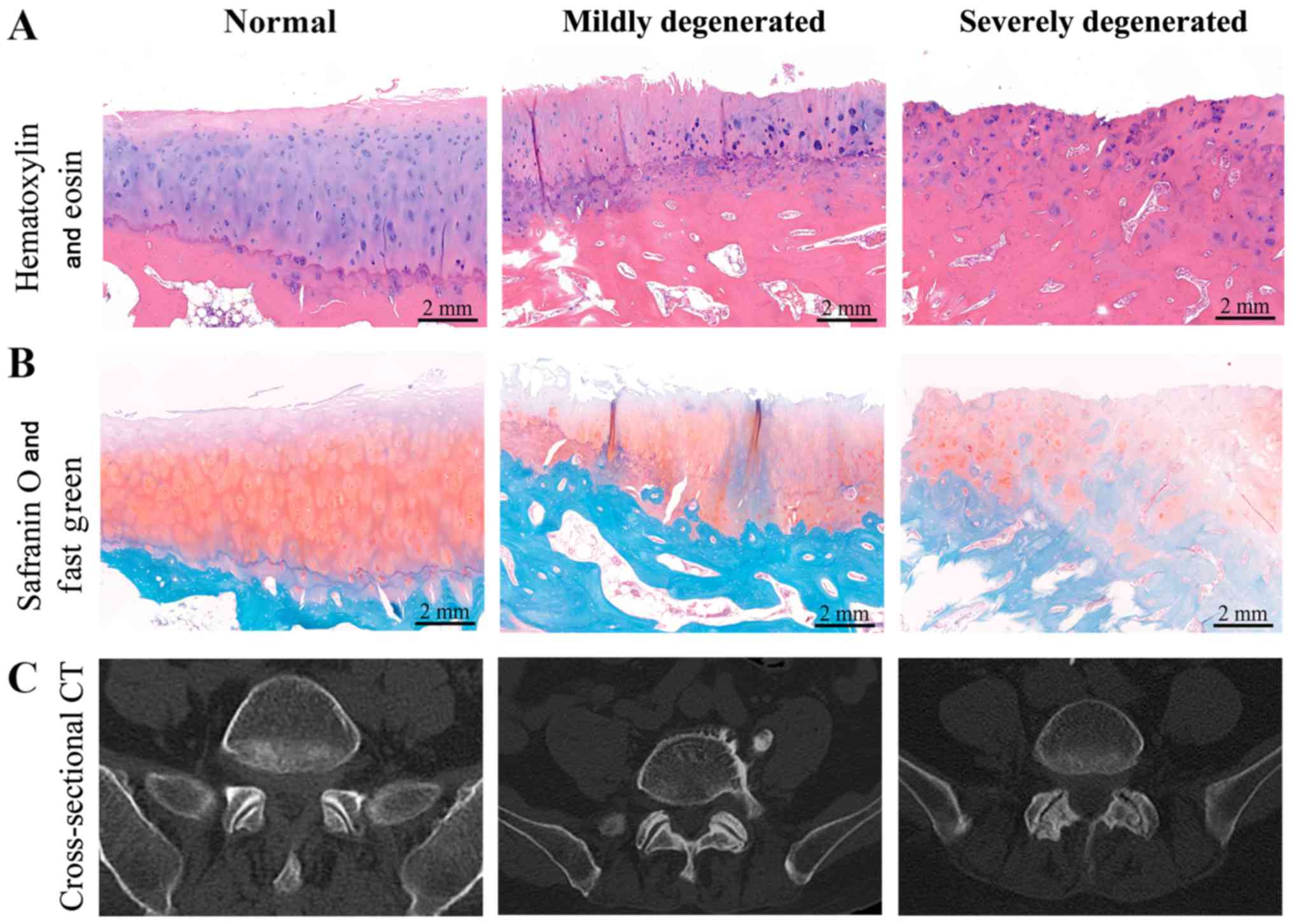
HE and safranin O staining were performed to
classify 36 LZJ articular tissues into three groups according to
the degree of degeneration, as follows: i) Normal group (n=5); ii)
mildly degenerated group (n=12); and iii) severely degenerated
group (n=19). In the normal group, the articular cartilage surfaces
were smooth, and the four-layered tissue structures were intact. By
contrast, the articular cartilage surfaces of the mildly
degenerated group were rough and exhibited longitudinal fissures,
while the separation lines of the four-layered structure were
unclear. In the severely degenerated group, erosion and defects
were clearly observed, with a thinner cartilaginous layer and
evident proliferation of osseous tissue. Furthermore, the boundary
between the subchondral bone and underlying bone tissues was
unclear in this group (Fig. 1A and
B).
Cross-sectional computed tomography scans of the
lumbar area were also compared among the three groups, and the
radiological characteristics for each group were in agreement with
the staining results (Fig. 1C). In
addition, no significant differences were detected in the clinical
data among the three patient groups (Table II).
| Table II.Comparison of clinical characteristics
among the three groups (n=36). |
Table II.
Comparison of clinical characteristics
among the three groups (n=36).
| Group | N | Sex
(male/female) | Age (years) | Chronic disease
(yes/no) |
|---|
| Normal | 5 | 3:2 | 48.20±12.07 |
3:2 |
| Mildly
degenerated | 12 |
8:4 | 53.25±7.71 |
6:6 |
| Severely
degenerated | 19 | 13:6 | 55.26±9.69 | 10:9 |
|
F/χ2 |
| 0.124 | 1.126 | 0.143 |
| P-value |
| 0.940 | 0.337 | 0.931 |
Stem cells from differentially
degenerated LZAC display MSC-like characteristics
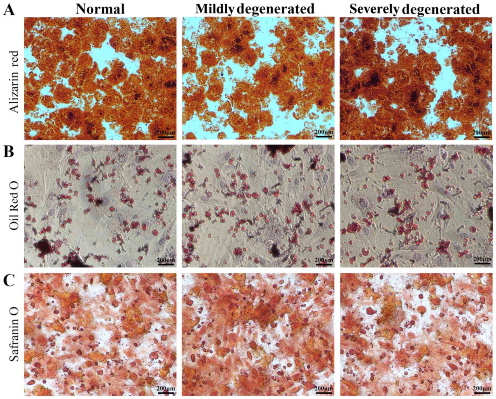
Subsequent to osteogenic differentiation of isolated
stem cells, rock salt gradually deposited and formed a plurality of
granular calcium nodules. Alizarin red staining result indicated
all stem cells were stained red (Fig.
2A). After adipogenic induction, Oil Red O staining results
demonstrated that 3 kinds of stem cells were all stained red
(Fig. 2B). Chondrocyte-specific
aggrecan were stained red with safranin O following chondrogenic
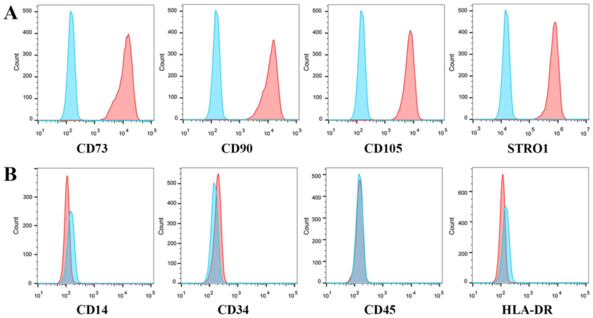
induction (Fig. 2C). Flow
cytometry analysis further revealed that isolated stem cells from
all groups were positive for the CD90, CD73, STRO-1 and CD105
surface markers, and negative for CD14, CD34, CD45 and HLA-DR
(Fig. 3).
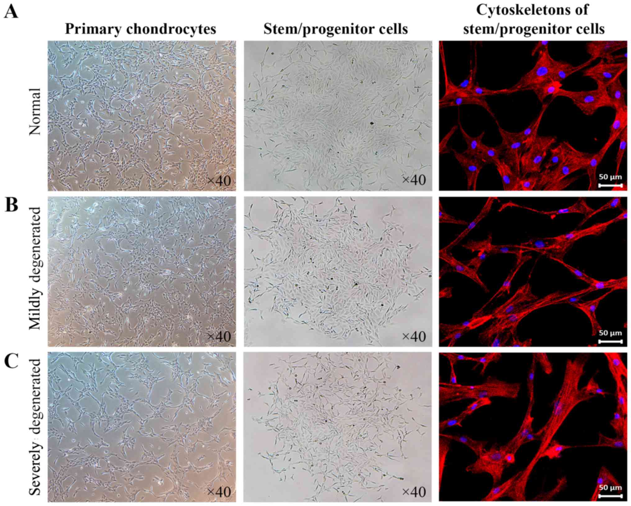
Morphological changes are observed in
stem cells from differentially degenerated LZACs
Primary chondrocytes from the normal and mildly
degenerated groups were polygonal and tightly arranged, while those
from the severely degenerated group were elongated and loosely
arranged. Stem cells isolated from monoclones were different to
primary chondrocytes; more specifically, they were smaller in size
and arranged in a whirlpool configuration. No evident differences
in the morphology were observed between the three cell groups
microscopically. As presented in Fig.
4, phalloidin staining revealed that the stem cell
cytoskeletons in the normal group were spindle-shaped, while those
in the mildly and severely degenerated groups were visibly
elongated. These results indicated that, compared with normal LZAC,
abnormal fibrosis was evident in stem cells isolated from mildly
and severely degenerated LZAC cases.
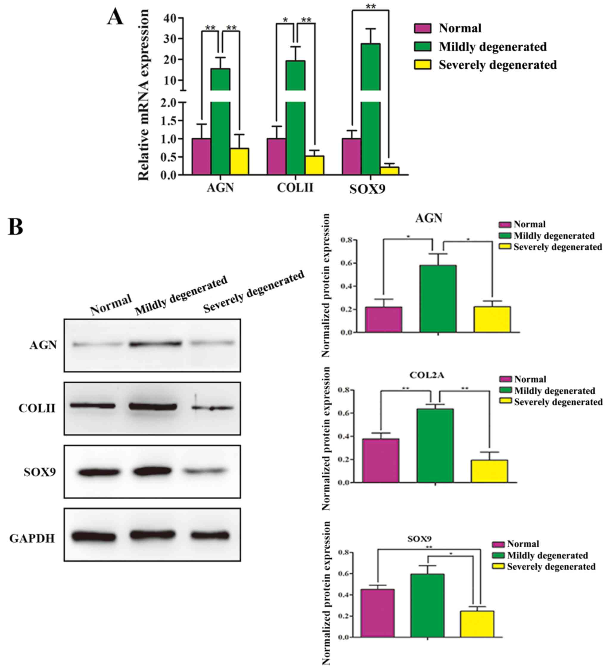
Chondrogenic ability differs among
stem cells isolated from differentially degenerated LZAC
RT-qPCR and western blot analysis confirmed that
stem cells from the mildly degenerated group had enhanced
chondrogenic potential, whereas those from the severely degenerated
group had reduced chondrogenic potential, compared with cells from
the normal group. The chondrogenic differentiational genes and
proteins (ACAN, COL-II and SOX9) of 3 kinds of stem cells were
analyzed respectively, and the results demonstrated that the
expression of the 3 genes and proteins by the mildly degenerated
group was stronger than the other two cells. And the 3 genes and
proteins expression of the normal group was the second highest and
that of the severely degenerated group was worse (Fig. 5). These results indicated that,
although stem cells with multilineage differentiation potential
existed in LZAC at different degrees of degeneration, stem cells
from mildly degenerated LZAC exhibited a stronger chondrogenic
potential.
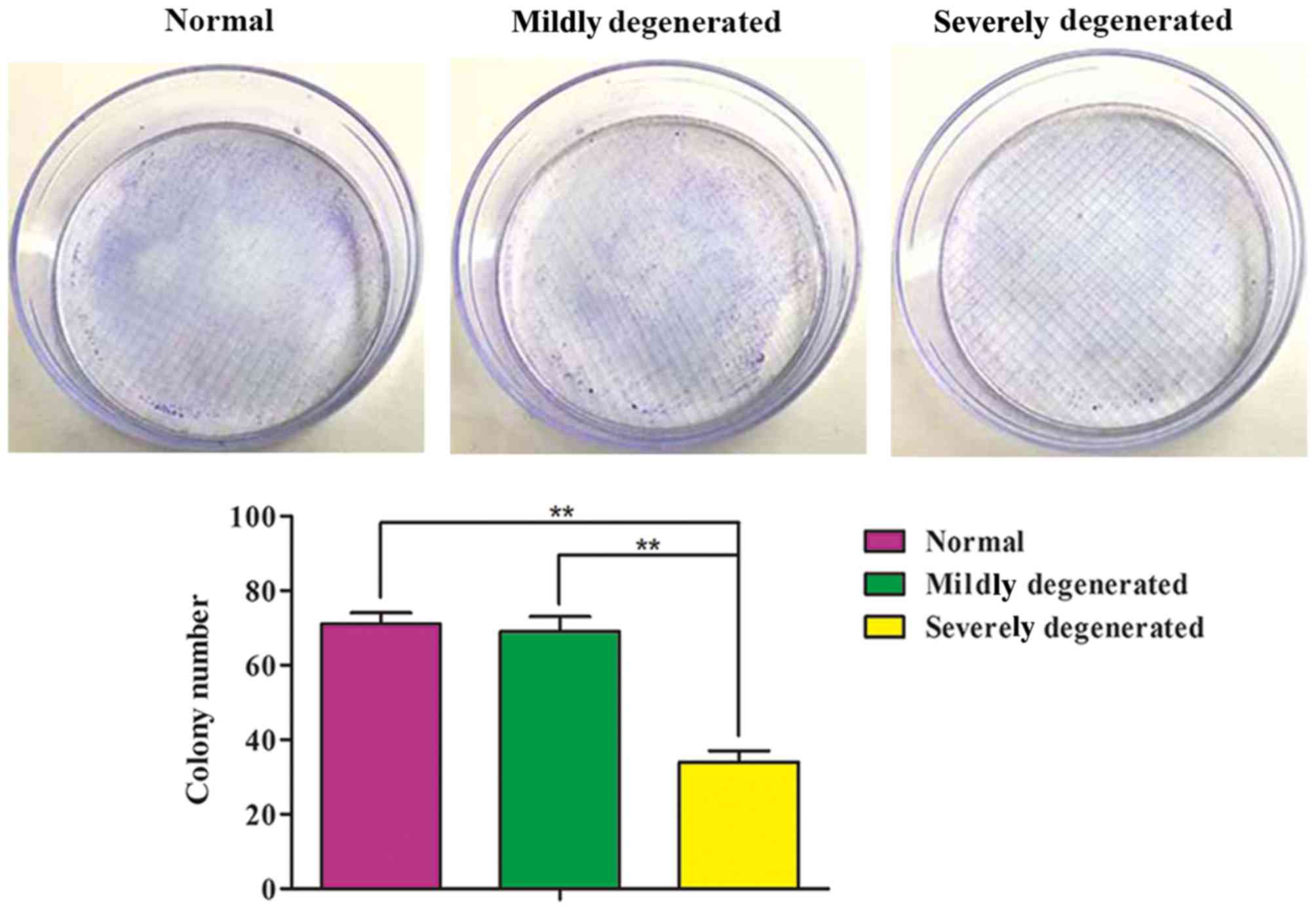
Stem cell clonal formation varies in
differentially degenerated LZAC
Stem cells isolated from all three groups were
observed to form colonies; however, the colony number was
significantly lower in the severely degenerated group. No
significant difference was observed between the normal and mildly
degenerated groups with respect to colony formation (Fig. 6). These results indicated that stem
cells from normal and mildly degenerated LZAC possess good clonal
formation ability.
Discussion
Osteoarthritis is characterized by an imbalance
between the dynamic processes of cartilaginous tissue decomposition
and repair (8). Similarly, the
main pathogenic manifestations of LFJOA include abrasion of the
arthrodial cartilage, responsive hypertrophy of the subchondral
bone and osteophyte formation in the joint verge (9,10).
Histological evaluation of osteoarthritis has confirmed increased
levels of chondrocyte apoptosis, loss of proteoglycans secreted by
chondrocytes and a denser arrangement of chondrocytes in
degenerated cartilaginous tissues (11).
Cartilaginous tissue has no blood vessels or nerves,
and is composed of 5% chondrocytes and 95% mesochondrium; thus, it
has a poor self-repairing and regenerative capacity (12). Regenerative medicine has become a
focal point, allowing regenerating of defective cartilaginous
tissues using autologous chondrocytes (13–15).
However, the application of chondrocytes in repair strategies has a
number of limitations, including the limited availability of adult
chondrocytes. Chondrocytes are terminally differentiated cells with
an extremely weak proliferative capacity, and dedifferentiation
often occurs during in vitro cultivation. Furthermore, the
chondrogenic potential of chondrocytes is lost in vitro,
making it difficult to obtain a large number of normally
functioning chondrocytes from a small number of adult chondrocytes
(16–18). Additionally, the extraction
efficiency of primary chondrocytes from cartilaginous tissues is
low (19).
Research in this field is currently focusing on the
repair of bone and cartilage using stem cells. Studies have
demonstrated that cells with differentiation capacities exist in
both normal and degenerated cartilage (20,21).
Clinical tests have verified that implanting or injecting MSCs can
promote cartilage formation in articulatio genus defect
areas (22). Although MSC
isolation from the endplate and LZAC remains under investigation,
the future application of these stem cells within spinal fusion
surgery and for degenerative disc disease is promising (23–26).
In the present study, the monoclonal cell culture
method was used to prove the existence of stem cells with clonal
ability and multidifferentiation potential in human LZAC. As part
of the LZA microenvironment, these cells have vital roles in the
initiation and progression of degenerative spinal diseases, and
thus may be a more desirable cell source for tissue repair.
Although stem cells with self-renewal and
multilineage differentiation capacities have been found to exist in
both normal and degenerated articular cartilage, their full
potential for application in tissue repair remains unclear. A
number of previous studies have suggested that stem cells only
exist on surfaces where the cartilage is rapidly growing and
developing, while others propose that stem cells are more prevalent
at the boundary area of cartilage injury (27,28).
These studies indicate, to a certain extent, that stem cells are
necessary for the supplementation and maintenance of mesochondrial
components. Conversely, the differentiation of stem cells also
depends on the extracellular microenvironment (29). In the present study, although stem
cells with induced differentiation and clonogenic abilities were
isolated from differentially degenerated LZAC, their chondrogenic
differentiation and clonogenic abilities differed significantly
among the groups.
Cytoskeleton staining revealed fibrosis in the stem
cells isolated from mildly and severely degenerated LZAC,
indicating that the mesochondrial environment altered the
characteristics of these cells to some extent (30,31).
Subsequent experiments suggested that stem cells isolated from
mildly degenerated LZAC exhibited enhanced chondrogenic
differentiation and clonogenic abilities compared with those from
normal LZAC. Furthermore, stem cells from severely degenerated LZAC
exhibited reduced chondrogenic differentiation and clonogenic
abilities.
The present study successfully isolated stem cells
from differentially degenerated LZAC using a monoclonal cell
culture method, and compared the chondrogenic and clonogenic
characteristics in the different groups. However, there are certain
limitations: i) The osteogenic and adipogenic differentiation
capacities of stem cells require further investigation, as do their
surface marker profiles, in comparison with the well-characterized
bone marrow-derived MSCs; and ii) although the differentiation
potential of stem cells from cartilage tissue is affected by
multiple factors, induction models based on nutrition, mechanics
and inflammation were not established or investigated in the
present study.
As the stem cells were obtained from differentially
degenerated LZAC tissues, it is hypothesized that the stem cells
from mildly degenerated articular cartilages are in a
‘transitional’ state, and that their increased differentiation and
clonal abilities are likely to be transiently enforced due to
external stimuli. During the aggravation of cartilage tissue
degeneration, stem cells gradually lose their normal functionality.
These findings are important for specific clinical treatment of
differentially degenerated cartilage in the future.
In conclusion, stem cells with multilineage
differentiation potential and clonal properties were identified in
human LZAC, and these characteristics were more prominent in mildly
degenerated, compared with severely degenerated, articular
cartilage.
Acknowledgements
Not applicable.
Funding
The present study was supported by the National
Natural Science Foundation of China (grant no. 81572185), the
Natural Science Foundation of Anhui Province of China (grant no.
1708085MH185) and the Natural Science Foundation for Young
Scientists of Anhui Province of China (grant no. 1808085QH275).
Availability of data and materials
The datasets used and/or analyzed during the current
study are available from the corresponding author on reasonable
request.
Authors' contributions
HX provided technical support on the study
conception and supervision, and LX, SX, XW and ZJ provided
technical support on cell-induced differentiation and staining. JW
provided technical support on RT-qPCR and western blot analysis,
and BY provided technical support on flow cytometry, colony-forming
assay and statistical analysis.
Ethics approval and consent to
participate
The present study was approved by the Ethics
Committee of our hospital. Informed consent was obtained from all
participants.
Patient consent for publication
No conflict of interest exits in the submission of
this manuscript, and manuscript is approved by all authors for
publication.
Competing interests
The authors have declared that no competing
interests exist.
References
|
1
|
Shelerud RA: Epidemiology of occupational
low back pain. Clin Occup Environ Med. 5:501–528. 2006.PubMed/NCBI
|
|
2
|
Sehgal N, Shah RV, McKenzie-Brown AM and
Everett CR: Diagnostic utility of facet (zygapophysial) joint
injections in chronic spinal pain: A systematic review of evidence.
Pain Physician. 8:211–224. 2005.PubMed/NCBI
|
|
3
|
Cosenza S, Ruiz M, Toupet K, Jorgensen C
and Noël D: Mesenchymal stem cells derived exosomes and
microparticles protect cartilage and bone from degradation in
osteoarthritis. Sci Rep. 7:162142017. View Article : Google Scholar : PubMed/NCBI
|
|
4
|
Kumar H, Ha DH, Lee EJ, Park JH, Shim JH,
Ahn TK, Kim KT, Ropper AE, Sohn S, Kim CH, et al: Safety and
tolerability of intradiscal implantation of combined autologous
adipose-derived mesenchymal stem cells and hyaluronic acid in
patients with chronic discogenic low back pain: 1-year follow-up of
a phase I study. Stem Cell Res Ther. 8:2622017. View Article : Google Scholar : PubMed/NCBI
|
|
5
|
Tian H, Wu A, Guo M, Zhang K, Chen C, Li
X, Cheng X, Zhou T, Murray SS, Sun X and Zhao J: Adequate
restoration of disc height and segmental lordosis by lumbar
interbody fusion decreases adjacent segment degeneration. World
Neurosurg. 118:e856–e864. 2018. View Article : Google Scholar : PubMed/NCBI
|
|
6
|
Kim JS, Ali MH, Wydra F, Li X, Hamilton
JL, An HS, Cs-Szabo G, Andrews S, Moric M, Xiao G, et al:
Characterization of degenerative human facet joints and facet joint
capsular tissues. Osteoarthritis Cartilage. 23:2242–2251. 2015.
View Article : Google Scholar : PubMed/NCBI
|
|
7
|
Livak KJ and Schmittgen TD: Analysis of
relative gene expression data using real-time quantitative PCR and
the 2(-Delta Delta C(T)) method. Methods. 25:402–408. 2001.
View Article : Google Scholar : PubMed/NCBI
|
|
8
|
Hunter DJ and Felson DT: Osteoarthritis.
BMJ. 332:639–642. 2006. View Article : Google Scholar : PubMed/NCBI
|
|
9
|
Shuang F, Zhou Y, Hou SX, Zhu JL, Liu Y,
Zhang CL and Tang JG: Indian Hedgehog signaling pathway members are
associated with magnetic resonance imaging manifestations and
pathological scores in lumbar facet joint osteoarthritis. Sci Rep.
5:102902015. View Article : Google Scholar : PubMed/NCBI
|
|
10
|
Xu D, Sun Y, Bao G, Liu W, Zhu X, Cui S,
Fan J and Cui Z: MMP-1 overexpression induced by IL-1beta: Possible
mechanism for inflammation in degenerative lumbar facet joint. J
Orthop Sci. 18:1012–1019. 2013. View Article : Google Scholar : PubMed/NCBI
|
|
11
|
Alsalameh S, Amin R, Gemba T and Lotz M:
Identification of mesenchymal progenitor cells in normal and
osteoarthritic human articular cartilage. Arthritis Rheum.
50:1522–1532. 2004. View Article : Google Scholar : PubMed/NCBI
|
|
12
|
Poole CA: Articular cartilage chondrons:
Form, function and failure. J Anat. 191:1–13. 1997. View Article : Google Scholar : PubMed/NCBI
|
|
13
|
Lopez-Alcorocho JM, Aboli L,
Guillen-Vicente I, Rodriguez-Iñigo E, Guillen-Vicente M,
Fernández-Jaén TF, Arauz S, Abelow S and Guillen-García P:
Cartilage defect treatment using high-density autologous
chondrocyte implantation: Two-year follow-up. Cartilage. 9:363–369.
2018. View Article : Google Scholar : PubMed/NCBI
|
|
14
|
Ma N, Wang H, Xu X, Wan Y, Liu Y, Wang M,
Yu W, Dai Y, Peng J, Guo Q, et al: Autologous-cell-derived,
tissue-engineered cartilage for repairing articular cartilage
lesions in the knee: Study protocol for a randomized controlled
trial. Trials. 18:5192017. View Article : Google Scholar : PubMed/NCBI
|
|
15
|
Wong CC, Chen CH, Chan WP, Chiu LH, Ho WP,
Hsieh FJ, Chen YT and Yang TL: Single-stage cartilage repair using
platelet-rich fibrin scaffolds with autologous cartilaginous
grafts. Am J Sports Med. 45:3128–3142. 2017. View Article : Google Scholar : PubMed/NCBI
|
|
16
|
Elima K and Vuorio E: Expression of mRNAs
for collagens and other matrix components in dedifferentiating and
redifferentiating human chondrocytes in culture. FEBS Lett.
258:195–198. 1989. View Article : Google Scholar : PubMed/NCBI
|
|
17
|
Schnabel M, Marlovits S, Eckhoff G,
Fichtel I, Gotzen L, Vécsei V and Schlegel J:
Dedifferentiation-associated changes in morphology and gene
expression in primary human articular chondrocytes in cell culture.
Osteoarthritis Cartilage. 10:62–70. 2002. View Article : Google Scholar : PubMed/NCBI
|
|
18
|
Diaz-Romero J, Gaillard JP, Grogan SP,
Nesic D, Trub T and Mainil-Varlet P: Immunophenotypic analysis of
human articular chondrocytes: Changes in surface markers associated
with cell expansion in monolayer culture. J Cell Physiol.
202:731–742. 2005. View Article : Google Scholar : PubMed/NCBI
|
|
19
|
Stoddart MJ, Grad S, Eglin D and Alini M:
Cells and biomaterials in cartilage tissue engineering. Regen Med.
4:81–98. 2009. View Article : Google Scholar : PubMed/NCBI
|
|
20
|
Williams R, Khan IM, Richardson K, Nelson
L, McCarthy HE, Analbelsi T, Singhrao SK, Dowthwaite GP, Jones RE,
Baird DM, et al: Identification and clonal characterisation of a
progenitor cell sub-population in normal human articular cartilage.
PLoS One. 5:e132462010. View Article : Google Scholar : PubMed/NCBI
|
|
21
|
Hattori S, Oxford C and Reddi AH:
Identification of superficial zone articular chondrocyte
stem/progenitor cells. Biochem Biophys Res Commun. 358:99–103.
2007. View Article : Google Scholar : PubMed/NCBI
|
|
22
|
Kristjánsson B and Honsawek S: Current
perspectives in mesenchymal stem cell therapies for osteoarthritis.
Stem Cells Int. 2014:1943182014. View Article : Google Scholar : PubMed/NCBI
|
|
23
|
Kristjánsson B, Limthongkul W,
Yingsakmongkol W, Thantiworasit P, Jirathanathornnukul N and
Honsawek S: Isolation and Characterization of Human Mesenchymal
Stem Cells From Facet Joints and Interspinous Ligaments. Spine
(Phila Pa 1976). 41:E1–E7. 2016. View Article : Google Scholar : PubMed/NCBI
|
|
24
|
Liu LT, Huang B, Li CQ, Zhuang Y, Wang J
and Zhou Y: Characteristics of stem cells derived from the
degenerated human intervertebral disc cartilage endplate. PLoS One.
6:e262852011. View Article : Google Scholar : PubMed/NCBI
|
|
25
|
McAnany SJ, Ahn J, Elboghdady IM,
Marquez-Lara A, Ashraf N, Svovrlj B, Overley SC, Singh K and
Qureshi SA: Mesenchymal stem cell allograft as a fusion adjunct in
one- and two-level anterior cervical discectomy and fusion: A
matched cohort analysis. Spine J. 16:163–167. 2016. View Article : Google Scholar : PubMed/NCBI
|
|
26
|
Pneumaticos SG, Triantafyllopoulos GK,
Chatziioannou S, Basdra EK and Papavassiliou AG: Biomolecular
strategies of bone augmentation in spinal surgery. Trends Mol Med.
17:215–222. 2011. View Article : Google Scholar : PubMed/NCBI
|
|
27
|
Dowthwaite GP, Bishop JC, Redman SN, Khan
IM, Rooney P, Evans DJ, Haughton L, Bayram Z, Boyer S, Thomson B,
et al: The surface of articular cartilage contains a progenitor
cell population. J Cell Sci. 117:889–897. 2004. View Article : Google Scholar : PubMed/NCBI
|
|
28
|
Khan IM, Williams R and Archer CW: One
flew over the progenitor's nest: Migratory cells find a home in
osteoarthritic cartilage. Cell Stem Cell. 4:282–284. 2009.
View Article : Google Scholar : PubMed/NCBI
|
|
29
|
Badylak S, Obermiller J, Geddes L and
Matheny R: Extracellular matrix for myocardial repair. Heart Surg
Forum. 6:E20–E26. 2003. View
Article : Google Scholar : PubMed/NCBI
|
|
30
|
Olvera D, Sathy BN, Carroll SF and Kelly
DJ: Modulating microfibrillar alignment and growth factor
stimulation to regulate mesenchymal stem cell differentiation. Acta
Biomater. 64:148–160. 2017. View Article : Google Scholar : PubMed/NCBI
|
|
31
|
Yang Q, Teng BH, Wang LN, Li K, Xu C, Ma
XL, Zhang Y, Kong DL, Wang LY and Zhao YH: Silk fibroin/cartilage
extracellular matrix scaffolds with sequential delivery of TGF-β3
for chondrogenic differentiation of adipose-derived stem cells. Int
J Nanomedicine. 12:6721–6733. 2017. View Article : Google Scholar : PubMed/NCBI
|