Introduction
Lung cancer, as is well known, is the most common
cause of cancer-related deaths all over the world. As a malignant
tumor, lung cancer kills countless patients worldwide (1,2).
Every year, 1.8 million individuals are diagnosed with lung cancer,
and 1.6 million patients die as a result of the disease. In
addition, the incidence rate of lung adenocarcinoma (LADC), the
most common histologic subtype of lung cancer, has continued to
increase in men and women (3).
However, due to the delay in diagnosis, the treatment results for
LADC remain unsatisfactory; 5-year survival rates vary from 4 to
17% depending on the stage and on regional differences (4). Moreover, the treatment and prognosis
of LADC are still public health issues characterized by no progress
in advanced diagnosis and treatment (1,5,6).
Therefore, there is an urgent need to discover new molecular
biomarkers for the early diagnosis and treatment of LADC.
The human complement factor H (CFH)-related protein
(CFHR) family is composed of five members: CFHR1, CFHR2,
CFHR3, CFHR4 and CFHR5, and each member of this group
can bind to the central complement component C3b. Mutations,
genetic deletions, duplications or rearrangements in the individual
CFHR genes are associated with many diseases, including
cancer (7,8). Recent research shows that CFH-related
genes (CFHR1-5) are associated with age-related macular
degeneration (AMD) (9). At the
same time, large international genome-wide association studies have
shown that deletion of CFHR1 is associated with a reduced
risk of developing IgA nephropathy (10). Fratelli et al (11) found that the germinal homozygous
deletion of the CFHR1 gene could act as a promising risk
factor for acute myelogenous leukemia. Another report also
indicated that CFHR1 was homozygously deleted in the
cisplatin-resistance glioma cell lines U251 and CP2 (12). However, few studies have reported
the relationship between the CFHR family and LADC, and the
influence of CFHR1 on the pathological process of LADC
remains unexplored.
The aim of the present study was to evaluate the
function of CFHR1 and its relationship with clinical
treatment and prognosis in human LADC. Our data indicate that
CFHR1 functions as a potential tumor suppressor in LADC
tissues and cell lines. Kaplan-Meier analysis suggests that
CFHR1 is an independent prognostic factor for LADC
patients.
Materials and methods
Data collection and reanalysis using
different bioinformatic methods
The expression levels of CFHR1 in LADC
tissues and cell lines are evidenced by a variety of bioinformatic
network resources (Table S1).
Oncomine, a bioinformatics project that analyzes
cancer transcriptome data to make it available to the biomedical
research community, contains 65 gene expression datasets (13). UALCAN provides a silicon-based
platform for validation of target genes and identification of tumor
subpopulation-specific candidate biomarkers (14). GEPIA is a web server for gene
expression profiling and interactive analyses in human cancers. It
provides several key interactive customization features, such as
differential expression analysis, patient survival analysis, and
similarity gene detection (15).
Through these public bioinformatics platforms, we clearly defined
the expression profile of CFHR1 in human LADC tissues and
cell lines.
Kaplan-Meier plotter is a web-based tool for
assessing the impact of 54,675 genes on the survival of 10,461
cancer samples and quickly determining the prognosis of the
diseases (16), such as overall
survival (OS) and post progressive survival (PPS) (17).
We downloaded the therapeutic transcriptome
microarray dataset from the Gene Expression Omnibus (GEO) database
under the login numbers GSE6400 (18) and GSE21656 (19). Then, we re-analyzed the original
data in these datasets to further understand the impact of
CFHR1 expression on the chemotherapy response of LADC
patients.
The web resource cBioPortal of cancer genomics
provides a desired strategy for the exploration, visualization and
analysis of multidimensional cancer genomics data (20). We used it to screen the coexpressed
genes of CFHR1 in LADC tissues. Protein-protein interaction
(PPI) networks of these coexpressed genes were constructed using
the STRING database (21). Then,
Cytoscape software (version 3.0) was used to perform detailed
visualization (22).
Next, we used the web-based Gene SeT AnaLysis
Toolkit (WebGestalt) to perform Gene Ontology (GO) enrichment
analysis (23). Meanwhile, the
Pathview algorithm was used to analyze the relevant KEGG pathways
(24).
Statistical analyses
The Student's t-test and the statistical software
package SPSS (SPSS12.0, IBM Analytics) were used to analyze the
differentially expressed mRNAs between cancer tissues and noncancer
tissues. Meanwhile, the relationship between CFHR1
expression and clinicopathological features of LADC patients was
analyzed using a Chi-square test. The variants with statistical
significance in single-factor analysis were further examined by
multiple-factor analysis using the COX regression model. Pearson
correlation coefficient was used to evaluate the correlation
between genes. If P≤0.05, then the result was considered
statistically significant.
Results
CFHR1 is downregulated in LADC
tissues
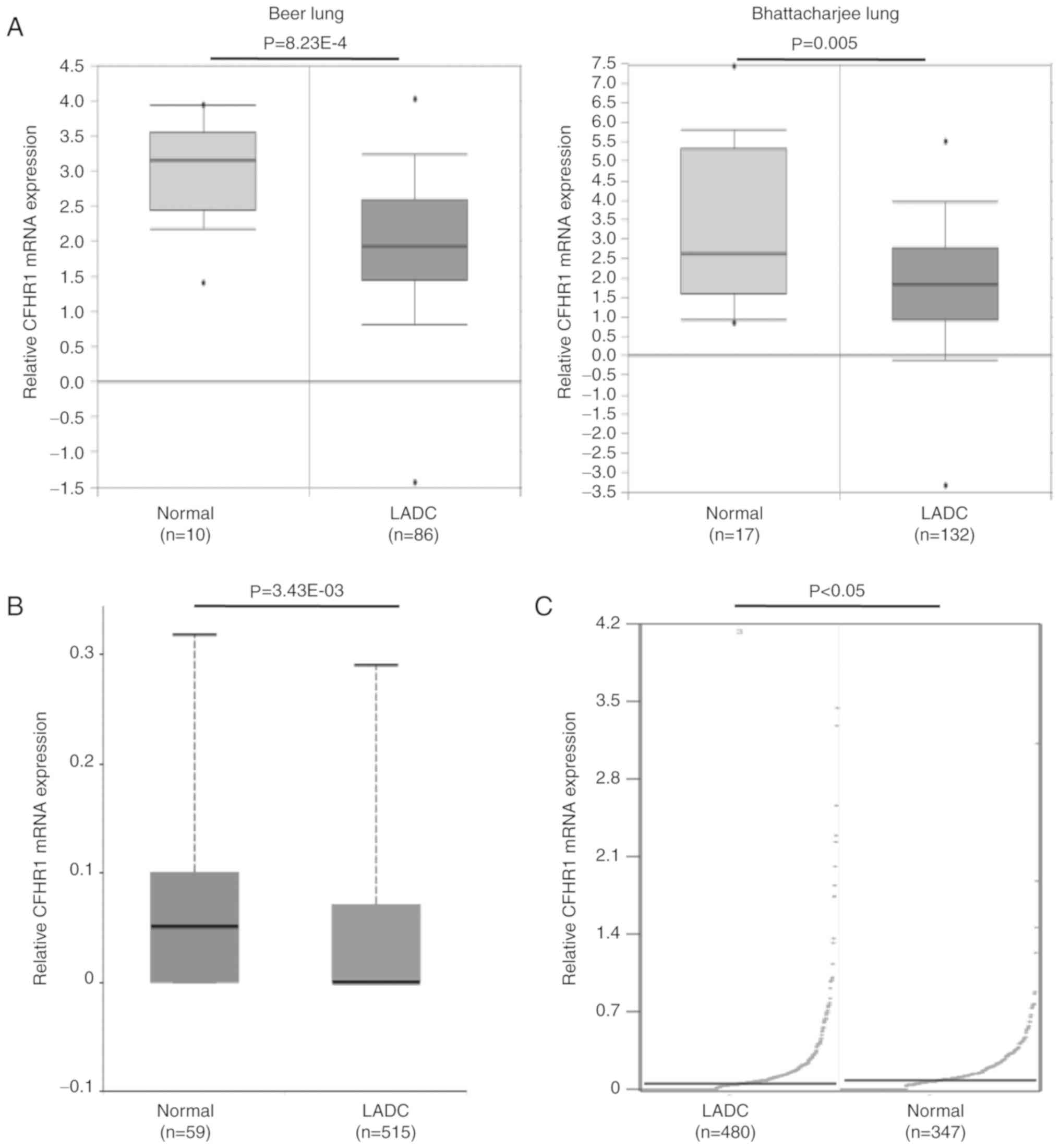
To detect changes in CFHR1 expression between
LADC and adjacent nontumor tissues, the expression profiles of
CFHR1 were analyzed using three independent bioinformatic
databases. First, as shown in Fig.
1A, it was found that CFHR1 transcription levels were
significantly reduced in tumor tissues based on two microarray
datasets from the Oncomine platform (25,26).
Furthermore, the downregulation of CFHR1 transcription was
confirmed in LADC tissues by using the UALCAN tool (Fig. 1B). Finally, to further confirm this
result, the expression of CFHR1 was re-analyzed in the GEPIA
database and the same above-mentioned trend was verified (Fig. 1C). This observation confirmed that
CFHR1 is downregulated in LADC tissues.
CFHR1 as a presumed prognostic factor
for adenocarcinoma
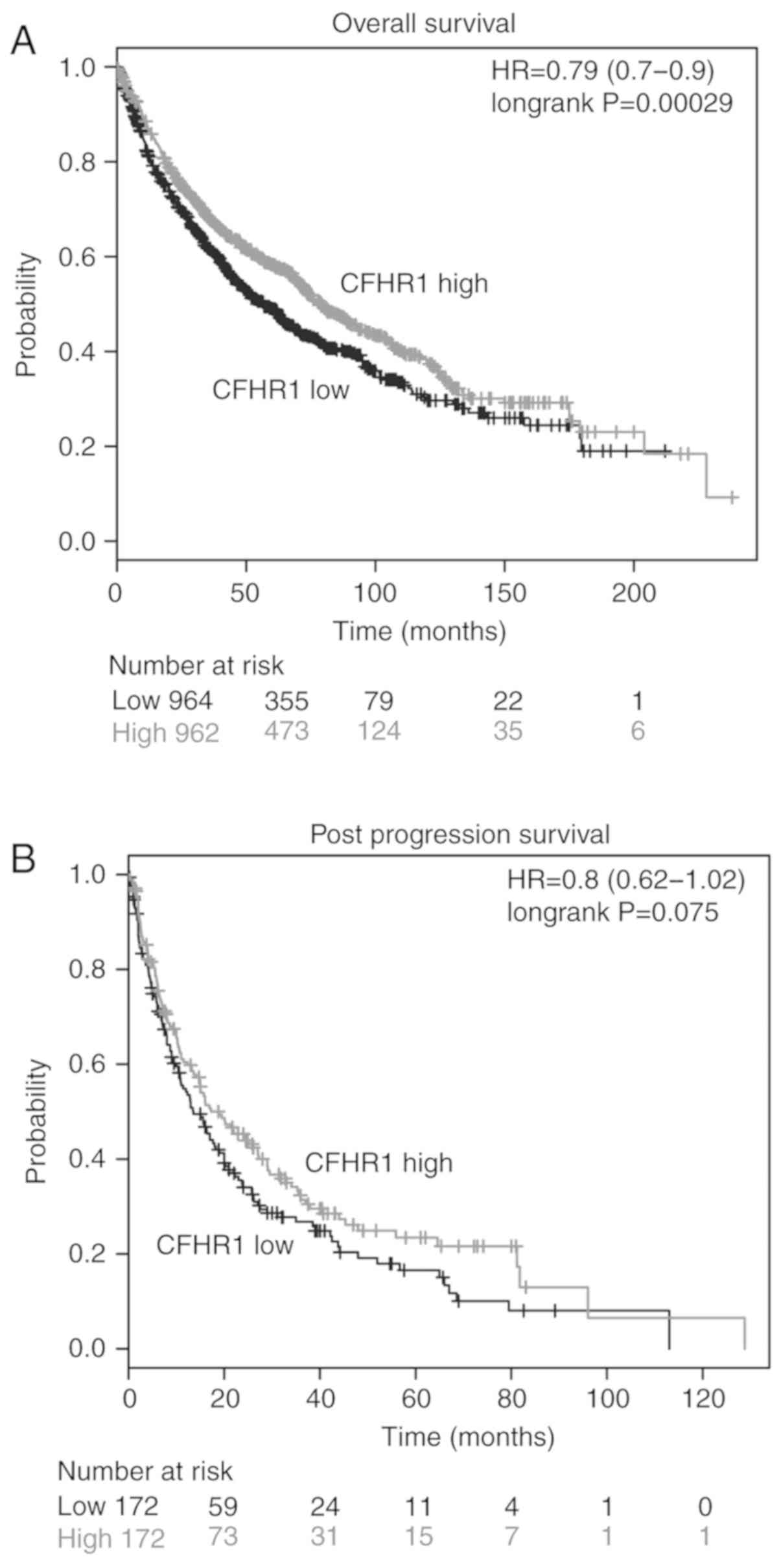
To date, almost no literature has reported the
relationship between the expression of CFHR1 and the
clinical prognosis of human LADC. Thus, we conducted a clinical
follow-up survey with the most commonly used monitoring indicators,
OS and PPS (17,27). By using the Kaplan-Meier plotter
platform, it was found that patients with downregulated
CFHR1 expression had a significantly shorter OS (P<0.01)
(Fig. 2A). Moreover, patients with
high levels of CFHR1 tended to have a longer PPS, although
this difference was not significant (P>0.05) (Fig. 2B). The reasons may be due to the
sample size, and future evaluations with larger datasets are
warranted. Furthermore, associations between CFHR1
expression and KRAS mutation or T stage were observed to be
statistically significant (P=0.013 and P=0.002, respectively)
(Table I). No correlation was
observed between CFHR1 expression and sex, age, race,
EGFR mutation, EML4-ALK translocation, lymph node
metastasis, distant metastasis, pathologic stage, smoking history
and Karnofsky performance score (Table
I). The multiple-factor analysis using the COX regression model
indicated that pathological T stage was independently associated
with CFHR1 transcription levels in LUAD samples (Table II). In summary, decreased
CFHR1 expression in patients with LADC is likely to be a
valuable prognostic factor.
 | Table I.Single factor clinical data analysis
related to CFHR1. |
Table I.
Single factor clinical data analysis
related to CFHR1.
| Source | No. | Mean ± SD | P-value |
|---|
| Sex |
|
| 0.350 |
|
Male | 207 | 0.959±1.47 |
|
|
Female | 248 | 0.830±1.46 |
|
| Kras_mutation |
|
| 0.013 |
| No | 34 | 1.22±1.84 |
|
|
Yes | 14 | 0.341±0.427 |
|
| EGFR_mutation |
|
| 0.301 |
| No | 171 | 0.842±1.18 |
|
|
Yes | 64 | 1.09±1.78 |
|
|
EML4_ALK_translocation |
|
| 0.773 |
| No | 183 | 0.888±1.38 |
|
|
Yes | 23 | 0.978±1.72 |
|
| Pathologic_T |
|
| 0.002 |
|
T1/T1a/T1b | 140 | 0.979±1.66 |
|
|
T2/T2a/T2b | 254 | 0.814±1.28 |
|
| T3 | 41 | 0.603±0.986 |
|
| T4 | 18 | 1.12±1.47 |
|
| TX | 2 | 7.69±0.868 |
|
| Race |
|
| 0.802 |
|
Caucasian | 355 | 0.907±1.43 |
|
|
Asian | 7 | 0.544±1.01 |
|
| Black
or African- | 25 | 0.882±1.63 |
|
|
American |
|
|
|
| Pathologic_N |
|
| 0.801 |
| N0 | 290 | 0.877±1.48 |
|
| N1 | 83 | 0.838±1.16 |
|
| N2 | 70 | 0.990±1.63 |
|
| N3 | 2 | 0 |
|
| NX | 9 | 1.22±2.23 |
|
| Pathologic_M |
|
| 0.560 |
| M0 | 311 | 0.886±1.50 |
|
|
M1/M1a/M1b | 22 | 0.936±1.61 |
|
| MX | 118 | 0.915±1.38 |
|
|
Pathologic_stage |
|
| 0.669 |
| Stage
I/IA/IB | 246 | 0.908±1.53 |
|
| Stage
IIA/IIB | 106 | 0.753±1.13 |
|
| Stage
IIIA/IIIB | 79 | 1.02±1.63 |
|
| Stage
IV | 23 | 0.895±1.58 |
|
| Karnofsky
performance score |
|
| 0.394 |
|
0–70 | 13 | 0.698±0.692 |
|
| 80 | 19 | 1.10±1.67 |
|
| 90 | 19 | 0.395±0.835 |
|
|
100 | 29 | 0.637±1.41 |
|
| Age (years) |
|
| 0.471 |
|
40–60 | 116 | 0.858±1.35 |
|
|
60–80 | 292 | 0.911±1.50 |
|
|
>80 | 27 | 0.562±0.637 |
|
| Table II.Clinical multivariate data related to
CFHR1. |
Table II.
Clinical multivariate data related to
CFHR1.
| Source | Type III sum of
squares | df | Mean square | F | P-value |
|---|
|
Kras_mutation_found |
4.627 | 1 | 4.627 | 2.513 | 0.120 |
| Pathologic_T | 36.867 | 4 | 9.217 | 5.005 | 0.002 |
Role of CFHR1 in the treatment of
adenocarcinoma
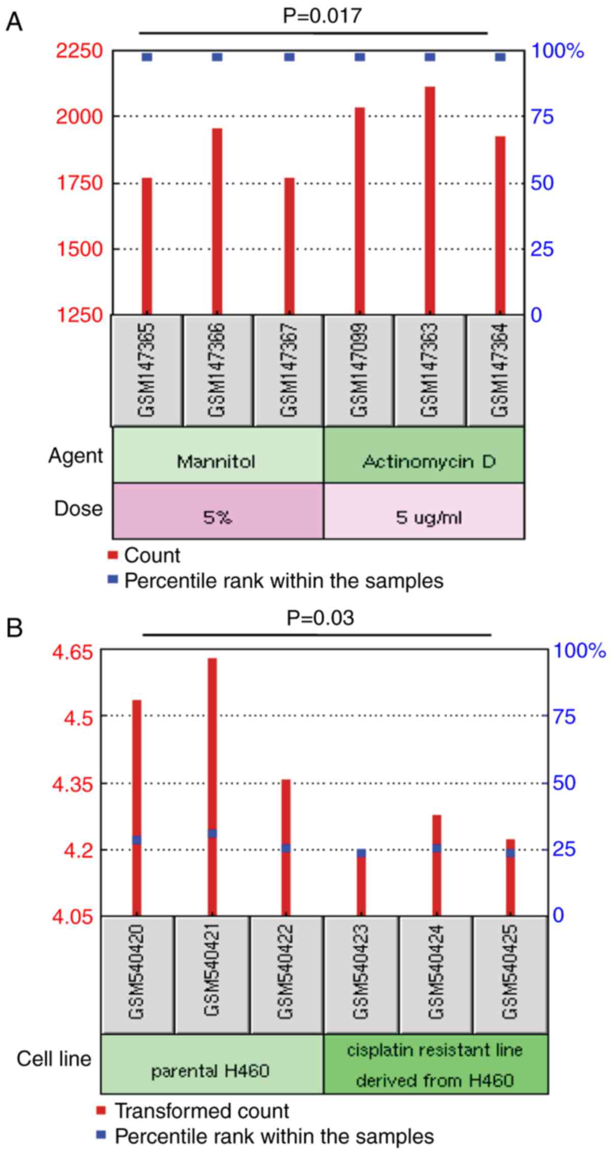
Next, from the GEO database, we screened two
microarray datasets related to chemotherapy to further determine
the effect of CFHR1 in the treatment of LADC patients. From
the data of GSE6400 (18), it was
found that treatment with the anticancer agent actinomycin D
obviously upregulated the expression of CFHR1, further
exerting this anti-proliferative activity in cultured A549 LADC
cells (P=0.017) (Fig. 3A).
Meanwhile, data from GSE21656 (19) indicated that the expression of
CFHR1 in a cisplatin-resistant LADC cell line (CDDP-R) was
significantly downregulated when compared with the parental cell
line H460 (P=0.03) (Fig. 3B).
These findings showed that changes in CFHR1 expression
levels may be involved in the therapeutic response to cancer.
Network analysis of coexpressed genes
of CFHR1
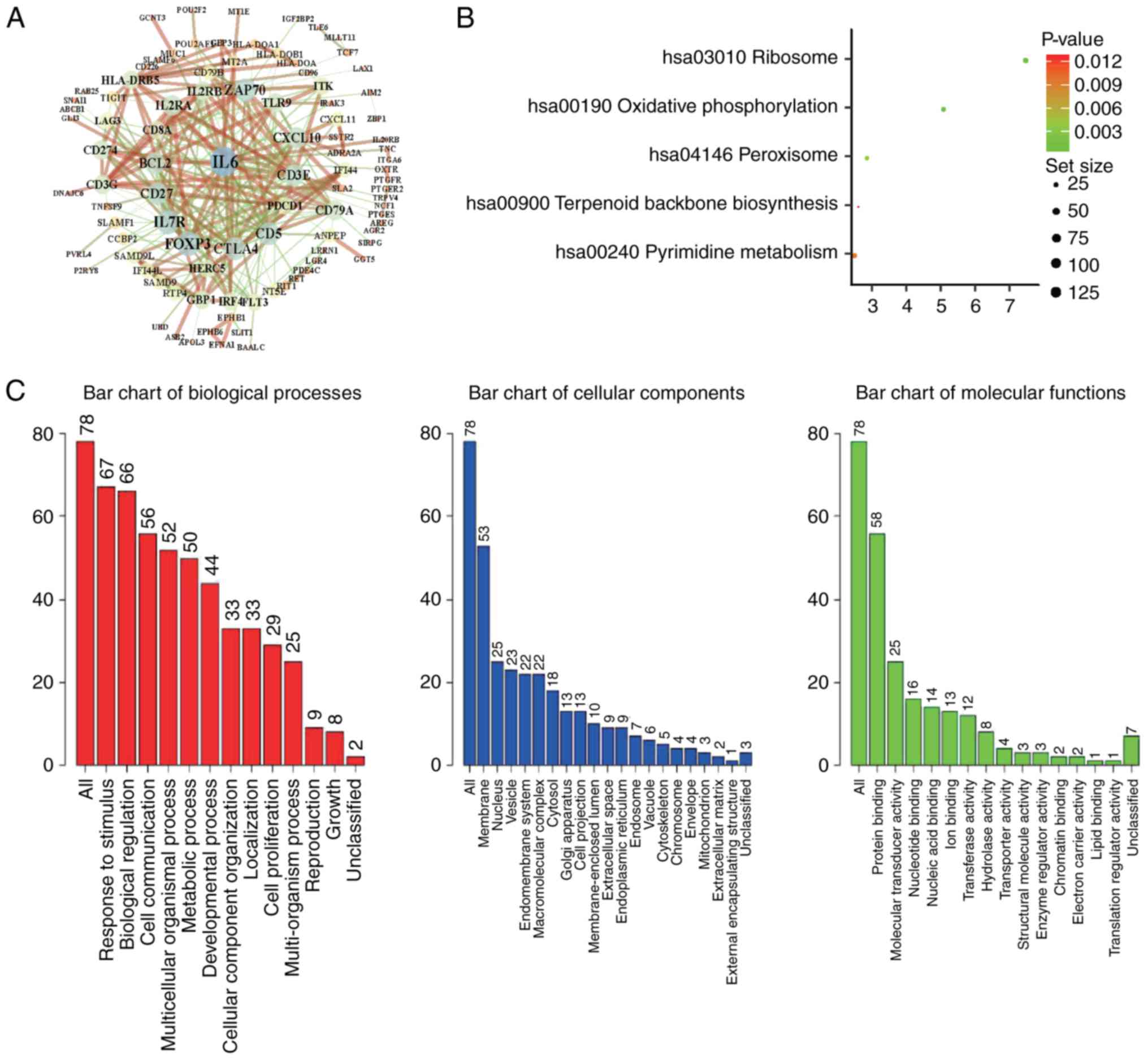
To further understand the biological function of
CFHR1, we performed functional enrichment annotation
analysis of its coexpressed genes. We downloaded the coexpressed
genes of CFHR1 from the cBioPortal database and screened 243
coexpressed genes with criteria of P≤0.05 and |LogFC| ≥0.7
(Table SII). Then, the PPI
network of 243 differentially coexpressed genes was performed by
two frequently used algorithms, STRING and Cytoscape (Fig. 4A). At the same time, the Pathview
database was used to analyze the KEGG pathways (Table SIII), and it was found that the
most significant pathway was ribosome (Fig. 4B). Finally, to further illuminate
the function among these 243 screened genes, WebGestalt was used to
conduct the GO annotations and to identify the main molecular
function (protein binding), biological process (response to
stimulus and biological regulation) and cellular component
(membrane) related to CFHR1 biology (Fig. 4C).
Discussion
The aim of the present study was to understand the
potential of human complement factor H-related protein 1
(CFHR1) in the development and treatment of lung
adenocarcinoma (LADC). The present study is the first to use
multiple public datasets to analyze the expression of CFHR1
in LADC tissues. At the same time, the coexpressed genes of
CFHR1 were analyzed and several possible signaling pathways
were identified that could determine its biological significance in
cancer development. Using the Oncomine, UALCAN and GEPIA datasets,
it was demonstrated that CFHR1 is significantly
downregulated in LADC tissues. Moreover, through statistical
analysis of clinical data from TCGA, it was found that the
expression level of CFHR1 was closely related to KRAS
mutation and pathological T stage in LADC patients.
CFHR1 is a complement modulator that regulates
complement by blocking complement C5 convertase activity and
interfering with C5b surface binding (28). In autoimmune atypical hemolytic
uremic syndrome (aHUS), CFH is blocked by FH autoantibodies, and
90% of patients carry homozygous deletions of CFHR1
(29). However, heterozygous
CFHR1/CFH hybrid genes were also identified in 4.5% of
patients with aHUS. These genomic rearrangements among CFH
and CFHR have been proven to be associated with a high risk
of posttransplant recurrence and poor clinical prognosis (30). In addition, Guo et al
(31) found that abnormally
expressed CFHR1 could act as a promising predictive
biomarker for cervical squamous cell carcinoma. Using an off-site
matrix-based electrochemistry platform, Arya and Estrela further
investigated CFHR1 as a bladder cancer protein marker (32). CFHR1 gene polymorphisms also
showed stronger associations with event-free survival in patients
with follicular lymphoma (33).
Although several studies have indicated the roles of CFHR1
in the pathological process of human diseases, including cancers,
no studies have revealed the functions of CFHR1 in LADC. In
the present study, we demonstrated that CFHR1 plays a
potential role in tumor inhibition in LADC samples. In addition, it
was also demonstrated that high expression of CFHR1 is
significantly associated with prolonged clinical OS and PPS in LADC
patients. This provides an idea for further comprehensive
exploration of the molecular mechanism of CFHR1 as a
promising therapeutic biomarker in LADC.
In the present study, the exact interaction between
CFHR1 and its coexpressed genes was not found; however, the
PPI that was constructed benefits the identification of the
function of CFHR1 to some extent. Jullien et al
(34) discovered that CFHR1
is connected with a decreased level of glomerular immune deposits.
Moreover, interleukin-6 (IL-6), located in the PPI network
(Fig. 4A), is considered as a
cytokine that essentially functions in immunoregulation via a
signal transducer and activator of transcription 3 (STAT3)
-dependent manner (35).
Therefore, a phenomenon may exist in which CFHR1 controls
the secretion of IL-6 to influence the immunoregulation of human
cancer cells. In addition, through the functional enrichment
annotation analysis, the main functional pathway of the coexpressed
genes of CFHR1 has been confirmed to be the ribosome
pathway. Previous research has demonstrated that the ribosome
signaling pathway is significantly related to the microenvironment
and metabolic changes of cancer cells (36,37).
However, according to the published literature, no relevant studies
have illuminated the detailed function and mechanism of
CFHR1 in the pathway modulation. Therefore, further studies
are needed to clarify the roles of CFHR1 in these KEGG
pathways.
Overall, our results suggest that CFHR1 is a
candidate tumor suppressor in human LADC disease. The public
database-based re-analysis methods also provide a novel research
strategy for screening potential biomarkers related to the
pathogenesis of malignant human diseases.
Supplementary Material
Supporting Data
Acknowledgements
Not applicable.
Funding
The study was supported by the Natural Science
Foundation of Hunan Province (grant no. 2019JJ50932), the National
Natural Science Foundation of China (grant nos. 81803035, 81703036
and 81572946), the China Postdoctoral Science Foundation (grant no.
2017M610510), the Open-End Fund for the Valuable and Precision
Instruments of Central South University (grant no. CSUZC201836),
the Youth Fund of Xiangya Hospital (grant no. 2017Q17) and
Postdoctoral Science Foundation of Central South University (grant
no. 185702).
Availability of data and materials
All data generated or analyzed during this study are
included in this published article.
Authors' contributions
GW, YY, JZ and ZX concieved and designed the study.
GW, XW, XR, XC, SZ, JW, LQ, XY, CO, WL and ZG acquired and
interpreted the data. GW, YY, JZ and ZX drafted the manuscript. YY
and ZX revised the manuscript. All authors read and approved the
final manuscript, and agree to be accountable for all aspects of
the research in ensuring that the accuracy or integrity of any part
of the work are appropriately investigated and resolved.
Ethics approval and consent to
participate
Not applicable.
Patient consent for publication
Not applicable.
Competing interests
The authors declare that they have no competing
interests.
Author information
Dr Zhijie Xu is now a Postdoctoral Fellow at the
Department of Pharmacy of Xiangya Hospital, Central South
University, Changsha, Hunan.
References
|
1
|
Hirsch FR, Scagliotti GV, Mulshine JL,
Kwon R, Curran WJ Jr, Wu YL and Paz-Ares L: Lung cancer: Current
therapies and new targeted treatments. Lancet. 389:299–311. 2017.
View Article : Google Scholar : PubMed/NCBI
|
|
2
|
Wei J, Yan Y, Chen X, Qian L, Zeng S, Li
Z, Dai S, Gong Z and Xu Z: The roles of plant-derived Triptolide on
non-small cell lung cancer. Oncol Res. 27:849–858. 2019. View Article : Google Scholar : PubMed/NCBI
|
|
3
|
Ulahannan D, Khalifa J, Faivre-Finn C and
Lee SM: Emerging treatment paradigms for brain metastasis in
non-small-cell lung cancer: An overview of the current landscape
and challenges ahead. Ann Oncol. 28:2923–2931. 2017. View Article : Google Scholar : PubMed/NCBI
|
|
4
|
Zhou S, Yan Y, Chen X, Wang X, Zeng S,
Qian L, Wei J, Yang X, Zhou Y, Gong Z and Xu Z: Roles of highly
expressed PAICS in lung adenocarcinoma. Gene. 692:1–8. 2019.
View Article : Google Scholar : PubMed/NCBI
|
|
5
|
Zhang R, Sun S, Ji F, Liu C, Lin H, Xie L,
Yang H, Tang W, Zhou Y, Xu J and Li P: CNTN-1 enhances
chemoresistance in human lung adenocarcinoma through induction of
epithelial-mesenchymal transition by targeting the PI3K/Akt
pathway. Cell Physiol Biochem. 43:465–480. 2017. View Article : Google Scholar : PubMed/NCBI
|
|
6
|
Yan Y, Su W, Zeng S, Qian L, Chen X, Wei
J, Chen N, Gong Z and Xu Z: Effect and mechanism of tanshinone i on
the radiosensitivity of lung cancer cells. Mol Pharm. 15:4843–4853.
2018. View Article : Google Scholar : PubMed/NCBI
|
|
7
|
Skerka C, Chen Q, Fremeaux-Bacchi V and
Roumenina LT: Complement factor H related proteins (CFHRs). Mol
Immunol. 56:170–180. 2013. View Article : Google Scholar : PubMed/NCBI
|
|
8
|
Rogers LM, Mott SL, Smith BJ, Link BK,
Sahin D and Weiner GJ: Complement-regulatory proteins CFHR1 and
CFHR3 and patient response to anti-CD20 monoclonal antibody
therapy. Clin Cancer Res. 23:954–961. 2017. View Article : Google Scholar : PubMed/NCBI
|
|
9
|
Hageman GS, Hancox LS, Taiber AJ, Gehrs
KM, Anderson DH, Johnson LV, Radeke MJ, Kavanagh D, Richards A,
Atkinson J, et al: Extended haplotypes in the complement factor H
(CFH) and CFH-related (CFHR) family of genes protect against
age-related macular degeneration: Characterization, ethnic
distribution and evolutionary implications. Ann Med. 38:592–604.
2006. View Article : Google Scholar :
|
|
10
|
Jullien P, Laurent B, Claisse G, Masson I,
Dinic M, Thibaudin D, Berthoux F, Alamartine E, Mariat C and
Maillard N: Deletion Variants of CFHR1 and CFHR3
associate with mesangial immune deposits but not with progression
of IgA nephropathy. J Am Soc Nephrol. 29:661–669. 2018. View Article : Google Scholar : PubMed/NCBI
|
|
11
|
Fratelli M, Bolis M, Kurosaki M, Dori M,
Guarnaccia V, Spinelli O, Alberti M, Valoti E, Pileggi S, Noris M,
et al: Association of CFHR1 homozygous deletion with acute
myelogenous leukemia in the European population. Leuk Lymphoma.
57:1234–1237. 2016. View Article : Google Scholar : PubMed/NCBI
|
|
12
|
Cui L, Fu J, Pang JC, Qiu ZK, Liu XM, Chen
FR, Shi HL, Ng HK and Chen ZP: Overexpression of IL-7 enhances
cisplatin resistance in glioma. Cancer Biol Ther. 13:496–503. 2012.
View Article : Google Scholar : PubMed/NCBI
|
|
13
|
Rhodes DR, Kalyana-Sundaram S, Mahavisno
V, Varambally R, Yu J, Briggs BB, Barrette TR, Anstet MJ,
Kincead-Beal C, Kulkarni P, et al: Oncomine 3.0: Genes, pathways
and networks in a collection of 18,000 cancer gene expression
profiles. Neoplasia. 9:166–180. 2007. View Article : Google Scholar : PubMed/NCBI
|
|
14
|
Chandrashekar DS, Bashel B, Balasubramanya
SAH, Creighton CJ, Ponce-Rodriguez I, Chakravarthi BVSK and
Varambally S: UALCAN: A portal for facilitating tumor subgroup gene
expression and survival analyses. Neoplasia. 19:649–658. 2017.
View Article : Google Scholar : PubMed/NCBI
|
|
15
|
Tang Z, Li C, Kang B, Gao G, Li C and
Zhang Z: GEPIA: A web server for cancer and normal gene expression
profiling and interactive analyses. Nucleic Acids Res. 45:W98–W102.
2017. View Article : Google Scholar : PubMed/NCBI
|
|
16
|
Lánczky A, Nagy Á, Bottai G, Munkácsy G,
Szabó A, Santarpia L and Győrffy B: miRpower: A web-tool to
validate survival-associated miRNAs utilizing expression data from
2178 breast cancer patients. Breast Cancer Res Treat. 160:439–446.
2016. View Article : Google Scholar : PubMed/NCBI
|
|
17
|
Yan Y, Xu Z, Hu X, Qian L, Li Z, Zhou Y,
Dai S, Zeng S and Gong Z: SNCA is a functionally low-expressed gene
in lung adenocarcinoma. Genes (Basel). 9:E162018. View Article : Google Scholar : PubMed/NCBI
|
|
18
|
Wang Z, Lecane PS, Thiemann P, Fan Q,
Cortez C, Ma X, Tonev D, Miles D, Naumovski L, Miller RA, et al:
Synthesis and biologic properties of hydrophilic sapphyrins, a new
class of tumor-selective inhibitors of gene expression. Mol Cancer.
6:92007. View Article : Google Scholar : PubMed/NCBI
|
|
19
|
Sun Y, Zheng S, Torossian A, Speirs CK,
Schleicher S, Giacalone NJ, Carbone DP, Zhao Z and Lu B: Role of
insulin-like growth factor-1 signaling pathway in
cisplatin-resistant lung cancer cells. Int J Radiat Oncol Biol
Phys. 82:e563–e572. 2012. View Article : Google Scholar : PubMed/NCBI
|
|
20
|
Gao J, Aksoy BA, Dogrusoz U, Dresdner G,
Gross B, Sumer SO, Sun Y, Jacobsen A, Sinha R, Larsson E, et al:
Integrative analysis of complex cancer genomics and clinical
profiles using the cBioPortal. Sci Signal. 6:pl12013. View Article : Google Scholar : PubMed/NCBI
|
|
21
|
Szklarczyk D, Morris JH, Cook H, Kuhn M,
Wyder S, Simonovic M, Santos A, Doncheva NT, Roth A, Bork P, et al:
The STRING database in 2017: Quality-controlled protein-protein
association networks, made broadly accessible. Nucleic Acids Res.
45:D362–D368. 2017. View Article : Google Scholar : PubMed/NCBI
|
|
22
|
Su G, Morris JH, Demchak B and Bader GD:
Biological network exploration with Cytoscape 3. Curr Protoc
Bioinformatics. 47:8.13.1–24. 2014. View Article : Google Scholar
|
|
23
|
Wang J, Vasaikar S, Shi Z, Greer M and
Zhang B: WebGestalt 2017: A more comprehensive, powerful, flexible
and interactive gene set enrichment analysis toolkit. Nucleic Acids
Res. 45:W130–W137. 2017. View Article : Google Scholar : PubMed/NCBI
|
|
24
|
Luo W, Pant G, Bhavnasi YK, Blanchard SG
Jr and Brouwer C: Pathview Web: User friendly pathway visualization
and data integration. Nucleic Acids Res. 45:W501–W508. 2017.
View Article : Google Scholar : PubMed/NCBI
|
|
25
|
Bhattacharjee A, Richards WG, Staunton J,
Li C, Monti S, Vasa P, Ladd C, Beheshti J, Bueno R, Gillette M, et
al: Classification of human lung carcinomas by mRNA expression
profiling reveals distinct adenocarcinoma subclasses. Proc Natl
Acad Sci USA. 98:13790–13795. 2001. View Article : Google Scholar : PubMed/NCBI
|
|
26
|
Beer DG, Kardia SL, Huang CC, Giordano TJ,
Levin AM, Misek DE, Lin L, Chen G, Gharib TG, Thomas DG, et al:
Gene-expression profiles predict survival of patients with lung
adenocarcinoma. Nat Med. 8:816–824. 2002. View Article : Google Scholar : PubMed/NCBI
|
|
27
|
Zhou H, Vallieres M, Bai HX, Su C, Tang H,
Oldridge D, Zhang Z, Xiao B, Liao W, Tao Y, et al: MRI features
predict survival and molecular markers in diffuse lower-grade
gliomas. Neuro Oncol. 19:862–870. 2017. View Article : Google Scholar : PubMed/NCBI
|
|
28
|
Hannan JP, Laskowski J, Thurman JM,
Hageman GS and Holers VM: Mapping the complement factor H-related
protein 1 (CFHR1):C3b/C3d interactions. PLoS One. 11:e01662002016.
View Article : Google Scholar : PubMed/NCBI
|
|
29
|
Trojnar E, Józsi M, Uray K, Csuka D,
Szilágyi Á, Milosevic D, Stojanović VD, Spasojević B, Rusai K,
Müller T, et al: Analysis of linear antibody epitopes on factor H
and CFHR1 using sera of patients with autoimmune atypical hemolytic
uremic syndrome. Front Immunol. 8:3022017. View Article : Google Scholar : PubMed/NCBI
|
|
30
|
Valoti E, Alberti M, Tortajada A,
Garcia-Fernandez J, Gastoldi S, Besso L, Bresin E, Remuzzi G,
Rodriguez de Cordoba S and Noris M: A novel atypical hemolytic
uremic syndrome-associated hybrid CFHR1/CFH gene encoding a fusion
protein that antagonizes factor H-dependent complement regulation.
J Am Soc Nephrol. 26:209–219. 2015. View Article : Google Scholar : PubMed/NCBI
|
|
31
|
Guo X, Hao Y, Kamilijiang M, Hasimu A,
Yuan J, Wu G, Reyimu H, Kadeer N and Abudula A: Potential
predictive plasma biomarkers for cervical cancer by 2D-DIGE
proteomics and ingenuity pathway analysis. Tumour Biol.
36:1711–1720. 2015. View Article : Google Scholar : PubMed/NCBI
|
|
32
|
Arya SK and Estrela P: Electrochemical
ELISA-based platform for bladder cancer protein biomarker detection
in urine. Biosens Bioelectron. 117:620–627. 2018. View Article : Google Scholar : PubMed/NCBI
|
|
33
|
Charbonneau B, Maurer MJ, Fredericksen ZS,
Zent CS, Link BK, Novak AJ, Ansell SM, Weiner GJ, Wang AH, Witzig
TE, et al: Germline variation in complement genes and event-free
survival in follicular and diffuse large B-cell lymphoma. Am J
Hematol. 87:880–885. 2012. View Article : Google Scholar : PubMed/NCBI
|
|
34
|
Jullien P, Laurent B, Claisse G, Masson I,
Dinic M, Thibaudin D, Berthoux F, Alamartine E, Mariat C and
Maillard N: Deletion variants of CFHR1 and CFHR3 associate with
mesangial immune deposits but not with progression of IgA
nephropathy. J Am Soc Nephrol. 29:661–669. 2018. View Article : Google Scholar : PubMed/NCBI
|
|
35
|
Lamano JB, Lamano JB, Li YD, DiDomenico
JD, Choy W, Veliceasa D, Oyon DE, Fakurnejad S, Ampie L,
Kesavabhotla K, et al: Glioblastoma-derived IL6 induces
immunosuppressive peripheral myeloid cell PD-L1 and promotes tumor
growth. Clin Cancer Res. 25:3643–3657. 2019. View Article : Google Scholar : PubMed/NCBI
|
|
36
|
Bustelo XR and Dosil M: Ribosome
biogenesis and cancer: basic and translational challenges. Curr
Opin Genet Dev. 48:22–29. 2018. View Article : Google Scholar : PubMed/NCBI
|
|
37
|
de la Cruz J, Gómez-Herreros F,
Rodríguez-Galán O, Begley V, de la Cruz Muñoz-Centeno M and Chávez
S: Feedback regulation of ribosome assembly. Curr Genet.
64:393–404. 2018. View Article : Google Scholar : PubMed/NCBI
|