Introduction
Cataract is defined as the formation of a dense and
cloudy area in the crystalline lens of the eye (1). Visual impairment due to cataract
affects ~40,000,000 of individuals worldwide and has become a major
cause of blindness globally (2).
Conventional phacoemulsification (CPCS) has been considered as the
most prevalent and effective surgical procedure for cataract in
past decades (3). However, while
this method has been widely used, more than 20 million patients
experience blindness due to bilateral cataracts, especially in
developing countries (4). In 2001,
for the first time, femtosecond laser technology was introduced in
clinical practice, and it was used for flap creation in laser
in-site keratomileusis (5). Since
this introduction, femtosecond laser technology has been expanded
to variety of clinical applications, including cataract surgery
(6). Femtosecond laser-assisted
cataract surgery (FLACS) is more reliable compared with CPCS
(7). For instance, more circular
and centered capsulorhexis can be created by FLACS to reduce the
intraocular lens (IOL) tilt, as well as decentration during IOL
implantation (8). FLACS also
allows for more effective control of the post-operative astigmatism
via the creation of improved quality corneal incisions (9). Moreover, femtosecond laser-assisted
pre-fragmentation of the crystalline lens can improve the
phacoemulsification power and time consumption during the surgery
(10). However, multiple
complications such as inflammation and miosis have been reported in
several patients with cataract treated by FLACS (11).
Clinically, both glucocorticoids (GCs) and
non-steroidal anti-inflammatory drugs (NSAIDs) are used frequently
due to their well-known anti-inflammatory effects (12). As these agents have different
mechanisms of action, combination therapy with GCs and NSAIDS may
provide extra benefits; this method has been routinely used for
patients undergoing cataract surgery (13). Bromfenac sodium (BS), a potent
NSAIDs, has been observed minimal adverse events in number studies,
although it was found the risk of corneal compromise when use of BS
to preexisting corneal disease (14). The present study evaluated the
curative effect of BS after cataract surgery with the replacement
of GCs, and the mechanisms of action by treatment of the patients
with BS were clarified.
Materials and methods
Clinical donors
A total of 90 patients (men, 45; women, 45; age,
50–89; mean age, 67.74±9.34 years) from The Fourth Affiliated
Hospital of China Medical University (Shenyang, China) between
August 2014 and January 2015 were enrolled in the present study.
All the experiments were approved by the Ethics Committee of The
Fourth Affiliated Hospital of China Medical University (approval
no. ChiCTR-TRC-14005114) and informed consent was signed by every
study participant.
The patient population did not include corneal
diseases, intraoperative complications and other types of eye
diseases, such as inflammation, uveitis and glaucoma, or any other
pathologies. Patients receiving systemic or topical
anti-inflammatory therapy within 1 month before surgery were also
excluded. The study participants were randomly divided into five
groups: Group I, CPCS + 0.1% dexamethasone treatment (DEX; Nitto
Medic Co., Ltd.)/0.3% tobramycin (TOB; Nitto Medic Co., Ltd.);
group II, CPCS + 0.1% BS treatment (Senju Pharmaceutical Co.,
Ltd.); group III, FLACS + 0.1% DEX/0.3% TOB; group IV, FLACS + 0.1%
BS; and group V, FLACS + 0.1% pranoprofen (Senju Pharmaceutical
Co., Ltd.). Each of the study groups (I–V) consisted of a total of
22, 22, 19, 24 and 23 patients, respectively. No significant
differences in the surgical data of these groups such as age, sex,
eye type and nucleus degree of the lens (based on the Emery-Little
classification) (15) were
observed. Additional clinical information for these patients is
presented in Table I.
 | Table I.Clinical information of the
patients. |
Table I.
Clinical information of the
patients.
| Parameter | Group I (n=22) | Group II
(n=22) | Group III
(n=19) | Group IV
(n=24) | Group V (n=23) | P-value |
|---|
| Age (years) | 70.5±9.70 | 65.79±10.47 | 65.79±8.21 | 65.36±8.29 | 65.96±7.19 | 0.181 |
| Sex
(male/female) | 10/12 | 13/9 | 11/8 | 10/14 |
9/14 | 0.577 |
| Eye
(left/right) |
8/14 |
7/15 |
8/11 | 12/12 | 11/12 | 0.196 |
| Cataract nuclear
grading (Level II) | 0 | 0 | 1 | 0 | 0 | – |
| Cataract nuclear
grading (Level III) | 22 | 21 | 17 | 24 | 23 | 0.279 |
| Cataract nuclear
grading (Level IV) | 0 | 1 | 1 | 0 | – | – |
| Preoperative
CDVA | 0.65±0.30 | 0.64±0.25 | 0.83±0.40 | 0.75±0.70 | 0.73±0.61 | 0.353 |
Surgical treatment
A total of ~150 µl of aqueous humor was collected
from patients undergoing routine and FLACS. The specimens were
collected at the beginning of the surgery and stored at −80°C until
used. Cataract surgery was performed by one surgeon and an
assistant. Before the surgery, the pupils were dilated using a
combination of 0.5% tropicamide (Mydrin-P; Santen Pharmaceutical
Co., Ltd.) with 5% phenylephrine hydrochloride eyedrops
(Neosynesin, Kowa Co., Ltd.), followed by anesthesia and washing
using 4% lidocaine hydrochloride eye drops (Xylocanine; AstraZeneca
Co., Ltd.). Patients in groups I and II were treated using a
standard CPCS protocol (16). In
brief, a 3.0-mm-wide clear corneal incision was made, followed by
an insertion into the clear cornea to create a self-sealing
incision. Subsequently, the foldable acrylic IOL (AcrySof IQ Toric;
Alcon, Inc.) was implanted in the capsular bag.
Surgery in patients of the other three groups
(III–V) was performed using FLACS with a LenSX Laser system
(AcrySof IQ Toric; Alcon, Inc.) and INFINTI Vision System (AcrySof
IQ Toric; Alcon, Inc.). In the case of capsulotomy, the diameter
was set at 5.0 mm and the programmed pulse energy was set at 6 µJ
with an incision depth of 300 µm. The lens was cut into quadrants
and the distance of dot/layer and pulse energy were set to 10/10 µm
and 12 µJ, respectively. The corneal primary incision was a
3.0-mm-wide triplanar incision (the distance of dot/layer, 6/6 µm)
with an anterior side cut angle of 70° and a posterior side cut
angle of 15°. The corneal auxiliary incision was a 1.0-mm-wide
incision (the distance of dot/layer, 5/5 µm) with 6 µJ of pulse
energy. Suction time was recorded by LenSX laser system. A total of
150 µl of aqueous flare was collected from the blunt opened clear
corneal primary incision under sterile conditions after the laser
treatment and immediately stored at −80°C for further analysis. A
standard CPCS was used for removing the broken crystalline lenses,
followed by implantation of the foldable acrylic IOL (AcrySof IQ
Toric; Alcon, Inc.). Then, the anterior chamber was closed by ~2 ml
of saline injection.
Perioperative period treatment
Patients in groups II and IV were treated with 0.1%
BS eye drops ≥1 h before the surgery. Post-operatively these
subjects were treated with 0.1% BS eye drops twice daily for 2
months. Patients in these two groups also received 0.5%
levofloxacin eye drops (Santen Pharmaceutical Co., Ltd.) four times
per day for 1 month post-operatively to protect from inflammation.
Patients from groups I and III were treated with 0.1% DEX/0.3% TOB
eye drops four times daily for 1 month after surgery followed by
replacement of 0.1% fluorometholone eye drops (Santen
Pharmaceutical Co., Ltd.) four times daily for 1 month. Subjects in
group V were treated with 0.1% pranoprofen eye drops (Senju
Pharmaceutical Co., Ltd.) four times per day, which was started 3
days before the surgery and continued up to 2 months post-surgery.
Moreover, patients in all the study groups received 0.5%
tropicamide phenylephrine eye drops (Santen Pharmaceutical Co.,
Ltd.) twice daily for 1 week after the surgery.
Clinical outcomes
Corrected distance visual acuity (CDVA) was measured
1 month before and after the surgery (17). In brief, position the patient at a
distance of 6 meters from the chart after ensure the illumination
on the testing chart. Ask the patient to read from the top to
bottom and from the left to right of the chart. Then, record the
visual acuity for each eye separately. The conversion of decimal
acuity values to logMAR was used for the calculation of CDVA.
Aqueous flare, intraocular pressure (IOP) and corneal thickness
measurements were performed postoperatively at 1, 7, 30, 45 and 60
days using FM-600 Laser Flare Meters (KOWA Company, Ltd.), NT-530P
Non-Contact Tonometer (Nidek Co. Ltd.) and Pentacam (Oculus GmbH),
respectively. Macular thickness was measured using the Spectralis
OCT BluePeak module (Heidelberg Engineering, Inc.). The equipment
was set as a 3D scan model (Frequency, 40,000 times/sec; Depth, 2.0
mm; Range, 8.8×8.8 mm2) and data analysis was
automatically performed using HEYEX 2 image management software
system (Heidelberg Engineering, Inc.). The severity of the pain was
scored from 0 to 4, where 0=no pain/best outcome; 1=mild pain/no
need for drug intervention/does not affect normal life; 2=moderate
pain/affects normal life; 3=severe pain/unable to live a normal
life/appropriate to drug intervention and 4=worst outcome/most
pain, and was based on the short form of the Brief Pain Inventory
(BPI) (18). Pupil diameter
measurement was performed by the same doctor under normal room
light illumination. The pupil diameters of patients were measured
the using a ruler in the horizontal and vertical directions. The
measurement was repeated three times and the average was taken. The
pupil reduction ratio after the surgery was calculated as: (Pupil
diameter before surgery-pupil diameter after surgery)/pupil
diameter before surgery ×100. To measure the size of macular
thickness, we reconstruct a surface map as a false-color
topographic image and divided into 9 map sectors as shown in below.
The central subfield macular thickness (CSMT) was defined as the
average of the mean thickness within the central 1,000 µm ring. The
inner macular ring and the outer macular ring were separated into
four quadrants with the diameters of 3,000 and 6,000 µm,
respectively. The macular thickness was calculated as the mean
standard ± deviation in these total 9 regions.
Cell culture and UV B treatment
SRA01/04 (cat. no. RCB1591; RIKEN), a human lens
epithelium-derived cell line, was cultured in DMEM (FUJIFILM Wako
Pure Chemical) supplemented with 20% heat-inactivated FBS (MP
Biomedicals, LLC) and 1% penicillin-streptomycin (Nacalai Tesque,
Inc.) in a 5% CO2 humidified incubator at 37°C. Cells
were seeded in 96-well plate at a density of 5×10⁵
cells/ml 1 day before the experiment, which was followed by
overnight incubation at 37°C. Cells were washed twice with
pre-warmed PBS and treated with UV irradiation at room temperature
for 30 sec using a CL-1000M UV lamp (Thermo Fisher Scientific,
Inc.). Most of the resulting wavelengths were in UVB range 250–365
nm. The used UVB energy sources were at 0, 20, 40, 60 and 80
mJ/cm2. SRA01/04 cells were irradiated for 30 sec in the
absence or presence of different concentrations (0–80 µg/ml) of BS
at 37°C. PBS was replaced with fresh DMEM post-UV irradiation.
MTT assay
Viability of SRA01/04 cells was determined using a
MTT assay based on mitochondrial reduction of MTT to formazan.
Cultured medium was replaced with 200 µl MTT-containing fresh
medium after the treatment. MTT (FUJIFILM Wako Pure Chemical)
solution (7.5 mg/ml) was added into each well (20 µl/well) followed
by incubation at 37°C for 1.5 h. Then, 150 µl culture supernatant
was removed from each well and the formazan crystal was lysed by
adding 100 µl MTT stop solution (0.4% HCl, 10% Triton X-100 in
Isopropanol). After 12-h incubation at 37°C, the absorbance was
measured at 570 nm, with 655 nm as reference wavelength, on a
microplate reader (Bio-Rad Laboratories, Inc.).
ELISA
The Prostaglandin E2 (PGE2)
production in the aqueous flare and culture supernatants after UV
exposure were measured using a PGE2 Expression ELISA kit
(cat. no. 500141, Cayman Chemical Company) as per the
manufacturer's instruction. Human interleukin IL-1β/IL-1F2 DuoSet
ELISA kit (cat. no. DY20105, R&D Systems, Inc.) and
Cytotoxicity lactate dehydrogenase (LDH) Assay kit (cat. no. CK12,
Dojindo Molecular Technologies, Inc.) were used to detect the
release of IL-1β and LDH, respectively, according to the
manufacturer's instructions.
Reverse transcription-quantitative PCR
(RT-qPCR)
Total RNA from SRA01/04 cells was isolated using
TRIzol® reagent (Invitrogen; Thermo Fisher Scientific,
Inc.) followed by the reverse transcription of extracted RNA at
37°C for 1 h and inactivation of the reaction at 95°C for 5 min
using a miScript II RT kit (QIAGEN GmbH) in a fluorescence thermal
cycler (Bio-Rad Laboratories, Inc.). In brief, the qPCR
amplification conditions consisted of pre-denaturation for 3 min at
94°C, followed by a total of 30 cycles of denaturation for 30 sec
at 94°C, annealing at 58°C for 30 sec and extension for 60 sec at
72°C. The expression of related genes was measured using a SYBR
Green PCR reagent kit (Applied Biosystems; Thermo Fisher
Scientific, Inc.) with the following primer sets on an ABIViiA7 RT
PCR system (Thermo Fisher Scientific, Inc., Waltham, MA, USA). The
relative expression of target genes was normalized to β-actin.
Expression of β-actin was used as internal control for the analysis
of other genes while the 2−ΔΔCq method was used for data
analysis (19). The primers
sequences were as following: COX-1 forward,
5′-CTTTTCACCGTAGGTGGCCT-3′ and reverse, 5′-AGTGGAAGTGGGCTACAACG-3′;
COX-2 forward, 5′-ACCGTCTGAACTATCCTGCC-3′ and reverse,
5′-AGATTAGTCCGCCGTAGTCG-3′; and β-actin forward,
5′-GTGGGGCGCCCCAGGCACCA-3′ and reverse,
5′-CTCCTTAATGTCACGCACGATTTC-3′.
Western blotting
SRA01/04 cells were homogenized in RIPA lysis and
extraction buffer (Thermo Fisher Scientific, Inc.). Bicinchoninic
acid Protein assay kit (FUJIFILM Wako Pure Chemical) was used for
determining protein concentrations. In total, 20 µg each sample
were separated by 15% SDS-PAGE and transferred to PVDF membrane
(Immobilon-P; EMD Millipore). Then, 5% skim milk was used to
blocked membranes for 1 h at room temperature, and membranes were
incubated with primary antibodies against cleaved human caspase-1
p20 (cat. no. AG-208-0042-C100; AdipoGen), COX-1 (cat. no. 4841;
Cell Signaling Technology, Inc.) and COX-2 (cat. no. 12282; Cell
Signaling Technology, Inc.; all 1:1,000 dilutions) overnight at
4°C. GAPDH (cat. no. 5174; Cell Signaling Technology, Inc.;
1:1,000) was used as an internal control. The secondary horseradish
peroxide-conjugated anti-rabbit IgG antibody (Cell Signaling
Technology, Inc.; cat. no. 5127; 1:5,000) was incubated at room
temperature for 1 h. Protein band intensity was analyzed using
Luminata Forte Western horseradish peroxidase Substrate (EMD
Millipore) with a Bio-Rad ChemiDox XRS+ imaging system
and Image Lab Software Version 6.0.1 (Bio-Rad Laboratories,
Inc.).
Statistical analysis
All experiments were conducted with at least three
independent replicates. Data are presented as the mean ± standard
deviation using GraphPad Prism 5.0 (GraphPad Software, Inc.). The
comparisons of paired samples (preoperative vs. postoperative
measurements of the same patient) in Figs. 1 and 2 were assessed using a repeated measures
ANOVA, which was used to analyse matched samples. Differences among
multiple groups were assessed with one-way ANOVA followed by
Bonferroni post hoc test or Scheffe's multiple comparison test
(SPSS 22.0; IBM Corp.), which was used to analyse unpaired samples.
The relations between categorical variables in Tables I and II were analyzed with the χ2
test. In addition, Fisher's exact test was used to detect
differences in levels of cataract between the groups. P<0.05 was
considered to indicate a statistically significant difference.
| Table II.Clinical patient data. |
Table II.
Clinical patient data.
|
|
|
|
|
|
|
|
|
| Intraocular
pressure (mmHg) |
|---|
|
|
|
|
|
|
|
|
|
|
|
|---|
| Group | n | Pupil diameter
before FLACS (mm) | Pupil diameter
before CPCS (mm) | Pupil reduction
ratio | Concentration of
PGE2 in aqueous humor (pg/ml) | CDE (%) | Femtosecond laser
action time ± SD (sec) | Suction time ± SD
(sec) | Before FLACS | After FLACS |
|---|
| I | 22 | – | 7.78±0.76 | – | 12.05±4.67 | 12.95±4.25 | – | – | – | – |
| II | 22 | – | 8.21±0.50 | – |
13.16±3.85ns |
11.89±4.43ns | – | – | – | – |
| III | 19 | 7.64±0.69 | 6.83±1.17 | 9/10 |
51.84±6.67a |
9.42±4.49a | 35.16±5.71 | 206.8±92.79 | 16.66±3.96 | 17.56±8.94 |
| IV | 24 | 7.68±0.56 | 7.45±0.91 | 4/20 |
18.41±4.45a |
7.44±4.73a | 34.5±6.19 | 190.3±85.68 | 16.12±4.12 | 18.44±6.60 |
| V | 23 | 7.72±0.61 | 7.36±0.83 | 5/19 |
18.54±4.32a |
7.42±4.56a | 34.71±6.12 | 193.3±86.13 | 16.31±3.94 | 18.12±6.47 |
| P-value |
| 0.86 | 0.042 | 0.046 | P<0.05 | P<0.05 | 0.722 | 0.549 | 0.666 | 0.711 |
Results
Beneficial effects of BS on the
perioperative period of cataract surgery
No statistically significant differences in the
clinical outcomes including CDVA, aqueous flare, IOP, central
corneal thickness (CCT) and macular morphology (Table I) among the patients were
identified before the surgery. Based on the data in Table II, it was found that the pupil
diameter was increased by BS treatment (group II) before the CPCS,
as compared with NSAID treatment (group I). However, the pupil
diameter was significantly reduced by DEX/TOB treatment when the
patients were receiving FLACS (group III). More important, BS
treatment strongly improved pupil reduction, as group IV indicated.
It was also observed similar protective effect of pranoprofen on
pupil reduction as BS. Additionally, cumulative dissipated energy
(CDE) in the patients who received FLACS was significantly reduced
compared with the patients who received CPCS (Table II), indicating that energy
delivered induced less damage in the FLACS group compared with the
CPCS group. In addition, no surgical complications in any of the
study subjects after the surgery were demonstrated.
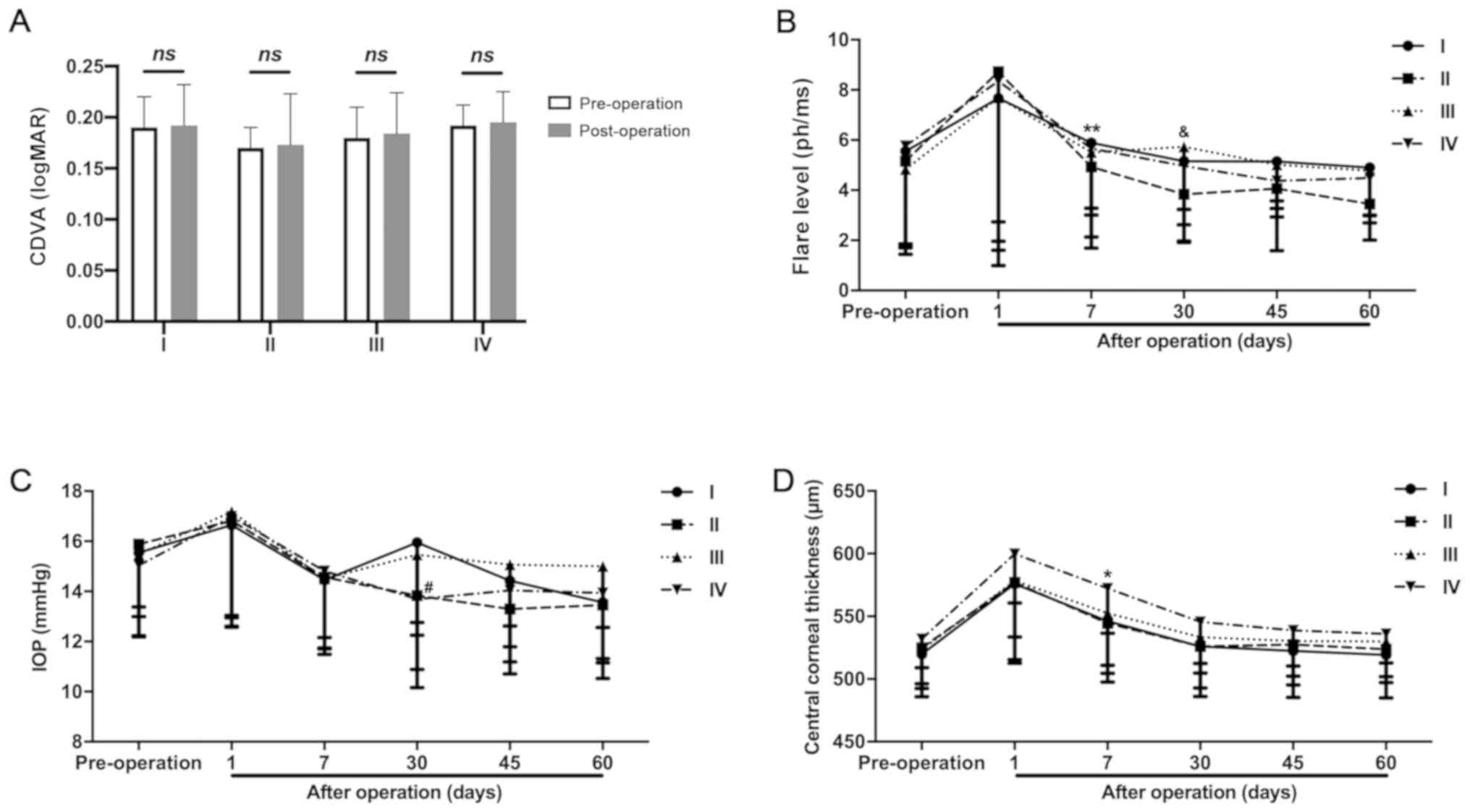
Compared with the results of the patients visual
acuity before surgery, no significant difference was observed in
the visual acuity in patients of each group 1-week post-surgery,
suggesting the surgical procedure was successful (Fig. 1A). However, aqueous flare was
observed in all the groups 1 day after the surgery (Fig. 1B). Moreover, reduction of the
aqueous flare was observed in the BS-treated groups but not in the
NSAID-treated groups after 7 days of treatment. It was also found
that the aqueous flare was significantly inhibited by BS compared
with NSAIDs after 30 days of treatment. Furthermore, significantly
higher IOP was observed in GC-treated groups 30 days after surgery
as compared with BS-treated subjects (Fig. 1C). It was demonstrated that the CCT
value gradually returned to the pre-operative state, although FLACS
treatment significantly increased CCT a day after the operation
(Fig. 1D).
Protective effects of BS on surgical
complications
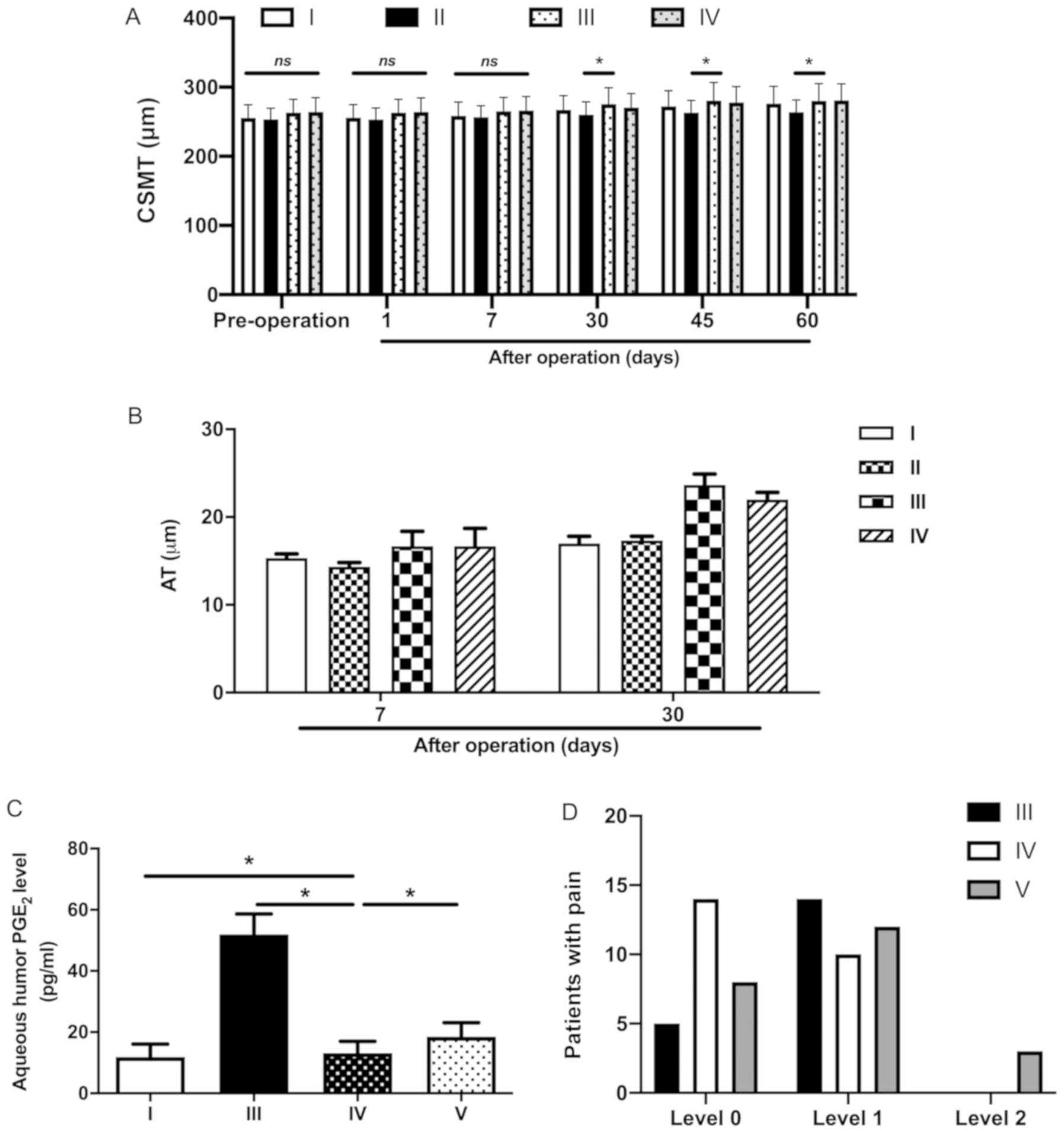
There were no significant changes of CSMT between
each group at 1-week after surgery (Fig. 2A). However, it was observed that BS
exerted a protective effect on the expansion of CSMT on days 30, 45
and 60 post-surgery (Fig. 2A).
Moreover, on day 30, the average thickness size of the outer
macular ring in FLACS was inhibited by BS (Fig. 2B). It was also found that the inner
macular ring AT demonstrated a similar trend to CSMT (data not
shown). Thus, BS also reduced the inner macular ring AT in CPCS and
FLACS groups.
It has been reported that FLACS can induce the
hyperproduction of PGE2, which is one of the major
causes of the miosis (20).
Therefore, PGE2 levels were measured in each group after
the surgery. A higher concentration of PGE2 was detected
in the aqueous humor of DEX/TOB-treated FLACS group (group III)
compared with that of CPCS group (group I). Additionally, compared
with the DEX/TOB-treated patients (group III), either BS treatment
(group IV) or pranoprofen treatment (group V) significantly
suppressed PGE2 production after the patients received
FLACS (Fig. 2C). After FLACS
surgery, no severe pain (more than level 2) was found in the
patients. As Fig. 2D illustrates,
most of patients treated by BS did not felt any pain. However, ten
patients received DEX/TOB and 5 patients treated by BS felt mild
pain after FLACS surgery. As compared with these two groups, more
severe pain (level 2) was found in 3 patients who received
pranoprofen treatment. Furthermore, there were 8 patients
distributed in level 0 of pain and 12 patients presented in level 1
of pain in pranoprofen group, respectively. Collectively, these
results demonstrated that BS has a higher protective effect on pain
compared with pranoprofen during the surgery (Fig. 2D).
Protective effects of BS on the
perioperative period of cataract surgery via modulating COX
To identify the mechanism underlying the protective
effects exerted by BS on the perioperative period of cataract
surgery, the present study established an in vitro model via
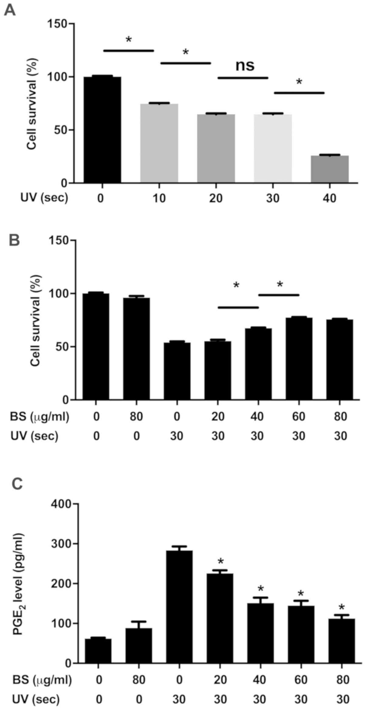
the irradiation of SRA01/04 cells with UV. As demonstrated in
Fig. 3A, the apoptosis of SRA01/04
cells occurred in a time-dependent manner post UV irradiation.
Additionally, it was identified that ~50% of cells died after
exposure to UV for 30 sec, and that most of the cells died after
exposure to UV for 40 sec (Fig.
3A). Moreover, the results suggested that BS prevented SRA01/04
cells from apoptosis in a dose-dependent manner when the cells
irradiated with UV for 30 sec (Fig.
3B). Similarly, it was also demonstrated that the
PGE2 level in the supernatant was increased by UV
exposure compared with non-UV irradiated cells, and this was
significantly reversed by BS treatment in a dose-dependent manner
(Fig. 3C).
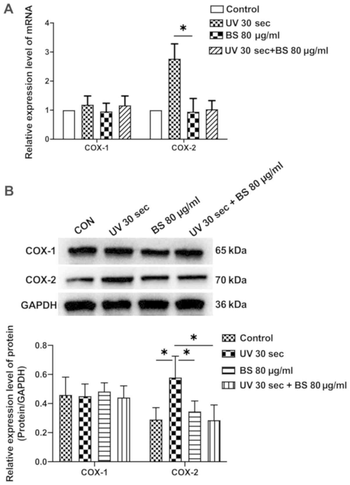
The mRNA expression level of COX-2, but not COX-1,
was significantly upregulated by UV irradiation (Fig. 4A). In addition, the protein
expression level of COX-2 was significantly increased by UV
irradiation (Fig. 4B). The results
also indicated that BS treatment significantly inhibited the
expression of COX-2 at both transcription and protein level
(Fig. 4). Under same condition,
the pyroptosis markers including IL-1β, LDH and cleaved caspase-1
were enhanced by UV-irradiated cells (Fig. 5). However, treatment of cells with
BS strongly suppressed pyroptosis, as accessed by the production of
IL-1β and LDH, expression of cleaved caspase-1 as well. These
results indicate that BS treatment protects the cell survival via
the suppression of pro-inflammatory factors and inhibition of
caspase-1 cleavage.
Discussion
CPCS has been routinely used for the treatment of
patients with cataract in the last decade (21). Tissue injuries such as endothelial
cell loss and macular edema are frequently induced during surgery
(22). Previous studies have
reported that the use of FLACS has fewer complications and is more
reliable compared with CPCS (23).
The present results suggested that there was no difference in CDVA,
as well as aqueous flare, between CPCS groups and FLACS groups.
Moreover, there was no significant difference in IOP between these
groups before or after the surgery, suggesting that the IOP rise
after surgery was not associated with several docking attempts,
vacuum time and treatment time; these results were consistent with
Kerr et al (24). In
GC-treated groups, it was found that the IOPs of two patients were
not within the normal range (12–22 mmHg) 30 days post-surgery.
Furthermore, both aqueous flare and IOP were significantly reduced
by BS treatment 30 days after the surgery. Therefore, the present
results indicated that CCT was increased by FLACS with BS
treatment, which can be interpreted by the frequent use of BS
before the surgery.
Macular edema appears usually postoperatively after
1–6 weeks, with a peak in the 4-6th week, and this is considered a
major factor for vision impairment after cataract surgery (25). Wittpenn et al (26) have reported that NSAIDs can protect
against the occurrence of macular edema and maintain the CCT. The
present findings further support this observation and suggest that
BS treatment can effectively prevent macular edema in the eyes
after cataract surgery. The present results also provided
supporting evidence for the effectiveness and safety of BS. Thus,
BS may be used as an alternative to GC and it has protective
effects on the complications of cataract surgery.
PGEs, a type of lipid autacoids derived from
arachidonic acid, have been implicated in a variety of inflammatory
diseases such as rheumatoid arthritis or allergic asthma (27). PGE2 is one of the most
abundant PGEs in mammals, and is synthesized by COX-1 and −2
(27). Previous studies have
reported that overproduction of PEG2 is observed in the
aqueous humor of patients after cataract surgery and can cause a
reduction in the size of the pupils (20,28,29).
PGE2 concentration is significantly reduced in
NSAIDs-treated patients as these drugs can directly bind and
inhibit the active site of COXs (30). The results of the present study
demonstrated that BS, as an NSAID drug, strongly suppresses
PGE2 production in patients and in UV-induced cells.
However, it should be noted that COX-2 expression, but not COX-1,
was inhibited by BS treatment, suggesting that BS mitigates miosis
via modulating COX-2.
In addition to PEG2-related inflammation,
it has been shown that pyroptosis can play a role in the formation
of cataract (31). Pyroptosis, a
type of inflammatory cell death, is induced by inflammasome
activation, which causes rapid rupture of the cell membrane and the
release of pro-inflammatory factors, such as IL-1β, IL-18 and LDH
(32). In line with these
observations, the results of the present study demonstrated that
SRA01/04 cells apoptosis was caused by UV irradiation, indicating
the presence of UV-induced pyroptosis. The anti-pyroptotoc effects
on UV-irradiated cells were observed when treatment of cells with
BS. To the best of our knowledge, the present study was the first
report the inhibitory effects of BS on pyroptosis.
In conclusion, the present study evaluated the
curative effect of BS during the perioperative period of cataract
surgery and examined its action mechanisms. It was demonstrated
that BS was more effective and safer compared with GC after
cataract surgery. Furthermore, BS can protect against postoperative
inflammation by inhibiting PGE2 production. In
vitro BS prevented SRA01/04 cells apoptosis after UV treatment
and also suppressed PGE2 release from UV-irradiated
SRA01/04 cells by modulating COS-2 expression. Collectively, the
present results suggested that BS could replace the existing GC as
a reliable drug for perioperative period of cataract surgery.
Acknowledgements
Not applicable.
Funding
This study was supported by the National Natural
Science Foundation of China (grant no. 81470617).
Availability of data and materials
The datasets used and/or analyzed during the current
study are available from the corresponding author on reasonable
request.
Authors' contributions
JingsongZ designed this study. LL, JiangyueZ, JW, YQ
and JingsongZ conducted the experiments. LL, JingsongZ and JW
analyzed the data. LL and JingsongZ wrote the manuscript. JingsongZ
edited the manuscript. All authors read and approved the final
manuscript.
Ethics approval and consent to
participate
All the experiments were approved by the Ethics
Committee of the Fourth Affiliated Hospital of China Medical
University (approval no. ChiCTR-TRC-14005114) and written informed
consent was obtained from the participants.
Patient consent for publication
Not applicable.
Competing interests
The authors declare that they have no competing
interests.
References
|
1
|
Thompson J and Lakhani N: Cataracts. Prim
Care. 42:409–423. 2015. View Article : Google Scholar : PubMed/NCBI
|
|
2
|
Khairallah M, Kahloun R, Bourne R, Limburg
H, Flaxman SR, Jonas JB, Keeffe J, Leasher J, Naidoo K, Pesudovs K,
et al: Number of people blind or visually impaired by cataract
worldwide and in world regions, 1990 to 2010. Invest Ophthalmol Vis
Sci. 56:6762–6769. 2015. View Article : Google Scholar : PubMed/NCBI
|
|
3
|
Dick HB and Schultz T: A review of
laser-assisted versus traditional phacoemulsification cataract
surgery. Ophthalmol Ther. 6:7–18. 2017. View Article : Google Scholar : PubMed/NCBI
|
|
4
|
Pascolini D and Mariotti SP: Global
estimates of visual impairment: 2010. Br J Ophthalmol. 96:614–618.
2012. View Article : Google Scholar : PubMed/NCBI
|
|
5
|
Nordan LT, Slade SG, Baker RN, Suarez C,
Juhasz T and Kurtz R: Femtosecond laser flap creation for laser in
situ keratomileusis: Six-month follow-up of initial U.S. clinical
series. J Refract Surg. 19:8–14. 2003.PubMed/NCBI
|
|
6
|
Farid M and Steinert RF: Femtosecond
laser-assisted corneal surgery. Curr Opin Ophthalmol. 21:288–292.
2010.PubMed/NCBI
|
|
7
|
Whang WJ, Yoo YS, Joo CK and Yoon G:
Comparison of refractive outcomes between femtosecond
laser-assisted cataract surgery and conventional cataract surgery.
Medicine (Baltimore). 97:e137842018. View Article : Google Scholar : PubMed/NCBI
|
|
8
|
Kránitz K, Miháltz K, Sándor GL, Takacs A,
Knorz MC and Nagy ZZ: Intraocular lens tilt and decentration
measured by Scheimpflug camera following manual or femtosecond
laser-created continuous circular capsulotomy. J Refract Surg.
28:259–263. 2012. View Article : Google Scholar : PubMed/NCBI
|
|
9
|
Pereira A, Somani S, Tam ES, Chiu H and
Maini R: Comparison of surgically induced astigmatism and corneal
morphological features between femtosecond laser and manual clear
corneal incisions. J Refract Surg. 35:796–802. 2019. View Article : Google Scholar : PubMed/NCBI
|
|
10
|
Nagy Z, Takacs A, Filkorn T and Sarayba M:
Initial clinical evaluation of an intraocular femtosecond laser in
cataract surgery. J Refract Surg. 25:1053–1060. 2009. View Article : Google Scholar : PubMed/NCBI
|
|
11
|
Nagy ZZ, Takacs AI, Filkorn T, Kránitz K,
Gyenes A, Juhász É, Sándor GL, Kovacs I, Juhász T and Slade S:
Complications of femtosecond laser-assisted cataract surgery. J
Cataract Refract Surg. 40:20–28. 2014. View Article : Google Scholar : PubMed/NCBI
|
|
12
|
Juthani VV, Clearfield E and Chuck RS:
Non-steroidal anti-inflammatory drugs versus corticosteroids for
controlling inflammation after uncomplicated cataract surgery.
Cochrane Database Syst Rev. 7:CD0105162017.PubMed/NCBI
|
|
13
|
Hirneiss C, Neubauer AS, Kampik A and
Schönfeld CL: Comparison of prednisolone 1%, rimexolone 1% and
ketorolac tromethamine 0.5% after cataract extraction: A
prospective, randomized, double-masked study. Graefes Arch Clin Exp
Ophthalmol. 243:768–773. 2005. View Article : Google Scholar : PubMed/NCBI
|
|
14
|
Jones J and Francis P: Ophthalmic utility
of topical bromfenac, a twice-daily nonsteroidal anti-inflammatory
agent. Expert Opin Pharmacother. 10:2379–2385. 2009. View Article : Google Scholar : PubMed/NCBI
|
|
15
|
Emery JM and Little JH: Patient selection.
Phacoemulsification and Aspiration of Cataract. C. V. Mosby. (St.
Louis, MO). 45–48. 1979.
|
|
16
|
Kelman CD: Phaco-Emulsification and
Aspiration: A New Technique of Cataract Removal: A Preliminary
Report. Am J Ophthalmol. 191:2018. View Article : Google Scholar : PubMed/NCBI
|
|
17
|
Marsden J, Stevens S and Ebri A: How to
measure distance visual acuity. Community Eye Health.
27:162014.PubMed/NCBI
|
|
18
|
Daut RL, Cleeland CS and Flanery RC:
Development of the Wisconsin Brief Pain Questionnaire to assess
pain in cancer and other diseases. Pain. 17:197–210. 1983.
View Article : Google Scholar : PubMed/NCBI
|
|
19
|
Livak KJ and Schmittgen TD: Analysis of
relative gene expression data using real-time quantitative PCR and
the 2(-Delta Delta C(T)) method. Methods. 25:402–408. 2001.
View Article : Google Scholar : PubMed/NCBI
|
|
20
|
Schultz T, Joachim SC, Kuehn M and Dick
HB: Changes in prostaglandin levels in patients undergoing
femtosecond laser-assisted cataract surgery. J Refract Surg.
29:742–747. 2013. View Article : Google Scholar : PubMed/NCBI
|
|
21
|
Parihar J, Sahoo PK, Dash RG and Kamath
AP: An advanced cataract surgery by phacoemulsification: An initial
experience. Med J Armed Forces India. 54:229–231. 1998. View Article : Google Scholar : PubMed/NCBI
|
|
22
|
Dick HB, Kohnen T, Jacobi FK and Jacobi
KW: Long-term endothelial cell loss following phacoemulsification
through a temporal clear corneal incision. J Cataract Refract Surg.
22:63–71. 1996. View Article : Google Scholar : PubMed/NCBI
|
|
23
|
Zhang X, Yu Y, Zhang G, Zhou Y, Zhao G,
Chen M, Wang Y, Zhu S, Zhang H and Yao K: Performance of
femtosecond laser-assisted cataract surgery in Chinese patients
with cataract: A prospective, multicenter, registry study. BMC
Ophthalmol. 19:772019. View Article : Google Scholar : PubMed/NCBI
|
|
24
|
Kerr NM, Abell RG, Vote BJ and Toh T:
Intraocular pressure during femtosecond laser pretreatment of
cataract. J Cataract Refract Surg. 39:339–342. 2013. View Article : Google Scholar : PubMed/NCBI
|
|
25
|
Flach AJ: The incidence, pathogenesis and
treatment of cystoid macular edema following cataract surgery.
Trans Am Ophthalmol Soc. 96:557–634. 1998.PubMed/NCBI
|
|
26
|
Wittpenn JR, Silverstein S, Heier J,
Kenyon KR, Hunkeler JD and Earl M; Acular LS for Cystoid Macular
Edema (ACME) Study Group, : A randomized, masked comparison of
topical ketorolac 0.4% plus steroid vs steroid alone in low-risk
cataract surgery patients. Am J Ophthalmol. 146:554–560. 2008.
View Article : Google Scholar : PubMed/NCBI
|
|
27
|
Ricciotti E and FitzGerald GA:
Prostaglandins and inflammation. Arterioscler Thromb Vasc Biol.
31:986–1000. 2011. View Article : Google Scholar : PubMed/NCBI
|
|
28
|
Solomon KD, Turkalj JW, Whiteside SB,
Stewart JA and Apple DJ: Topical 0.5% ketorolac vs 0.03%
flurbiprofen for inhibition of miosis during cataract surgery. Arch
Ophthalmol. 115:1119–1122. 1997. View Article : Google Scholar : PubMed/NCBI
|
|
29
|
Gimbel HV: The effect of treatment with
topical nonsteroidal anti-inflammatory drugs with and without
intraoperative epinephrine on the maintenance of mydriasis during
cataract surgery. Ophthalmology. 96:585–588. 1989. View Article : Google Scholar : PubMed/NCBI
|
|
30
|
Bucci FA Jr and Waterbury LD: Aqueous
prostaglandin E(2) of cataract patients at trough ketorolac and
bromfenac levels after 2 days dosing. Adv Ther. 26:645–650. 2009.
View Article : Google Scholar : PubMed/NCBI
|
|
31
|
Jin X, Jin H, Shi Y, Guo Y and Zhang H:
Pyroptosis, a novel mechanism implicated in cataracts. Mol Med Rep.
18:2277–2285. 2018.PubMed/NCBI
|
|
32
|
Davis BK, Wen H and Ting JP: The
inflammasome NLRs in immunity, inflammation, and associated
diseases. Annu Rev Immunol. 29:707–735. 2011. View Article : Google Scholar : PubMed/NCBI
|