Introduction
Endometriosis (EMs) is a chronic inflammatory
sickness marked by the presence of endometrial glands and matrices
outside the uterine cavity (1).
The common clinical symptoms are progressive dysmenorrhea, chronic
pelvic pain and difficulty in sexual intercourse in women of
childbearing age. In addition, it can also cause menstrual
disorders, infertility and other adverse consequences, which
seriously affect the living standards and lifestyle of patients
with EMs. However, the pathogenesis and pathophysiology of EMs are
currently unknown. In recent years, research has concentrated on
the theories of inflammatory stimulation (2), cellular immune monitoring deficiency
(3), retrograde menstruation and
coelomic metaplasia (4). According
to the classical implantation theory, ectopic endometrial adhesion
and implantation, angiogenesis and proliferation are the most
prominent pathomorphological features of EMs (5). Increasing evidence shows that cell
adhesion and invasive molecules involved in inflammatory and immune
processes play an important role in endometrial pathophysiology.
Researchers have also proposed the ‘3A model’, which hypothesizes
that the progression of EMs is inevitably accompanied by
pathological processes of adhesion, invasion and angiogenesis
(6), thereby promoting the
implantation and growth of counter-current endometrial cells in the
pelvic and abdominal cavities and then infiltrating to form lesions
under the influence of the immune response and hormones.
In recent decades, the onset age of EMs has
gradually become younger with the change of social life style and
the increase of uterine surgical procedures such as abortion and
caesarean section (7). EMs is
challenging to treat at present due to its unique pathogenesis and
estrogen-dependent characteristics (8). Pharmacological treatment is often the
first choice for patients with EMs due to its features such as ease
of use, safety, quick effect and minimal trauma, among which
hormonal agents such as combined oral contraceptives, danazol,
gonadotropin-releasing hormone agonists (GnRH-a) and non-steroidal
anti-inflammatory drugs are often used as first-line treatment for
EMs (9). However, a number of
patients often have to discontinue treatment due to inevitable side
effects such as breakthrough bleeding, peptic ulcers and
osteoporosis as well as short-term curative effect and frequent
relapses (10). Therefore, surgery
is frequently used as the standard treatment for EMs, with complete
removal of the ectopic lesion, relief of pelvic pain and
improvement in quality of life as the clinical outcomes (11). However, postoperative complications
and a high recurrence rate are also significant issues that trouble
clinicians today (12).
Consequently, it is crucial to seek an EMs treatment strategy with
low recurrence rate, few side effects and broad adaptability.
Traditional Chinese medicine (TCM) has a long
history of clinical application in China and is able to treat a
number of complex diseases through multi-target action. Tanshinone
IIA is a lipid-soluble component of the Chinese medicine Danshen
(Salvia miltiorrhiza Bge.), which has anti-inflammatory,
antioxidant, antibacterial, anti-tumour, anti-fibrosis and
neuroprotective effects (13). It
is mainly used for the treatment of cardiovascular and
cerebrovascular diseases, cancer, bone and joint disease (14–16).
Studies have found that tanshinone IIA is also effective in
treating polycystic ovary syndrome (17), adenomyosis (18), EMs (19) and other diseases. For the formation
of EMs ectopic lesions, tanshinone IIA can significantly inhibit
the levels of HAS2, CA-125, IL-8 and TNF-α, and impede the ectopic
endometrial epithelial-mesenchymal transformation (EMT) process,
thereby inducing apoptosis to achieve antifibrosis, and reducing
endothelial cell proliferation and lesion infiltration (20,21).
In addition, our previous study demonstrated that tanshinone IIA
can reduce mechanical hyperalgesia in EMs rats by lowering the
level of E2 and inhibiting the endogenous expression of dorsal root
ganglion renin-angiotensin system, while preventing the invasion
and growth of ectopic lesions (22). However, the potential therapeutic
effect of tanshinone IIA on EMs and its detailed pharmacological
mechanisms still need to be further investigated.
Network pharmacology is a new subject based on the
theory of systems biology, which analyzes biological systems and
selects specific signal nodes for multi-target drug molecular
design (23). Network pharmacology
explains the occurrence and development of diseases from the
perspective of system biology and biological network balance,
understands the interaction between drugs and the body from the
perspective of improving or restoring the balance of biological
network, and guides the discovery of new drugs (24). The research strategy of network
pharmacology is holistic and systematic, which conforms to the
principle of holistic view of TCM, so it is widely used to
elucidate the mechanism of TCM in the treatment of diseases
(25). The core targets and
pathways obtained through network pharmacological screening can
provide an improved basis for follow-up experiment. The present
study combined network pharmacology with experimental validation to
clarify the main targets and potential mechanisms of tanshinone IIA
in treatment of EMs.
Materials and methods
Acquisition of target genes related to
tanshinone IIA and EMs
The related targets of tanshinone IIA were predicted
by using the TCM systematic pharmacology database
(old.tcmsp-e.com/tcmsp.php) (26).
In order to collect targets of tanshinone IIA in a more
comprehensive way, target prediction was further performed through
the PharmMapper database (https://lilabecust.cn/pharmmapper/). Human protein
targets were reset only in the PharmMapper database and the default
settings kept for other parameters. The PharmMapper server
automatically matches small ligand molecules with pharmacophore
models in the database and ranks them based on their match scores.
Normalized fit score >0.8 was used as the filtering criterion.
In addition, the targets of tanshinone IIA were predicted by Swiss
Target Prediction database (https://www.swisstargetprediction.ch/) and probability
value ≥0.10 was employed as criteria to filter for targets. The
selected targets from the three databases were combined for
deduplication, and then the targets were converted into
standardized gene symbols using the UniProt database (https://www.uniprot.org/).
All gene targets related to EMs were collected from
Genecards database (genecards.org/), OMIM database (https://www.omim.org/) and DrugBank database
(https://www.ncbi.nlm.nih.gov/gene).
Key word ‘endometriosis’ was entered into the search column to
obtain target information. The targets obtained from the three
databases were combined and deduplicated, sorted by score, and
those with higher than the median score were considered as
endometriosis targets. The targets of tanshinone IIA and
endometriosis were uploaded to the online analysis tool of Venny2.1
(https://bioinfogp.cnb.csic.es/tools/venny/) to screen
out the common targets.
Construction of the protein-protein
interaction (PPI) network and selection of core genes
The intersection targets of tanshinone IIA and EMs
were submitted to the STRING database (https://string-db.org/) with the species set to
Homo sapiens. Then, the highest confidence level was set to
the minimum necessary interaction score (0.9) for the filter,
hiding the discrete points in the network and leaving the remaining
parameters as defaults. The PPI network download was saved in tsv
format and imported into Cytoscape 3.7.2 software (cytoscape.org)
for visualization. Cytohubba can assign a score to each gene in the
PPI network through its 12 algorithms, and then select the top gene
as the key gene based on the gene score. Therefore, cytohubba
network analysis plug-in was used to calculate the degree of
connectivity between proteins. The greater the degree value, the
more interacting protein nodes and the greater influence on the
network, Finally, target nodes with degree centrality, betweenness
centrality and closeness centrality values higher relative to the
corresponding median values in the PPI network were chosen for
subsequent analysis.
Enrichment analysis of Gene Ontology
(GO) and Kyoto Encyclopedia of Genes and Genomes (KEGG)
Pathway enrichment analysis for GO and the KEGG were
achieved by the cluster profiler program in R software (27). The screening conditions were set to
P-value cutoff=0.05 and q-value cutoff=0.05 and the items with the
highest P-values selected for data visualization analysis. GO
analysis was used to screen the biological process (BP), cell
components (CC) and molecular function (MF). KEGG enrichment
primarily revealed pathway related content.
Animals
A total of 36 mature female Sprague Dawley rats
(age, 6–7 weeks) at SPF level weighing between 180–200 g were
donated by Beijing Vital River Laboratory Animal Technology Co.,
Ltd. (license no. SCXK 2016–0006). All rats were housed in standard
cages at 22–24 °C and relative humidity (40–60%), with a regular 12
h light/dark cycle and ad libitum access to food and water in
accordance with rat care protocols. The Animal Care and Welfare
Committee of the Capital Medical University (approval no.
AEEI-2018-031) reviewed and approved all experimental
procedures.
Experimental design and treatment
According to our previous study, the EMs rats model
was successfully prepared by autologous transplantation (22). Rats were randomly designated to
five groups: Sham, model, positive, tanshinone IIA low dose and
tanshinone IIA high dose (n=6 in each group). Rats in both sham
group and model group were administered solvent. Positive group
rats were intragastrically given 2 mg/kg/d medroxyprogesterone
acetate (Zhejiang Xianju Pharmaceutical Co., Ltd.). Rats in the
tanshinone IIA low dose and high dose groups received 3 and 12
mg/kg/day tanshinone IIA (Shanghai No.1 biochemical pharmaceutical
Co., Ltd.) respectively via intraperitoneal injection. At 21 days
after medication, blood samples were taken from the abdominal aorta
under anesthesia by intraperitoneal administration of 40 mg/kg
pentobarbital sodium and serum was extracted and kept at −20°C.
After blood collection, the rats were deeply anesthetized by
intraperitoneal injection of pentobarbital sodium 150 mg/kg and
subsequently sacrificed by cervical dislocation. Following
sacrifice, the animal's death was confirmed from loss of pain
response, failure to respond to toe pressure with hands or tweezers
and observation of cardiac and respiratory arrest. The ectopic
lesion was resected and part of the tissue was fixed in 4%
paraformaldehyde for 24 h at room temperature, while the remainder
was frozen at −80°C.
Hematoxylin and eosin (H&E)
staining
The tissue that had been fixed was withdrawn from
the paraformaldehyde solution, dehydrated by gradient ethanol,
transparent by xylene, embedded in paraffin and then cut into 4-µm
pieces. After xylene deparaffinization and gradient alcohol
dehydration, the sections were stained with H&E staining at
room temperature for 3 min and evaluated under a BX53 light
microscope (Olympus Corporation) to investigate pathological
morphological alterations.
Enzyme-linked immunosorbent assay
After centrifuging the blood for 10 min at 1,200 × g
at −4°C, the supernatant was gathered and stored at −20°C for
further examination. The concentrations of TNF-α and IL-1β in the
serum were determined using ELISA (CSB-E11987r, CSB-E08055r) as the
protocol provided by the manufacturer (Wuhan Huamei Biological
Engineering Co., LTD, China). The luminescent signal produced was
measured at 450 nm using a microplate reader (Thermo Fisher
Scientific, Inc.).
Immunohistochemical staining
Following xylene deparaffinization, the sections
were rehydrated and treated with a buffer containing 0.01 mol/l
sodium citrate. Next, the sections were treated with 3% hydrogen
peroxide for 20 min to block the activity of endogenous peroxidase.
Incubation with 2% bovine serum albumin (Beijing Dingguo Changsheng
Biotechnology Co., LTD) inhibited non-specific binding. After the
non-specific antibodies were blocked, the primary antibody
anti-MMP9 (diluted 1:400; Bioss, bs-4593R), anti-ICAM-1 (diluted
1:400; Bioss, bs-0608R) and anti-VEGF (diluted 1:400; Abcam,
ab46154) was incubated with the tissues overnight at 4°C. The
antibody labeled with horseradish peroxidase HRP (1:200; Beijing
Dingguo Changsheng Biotechnology Co., LTD, IH-0061) was treated for
60 min at 37°C. Following that, the sections were rinsed with PBS
and stained with 0.01% DAB. Finally, the optical microscope image
was captured under a BX53 light microscope (Olympus) by randomly
selecting five fields from each section. The average integral
optical density was measured using Image-Pro Plus 6.0 software
(Media Cybernetics, Inc.).
Western blotting analysis
The eutopic and ectopic endometria were lysed in
RIPA) lysis buffer with protease inhibitors (Beijing Dingguo
Changsheng Biotechnology Co., LTD, Beijing, China). Then, lysates
were centrifuged at 12,000 × g for 10 min and the protein
concentration of the supernatants were measured by BCA assay kit
(Beijing Dingguo Changsheng Biotechnology Co., LTD, Beijing,
China). Each lysate supernatant was loaded onto 10%
SDS-polyacrylamide gels with 80 µg total protein. After the protein
samples had been transferred to PVDF membranes, they were blocked
for 1 h at room temperature with 5% nonfat milk. The principal
antibodies used overnight at 4°C were: β-actin (diluted 1:5000;
Immunoway, YM3028), PI3K (diluted 1:500; Bioss, bs-0128R), p-PI3K
(diluted 1:1,000; Bioss, bs-3332R), Akt (diluted 1:1,000; Bioss,
bs-6951R), phosphorylated (p-)Akt (diluted 1:1,000; Bioss,
bs-5182R), mTOR (1:1,000; Bioss, bs-1992R) and p-mTOR (diluted
1:1,000; Bioss, bs-3495R). The internal reference was β-actin.
After 1 h incubation at room temperature with a HRP-labeled goat
anti-rabbit IgG secondary antibody, the bands were detected using
superenhanced chemiluminescence detection tools. Images were
captured immediately after color development using a gel imaging
system (Bio-Rad Laboratories, Inc.). ImageJ was used to analyze the
band intensities (v1.46; National Institutes of Health).
Reverse transcription-quantitative
(RT-q) PCR
TRIzol® reagent (Invitrogen; Thermo
Fisher Scientific, Inc.) was used to extract total RNA from the
tissues of the eutopic and ectopic endometria according to the
manufacturer's instructions., and the microultraviolet
spectrophotometer (Quawell Q5000) was utilized to assess the RNA
content. cDNA synthesis was conducted with
M-MLVReverseTranscriptase reagent kit (Invitrogen, AM2044) in a
reaction volume of 20 µl. Following the guidelines provided by the
manufacturer, RT-qPCR was conducted utilizing an Applied Biosystems
7500 system (Applied Biosystems) and SYBR Green Master Mix
(Invitrogen; Thermo Fisher Scientific, Inc.). The conditions were
as follows: 35 cycles of pre-denaturation at 95°C for 2 min,
denaturation at 95°C for 15 sec, annealing at 62°C for 30 sec and
extension at 72°C for 30 sec. GAPDH served as the internal
standard. Each sample was run in triplicate and GAPDH was used as
the reference. Foldchange of gene expression was calculated using
2−ΔΔCq method (28).
Table I lists the RT-qPCR
primers.
 | Table I.Reverse transcription-quantitative
PCR primer sequences. |
Table I.
Reverse transcription-quantitative
PCR primer sequences.
| Gene | Primer sequences
(5′→3′) | Product size
(bp) |
|---|
| GAPDH | F:
CCTGGGCTACACTGAGGA | 164 |
|
| R:
TGAGGTCCACCACCCTGT |
|
| PI3K | F:
GTAGGCCCGAGTAAGCTGAA | 206 |
|
| R:
CCGTAGGTGAGACCCCAAGT |
|
| mTOR | F:
ATTCGCATTCAGTCCATAGCC | 179 |
|
| R:
AAACAAACTCGTGCCCATTGC |
|
Statistical analysis
SPSS 22.0 software (IBM Corp.) was used for data
analysis and the measurements were presented as the mean ± standard
deviation. One-way analysis of variance (ANOVA) and the Bonferroni
post hoc test were used to observe the difference between multiple
groups. The Mann-Whitney rank sum test was used to perform a
nonparametric analysis. P<0.05 was considered to indicate a
statistically significant difference.
Results
Common targets for tanshinone IIA and
EMs
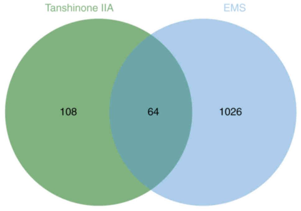
The retrieval process yielded information on 172
drug targets and 1,090 disease targets. A Venn diagram showed that
there were 64 overlapping genes between EMs targets and tanshinone
IIA action targets (Fig. 1).
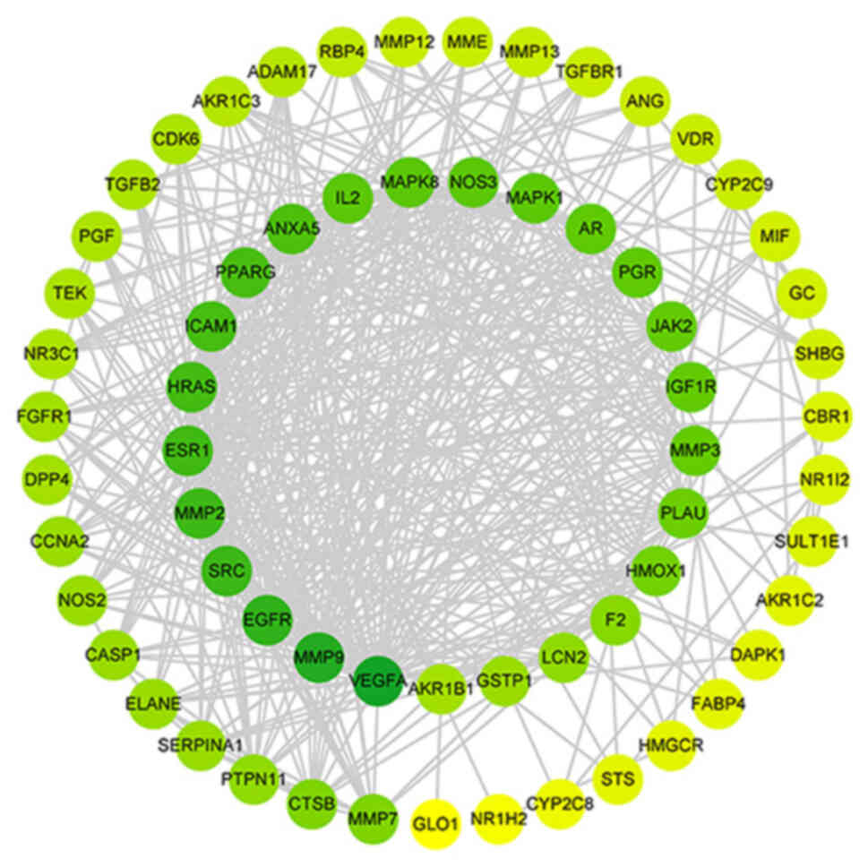
PPI network construction and
analysis
Based on the species Homo sapiens, a PPI
network with 64 nodes and 454 edges was created in the STRING
database, and the analysis results were loaded into Cytoscape 3.7.2
for visualization. As shown in Table
II, the cytohubba plugin was used to analyze the nodes in the
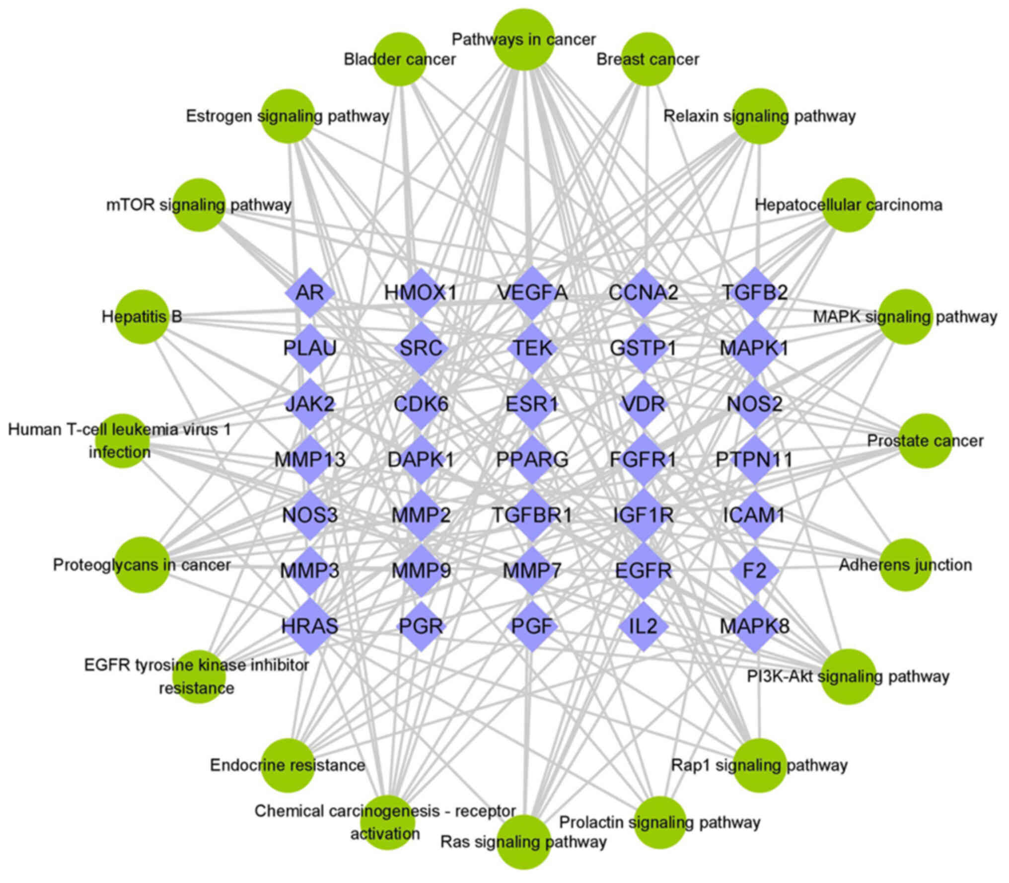
network diagram and finally selected 14 core genes; VEGFA, MMP9,
EGFR, SRC, MMP2, ESR1, HRAS, ICAM1, PPARG, ANXA5, MAPK8, IL2, NOS3
and MAPK1. Degree value of the node and the number of biological
functions in the network increase with the color of the node and
the greener the color, the more important it is in the network
(Fig. 2).
| Table II.Core targets of tanshinone IIA in the
treatment of endometriosis. |
Table II.
Core targets of tanshinone IIA in the
treatment of endometriosis.
| Gene | Degree |
BetweennessCentrality |
ClosenessCentrality |
ClusteringCoefficient |
|---|
| VEGFA | 47 | 0.14 | 0.80 | 0.32 |
| MMP9 | 41 | 0.08 | 0.72 | 0.37 |
| EGFR | 36 | 0.06 | 0.70 | 0.40 |
| SRC | 33 | 0.04 | 0.67 | 0.46 |
| MMP2 | 32 | 0.03 | 0.66 | 0.46 |
| ESR1 | 31 | 0.11 | 0.65 | 0.37 |
| HRAS | 30 | 0.03 | 0.65 | 0.49 |
| ICAM1 | 29 | 0.03 | 0.62 | 0.46 |
| PPARG | 28 | 0.09 | 0.64 | 0.40 |
| ANXA5 | 26 | 0.01 | 0.62 | 0.60 |
| MAPK8 | 22 | 0.01 | 0.59 | 0.65 |
| IL2 | 22 | 0.01 | 0.57 | 0.57 |
| NOS3 | 22 | 0.02 | 0.59 | 0.53 |
| MAPK1 | 21 | 0.01 | 0.59 | 0.62 |
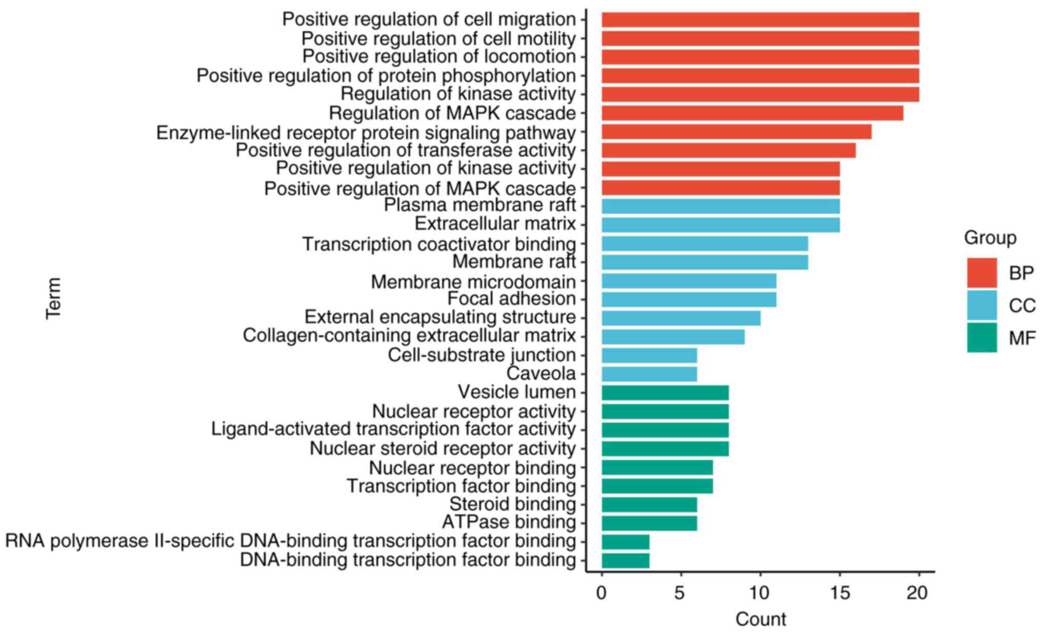
GO enrichment and KEGG pathway
analysis
The GO functions of common targets were enriched in
182 BP, 109 CC and 64 MF (P<0.05). The top 10 items of the BP,
CC, and MF gene ratio were selected to make a stick chart (Fig. 3). As shown in Fig. 3, BP mainly involved ‘positive
regulation of cell migration’, ‘positive regulation of cell
motility’, ‘positive regulation of locomotion’, ‘positive
regulation of protein phosphorylation’ and ‘regulation of kinase
activity’. CC mainly involved ‘plasma membrane raft’,
‘extracellular matrix’, ‘transcription coactivator binding’,
‘membrane raft’ and ‘membrane microdomain’. MF mainly involved
‘vesicle lumen’, ‘nuclear receptor activity’, ‘ligand-activated
transcription factor activity’, ‘nuclear steroid receptor activity’
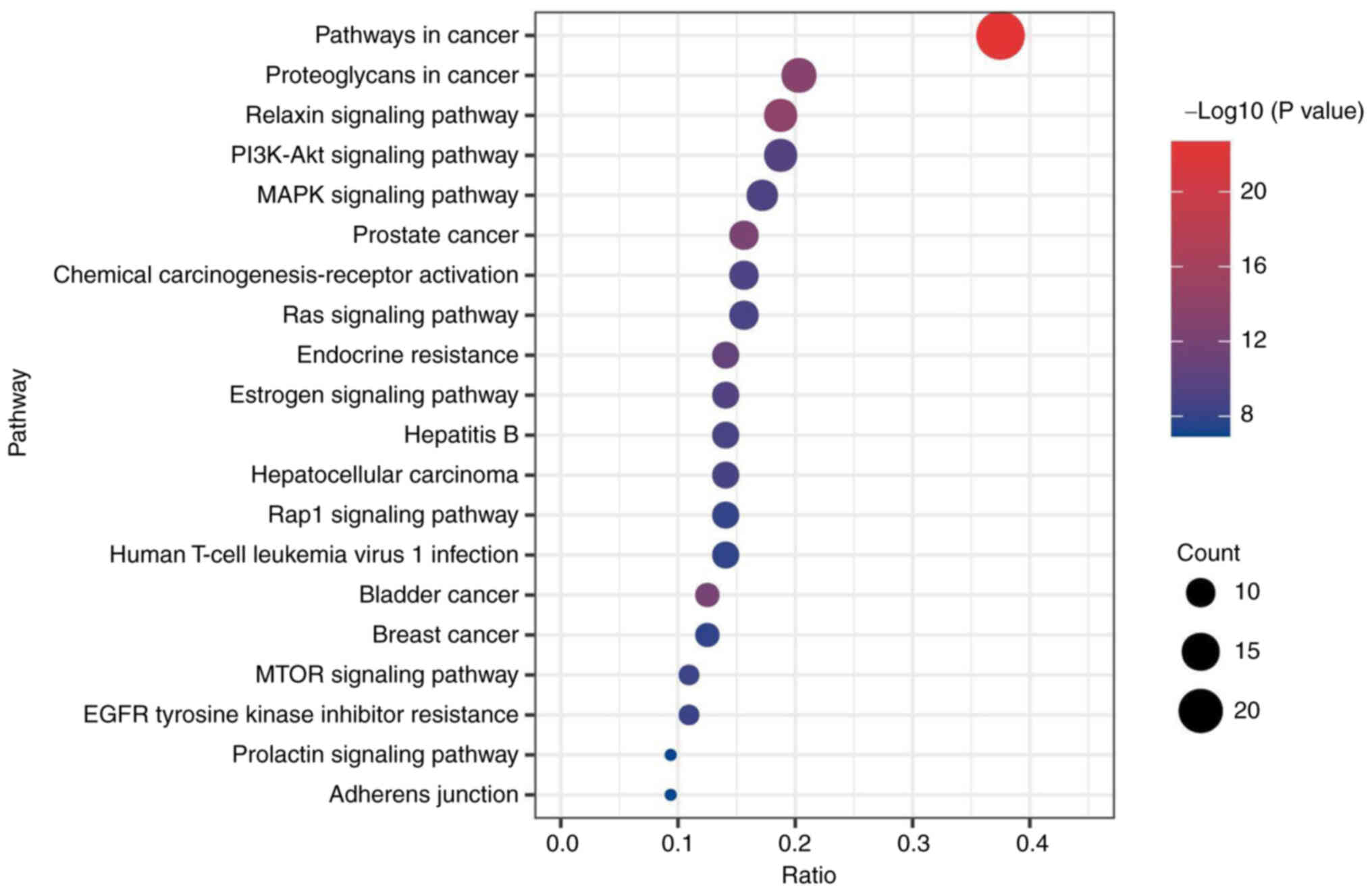
and ‘nuclear receptor binding’. In addition, a total of 198 items
were enriched by KEGG analysis (P<0.05). Fig. 4 depicts the initial 20 signaling
pathways, mainly including ‘relaxin signaling pathway’, ‘PI3K-Akt
signaling pathway’, ‘MAPK signaling pathway’, ‘Ras signaling
pathway’ and ‘mTOR signaling pathway’.
Construction of
compound-target-pathway network
Traditional Chinese medicine compounds are
characterized by multiple components and multiple targets, and the
relationship among the components, core targets and pathways can be
more clearly demonstrated by constructing the network of
compound-target-pathway network (Fig.
5). A total of 35 tanshinone IIA targets were implicated in
network regulation and the effect of tanshinone IIA on EMs may be
mediated by the PI3K-Akt, MAPK, Ras and mTOR signaling pathway.
Based on the above results, it was hypothesized that tanshinone IIA
participates in the treatment of EMs by regulating adhesion,
invasion, angiogenesis and inhibition of PI3K/Akt/mTOR signaling
pathway. This hypothesis was further tested with animal
experiments.
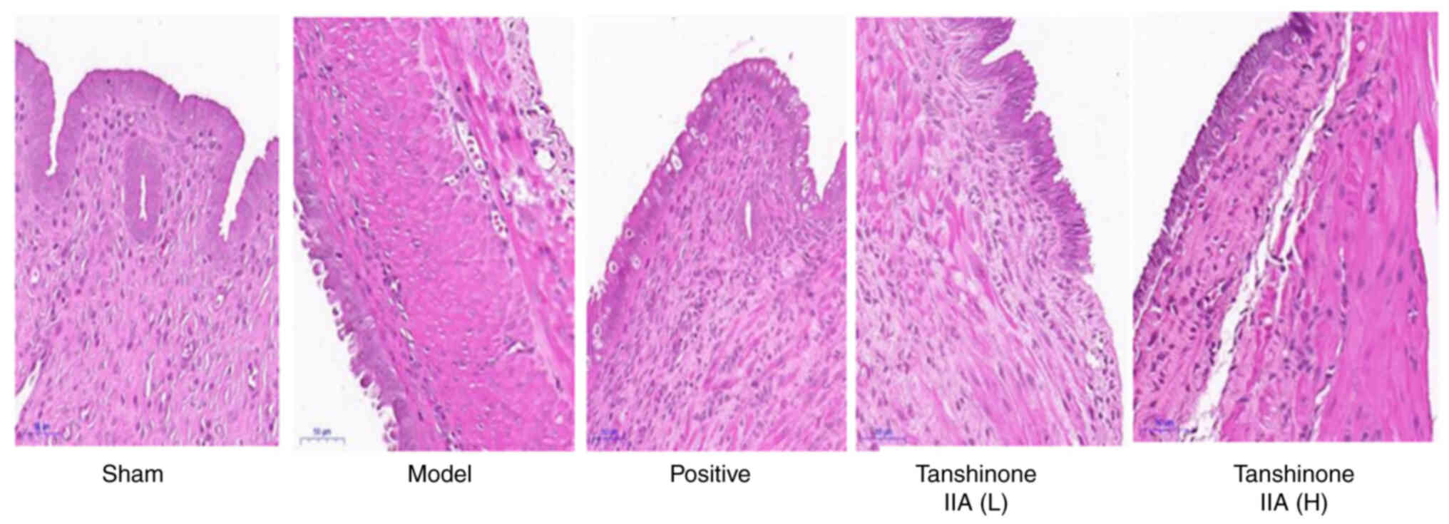
Histologic analysis
As depicted in Fig.
6, the endometrial epithelial cells in the sham group were
organized in an ordered fashion, with recognizable glandular
structure and noticeable glandular lumen. Interstitial changes such
as edema, hemorrhage and inflammatory cell infiltration were not
observed. In the model group, the epithelial cells were disordered
and appeared lamellar or pseudolamellar. In addition, subnuclear
vacuoles could be observed in some epithelial cells. In the
positive group, endothelial epithelial cells showed more necrosis
and apoptosis, but the glandular structure was clearer than in the
model group. The epithelial cells in the tanshinone IIA (L) and
tanshinone IIA (H) groups were arranged as a single layer, the
number of glands was diminished, and the ectopic endothelium was
atrophied and thinning. The effect of tanshinone IIA (H) group was
more prominent than that of tanshinone IIA (L) group.
Effect of tanshinone IIA on the
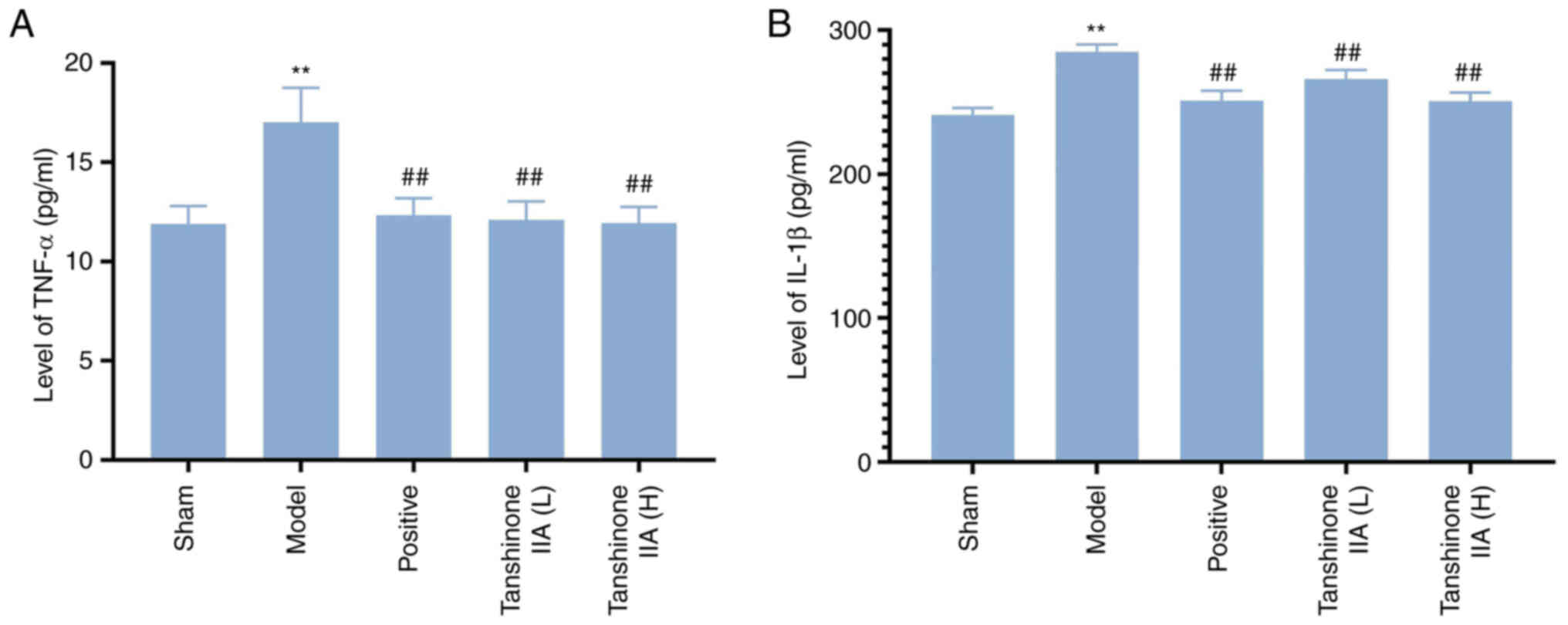
expression of TNF-α and IL-1β
The levels of TNF-α and IL-1β in the model group
were greater than that in the sham group (P<0.01). Compared with
the model group, serum levels of TNF-α and IL-1β in the positive
group and tanshinone IIA (L, H) groups were significantly decreased
(all P<0.01; Fig. 7).
Effect of tanshinone IIA on the
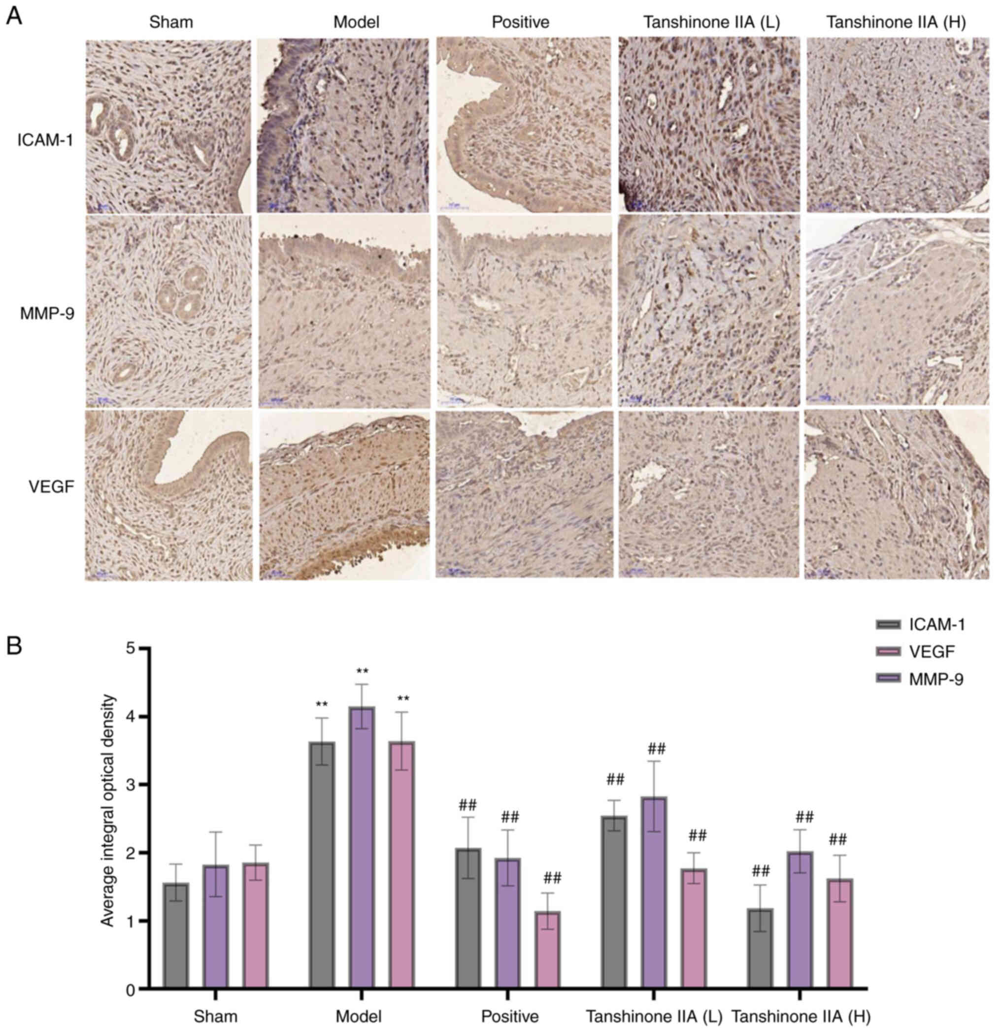
expression of MMP-9, ICAM-1 and VEGF by immunohistochemistry
When compared with the sham group, the expression
levels of MMP-9, ICAM-1 and VEGF were considerably greater in the
model group (P<0.01). When compared with the model group, the
expression levels of MMP-9, ICAM-1 and VEGF were considered to be
particularly lower in the positive group (P<0.01). It was also
discovered that tanshinone IIA (L and H) therapy resulted in a
considerable decrease in the expression levels of MMP-9, ICAM-1 and
VEGF compared with the model group. Moreover, the tanshinone IIA
(H) group experienced a higher reduction than the tanshinone IIA
(L) group (Fig. 8).
Effect of tanshinone IIA on the PI3K,
p-PI3K, Akt, p-Akt, mTOR and p-mTOR expression assessed by western
blotting
The results showed that the expression levels of
PI3K, p-PI3K, Akt, p-Akt, mTOR and p-mTOR were considerably greater
in the model group than those in the sham group. Positive and
tanshinone IIA (L and H) groups had substantially lower relative
expression levels of PI3K, p-PI3K, Akt, p-Akt, mTOR and p-mTOR
compared with the model group (Fig.
9).
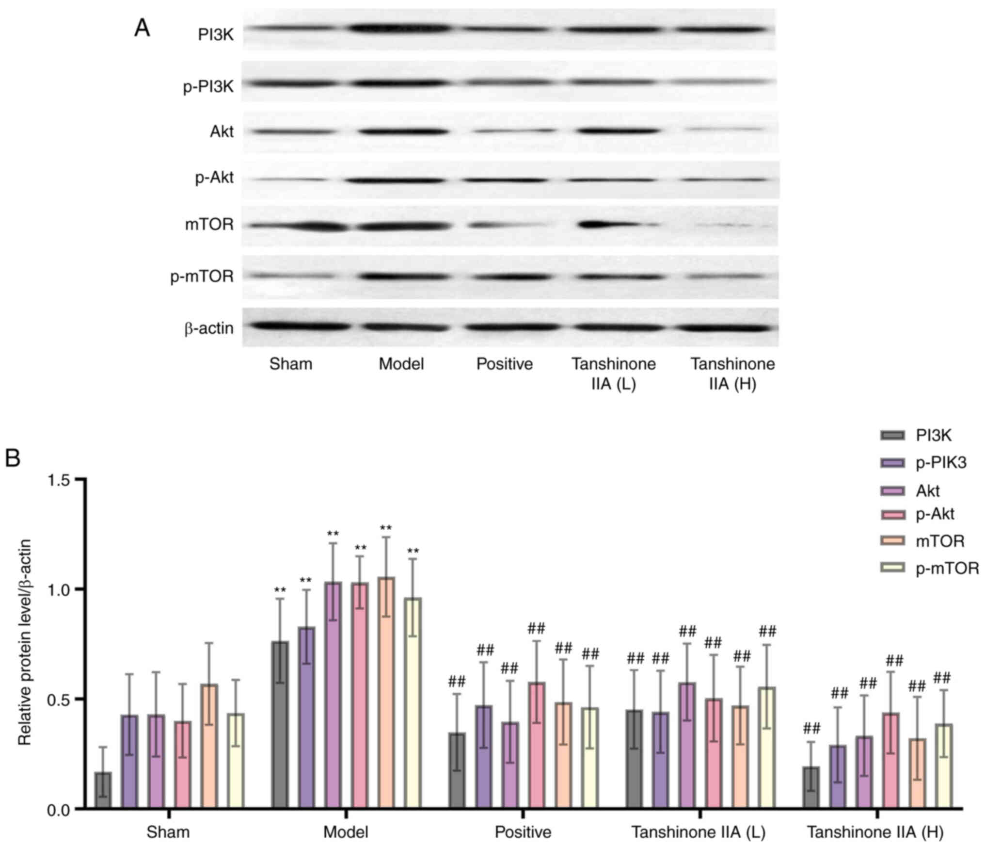 | Figure 9.Protein levels of PI3K, p-PI3K, Akt,
p-Akt, mTOR and p-mTOR in rats (n=6). (A) Representing western
blotting analysis of PI3K, p-PI3K, Akt, p-Akt, mTOR and p-mTOR. (B)
Quantification of PI3K, p-PI3K, Akt, p-Akt, mTOR and p-mTOR
expression. β-actin was used as a loading control. Data are
presented as mean ± standard deviation. **P<0.01 vs. sham group;
##P<0.01 vs. model group. p-, phosphorylated. |
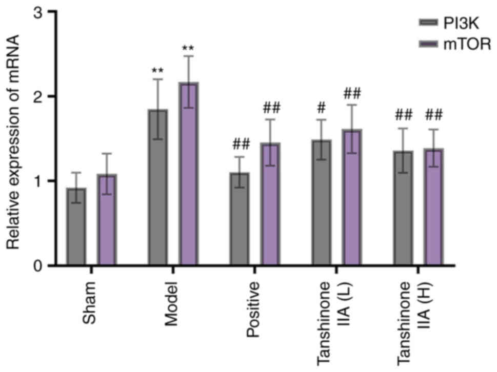
Effect of tanshinone IIA on gene
expression of PI3K and mTOR
RT-qPCR was used to investigate PI3K and mTOR mRNA
levels. The PI3K and mTOR mRNA levels in the model group were
notably greater than those in the sham group. Treatment with
medroxyprogesterone acetate or tanshinone IIA (L and H)
substantially lowered PI3K and mTOR gene expression levels when
compared with the model group (Fig.
10).
Discussion
EMs is a multifactorial benign gynaecological
disease with a high incidence and complex pathogenesis for which
there is no well-established clinical treatment. The traditional
surgical treatment is usually to remove the ectopic lesion as the
ultimate goal, although it is easy to relapse after surgery
(29). Commonly used medications
include oral contraceptives, danazol, GnRH-a and aromatase
inhibitors. However, long-term use of these drugs can cause a
variety of adverse reactions, such as bleeding, pelvic pain,
hirsutism, acne and osteoporosis (30,31).
As a result, researchers are also exploring new strategies for
treating EMs. The present study revealed the regulatory network in
EMs and identified tanshinone IIA as a candidate drug for EMs. The
network pharmacology analysis revealed the tanshinone IIA related
molecular functions and pharmacological targets for treating EMs
and the therapeutic effect and mechanism of tanshinone IIA were
verified in vivo.
Tanshinone IIA is a lipid-soluble component
extracted from the Chinese herb Danshen (Salvia miltiorrhiza
Bge.), which can effectively inhibit cell invasion, proliferation
and apoptosis. Previous research by our team demonstrated that
tanshinone IIA could significantly inhibit the growth of EMs
lesions, inhibit the expression of proteins including
angiotensinogen and Ang II in the dorsal root ganglion by reducing
E2 levels, and alleviate the general hyperalgesia of EMs by
regulating dorsal root ganglion sprouting via renin-angiotensin
system (22). Network pharmacology
combined with bioinformatics technology is a popular tool for
studying the pharmacological mechanism of drugs in recent years.
Through the construction of drug-target-disease interaction
network, it provides valuable predictive guidance for further
elucidation of the mechanism of drugs for disease treatment
(32,33). The present study analyzed the
relevant targets and mechanisms of tanshinone IIA in the management
of EMs using network pharmacology.
The present study first used the network
pharmacology approach to screen the target genes related to
tanshinone IIA and EMs. After screening and matching, 64 common
targets were obtained and used for further study. Through PPI
network construction, it was predicted that the core biological
targets of tanshinone IIA for EMs treatment such as VEGFA, MMP-9,
ESR1, ICAM-1 and IL-2, involving the pathological effects of
adhesion, invasion, angiogenesis, inflammatory response and
cellular immune response. To illustrate the functional and
signaling pathways of the 14 protein targets associated with
tanshinone IIA for EMs, GO and KEGG analyses were performed. The
results showed that the target genes were mainly enriched in the
positive regulation of cell migration, positive regulation of cell
motility, positive regulation of protein phosphorylation,
regulation of kinase activity and other biological pathways. KEGG
analysis showed that 14 target genes were enriched in 198 signaling
pathways, indicating that tanshinone IIA acts as a treatment for
EMs by regulating multiple signaling pathways. Among them, the
PI3K-AKT pathway and mTOR pathway showed high enrichment.
Therefore, it was hypothesized that tanshinone IIA might regulate
the adhesion, invasion and angiogenesis of EMs via PI3K/Akt/mTOR
signaling pathway. Based on the above results, the therapeutic role
of tanshinone IIA on EMs was detected.
EMs is known to be an inflammatory disease. Studies
have found that the presence of a large number of macrophages in
the peritoneal fluid of patients with EMs can release more
pro-inflammatory cytokines such as IL and TNF-α to participate in
the intercellular communication modification, promote fibroblast
mitosis, adhesion and maintain the adhesion of ectopic endometrial
tissue (2,34). In the present study, the serum
levels of TNF-α and IL-1β were upregulated in the model group.
After intervention with tanshinone IIA (3 or 12 mg/kg), the serum
levels of TNF-α and IL-1β were downregulated, suggesting that
tanshinone IIA may play an anti-inflammatory role in EMs.
ICAM-1, MMP-9 and VEGF are essential cytokines for
the adhesion, invasion and angiogenesis of ectopic endometrial
tissue in the pelvic and abdominal cavities of EMs. As the most
potent cell adhesion molecule in the immunoglobulin superfamily,
ICAM-1 plays a crucial role in regulating cell-to-cell and
cell-extracellular matrix adhesion (35). In EMs pathogenesis, the
concentration of ICAM-1 increases significantly and reduces the
toxicity of natural killer cells, which mediate a defective immune
surveillance response mechanism and promote the adhesion,
implantation and proliferation of ectopic tissue (36). MMP-9, an inflammatory mediator
associated with inflammation and angiogenic remodeling, can be
significantly upregulated in EMs progression by TNF-α. MMP-9
promotes ectopic tissue invasion and ectopic lesion formation by
degrading extracellular matrix components (37). It has been found that macrophages
in EMs peritoneal fluid are the primary source of VEGF (38). Macrophages release pro-inflammatory
cytokines TNF-α and IL-1β, which induce upregulation of VEGF
expression through the COX-2 signaling pathway, thereby stimulating
peripheral endometrial angiogenesis and proliferation of ectopic
endometrium (39,40). Immunohistochemistry was employed in
the present study to detect alterations in the expression of
ICAM-1, MMP-9 and VEGF. Results showed that the expression levels
of ICAM-1, MMP-9 and VEGF were significantly elevated in the EMs
lesions of rats in the model group, whereas they were downregulated
in the tanshinone IIA (3 or 12 mg/kg) treatment groups. There were
no statistically significant changes between the positive group
(medroxyprogesterone acetate) and the tanshinone IIA group. This
suggested that tanshinone IIA can limit the infiltration of ectopic
lesions in EMs by modulating adhesion, invasion and
angiogenesis.
Angiogenesis plays a crucial role in the formation
of EMs. As the most important angiogenic factor, VEGF and its
receptors can upregulate CD62E and CD105 levels by inducing the
PI3K/Akt signaling pathway, thus leading to a large number of
neovascularization of vascular endothelial cells in an EMs
environment, which is conducive to the occurrence and development
of EMs (41,42). Other studies have shown that
blocking the mTOR signaling pathway can significantly inhibit the
formation and progression of ectopic lesions in EMs rats (43,44).
As is well known, EMs is also an estrogen-dependent disease, and
studies have proven that the estrogen signaling pathway is
implicated in the formation of EMs (10,45).
By participating in EMs estrogen-signal transduction pathway, the
PI3K/Akt/mTOR signaling pathway affects endometrial homeostasis and
increases vascular permeability, participates in cell adhesion,
invasion and angiogenesis in ectopic endometrium, and ultimately
leads to the formation of EMs (46,47).
The present study found that the expression of PI3K, p-PI3K, Akt,
p-Akt, mTOR and p-mTOR proteins in the ectopic endometrium of rats
in the model group was significantly increased, suggesting that the
PI3K/Akt/mTOR signaling pathway may be involved in the progression
of EMs. Following treatment with tanshinone IIA, the protein
expressions of PI3K, p-PI3K, Akt, p-Akt, mTOR and p-mTOR were
significantly downregulated. RT-qPCR also showed that the mRNA
expressions of PI3K and mTOR in the ectopic endometrium of rats
treated with tanshinone IIA was downregulated. Therefore, it was
hypothesized that tanshinone IIA may prevent the excitation of
PI3K/Akt/mTOR signaling pathway, thus further restricting the
progression of EMs.
At present, the pathogenesis of EMs is not fully
understood and its treatment options are still limited. TCM is
effective in treating EMs and deserves further study. There are
several suggestions for future work. First, make good use of
network pharmacology tools to predict the core targets of TCM and
combine experimental verification to promote the development of new
TCM drugs. Second, combined with the latest progress in the
pathological mechanism of EMs, the principle of multi-pathway and
multi-target action of TCM should be expanded as far as possible.
Finally, combined with clinical trials to further confirm the
relevant efficacy of TCM, to form expert consensus, to provide
reference and basis for the formulation of disease guidelines.
In conclusion, the present study used a combination
of network pharmacology and animal experiments to explore the
mechanism of tanshinone IIA in treating EMs. It found that
tanshinone IIA can inhibit secretion of inflammatory factors TNF-α
and IL-1β, inhibit the expression of ICAM-1, MMP-9 and VEGF and
regulate the adhesion, invasion and angiogenesis of ectopic
endometrial tissue, thereby preventing the formation of ectopic
lesions. In addition, the PI3K/Akt/mTOR signaling pathway plays an
important role in regulating this effect. The present study
provided a new therapeutic strategy for EMs and provided a
reference for further study of the pharmacological mechanism of
tanshinone IIA.
Acknowledgements
Not applicable.
Funding
The present study was supported by the Capital Health
Development Scientific Research Special Project (grant no. Shou-fa
2018-4-4204) and the Beijing TCM Science and Technology Development
Funding Project (grant no. QN-2020-18).
Availability of data and materials
The datasets used and/or analyzed during the current
study are available from the corresponding author upon reasonable
request.
Authors' contributions
XG and ZC designed and conceived the study. XZ, SL,
WL, SP and FG conducted the experiments. ZJ and ZG analyzed the
experimental data. XZ and SL drafted the manuscript, which was
reviewed and edited by XG and ZC. XG and ZC confirm the
authenticity of all the raw data. All authors have read and
approved the final version of the manuscript.
Ethics approval and consent to
participate
The present study was approved (approval no.
AEEI-2018-031) by and followed the guidelines of the Ethics
Committee for Experimental Animals of Capital Medical University
(Beijing, China).
Patient consent for publication
Not applicable.
Competing interests
The authors declare that they have no competing
interests.
References
|
1
|
Chen LH, Lo WC, Huang HY and Wu HM: A
Lifelong impact on endometriosis: Pathophysiology and
pharmacological treatment. Int J Mol Sci. 24:75032023. View Article : Google Scholar : PubMed/NCBI
|
|
2
|
Donnez J and Cacciottola L: Endometriosis:
An inflammatory disease that requires new therapeutic options. Int
J Mol Sci. 23:15182022. View Article : Google Scholar : PubMed/NCBI
|
|
3
|
Abramiuk M, Grywalska E, Małkowska P,
Sierawska O, Hrynkiewicz R and Niedźwiedzka-Rystwej P: The role of
the immune system in the development of endometriosis. Cells.
11:20282022. View Article : Google Scholar : PubMed/NCBI
|
|
4
|
Vercellini P, Viganò P, Somigliana E and
Fedele L: Endometriosis: Pathogenesis and treatment. Nat Rev
Endocrinol. 10:261–275. 2014. View Article : Google Scholar : PubMed/NCBI
|
|
5
|
Amro B, Ramirez Aristondo ME, Alsuwaidi S,
Almaamari B, Hakim Z, Tahlak M, Wattiez A and Koninckx PR: New
understanding of diagnosis, treatment and prevention of
endometriosis. Int J Environ Res Public Health. 19:67252022.
View Article : Google Scholar : PubMed/NCBI
|
|
6
|
Vallée A, Vallée JN, Le Blanche A and
Lecarpentier Y: PPARγ Agonists: Emergent Therapy in Endometriosis.
Pharmaceuticals (Basel). 14:5432021. View Article : Google Scholar : PubMed/NCBI
|
|
7
|
Nominato NS, Prates LF, Lauar I, Morais J,
Maia L and Geber S: Caesarean section greatly increases risk of
scar endometriosis. Eur J Obstet Gynecol Reprod Biol. 152:83–85.
2010. View Article : Google Scholar : PubMed/NCBI
|
|
8
|
Horne AW and Missmer SA: Pathophysiology,
diagnosis, and management of endometriosis. Bmj. 14:2022–070750.
2022.
|
|
9
|
Chapron C, Marcellin L, Borghese B and
Santulli P: Rethinking mechanisms, diagnosis and management of
endometriosis. Nat Rev Endocrinol. 15:666–682. 2019. View Article : Google Scholar : PubMed/NCBI
|
|
10
|
Taylor HS, Kotlyar AM and Flores VA:
Endometriosis is a chronic systemic disease: Clinical challenges
and novel innovations. Lancet. 397:839–852. 2021. View Article : Google Scholar : PubMed/NCBI
|
|
11
|
Leonardi M, Gibbons T, Armour M, Wang R,
Glanville E, Hodgson R, Cave AE, Ong J, Tong YYF, Jacobson TZ, et
al: When to Do Surgery and When Not to Do Surgery for
Endometriosis: A Systematic Review and Meta-analysis. J Minim
Invasive Gynecol. 27:390–407. 2020. View Article : Google Scholar : PubMed/NCBI
|
|
12
|
Yela DA, Vitale SG, Vizotto MP and
Benetti-Pinto CL: Risk factors for recurrence of deep infiltrating
endometriosis after surgical treatment. J Obstet Gynaecol Res.
47:2713–2719. 2021. View Article : Google Scholar : PubMed/NCBI
|
|
13
|
Guo R, Li L, Su J, Li S, Duncan SE, Liu Z
and Fan G: Pharmacological activity and mechanism of Tanshinone IIA
in Related Diseases. Drug Des Devel Ther. 14:4735–4748. 2020.
View Article : Google Scholar : PubMed/NCBI
|
|
14
|
Zhang X, Wang Q, Wang X, Chen X, Shao M,
Zhang Q, Guo D, Wu Y, Li C, Wang W and Wang Y: Tanshinone IIA
protects against heart failure post-myocardial infarction via
AMPKs/mTOR-dependent autophagy pathway. Biomed Pharmacother.
112:1085992019. View Article : Google Scholar : PubMed/NCBI
|
|
15
|
Miao Q, Wang R, Sun X, Du S and Liu L:
Combination of puerarin and tanshinone IIA alleviates ischaemic
stroke injury in rats via activating the Nrf2/ARE signalling
pathway. Pharm Biol. 60:1022–1031. 2022. View Article : Google Scholar : PubMed/NCBI
|
|
16
|
Ni H, Ruan G, Sun C, Yang X, Miao Z, Li J,
Chen Y, Qin H, Liu Y, Zheng L, et al: Tanshinone IIA inhibits
gastric cancer cell stemness through inducing ferroptosis. Environ
Toxicol. 37:192–200. 2022. View Article : Google Scholar : PubMed/NCBI
|
|
17
|
Jin J, Hu QY, Xu WW, Zhu WJ, Liu B, Liu J,
Wang W and Zhou HF: Tanshinone IIA attenuates estradiol-induced
polycystic ovarian syndrome in mice by ameliorating FSHR expression
in the ovary. Exp Ther Med. 17:3501–3508. 2019.PubMed/NCBI
|
|
18
|
Luo Y, Li ZM, Li LP, Zou Y, Xu XY, Zhang
ZY, Liu FY, Xiong Y and Wan L: ITRAQ-based proteomics analysis of
tanshinone IIA on human ectopic endometrial stromal cells of
adenomyosis. Arch Gynecol Obstet. 303:1501–1511. 2021. View Article : Google Scholar : PubMed/NCBI
|
|
19
|
Luo M, Cai X, Yan D, Liu X and Guo SW:
Sodium tanshinone IIA sulfonate restrains fibrogenesis through
induction of senescence in mice with induced deep endometriosis.
Reprod Biomed Online. 41:373–384. 2020. View Article : Google Scholar : PubMed/NCBI
|
|
20
|
Zhang Q, Liu X and Guo SW: Progressive
development of endometriosis and its hindrance by anti-platelet
treatment in mice with induced endometriosis. Reprod Biomed Online.
34:124–136. 2017. View Article : Google Scholar : PubMed/NCBI
|
|
21
|
Zhou ZH, Weng Q, Zhou JH and Zhou J:
Extracts of Salvia miltiorrhiza Bunge on the cytokines of
rat endometriosis models. Afr J Tradit Complement Altern Med.
9:303–314. 2012. View Article : Google Scholar : PubMed/NCBI
|
|
22
|
Chen ZZ and Gong X: Tanshinone IIA
contributes to the pathogenesis of endometriosis via renin
angiotensin system by regulating the dorsal root ganglion axon
sprouting. Life Sci. 240:1170852020. View Article : Google Scholar : PubMed/NCBI
|
|
23
|
Jiashuo WU, Fangqing Z, Zhuangzhuang LI,
Weiyi J and Yue S: Integration strategy of network pharmacology in
traditional Chinese medicine: A narrative review. J Tradit Chin
Med. 42:479–486. 2022.PubMed/NCBI
|
|
24
|
Li X, Wei S, Niu S, Ma X, Li H, Jing M and
Zhao Y: Network pharmacology prediction and molecular docking-based
strategy to explore the potential mechanism of Huanglian Jiedu
Decoction against sepsis. Comput Biol Med. 144:1053892022.
View Article : Google Scholar : PubMed/NCBI
|
|
25
|
Zhang R, Zhu X, Bai H and Ning K: Network
Pharmacology Databases for Traditional Chinese Medicine: Review and
Assessment. Front Pharmacol. 10:1232019. View Article : Google Scholar : PubMed/NCBI
|
|
26
|
Ru J, Li P, Wang J, Zhou W, Li B, Huang C,
Li P, Guo Z, Tao W, Yang Y, et al: TCMSP: A database of systems
pharmacology for drug discovery from herbal medicines. J
Cheminform. 6:1758–2946. 2014. View Article : Google Scholar : PubMed/NCBI
|
|
27
|
Yu G, Wang LG, Han Y and He QY:
clusterProfiler: An R package for comparing biological themes among
gene clusters. OMICS. 16:284–287. 2012. View Article : Google Scholar : PubMed/NCBI
|
|
28
|
Ding J, Tan X, Song K, Ma W, Xiao J, Song
Y and Zhang M: Bushen huoxue recipe alleviates implantation loss in
mice by enhancing estrogen-progesterone signals and promoting
decidual angiogenesis through FGF2 during early pregnancy. Front
Pharmacol. 9:4372018. View Article : Google Scholar : PubMed/NCBI
|
|
29
|
Saunders PTK and Horne AW: Endometriosis:
Etiology, pathobiology, and therapeutic prospects. Cell.
184:2807–2824. 2021. View Article : Google Scholar : PubMed/NCBI
|
|
30
|
Casper RF: Progestin-only pills may be a
better first-line treatment for endometriosis than combined
estrogen-progestin contraceptive pills. Fertil Steril. 107:533–536.
2017. View Article : Google Scholar : PubMed/NCBI
|
|
31
|
Qin Z, Dong Z, Liu J, Zhong A, Bao M, Wang
H, Yu H, Zhang S, Zhang W, Shen L, et al: A Preliminary study on
the effects of black cohosh preparations on bone metabolism of rat
models With GnRH-a-Induced Peri-Menopausal symptoms. Front
Endocrinol. 13:8543452022. View Article : Google Scholar : PubMed/NCBI
|
|
32
|
Zhao L, Zhang H, Li N, Chen J, Xu H, Wang
Y and Liang Q: Network pharmacology, a promising approach to reveal
the pharmacology mechanism of Chinese medicine formula. J
Ethnopharmacol. 309:1163062023. View Article : Google Scholar : PubMed/NCBI
|
|
33
|
Han Y, Xiao Y, Yu L, Chen J, Yang X, Cui H
and Liang J: Advances in the mechanism of luteolin against
hepatocellular carcinoma based on bioinformatics and network
pharmacology. J Cancer. 14:966–980. 2023. View Article : Google Scholar : PubMed/NCBI
|
|
34
|
Wang Y, Dragovic RA, Greaves E, Becker CM
and Southcombe JH: Macrophages and small extracellular vesicle
mediated-intracellular communication in the peritoneal
microenvironment: Impact on endometriosis development. Front Reprod
Health. 5:11308492023. View Article : Google Scholar : PubMed/NCBI
|
|
35
|
Kim KH, Lee EN, Park JK, Lee JR, Kim JH,
Choi HJ, Kim BS, Lee HW, Lee KS and Yoon S: Curcumin attenuates
TNF-α-induced expression of intercellular adhesion molecule-1,
vascular cell adhesion molecule-1 and proinflammatory cytokines in
human endometriotic stromal cells. Phytother Res. 26:1037–1047.
2012. View Article : Google Scholar : PubMed/NCBI
|
|
36
|
Chopyak VV, Koval HD, Havrylyuk AM,
Lishchuk-Yakymovych KA, Potomkina HA and Kurpisz MK:
Immunopathogenesis of endometriosis-a novel look at an old problem.
Cent Eur J Immunol. 47:109–116. 2022. View Article : Google Scholar : PubMed/NCBI
|
|
37
|
Cheng J, Li C, Ying Y, Lv J, Qu X, McGowan
E, Lin Y and Zhu X: Metformin alleviates endometriosis and
potentiates endometrial receptivity via decreasing VEGF and MMP9
and increasing leukemia inhibitor factor and HOXA10. Front
Pharmacol. 13:7502082022. View Article : Google Scholar : PubMed/NCBI
|
|
38
|
Zhang F, Liu XL, Wang W, Dong HL, Xia YF,
Ruan LP and Liu LP: Expression of MMIF, HIF-1α and VEGF in Serum
and Endometrial Tissues of Patients with Endometriosis. Curr Med
Sci. 38:499–504. 2018. View Article : Google Scholar : PubMed/NCBI
|
|
39
|
Ansariniya H, Hadinedoushan H, Javaheri A
and Zare F: Vitamin C and E supplementation effects on secretory
and molecular aspects of vascular endothelial growth factor derived
from peritoneal fluids of patients with endometriosis. J Obstet
Gynaecol. 39:1137–1142. 2019. View Article : Google Scholar : PubMed/NCBI
|
|
40
|
Hattori K, Ito Y, Honda M, Sekiguchi K,
Hosono K, Shibuya M, Unno N and Majima M: Lymphangiogenesis induced
by vascular endothelial growth factor receptor 1 signaling
contributes to the progression of endometriosis in mice. J
Pharmacol Sci. 143:255–263. 2020. View Article : Google Scholar : PubMed/NCBI
|
|
41
|
Chang KK, Liu LB, Jin LP, Meng YH, Shao J,
Wang Y, Mei J, Li MQ and Li DJ: NME1 suppression of endometrial
stromal cells promotes angiogenesis in the endometriotic milieu via
stimulating the secretion of IL-8 and VEGF. Int J Clin Exp Pathol.
6:2030–2038. 2013.PubMed/NCBI
|
|
42
|
Samimi M, Pourhanifeh MH, Mehdizadehkashi
A, Eftekhar T and Asemi Z: The role of inflammation, oxidative
stress, angiogenesis, and apoptosis in the pathophysiology of
endometriosis: Basic science and new insights based on gene
expression. J Cell Physiol. 234:19384–19392. 2019. View Article : Google Scholar : PubMed/NCBI
|
|
43
|
Madanes D, Bilotas MA, Bastón JI, Singla
JJ, Meresman GF, Barañao RI and Ricci AG: PI3K/AKT pathway is
altered in the endometriosis patient's endometrium and presents
differences according to severity stage. Gynecol Endocrinol.
36:436–440. 2020. View Article : Google Scholar : PubMed/NCBI
|
|
44
|
Liu Y, Qin X, Lu X and Jiang J: Effects of
inhibiting the PI3K/Akt/mTOR signaling pathway on the pain of
sciatic endometriosis in a rat model. Can J Physiol Pharmacol.
97:963–970. 2019. View Article : Google Scholar : PubMed/NCBI
|
|
45
|
Peiris AN, Chaljub E and Medlock D:
Endometriosis. JAMA. 320:26082018. View Article : Google Scholar : PubMed/NCBI
|
|
46
|
Jin X, Feng J and Cheng X: LncRNA IGF2-AS
promotes endometriosis progression through targeting
miR-370-3p/IGF2 axis and activating PI3K/AKT/mTOR signaling
pathway. J Assist Reprod Genet. 39:2699–2710. 2022. View Article : Google Scholar : PubMed/NCBI
|
|
47
|
Qin R, Zheng F, Qin W, Wang J, Ma N, Tian
W, Li J, Liao M and Qin A: Progranulin promotes proliferation,
migration and invasion via the PI3K/Akt signalling pathway in a
model of endometriosis. Reprod Biomed Online. 46:425–435. 2023.
View Article : Google Scholar : PubMed/NCBI
|