The developmental process of epithelial-mesenchymal
transition (EMT) occurs when epithelial cells acquire invasive
mesenchymal cell characteristics, and the activation of this
process has been indicated to be involved in tumor progression
(1–3).
EMT is associated with decreased expression of epithelial-specific
genes, such as E-cadherin, and an increase in the expression of
mesenchymal-specific genes, including N-cadherin and vimentin
(4,5).
EMT is believed to ultimately promote tumor metastasis by promoting
the migration of tumor cells across the basement membrane and their
invasion into the surrounding microenvironment (2,6).
Understanding the mechanism underlying EMT has profound results
with regard to the responsiveness of a tumor to a range of
available treatments.
Numerous studies have demonstrated that the
interaction of tumor cells with their microenvironment can induce
the expression of growth factors, cytokines and matrix
metalloproteinases (MMPs), further leading to EMT (7–9). The
transforming growth factor-β (TGF-β), Wnt, Notch and nuclear
factor-κB (NF-κB) signaling pathways have been found to be critical
for EMT induction (10,11). Sphingosine-1-phosphate (S1P), a
biologically-active lipid, has been found to play a vital role in
inflammatory diseases and cancer. It has been shown that S1P
contributes to tumor metastasis by modifying the extracellular
environment and via the induction of the invasion, motility and
migration of cells to other locations, as well as by EMT (12,13).
The tumor extracellular environment provides various
stimuli (such as interstitial fluid shear stress and traction
force) and the matrix for adjacent cells to contact. An early and
decisive event during tumor development is hypoxia, which triggers
a metabolic shift and induces processes such as coagulation,
angiogenesis and extracellular matrix (ECM) remodeling (14,15). The
glycocalyx localizes at the surface of stromal and malignant tumor
cells, and regulates a diverse range of molecular activities
involved in cell-cell and cell-matrix interactions, as well as ECM
remodeling. Recently, the glycosaminoglycans (GAGs) of the
glycocalyx were indicated to play important roles in the
mechanotransduction pathways involved in flow-regulated tumor
invasion and metastasis (16). In the
present review, we postulate that the glycocalyx takes center stage
in S1P-induced EMT.
We hypothesize that S1P promotes EMT in cancer by
remodelling the glycocalyx and inhibiting the Snail-MMP signaling
pathway. S1P plays a vital role in EMT, and the identification of
an S1P-glycocalyx-Snail-MMP signaling axis could provide insight
into novel anticancer therapeutic strategies.
The glycocalyx is a complex layer of numerous
membrane-bound macromolecules that covers the mammalian cell
surface (17). The glycocalyx is
mainly formed from glycoproteins that bear acidic oligosaccharides
and terminal sialic acids, as well as proteoglycans with their
associated GAG side chains (17).
Distinct disaccharide unit repeats characterize the GAGs and give
rise to a variety of components, including chondroitin sulfate
(CS), heparan sulfate (HS) and hyaluronic acid (HA). Sulfated GAGs
covalently attach themselves to specific sites within proteoglycans
(18). The syndecan family and
glypican family are two major protein core families of HS
proteoglycans that occur in almost all mammal cells. In syndecan-1,
two extra sites positioned closer to the membrane are reserved for
CS (18). HA is a disaccharide
polymer of greater length (1,000–10,000 kDa), which is synthesized
on the cell surface and interacts with HA receptors, such as the
transmembrane glycoprotein CD44, and CS chains (19).
The changes in the structure and function of the
glycocalyx are associated with disease occurrence. The components
of the glycocalyx, including syndecans, glypicans and HA, serve as
potential prognostic markers. Under specific pathophysiological
conditions, including tumor onset, progression and metastasis, the
expression and shedding of the glycocalyx components can be changed
(20). As the most well-characterized
syndecan family member, syndecan-1 is mainly expressed by
epithelial cells (21). In breast
carcinoma, the loss and overexpression of syndecan-1 correlates
with a poor prognosis and an aggressive phenotype (22–24). In
in vitro models of breast cancer, syndecan-1 is able to
promote tumorigenesis via the regulation of tumor cell spreading
and adhesion, proliferation and angiogenesis (25,26).
Syndecan-4 is widely expressed in the normal human mammary
epithelium, albeit typically at low levels, and it is the second
most prolific HS proteoglycan that is produced by the majority of
breast carcinoma cell lines (27).
Excess focal adhesion formation is promoted by the overexpression
of syndecan-4, resulting in a reduced level of cell migration
(28). Additionally,
syndecan-4-deficient mice and cells exhibit impaired wound repair
and mesenchymal cell migration (29,30).
Glypicans that are localized on the cell surface via a
glycosylphosphatidylinositol moiety may regulate the cell responses
to cell adhesion molecules and the ECM. Human breast and pancreatic
cancer cells strongly express glypican-1 (31,32), and
it is required by pancreatic cancer cells for efficient TGF-β
signaling (33). HA is also closely
correlated with tumor cell growth, proliferation and metastasis.
Recent studies have shown elevated serum HA levels in breast cancer
patients (34).
S1P maintains the integrity of the endothelial
glycocalyx structure and inhibits MMP activity. Recent studies
showed that the release of MMPs degrades the syndecan-1 ectodomain
and its associated GAGs when S1P levels fall below a critical range
of 100–300 nM and S1P1 is vacated (35), and that S1P induces the recovery of
the glycocalyx via the phosphoinositide 3-kinase (PI3K) signaling
pathway (36). In another study, HA
increased the secretion of MMP-2 and MMP-9 in multidrug-resistant
MCF-7 cells, and such an effect was blocked by the NF-κB inhibitor
BMS-345541 (37). Furthermore, HA has
been shown to activate the secretion of MMP-2 in time- and focal
adhesion kinase-dependent manners in a QG90 cell line derived from
human small cell lung carcinoma (38).
A study showed that the physiological levels of
interstitial flow shear stress upregulated MMP levels and enhanced
the motility of metastatic cells (16). The degradation of the glycocalyx on
the tumor cell surface by hyaluronidase and heparinase blocked the
flow-induced cell invasion. This study suggested that HA and HS
play important roles in tumor invasion and metastasis.
S1P is formed by phosphorylation of sphingosine,
catalyzed by sphingosine kinases 1 and 2 (39). S1P is a ligand for the S1P-specific
G-protein coupled receptors, termed S1P1–5. The effects
of S1P on cell invasion, motility and migration are mediated via
receptor-dependent pathways (12,39–41). In
general, S1P1 is exclusively coupled with Gi
protein to activate cell migration through extracellular
signal-regulated kinase, PI3K, Akt, phospholipase C and Rac
signaling (42–44). The S1P2 and S1P3
receptors could couple with the Gi, Gq and
G12/13 proteins to inhibit cell migration via ρ
signaling (41,45).
S1P modulates the levels of MMPs, such as MMP-2 and
MMP-9, regulating cell invasion (46–48).
Recent findings have suggested that EMT-associated MMPs are
involved with the progression of cancer via three distinct
mechanisms: i) Elevated MMPs levels in the tumor microenvironment
are able to directly induce EMT in epithelial cells; ii) cancer
cells that undergo EMT are able to generate more MMPs, facilitating
cell invasion and metastasis; and iii) EMT is able to produce
activated stromal-like cells that induce cancer progression through
further production of MMPs (49).
Previously, transcriptional profiling studies of Ras-transformed
mouse mammary epithelial cells that were induced to undergo EMT by
TGF-β treatments demonstrated the upregulation of MMP-2, −12 and
−13 (50,51). The TGF-β-induced EMT of MCF10A cells
stimulated the expression of MMP-2 (52). Additionally, the expression of Snail
in MCF-7 cells induced a MT1-MMP- and MT2-MMP-dependent invasion
program (53). In SCp2 cells, Snail
expression was MMP-3-dependent, as Snail levels decreased rapidly
after MMP-3 withdrawal (54). The
TGF-β signaling pathway is the most extensively studied (55). TGF-β-induced EMT involves smad and
non-smad pathway activation and is mediated by the transcriptional
repressors and master regulators of EMT, such as Snail (56). Snail-knockdown inhibits cell migration
and invasion induced by NF-κB and causes the suppression of
inflammation-mediated breast cancer metastasis (10). Thus, Snail plays a vital role in TGF-β
and NF-κB signaling, as well as in MMP-induced EMT. Overall, it is
possible that S1P-induced EMT is Snail-MMP signaling
pathway-dependent.
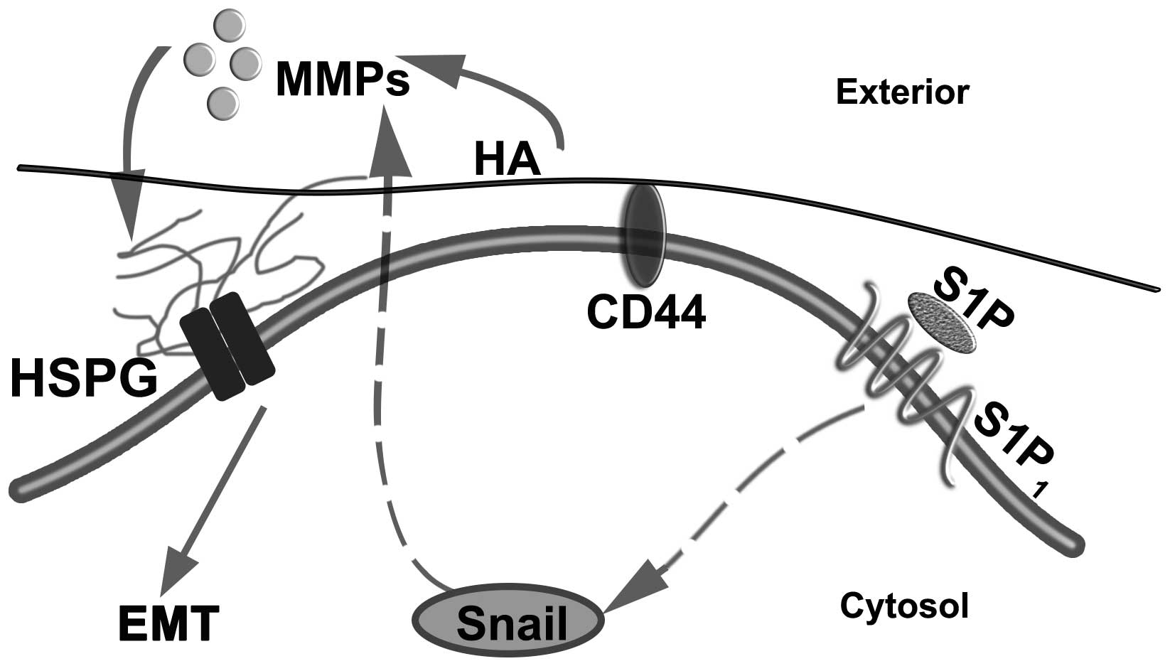
EMT is a central process in tumor metastasis. S1P
plays important roles in cell migration, motility and invasion.
Numerous studies have shown the close association among S1P, the
glycocalyx and the Snail-MMP signaling pathway, suggesting that a
glycocalyx-Snail-MMP signaling axis mediates the S1P-regulated EMT
in tumor progression and malignancy (Fig.
1). Once validated, the identification of this novel
S1P-glycocalyx-Snail-MMP signaling axis may provide insight into
novel diagnostic and anticancer therapeutic strategies.
This study was supported by the National Natural
Science Foundation of China (grant no. 11402153) and the Talent
Introduction Scientific Research Projects Funded Start-Up Funds of
Sichuan University of China (grant no. 2082204174089).
|
1
|
Thiery JP and Sleeman JP: Complex networks
orchestrate epithelial-mesenchymal transitions. Nat Rev Mol Cell
Biol. 7:131–142. 2006. View
Article : Google Scholar : PubMed/NCBI
|
|
2
|
Voulgari A and Pintzas A:
Epithelial-mesenchymal transition in cancer metastasis: Mechanisms,
markers and strategies to overcome drug resistance in the clinic.
Biochim Biophys Acta. 1796:75–90. 2009.PubMed/NCBI
|
|
3
|
Trimboli AJ, Fukino K, de Bruin A, Wei G,
Shen L, Tanner SM, Creasap N, Rosol TJ, Robinson ML, Eng C, et al:
Direct evidence for epithelial-mesenchymal transitions in breast
cancer. Cancer Res. 68:937–945. 2008. View Article : Google Scholar : PubMed/NCBI
|
|
4
|
Takeichi M: Cadherin cell adhesion
receptors as a morphogenetic regulator. Science. 251:1451–1455.
1991. View Article : Google Scholar : PubMed/NCBI
|
|
5
|
Thiery JP, Acloque H, Huang RY and Nieto
MA: Epithelial-mesenchymal transitions in development and disease.
Cell. 139:871–890. 2009. View Article : Google Scholar : PubMed/NCBI
|
|
6
|
Kalluri R and Weinberg RA: The basics of
epithelial-mesenchymal transition. J Clin Invest. 119:1420–1428.
2009. View
Article : Google Scholar : PubMed/NCBI
|
|
7
|
Orimo A, Gupta PB, Sgroi DC,
Arenzana-Seisdedos F, Delaunay T, Naeem R, Carey VJ, Richardson AL
and Weinberg RA: Stromal fibroblasts present in invasive human
breast carcinomas promote tumor growth and angiogenesis through
elevated SDF-1/CXCL12 secretion. Cell. 121:335–348. 2005.
View Article : Google Scholar : PubMed/NCBI
|
|
8
|
Barr S, Thomson S, Buck E, Russo S, Petti
F, Sujka-Kwok I, Eyzaguirre A, Rosenfeld-Franklin M, Gibson NW,
Miglarese M, et al: Bypassing cellular EGF receptor dependence
through epithelial-to-mesenchymal-like transitions. Clin Exp
Metastasis. 25:685–693. 2008. View Article : Google Scholar : PubMed/NCBI
|
|
9
|
Przybylo JA and Radisky DC: Matrix
metalloproteinase-induced epithelial-mesenchymal transition: Tumor
progression at Snail's pace. Int J Biochem Cell Biol. 39:1082–1088.
2007. View Article : Google Scholar : PubMed/NCBI
|
|
10
|
Huber MA, Azoitei N, Baumann B, Grünert S,
Sommer A, Pehamberger H, Kraut N, Beug H and Wirth T: NF-kappaB is
essential for epithelial-mesenchymal transition and metastasis in a
model of breast cancer progression. J Clin Invest. 114:569–581.
2004. View Article : Google Scholar : PubMed/NCBI
|
|
11
|
Fuxe J and Karlsson MC: TGF-β-induced
epithelial-mesenchymal transition: A link between cancer and
inflammation. Semin Cancer Biol. 22:455–461. 2012. View Article : Google Scholar : PubMed/NCBI
|
|
12
|
Tabasinezhad M, Samadi N, Ghanbari P,
Mohseni M, Saei AA, Sharifi S, Saeedi N and Pourhassan A:
Sphingosin 1-phosphate contributes in tumor progression. J Cancer
Res Ther. 9:556–563. 2013. View Article : Google Scholar : PubMed/NCBI
|
|
13
|
Milara J, Navarro R, Juan G, Peiró T,
Serrano A, Ramón M, Morcillo E and Cortijo J:
Sphingosine-1-phosphate is increased in patients with idiopathic
pulmonary fibrosis and mediates epithelial to mesenchymal
transition. Thorax. 67:147–156. 2012. View Article : Google Scholar : PubMed/NCBI
|
|
14
|
Belting M: Glycosaminoglycans in cancer
treatment. Thromb Res. 133(Suppl 2): S95–S101. 2014. View Article : Google Scholar : PubMed/NCBI
|
|
15
|
Pouysségur J, Dayan F and Mazure NM:
Hypoxia signalling in cancer and approaches to enforce tumour
regression. Nature. 441:437–443. 2006. View Article : Google Scholar : PubMed/NCBI
|
|
16
|
Qazi H, Palomino R, Shi ZD, Munn LL and
Tarbell JM: Cancer cell glycocalyx mediates mechanotransduction and
flow-regulated invasion. Integr Biol (Camb). 5:1334–1343. 2013.
View Article : Google Scholar : PubMed/NCBI
|
|
17
|
Tarbell JM, Simon SI and Curry FR:
Mechanosensing at the vascular interface. Annu Rev Biomed Eng.
16:505–532. 2014. View Article : Google Scholar : PubMed/NCBI
|
|
18
|
Jackson RL, Busch SJ and Cardin AD:
Glycosaminoglycans: Molecular properties, protein interactions, and
role in physiological processes. Physiol Rev. 71:481–539.
1991.PubMed/NCBI
|
|
19
|
Henry CB and Duling BR: Permeation of the
luminal capillary glycocalyx is determined by hyaluronan. Am J
Physiol. 277:H508–H514. 1999.PubMed/NCBI
|
|
20
|
Choi S, Lee H, Choi JR and Oh ES:
Shedding; towards a new paradigm of syndecan function in cancer.
BMB Rep. 43:305–310. 2010. View Article : Google Scholar : PubMed/NCBI
|
|
21
|
Couchman JR: Syndecans: Proteoglycan
regulators of cell-surface microdomains? Nat Rev Mol Cell Biol.
4:926–937. 2003. View
Article : Google Scholar : PubMed/NCBI
|
|
22
|
Leivonen M, Lundin J, Nordling S, von
Boguslawski K and Haglund C: Prognostic value of syndecan-1
expression in breast cancer. Oncology. 67:11–18. 2004. View Article : Google Scholar : PubMed/NCBI
|
|
23
|
Barbareschi M, Maisonneuve P, Aldovini D,
Cangi MG, Pecciarini L, Mauri Angelo F, Veronese S, Caffo O,
Lucenti A, Palma PD, et al: High syndecan-1 expression in breast
carcinoma is related to an aggressive phenotype and to poorer
prognosis. Cancer. 98:474–483. 2003. View Article : Google Scholar : PubMed/NCBI
|
|
24
|
Lendorf ME, Manon-Jensen T, Kronqvist P,
Multhaupt HA and Couchman JR: Syndecan-1 and syndecan-4 are
independent indicators in breast carcinoma. J Histochem Cytochem.
59:615–629. 2011. View Article : Google Scholar : PubMed/NCBI
|
|
25
|
Maeda T, Desouky J and Friedl A:
Syndecan-1 expression by stromal fibroblasts promotes breast
carcinoma growth in vivo and stimulates tumor angiogenesis.
Oncogene. 25:1408–1412. 2006. View Article : Google Scholar : PubMed/NCBI
|
|
26
|
Beauvais DM and Rapraeger AC: Syndecans in
tumor cell adhesion and signaling. Reprod Biol Endocrinol. 2:32004.
View Article : Google Scholar : PubMed/NCBI
|
|
27
|
Baba F, Swartz K, van Buren R, Eickhoff J,
Zhang Y, Wolberg W and Friedl A: Syndecan-1 and syndecan-4 are
overexpressed in an estrogen receptor-negative, highly
proliferative breast carcinoma subtype. Breast Cancer Res Treat.
98:91–98. 2006. View Article : Google Scholar : PubMed/NCBI
|
|
28
|
Longley RL, Woods A, Fleetwood A, Cowling
GJ, Gallagher JT and Couchman JR: Control of morphology,
cytoskeleton and migration by syndecan-4. J Cell Sci.
112:3421–3431. 1999.PubMed/NCBI
|
|
29
|
Echtermeyer F, Streit M, Wilcox-Adelman S,
Saoncella S, Denhez F, Detmar M and Goetinck P: Delayed wound
repair and impaired angiogenesis in mice lacking syndecan-4. J Clin
Invest. 107:R9–R14. 2001. View
Article : Google Scholar : PubMed/NCBI
|
|
30
|
Midwood KS, Valenick LV, Hsia HC and
Schwarzbauer JE: Coregulation of fibronectin signaling and matrix
contraction by tenascin-C and syndecan-4. Mol Biol Cell.
15:5670–5677. 2004. View Article : Google Scholar : PubMed/NCBI
|
|
31
|
Kleeff J, Ishiwata T, Kumbasar A, Friess
H, Büchler MW, Lander AD and Korc M: The cell-surface heparan
sulfate proteoglycan glypican-1 regulates growth factor action in
pancreatic carcinoma cells and is overexpressed in human pancreatic
cancer. J Clin Invest. 102:1662–1673. 1998. View Article : Google Scholar : PubMed/NCBI
|
|
32
|
Matsuda K, Maruyama H, Guo F, Kleeff J,
Itakura J, Matsumoto Y, Lander AD and Korc M: Glypican-1 is
overexpressed in human breast cancer and modulates the mitogenic
effects of multiple heparin-binding growth factors in breast cancer
cells. Cancer Res. 61:5562–5569. 2001.PubMed/NCBI
|
|
33
|
Li J, Kleeff J, Kayed H, Felix K, Penzel
R, Büchler MW, Korc M and Friess H: Glypican-1 antisense
transfection modulates TGF-beta-dependent signaling in Colo-357
pancreatic cancer cells. Biochem Biophys Res Commun. 320:1148–1155.
2004. View Article : Google Scholar : PubMed/NCBI
|
|
34
|
Yahya RS, El-Bindary AA, El-Mezayen HA,
Abdelmasseh HM and Eissa MA: Biochemical evaluation of hyaluronic
acid in breast cancer. Clin Lab. 60:1115–1121. 2014.PubMed/NCBI
|
|
35
|
Zeng Y, Adamson RH, Curry FR and Tarbell
JM: Sphingosine-1-phosphate protects endothelial glycocalyx by
inhibiting syndecan-1 shedding. Am J Physiol Heart Circ Physiol.
306:H363–H372. 2014. View Article : Google Scholar : PubMed/NCBI
|
|
36
|
Zeng Y, Liu XH, Tarbell J and Fu BM:
Sphingosine 1-phosphate induced synthesis of glycocalyx on
endothelial cells. Exp Cell Res. 339:90–95. 2015. View Article : Google Scholar : PubMed/NCBI
|
|
37
|
Fang XJ, Jiang H, Zhu YQ, Zhang LY, Fan QH
and Tian Y: Doxorubicin induces drug resistance and expression of
the novel CD44st via NF-kB in human breast cancer MCF-7 cells.
Oncol Rep. 31:2735–2742. 2014.PubMed/NCBI
|
|
38
|
Zhang Y, Thant AA, Hiraiwa Y, Naito Y,
Sein TT, Sohara Y, Matsuda S and Hamaguchi M: A role for focal
adhesion kinase in hyluronan-dependent MMP-2 secretion in a human
small-cell lung carcinoma cell line, QG90. Biochem Biophys Res
Commun. 290:1123–1127. 2002. View Article : Google Scholar : PubMed/NCBI
|
|
39
|
Pyne NJ and Pyne S: Sphingosine
1-phosphate and cancer. Nat Rev Cancer. 10:489–503. 2010.
View Article : Google Scholar : PubMed/NCBI
|
|
40
|
Payne SG, Milstien S and Spiegel S:
Sphingosine-1-phosphate: Dual messenger functions. FEBS Lett.
531:54–57. 2002. View Article : Google Scholar : PubMed/NCBI
|
|
41
|
Huang YL, Huang WP and Lee H: Roles of
sphingosine 1-phosphate on tumorigenesis. World J Biol Chem.
2:25–34. 2011. View Article : Google Scholar : PubMed/NCBI
|
|
42
|
Takabe K, Paugh SW, Milstien S and Spiegel
S: ‘Inside-out’ signaling of sphingosine-1-phosphate: Therapeutic
targets. Pharmacol Rev. 60:181–195. 2008. View Article : Google Scholar : PubMed/NCBI
|
|
43
|
Rosen H, Gonzalez-Cabrera PJ, Sanna MG and
Brown S: Sphingosine 1-phosphate receptor signaling. Annu Rev
Biochem. 78:743–768. 2009. View Article : Google Scholar : PubMed/NCBI
|
|
44
|
Pchejetski D, Böhler T, Stebbing J and
Waxman J: Therapeutic potential of targeting sphingosine kinase 1
in prostate cancer. Nat Rev Urol. 8:569–678. 2011. View Article : Google Scholar : PubMed/NCBI
|
|
45
|
Lepley D, Paik JH, Hla T and Ferrer F: The
G protein-coupled receptor S1P2 regulates Rho/Rho kinase pathway to
inhibit tumor cell migration. Cancer Res. 65:3788–3795. 2005.
View Article : Google Scholar : PubMed/NCBI
|
|
46
|
Kim ES, Kim JS, Kim SG, Hwang S, Lee CH
and Moon A: Sphingosine 1-phosphate regulates matrix
metalloproteinase-9 expression and breast cell invasion through
S1P3-Gαq coupling. J Cell Sci. 124:2220–2230. 2011. View Article : Google Scholar : PubMed/NCBI
|
|
47
|
Moon A, Kim MS, Kim TG, Kim SH, Kim HE,
Chen YQ and Kim HR: H-ras, but not N-ras, induces an invasive
phenotype in human breast epithelial cells: A role for MMP-2 in the
H-ras-induced invasive phenotype. Int J Cancer. 85:176–181. 2000.
View Article : Google Scholar : PubMed/NCBI
|
|
48
|
Xin C, Ren S, Kleuser B, Shabahang S,
Eberhardt W, Radeke H, Schäfer-Korting M, Pfeilschifter J and
Huwiler A: Sphingosine 1-phosphate cross-activates the Smad
signaling cascade and mimics transforming growth
factor-beta-induced cell responses. J Biol Chem. 279:35255–35262.
2004. View Article : Google Scholar : PubMed/NCBI
|
|
49
|
Radisky ES and Radisky DC: Matrix
metalloproteinase-induced epithelial-mesenchymal transition in
breast cancer. J Mammary Gland Biol Neoplasia. 15:201–212. 2010.
View Article : Google Scholar : PubMed/NCBI
|
|
50
|
Janda E, Lehmann K, Killisch I, Jechlinger
M, Herzig M, Downward J, Beug H and Grünert S: Ras and TGF [beta]
cooperatively regulate epithelial cell plasticity and metastasis:
Dissection of Ras signaling pathways. J Cell Biol. 156:299–313.
2002. View Article : Google Scholar : PubMed/NCBI
|
|
51
|
Jechlinger M, Grunert S, Tamir IH, Janda
E, Lüdemann S, Waerner T, Seither P, Weith A, Beug H and Kraut N:
Expression profiling of epithelial plasticity in tumor progression.
Oncogene. 22:7155–7169. 2003. View Article : Google Scholar : PubMed/NCBI
|
|
52
|
Kim ES, Sohn YW and Moon A:
TGF-beta-induced transcriptional activation of MMP-2 is mediated by
activating transcription factor (ATF) 2 in human breast epithelial
cells. Cancer Lett. 252:147–156. 2007. View Article : Google Scholar : PubMed/NCBI
|
|
53
|
Ota I, Li XY, Hu Y and Weiss SJ: Induction
of a MT1-MMP and MT2-MMP-dependent basement membrane transmigration
program in cancer cells by Snail1. Proc Natl Acad Sci USA.
106:20318–20323. 2009. View Article : Google Scholar : PubMed/NCBI
|
|
54
|
Radisky DC, Levy DD, Littlepage LE, Liu H,
Nelson CM, Fata JE, Leake D, Godden EL, Albertson DG, Nieto MA, et
al: Rac1b and reactive oxygen species mediate MMP-3-induced EMT and
genomic instability. Nature. 436:123–127. 2005. View Article : Google Scholar : PubMed/NCBI
|
|
55
|
Moustakas A and Heldin P: TGFβ and
matrix-regulated epithelial to mesenchymal transition. Biochim
Biophys Acta. 1840:2621–2634. 2014. View Article : Google Scholar : PubMed/NCBI
|
|
56
|
Xu J, Lamouille S and Derynck R:
TGF-beta-induced epithelial to mesenchymal transition. Cell Res.
19:156–172. 2009. View Article : Google Scholar : PubMed/NCBI
|