Introduction
Colorectal cancer (CRC) represents the third most
frequent tumor worldwide and the second cause of cancer-related
death, with 1.8 million cases and 862,000 deaths in 2018 according
to the World Health Organization estimates (1). Despite advances in early diagnosis and
treatment, the mortality rate is still high due to the development
of distant metastases as synchronous (in ~20.00% of patients)
(2) or metachronous (~40.00% of
patients) disease (3). Patients with
metastatic (m)CRC are generally treated with surgery and/or
systemic therapy carried out using either standard chemotherapy,
biological agents or a combination of the two methods (4). Considering targeted therapy with
biological agents, certain patients will respond to therapy,
whereas others will not due to their genetic features (5).
A previous study has demonstrated that mCRC is
characterized by a high frequency of mutations in the RAS
gene that are the main determinants of the failure of
anti-EGFR-based therapy such as cetuximab and panitumumab (6). Therefore, patients with mCRC are
routinely screened for BRAF, KRAS and NRAS mutations
to select the appropriate patients who are more likely to have a
positive response when treated with anti-EGFR (7–9). The
most frequent mutations associated with a poor response to EGFR
therapy are located in KRAS exon 2 (codons 12 and 13, 40.00%
of patients), although other codons (59 and 61 in exon 3, 117 and
146 in exon 4) may be affected, as well as mutations in NRAS
(codons 12, 13, 59, 61, 117 and 146) (10–14).
RAS status is currently determined in tissue samples, either
from the primary tumor or from metastasis obtained during biopsy or
surgery.
Since patients with mCRC patients are characterized
by poor health conditions, multiple biopsies should be avoided;
however, it has been demonstrated that a single tissue biopsy may
not be representative of the whole tumor due to intratumoral
heterogeneity (15). In addition,
tissue biopsy cannot be used in these patients for disease
monitoring for the same reason. Based on this, the concept of
liquid biopsy has been proposed as a surrogate for a tissue sample
(16–22). In previous years technological
improvements have enabled the study of circulating tumor cells
(CTCs) and circulating tumor DNA (ctDNA) in the peripheral blood
samples of patients with advanced cancers (23,24).
Previous studies have demonstrated that ctDNA levels are associated
with clinicopathological and biological features such as tumor
histological type, stage, burden, blood vessel proximity, apoptotic
rate and metastatic potential (17,25–28).
Among patients with mCRC, it has been demonstrated that a high
proportion of patients (86.00–100.00%) is characterized by
detectable ctDNA in plasma, and that 1.90–27.00% of total ctDNA
harbor different mutations (26).
The aims of the present study were as follows: i) To
analyze the concordance between KRAS and NRAS
mutational status evaluated in the tissue and the plasma from a
cohort of patients with mCRC (synchronous or metachronous); ii) to
evaluate the association between KRAS and NRAS
mutations in the ctDNA and patient clinicopathological features;
and iii) to analyze the mutant allele fraction (MAF) distribution
in the plasma samples and identify potential clinical
associations.
Materials and methods
Patients
The study cohort included 31 patients with mCRC
enrolled at Medical Oncology Unit, Azienda
Ospedaliero-Universitaria Careggi (Florence, Italy) between January
2017 and August 2018 following written informed consent and
approval from the Ethical Committee of Azienda
Ospedaliero-Universitaria Careggi (approval no. BIO.16.028,
25/10/2016). All patients were previously diagnosed with clinical
Tumor-Node-Metastasis stage IV (29)
and had measurable disease according to the Response Evaluation
Criteria in Solid Tumors version 1.1 (30).
Tissue samples
For all patients, KRAS and NRAS status
had been previously determined in tissue biopsies of either primary
tumors or metastases by using the Myriapod® Colon Status
kit (Diatech Pharmacogenetics Srl), which allows the detection of
216 mutations in oncogenes responsible for colorectal
cancerogenesis (KRAS, BRAF, PIK3CA, NRAS) using the
genotyping platform MassARRAY® system (Sequenom, Inc.)
based on matrix-assisted laser desorption ionization-time of flight
(MALDI-TOF) mass spectrometry.
Selection of biological samples and
DNA extraction
Hematoxylin-eosin sections from biopsies of
neoplastic colorectal tissues were obtained from the archive of the
Pathological Anatomy department of Azienda
Ospedaliero-Universitaria Careggi and then revised by two
experienced pathologists. Only sections containing ≥100 neoplastic
cells were selected. Corresponding formalin-fixed paraffin-embedded
blocks were obtained and 10-µm tissue sections were cut,
deparaffinized with solvent, rinsed in alcohol and dried before
processing. DNA was extracted from tissue sections a using MagCore
Genomic DNAFFPE One-Step kit (RBC Bioscience Corp.) and then
analyzed for quality and concentration using a spectrophotometer.
If the quality was optimal and the concentration was between
2.5–25.0 ng/ml, the samples were amplified.
DNA Amplification
DNA was amplified by multiplex-PCR using the Master
Amp-Mix amplification mixture (Diatech Pharmacogenetics Srl) in
order to obtain fragments comprising all polymorphic sites of
interest. The reaction mixture contained 1.3 µl water, 0.5 µl PCR
buffer, 0.4 µl MgCl2, 0.1 µl dNTP mix and 0.2 µl PCR
enzyme. All PCR mixes were placed on a plate and added to a
negative control (2 µl water), a sample (2 µl extracted DNA) and a
positive control (2 µl human wild-type control DNA provided with
the kit). Following spinning the reaction mixture by brief
centrifugation at maximum speed, the plate was put in a
thermocycler with the following amplification profile: 95°C for 2
min; followed by 45 cycles of 95°C for 30 sec, 56°C for 30 sec and
72°C for 60 sec; 72°C for 5 min; 4°C for 5 min; and hold at
10°C.
Treatment with shrimp alkaline
phosphatase (SAP) and iPLEX extension
Following the amplification, the amplification
products were treated with SAP (provided with the kit) to remove
nucleotide residues. A SAP cocktail (SAP-Mix) containing 1.53 ml
water, 0.17 ml SAP Buffer and 0.30 ml SAP Enzyme was prepared for
each sample. The samples were added to the SAP-Mix and placed in a
thermocycler with the following conditions: 37°C for 40 min; 85°C
for 5 min; 4°C for 5 min; and hold at 10°C. Subsequently, each
sample was extended using a Master Ext-Mix extension mixture. Each
mixture comprised 0.56 ml water, 0.20 µl Buffer Plus, 0.20 ml
Termination mix and 0.04 ml Thermosequenase for each sample
reaction. The samples were placed in a thermocycler with the Master
Ext-Mix and processed under the following thermocycling conditions:
94°C for 30 sec; followed by 40 cycles of 94° for 5 sec, 5 cycles
of 52°C for 5 sec and 5 cycles of 80°C for 5 sec; 72°C for 3 min;
4°C for 5 min; and hold at 10°C.
Spectra acquisition and analysis
Once the extension reaction was completed, the
reaction products were dispensed on a SpectroCHIP® II
G96 using the Nanodispenser RS-1000® Instrument. The
SpectroCHIP was then placed in the MassARRAY® Analyzer 4
Instrument for spectra acquisition. The generated spectra were
analyzed using the MassARRAY® database with
iGenetics® Myriapod® software version 5.1
(Diatech Pharmacogenetics Srl).
Blood sample collection
Peripheral blood (8 ml) was collected from each
patient enrolled in the study in K2 EDTA BD
Vacutainer® collection tubes (BD Biosciences)
immediately before starting therapy. The plasma was isolated within
4 h according to the Sysmex-Inostics protocol for the determination
of KRAS and NRAS status with the OncoBEAM®
RAS CRC assay (Sysmex Inostics). The plasma samples were stored at
−80°C.
ctDNA extraction and purification
ctDNA was extracted from the plasma and purified
using a QIAamp® Circulating Nucleic Acid kit and
QIAvac24 plus (both from Qiagen GmbH) according to the
manufacturer's protocol with slight modifications as indicated by
Sysmex Inostics.
BEAMing assay
For the detection of RAS mutations in the
ctDNA, OncoBEAM® RAS CRC kit (Sysmex Inostics) was used
according to the manufacturer's instructions. The ctDNA samples
were amplified with a multiplex PCR performed as follows: 98°C for
30 sec; followed by 21 cycles of 98°C for 15 sec, 60°C for 25 sec
and 72°C for 25 sec; 72°C for 35 sec; and hold at 4°C. The
multiplex PCR was followed by pooling and dilution with 1X pH 8.0
TE buffer to get the optimal amount of PCR product for each codon.
The diluted samples together with specific positive and negative
controls [part of the OncoBEAM® RAS CRC kit, (Sysmex
Inostics GmbH) and carrying specific mutations and no template,
respectively] were used for emulsion PCR. Following cycling,
magnetic beads bound to the amplicons were retrieved through a
breaking phase followed by the hybridization step carried out with
specific fluorescent probes for further detection by Cube6i flow
cytometer (Sysmex Inostics). The breaking phase was carried out
according to the manufacturer's protocol provided with the
OncoBEAM® RAS CRC kit using a magnet and two buffers,
Breaking Buffer 1 and 2, provided with the kit. The hybridization
step performed as follows: 70°C for 30 sec; gradual decrease from
70°C to 24°C with a 3% ramp rate; 24°C for 1 min; and hold at 21°C.
After the completion of the run, the data were exported and
analyzed by FCS Express version 5.0 software (Denovo software;
Sysmex Inostics).
Statistical analysis
The demographic, clinical and biological
characteristics of the patients are presented as frequencies and
percentages. Continuous variables are presented as the median
(range of variation). To evaluate the associations between
RAS mutational status and clinicopathological features,
parametric (Fisher's exact) and non-parametric (Kruskal-Wallis)
tests were used as appropriate. Data were analyzed using the
statistical software Stata 9.1 (StataCorp LP). A two-sided P
value ≤0.05 was considered to indicate a statistically significant
difference. For categorical variables, the samples were classified
as follows: Sex, male vs. female; localization of the primary
tumor, right colon vs. left colon vs. rectum; pathological stage at
the time of ctDNA analysis, relapse vs. newly diagnosed stage IV
tumor; histological type, adenocarcinoma vs. mucinous
adenocarcinoma; grading, G2 vs. G3; number of metastatic sites at
the time of ctDNA analysis, 1 vs. 2 vs. 3 vs. >3; surgery for
primary tumor, yes vs. no. The percentage of agreement was
evaluated according to the Cohen's κ of concordance and its 95.00%
confidence interval.
Results
Clinical characteristics
In the present study, a total of 31 patients were
enrolled (16 female, 15 male) with a mean age 67 years (range,
46–85 years) and a histologically diagnosed mCRC not treated for
the metastatic disease. The primary tumor was mainly localized in
the colon (80.64%) with a similar distribution between the left and
right colon. Among the patients, 64.52% received surgery with
curative intent for the primary disease and developed metachronous
metastases. The main demographic and clinicopathological features
of the cohort are presented in Table
I.
 | Table I.Demographic and clinicopathological
characteristics of the study cohort. |
Table I.
Demographic and clinicopathological
characteristics of the study cohort.
| Characteristic | No. of patients
(%) |
|---|
| Sex |
|
|
Female | 16 (51.61) |
|
Male | 15 (48.39) |
| Localization |
|
| Left
colon | 12 (38.71) |
| Right
colon | 13 (41.94) |
|
Rectum | 6 (19.35) |
| Pathological stage
at the time of ctDNA analysis |
|
|
Synchronous | 18 (58.06) |
|
Metachronous | 13 (41.94) |
| Histological
type |
|
|
Adenocarcinoma | 24 (87.10) |
|
Mucinous adenocarcinoma | 7 (12.90) |
| Grading |
|
| G2 | 19 (61.29) |
| G3 | 3 (9.68) |
|
Undefined | 9 (29.03) |
| Metastatic sites at
the time of ctDNA analysis |
|
| Only
liver | 8 (25.81) |
| Only
lung | 1 (3.23) |
|
Peritoneum | 4 (12.90) |
|
Multiple | 18 (58.06) |
| Number of
metastatic sites at the time of ctDNA analysis |
|
| 1 | 13 (41.93) |
| 2 | 12 (38.71) |
| ≥3 | 6 (19.36) |
| Surgery for primary
tumor |
|
| No | 11 (35.48) |
|
Yes | 20 (64.52) |
| Tissue used for
molecular analysis of RAS mutational status |
|
| Primary
tumor | 23 (74.19) |
|
Metastasis | 8 (25.81) |
RAS mutational status in tissue
samples
Molecular determination of RAS status in
paraffin-embedded samples was performed using primary tumor samples
in 74.19% of the patients and in metastatic tissues in the
remaining 25.81% of the patients. The analysis was performed by
MALDI-TOF mass spectrometry associated to single base extension
technology and provided conclusive results for all patients; six
samples (19.35%) expressed the wild-type form of KRAS, and
25 samples (80.65%) harbored mutations (Table II). The same analysis was performed
for NRAS, showing the presence of a mutation in only one
patient (96.77% wild-type and 3.23% mutated) (Table II).
| Table II.KRAS and NRAS analysis
of tissue samples. |
Table II.
KRAS and NRAS analysis
of tissue samples.
| RAS
status | No. of patients
(%) |
|---|
| Wild-type
KRAS | 6 (19.35) |
| Mutated
KRAS | 25 (80.65) |
| Exon 2
codon 12 | 19 (76.00) |
|
G12A | 4 |
|
G12C | 3 |
|
G12D | 10 |
|
G12V | 2 |
| Exon 2
codon 13 | 5 (20.00) |
|
G13D | 4 |
|
G13V | 1 |
| Exon 3
codon 59 | 0 (0.00) |
| Exon 3
codon 61 | 0 (0.00) |
| Exon 4
codon 117 | 0 (0.00) |
| Exon 4
codon 146 | 1 (4.00) |
|
A146P | 1 |
| Wild-type
NRAS | 30 (96.77) |
| Mutated
NRAS | 1 (3.23) |
| Exon 2
codon 12 | 1 (100.00) |
|
G12D | 1 |
| Exon 2
codon 13 | 0 (0.00) |
| Exon 3
codon 59 | 0 (0.00) |
| Exon 3
codon 61 | 0 (0.00) |
| Exon 4
codon 117 | 0 (0.00) |
| Exon 4
codon 146 | 0 (0.00) |
|
Undefined | 0 (0.00) |
RAS mutational status in plasma
samples
RAS mutational status was analyzed using
BEAMing technology. Of the 31 samples analyzed, wild-type
KRAS was identified in seven patients, whereas mutations
were detected in 22 patients (17 in codon 12, four in codon 13, one
in codon 146); for two patients, an informative result could not be
obtained. Regarding NRAS analysis, the wild-type form of the
gene was observed in 24 patients, two patients harbored a mutation
in codon 12, four harbored a mutation in codon 61 and the data for
one patient was not informative (Table
III). Fig. 1 demonstrates
representative plots obtained using the BEAMing assay (left plots,
‘universal signal’ vs. ‘mutant signal’; right plots, ‘wild-type
signal’ vs. ‘mutant signal’). In Fig.
1A, the results of a no template control are presented, and no
signal is present in the right plot. By contrast, in the two plots
in Fig. 1B representing the positive
control, an intense signal in the gate corresponding to mutant
beads (mtP3) can be observed. In Fig. 1C
and D, two representative samples classified as wild-type and
mutated, respectively, are demonstrated: In the former, almost no
signal is present in mtP3 gate, whereas in the latter, the presence
of the mutation is marked by the black dots in the mrP3 quadrant
(see arrow).
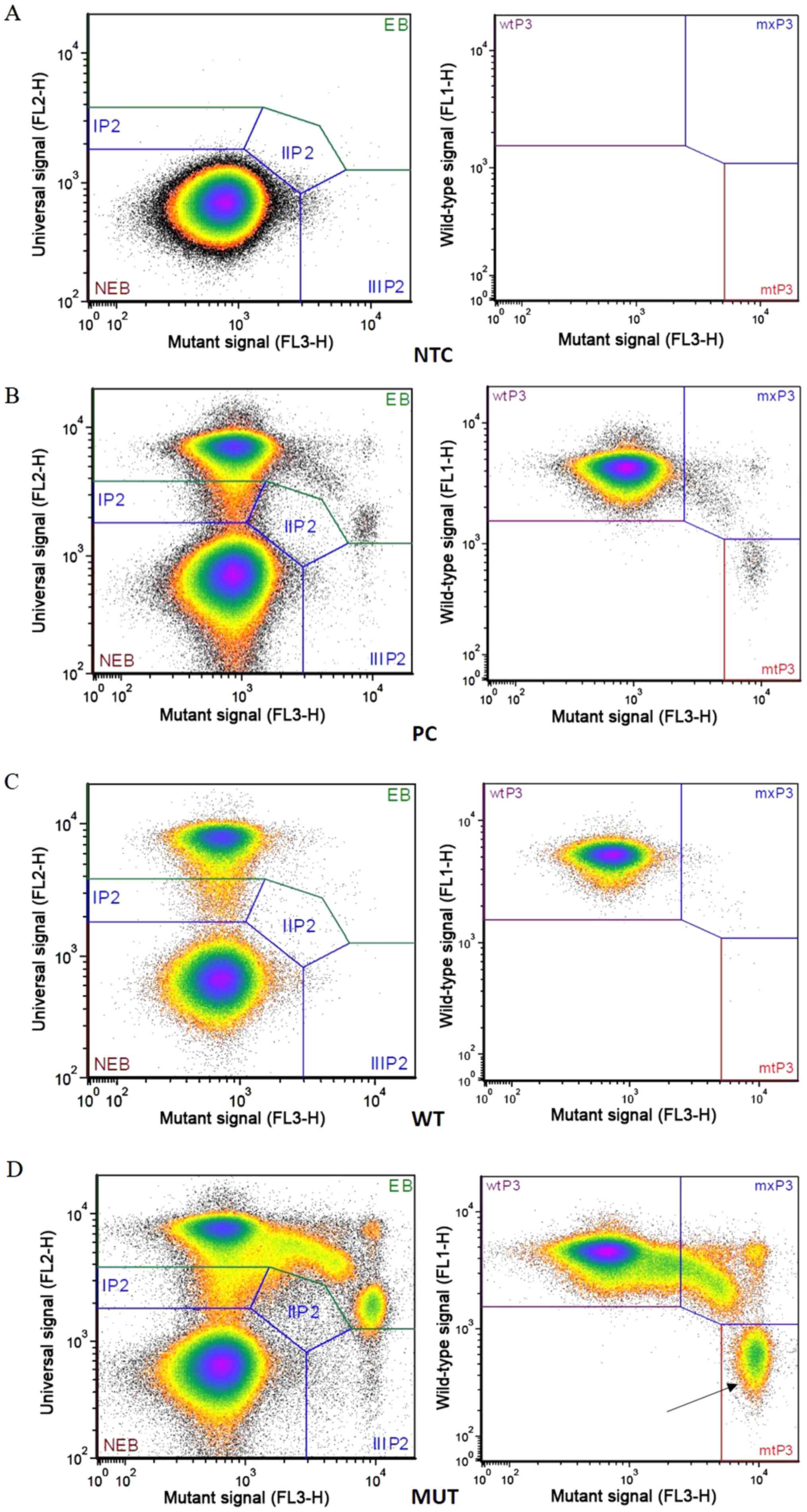 | Figure 1.Representative plots obtained by
BEAMing assay. In the left plot of each panel ‘universal signal’
vs. ‘mutant signal’ is reported, whereas in the right plot,
‘wild-type signal’ vs. ‘mutant signal’ is presented. (A) Negative
control; (B) PC; mutant beads, 844; mutant fraction, 0.46%; (C) WT;
mutant beads, 14; mutant fraction, 0.02%; (D) MUT; mutant beads,
14,746; mutant fraction, 7.645 (see arrow). EB, extended beads;
NEB, non-extended beads; wt, wild-type; mx, mutant and wild-type;
mt, mutant; NTC, no template control; PC, positive control; WT,
wild-type sample; MUT, mutant sample. |
| Table III.KRAS and NRAS analysis
of plasma samples. |
Table III.
KRAS and NRAS analysis
of plasma samples.
| RAS
status | No. of patients
(%) |
|---|
| Wild-type
KRAS | 7 (22.58) |
| Mutated
KRAS | 22 (77.42) |
| Exon 2
codon 12 | 17 (77.27) |
| Exon 2
codon 13 | 4 (18.18) |
| Exon 3
codon 59 | 0 (0.00) |
| Exon 3
codon 61 | 0 (0.00) |
| Exon 4
codon 117 | 0 (0.00) |
| Exon 4
codon 146 | 1 (4.55) |
| Not
informative | 2 |
|
Total | 31 |
| Wild-type
NRAS | 24 (77.42) |
| Mutated
NRAS | 6 (22.58) |
| Exon 2
codon 12 | 2 (33.33) |
| Exon 2
codon 13 | 0 (0.00) |
| Exon 3
codon 59 | 0 (0.00) |
| Exon 3
codon 61 | 4 (66.67) |
| Exon 4
codon 117 | 0 (0.00) |
| Exon 4
codon 146 | 0 (0.00) |
| Not
informative | 1 |
|
Total | 31 |
The technique used to analyze these plasma samples
from patients with mCRC did not provide the result of the test as
an absolute number of RAS mutated alleles, but as the mutant
allele fraction (MAF), which is the ratio (expressed as a
percentage) of mutated and wild-type RAS alleles (Table IV). Analysis of the mutated samples
(mutated beads ≥40) revealed large variability among patients, and
the mean number of mutant beads was 6,047.909±1.594 (n=22) for
KRAS and 281±0.103 (n=5) for NRAS (Table IV, last row).
| Table IV.OncoBEAM® RAS
colorectal cancer test results. |
Table IV.
OncoBEAM® RAS
colorectal cancer test results.
|
| KRAS | NRAS |
|---|
|
|
|
|
|---|
| Patient ID | Mutant beads
(n) | MAF | Test result | Mutant beads
(n) | MAF | Test result |
|---|
| COL001 | 8 | 0.013 | Wild-type | 13 | 0.041 | Wild-type |
| COL002 | 97,951 | 21.226 | Mutation
detected | 65 | 0.031 | Mutation
detected |
| COL003 | 6,862 | 5.610 | Mutation
detected | 6 | 0.011 | Wild-type |
| COL004 | 36,785 | 13.006 | Mutation
detected | 8 | 0.009 | Wild-type |
| COL005 | 12,879 | 6.759 | Mutation
detected | 22 | 0.020 | Wild-type |
| COL006 | 1,516 | 0.998 | Mutation
detected | 17 | 0.058 | Wild-type |
| COL007 | 32,095 | 5.262 | Mutation
detected | 4 | 0.002 | Wild-type |
| COL008 | 6 | 0.005 | Wild-type | 49 | 0.053 | Mutation
detected |
| COL009 | 43,649 | 9.861 | Mutation
detected | 7 | 0.002 | Wild-type |
| COL010 | 7 | 0.009 | Wild-type | 2 | 0.005 | Wild-type |
| COL011 | 691 | 0.653 | Mutation
detected | 0 | 0.000 | Wild-type |
| COL012 | 51,794 | 11.216 | Mutation
detected | 4 | 0.001 | Wild-type |
| COL013 | 137 | 0.144 | Mutation
detected | 6 | 0.015 | Wild-type |
| COL014 | 70 | 0.068 | Mutation
detected | 9 | 0.022 | Wild-type |
| COL015 | 29 | 0.012 | Wild-type | 10 | 0.007 | Wild-type |
| COL016 | 292 | 0.045 | Mutation
detected | 7 | 0.002 | Wild-type |
| COL017 | 29,038 | 28.149 | Mutation
detected | 2 | 0.002 | Wild-type |
| COL018 | 14,746 | 7.645 | Mutation
detected | 164 | 0.344 | Mutation
detected |
| COL019 | 9,023 | 5.455 | Mutation
detected | 3 | 0.017 | Wild-type |
| COL020 | 45 | 0.056 | Mutation
detected | NI | NI | NI |
| OB3 | 61 | 0.053 | Mutation
detected | 22 | 0.025 | Wild-type |
| OB4 | 7,861 | 11.211 | Mutation
detected | 1 | 0.002 | Wild-type |
| OB5 | NI | NI | NI | 1 | 0.003 | Wild-type |
| OB7 | 2 | 0.005 | Wild-type | 0 | 0.000 | Wild-type |
| OB8 | 1,178 | 0.918 | Mutation
detected | 6 | 0.005 | Wild-type |
| OB9 | 10,639 | 4.693 | Mutation
detected | 5 | 0.004 | Wild-type |
| OB10 | 42 | 0.017 | Mutation
detected | 429 | 0.469 | Mutation
detected |
| OB11 | 24 | 0.028 | Wild-type | 246 | 0.516 | Mutation
detected |
| OB12 | 15 | 0.013 | Wild-type | 5 | 0.009 | Wild-type |
| OB14 | NI | NI | NI | 9 | 0.023 | Wild-type |
| OB15 | 0 | 0.000 | Wild-type | 7 | 0.012 | Wild-type |
| Mean ± SEM |
6,047.909±1.594 |
|
| 281±0.103 |
|
|
For the majority of the patients, a single mutation
either in KRAS or NRAS was detected (28/31), whereas
three patients presented with mutations in both genes. Notably, in
two of these three patients (patients COL002 and COL018), a
KRAS mutation was also identified in the tissue sample,
whereas NRAS mutations were detected only in the plasma. In
both cases, the allele frequency of KRAS (MAF, 21.226 and
7.645) was higher compared with that of NRAS (MAF, 0.031 and
0.344); this result was not unexpected since the BEAMing technique
exhibited higher sensitivity compared with that of MALDI-TOF mass
spectrometry, and thus it is possible that a number of the
mutations were not detected in the tissue samples if their
frequency was low, as observed in these patients.
The concordance between the results obtained in the
tissue and plasma samples was subsequently evaluated (Table V). Analysis of the data using Cohen's
κ revealed a moderate concordance between tissue and plasma
analysis of KRAS mutational status (agreement, 85.18%;
Cohen's κ, 0.513); for NRAS mutational status a fair
agreement was found (agreement, 83.33%; Cohen's κ, 0.242)
| Table V.Concordance between tissue and plasma
analysis of RAS mutational status. |
Table V.
Concordance between tissue and plasma
analysis of RAS mutational status.
|
| KRAS | NRAS |
|---|
|
|
|
|
|---|
| Patient ID | Tissue | Plasma | Concordance | Tissue | Plasma | Concordance |
|---|
| COL001 | G12D | wt | no | wt | wt | yes |
| COL002 | G12C | Cd12 | yes | wt | Cd12 | no |
| COL003 | G12A | Cd12 | yes | wt | wt | yes |
| COL004 | G12D | Cd12 | yes | wt | wt | yes |
| COL005 | G13D | Cd13 | yes | wt | wt | yes |
| COL006 | G12C | Cd12 | yes | wt | wt | yes |
| COL007 | G12D | Cd12 | yes | wt | wt | yes |
| COL008 | wt | wt | yes | G12D | CD12 | yes |
| COL009 | G13D | Cd13 | yes | wt | wt | yes |
| COL010 | G13D | Cd13 | yes | wt | wt | yes |
| COL011 | G12D | Cd12 | yes | wt | wt | yes |
| COL012 | G12A | Cd12 | yes | wt | wt | yes |
| COL013 | G13D | Cd13 | yes | wt | wt | yes |
| COL014 | G12A | Cd12 | yes | wt | wt | yes |
| COL015 | G13V | wt | no | wt | wt | yes |
| COL016 | G12D | Cd12 | yes | wt | wt | yes |
| COL017 | G146P | Cd146 | yes | wt | wt | yes |
| COL018 | G12D | Cd12 | yes | wt | CD61 | no |
| COL019 | G12C | Cd12 | yes | wt | wt | yes |
| COL020 | G12D | Cd12 | yes | wt | NI | NA |
| OB3 | G12D | Cd12 | yes | wt | wt | yes |
| OB4 | G12D | Cd12 | yes | wt | wt | yes |
| OB5 | wt | NI | NA | wt | wt | yes |
| OB7 | G12D | wt | no | wt | wt | yes |
| OB8 | G12V | Cd12 | yes | wt | wt | yes |
| OB9 | G12A | Cd12 | yes | wt | wt | yes |
| OB10 | wt | Cd12 | no | wt | CD61 | no |
| OB11 | wt | wt | yes | wt | CD61 | no |
| OB12 | wt | wt | yes | wt | wt | yes |
| OB14 | wt | NI | NA | wt | CD61 | no |
| OB15 | G12V | wt | no | wt | wt | yes |
In order to evaluate the association between
RAS mutational status and patient clinicopathological
features, the samples were divided into two groups: Wild-type and
mutated (either K- or NRAS). No significant
associations were identified between the two groups (Table VI).
| Table VI.Associations between RAS
mutational status and clinicopathological features. |
Table VI.
Associations between RAS
mutational status and clinicopathological features.
|
|
| RAS
mutational status, n (%) |
|
|---|
|
|
|
|
|
|---|
| Clinical
feature | n | Wild-type | Mutated | P-value |
|---|
| Sex |
|
|
| 0.355a |
|
Male | 15 | 3 (20.00) | 12 (80.00) |
|
|
Female | 16 | 3 (18.75) | 13 (81.25) |
|
| Age, years |
|
|
| 0.221b |
| Mean,
67 (range, 46–85) | 31 | 6 (19.35) | 25 (80.65) |
|
| Localization |
|
|
| 0.333a |
| Right
colon | 13 | 1 (7.69) | 12 (92.31) |
|
| Left
colon | 12 | 3 (25.00) | 9 (75.00) |
|
|
Rectum | 6 | 2 (33.33) | 4 (66.67) |
|
| Pathological stage
at the time of ctDNA analysis |
|
|
|
>0.999a |
|
Synchronous | 18 | 3 (16.67) | 15 (83.33) |
|
|
Metachronous | 13 | 3 (23.08) | 10 (76.92) |
|
| Histological
type |
|
|
| 0.287a |
|
Adenocarcinoma | 24 | 3 (12.50) | 21 (87.50) |
|
|
Mucinous adenocarcinoma | 7 | 3 (42.86) | 4 (57.14) |
|
| Grading |
|
|
| 0.422a |
| G1 | 0 | 0 (0.00) | 0 (0.00) |
|
| G2 | 19 | 3 (15.79) | 16 (84.21) |
|
| G3 | 3 | 1 (33.33) | 2 (66.67) |
|
|
Undefined | 9 | 2 (22.22) | 7 (77.78) |
|
| Number of
metastatic sites at the time of ctDNA analysis |
|
|
| 0.413a |
| 1 | 13 | 1 (7.69) | 12 (92.31) |
|
| 2 | 12 | 5 (41.67) | 7 (58.33) |
|
| ≥3 | 6 | 0 (0.00) | 6 (100.00) |
|
| Surgery for primary
tumor |
|
|
|
>0.999a |
| No | 11 | 3 (27.27) | 8 (72.73) |
|
|
Yes | 20 | 3 (15.00) | 17 (85.00) |
|
Discussion
Emerging evidence has demonstrated the importance of
liquid biopsy as a surrogate of standard tissue biopsies for
diagnostic purposes as well as for monitoring patients with mCRC
(16–21). In particular, in mCRC patients,
multiple biopsies should be avoided due to their poor general
health conditions.
In 2016, a meta-analysis reported that ctDNA may
represent an indicator for poor prognosis, including both
recurrence-free survival and overall survival (OS), in patients
with stage I–IV CRC (28). A study
performed by Spindler et al (31) demonstrated that an increase in ctDNA
reduced the progression-free survival (PFS) and OS (defined as the
months elapsed from the first diagnosis and the progression of the
disease or death of the patient, respectively) time with a hazard
ratio of 1.4 (95% CI, 1.1–1.7; P=0.03) and 1.6 (95% CI, 1.3–2.0;
P<0.0001), respectively. In addition, their study revealed that
the evaluation of KRAS mutations in the plasma provided
additional information on the patient outcome (31). A recent study performed a parallel
analysis of ctDNA and circulating tumor cells (CTCs) and
demonstrated that the former represented an improved tool for the
management of patients with CRC since CTCs were not detected in all
samples in contrast to ctDNA, and a low volume of blood was
sufficient for the molecular analysis (32).
Overall, the frequency of K- and N-RAS
mutations in the present study was higher compared with the one
expected to be observed in the general population, although
notably, for a part of the cohort, one of the inclusion criteria
was the presence of KRAS mutations.
In the cohort of patients analyzed in this study,
coexistence of K- and N-RAS mutations was observed in
3/31 patients (9.70%). For two patients, MAF values were higher for
KRAS compared with those for NRAS, whereas an
opposite trend was observed in the third patient. Although
K- and N-RAS mutations are generally mutually
exclusive, the high sensitivity of BEAMing may allow the
identification of subclonal mutations that may have been missed
with other techniques. However, since these mutations are present
at in a small proportion of patients their biological and clinical
relevance requires further investigation. It may have been be
interesting to apply the BEAMing technology to the tissue samples
to evaluate whether such subclonal mutations were present in the
tumor tissues as well as in the plasma, but it was not possible to
identify them due to the lower sensitivity of the standard
techniques applied in the analyses of tissue samples.
The results of the present study identified a
concordance between BEAMing and MALDI-TOF, as previously reported
in other studies (33,34). Overall, for the KRAS
mutational status, five samples were not in accordance. The
possible causes of such discordance were subsequently explored. In
four patients, a mutation was detected in the tissue, but not in
the plasma; this observation may be explained by the elimination of
sensitive clones by the treatment if the two analyses were
performed at different times (as in patients OB7 and OB15); on the
other hand, the discrepancy may also have occurred due to the low
tumor burden if the two analyses were performed at similar times
(as in patients COL001 and COL015). COL001 and COL015, harboring
KRAS mutations in the tissue, but not in the plasma, had
mucinous tumors and underwent surgical resection with curative
intent. In addition, the analysis of radiographic images captured
on a date close to the blood collection demonstrated that the two
patients exhibited a low tumor burden (localization of the disease
limited to the peritoneum in one patient and relatively small
metastases in the liver in the other). These results were in
accordance with those obtained in a large multicenter prospective
cohort, which demonstrated that surgery of the primary tumor,
absence of liver metastases and peritoneal localization were
significantly associated with inconclusive results in the plasma
(35).
In one patient (OB10), a mutation was detected in
the plasma, but not in the tissue. Since the BEAMing assay is more
sensitive compared with MALDI-TOF mass spectrometry, the mutation
may have been missed due to the low MAF (0.017). Another
explanation may be that mutations may have developed a long time
after diagnosis and evaluation of RAS mutational status in
tissue samples, and if tissue analysis was performed using tissues
obtained during a biopsy, the mutation may have been missed due to
the heterogeneity and the low tumor cell fraction in the specimen.
This patient had a long and satisfactory response (2 years) to
therapy that has been recently published as a case report (36) and may represent an example of
rechallenge failure due to molecular determinants. In particular,
since the patient exhibited a complete response to Cetuximab, after
two years, the treatment was repeated using the same drug, but the
disease rapidly progressed; the analysis of plasma in this case may
have helped determine a different schedule of treatment.
In addition, for NRAS, five samples were not
in accordance; a mutation was detected in the plasma, whereas the
tissues was classified as harboring wild-type NRAS. This
discrepancy may be explained as aforementioned.
A potential limitation of the present study was the
absence of validation of the data obtained through BEAMing with a
different technique. In the present study, it was not possible to
obtain such confirmation, since the amount DNA available from each
patient was not enough to perform other experiments with various
methodologies, and no additional blood collection was performed due
to the general conditions of the patients to avoid enhancing their
discomfort. Such evaluation represents a future perspective and
will be performed in a further study. However, in patients with low
MAF (such as patient OB10) with a value close to the cut-off,
BEAMing analysis was performed twice to verify the result since
additional methods could not be performed.
A recent multicenter clinical study performed in
Japan provided clinical validation of the OncoBEAM® RAS
CRC assay (37), paving the road to
the incorporation of BEAMing into clinical practice. The results of
the aforementioned study, although it was performed on a larger
cohort compared to the one used in the present study, were in
concert with the findings of the present study since they obtained
an overall concordance between tissue and plasma analyses equal to
86.4%.
Overall, the results of the present study identified
a certain degree of agreement between the two techniques. Based on
these preliminary data, it is suggested that both analyses should
be routinely performed to provide clinicians an additional tool for
the management of patients with mCRC. To confirm these results,
validation studies on larger cohorts are warranted, as well as
studies aimed at determining the RAS status to monitor
therapy and during follow-up.
Acknowledgements
The authors would like to thank Dr Caterina
Fattorini (Section of Pathological Anatomy, Department of Health
Sciences, University of Florence, Azienda Ospedaliero-Universitaria
Careggi) for integrating the methods concerning tissue analysis.
Part of these data were presented at EACR-ESMO Joint Conference on
Liquid Biopsies, held in Bergamo (Italy) on May 15th-17th 2019.
Funding
No funding was received.
Availability of data and materials
The datasets used and/or analyzed during the current
study are available from the corresponding author on reasonable
request.
Authors' contributions
EL performed the experiments, analyzed the data and
wrote the manuscript. LA enrolled and followed up the patients,
retrieved clinical data and wrote the manuscript. BF enrolled and
followed up the patients, contributed to analysis of the data and
writing the manuscript. LDC collected the samples and participated
in data analysis. ADC retrieved clinical data and participated in
data analysis. DL enrolled and followed up the patients and
contributed to interpreting and analyzing data. MA participated in
performing the experiments. AA participated in the study design and
revised the manuscript. FC performed molecular analysis on tissue
specimens. LM performed histological diagnoses. FDC designed and
supervised the study and revised the manuscript. All authors read
and approved the final manuscript.
Ethics approval and consent to
participate
The present study was approved by the Ethical
Committee of Azienda Ospedaliero-Universitaria Careggi (approval
no. BIO.16.028, 25/10/2016). All patients were enrolled after
providing written informed consent.
Patient consent for publication
Not applicable.
Competing interests
The authors declare that they have no competing
interests.
References
|
1
|
International Agency for Research on
Cancer, . Colorectal cancer. Source: Globocan 2018. The Global
Cancer Observatory. 2019.
|
|
2
|
Cook AD, Single R and McCahill LE:
Surgical resection of primary tumors in patients who present with
stage IV colorectal cancer: An analysis of surveillance,
epidemiology, and end results data, 1988 to 2000. Ann Surg Oncol.
12:637–645. 2005. View Article : Google Scholar : PubMed/NCBI
|
|
3
|
van Gestel YR, de Hingh IH, van Herk-Sukel
MP, van Erning FN, Beerepoot LV, Wijsman JH, Slooter GD, Rutten HJ,
Creemers GJ and Lemmens VE: Patterns of metachronous metastases
after curative treatment of colorectal cancer. Cancer Epidemiol.
38:448–454. 2014. View Article : Google Scholar : PubMed/NCBI
|
|
4
|
Van Cutsem E, Cervantes A, Nordlinger B
and Arnold D; ESMO Guidelines Working Group, : Metastatic
colorectal cancer: ESMO Clinical Practice Guidelines for diagnosis,
treatment and follow-up. Ann Oncol. 25 (Suppl 3):iii1–9. 2014.
View Article : Google Scholar : PubMed/NCBI
|
|
5
|
Xie YH, Chen YX and Fang JY: Comprehensive
review of targeted therapy for colorectal cancer. Signal Transduct
Target Ther. 5:222020. View Article : Google Scholar : PubMed/NCBI
|
|
6
|
Qiu LX, Mao C, Zhang J, Zhu XD, Liao RY,
Xue K, Li J and Chen Q: Predictive and prognostic value of KRAS
mutations in metastatic colorectal cancer patients treated with
cetuximab: a meta-analysis of 22 studies. Eur J Cancer.
46:2781–2787. 2010. View Article : Google Scholar : PubMed/NCBI
|
|
7
|
Benson AB III, Venook AP, Bekaii-Saab T,
Chan E, Chen YJ, Cooper HS, Engstrom PF, Enzinger PC, Fenton MJ,
Fuchs CS, et al: Colon cancer, version 3. 2014. J Natl Compr Canc
Netw. 12:1028–1059. 2014. View Article : Google Scholar : PubMed/NCBI
|
|
8
|
Van Cutsem E, Cervantes A, Adam R, Sobrero
A, Van Krieken JH, Aderka D, Aranda Aguilar E, Bardelli A, Benson
A, Bodoky G, et al: ESMO consensus guidelines for the management of
patients with metastatic colorectal cancer. Ann Oncol.
27:1386–1422. 2016. View Article : Google Scholar : PubMed/NCBI
|
|
9
|
Allegra CJ, Rumble RB and Schilsky RL:
Extended RAS gene mutation testing in metastatic colorectal
carcinoma to predict response to anti epidermal growth factor
receptor monoclonal antibody therapy: American society of clinical
oncology provisional clinical opinion update 2015 summary. J Oncol
Pract. 12:180–181. 2016. View Article : Google Scholar : PubMed/NCBI
|
|
10
|
Douillard JY, Oliner KS, Siena S,
Tabernero J, Burkes R, Barugel M, Humblet Y, Bodoky G, Cunningham
D, Jassem J, et al: Panitumumab-FOLFOX4 treatment and RAS mutations
in colorectal cancer. N Engl J Med. 369:1023–1034. 2013. View Article : Google Scholar : PubMed/NCBI
|
|
11
|
Heinemann V, von Weikersthal LF, Decker T,
Kiani A, Vehling-Kaiser U, Al-Batran SE, Heintges T, Lerchenmüller
C, Kahl C, Seipelt G, et al: FOLFIRI plus cetuximab versus FOLFIRI
plus bevacizumab as first-line treatment for patients with
metastatic colorectal cancer (FIRE-3): A randomised, open-label,
phase 3 trial. Lancet Oncol. 15:1065–1075. 2014. View Article : Google Scholar : PubMed/NCBI
|
|
12
|
Bokemeyer C, Köhne CH, Ciardiello F, Lenz
HJ, Heinemann V, Klinkhardt U, Beier F, Duecker K, van Krieken JH
and Tejpar S: FOLFOX4 plus cetuximab treatment and RAS mutations in
colorectal cancer. Eur J Cancer. 51:1243–1252. 2015. View Article : Google Scholar : PubMed/NCBI
|
|
13
|
Peeters M, Oliner KS, Price TJ, Cervantes
A, Sobrero AF, Ducreux M, Hotko Y, André T, Chan E, Lordick F, et
al: Analysis of KRAS/NRAS mutations in a phase III study of
panitumumab with FOLFIRI compared with FOLFIRI alone as second-line
treatment for metastatic colorectal cancer. Clin Cancer Res.
21:5469–5479. 2015. View Article : Google Scholar : PubMed/NCBI
|
|
14
|
Van Cutsem E, Lenz HJ, Köhne CH, Heinemann
V, Tejpar S, Melezínek I, Beier F, Stroh C, Rougier P, van Krieken
JH and Ciardiello F: Fluorouracil, leucovorin, and irinotecan plus
cetuximab treatment and RAS mutations in colorectal cancer. J Clin
Oncol. 33:692–700. 2015. View Article : Google Scholar : PubMed/NCBI
|
|
15
|
Jamal-Hanjani M, Quezada SA, Larkin J and
Swanton C: Translational implications of tumor heterogeneity. Clin
Cancer Res. 21:1258–1266. 2015. View Article : Google Scholar : PubMed/NCBI
|
|
16
|
Gormally E, Caboux E, Vineis P and Hainaut
P: Circulating free DNA in plasma or serum as biomarker of
carcinogenesis: Practical aspects and biological significance.
Mutat Res. 635:105–117. 2007. View Article : Google Scholar : PubMed/NCBI
|
|
17
|
Diehl F, Schmidt K, Durkee KH, Moore KJ,
Goodman SN, Shuber AP, Kinzler KW and Vogelstein B: Analysis of
mutations in DNA isolated from plasma and stool of colorectal
cancer patients. Gastroenterology. 135:489–498. 2008. View Article : Google Scholar : PubMed/NCBI
|
|
18
|
Hodgson DR, Wellings R, Orr MC McCormack
R, Malone M, Board RE and Cantarini MV: Circulating tumour-derived
predictive biomarkers in oncology. Drug Discov Today. 15:98–101.
2010. View Article : Google Scholar : PubMed/NCBI
|
|
19
|
Jung K, Fleischhacker M and Rabien A:
Cell-free DNA in the blood as a solid tumor biomarker-a critical
appraisal of the literature. Clin Chim Acta. 411:1611–1624. 2010.
View Article : Google Scholar : PubMed/NCBI
|
|
20
|
Schwarzenbach H, Stoehlmacher J, Pantel K
and Goekkurt E: Detection and monitoring of cell-free DNA in blood
of patients with colorectal cancer. Ann N Y Acad Sci. 1137:190–196.
2008. View Article : Google Scholar : PubMed/NCBI
|
|
21
|
De Mattos-Arruda L, Olmos D and Tabernero
J: Prognostic and predictive roles for circulating biomarkers in
gastrointestinal cancer. Future Oncol. 7:1385–1397. 2011.
View Article : Google Scholar : PubMed/NCBI
|
|
22
|
Pantel K and Alix-Panabières C: Real-time
liquid biopsy in cancer patients: Fact or fiction? Cancer Res.
73:6384–6388. 2013. View Article : Google Scholar : PubMed/NCBI
|
|
23
|
Haber DA and Velculescu VE: Blood-based
analyses of cancer: Circulating tumor cells and circulating tumor
DNA. Cancer Discov. 4:650–661. 2014. View Article : Google Scholar : PubMed/NCBI
|
|
24
|
Ma M, Zhu H, Zhang C, Sun X, Gao X and
Chen G: ‘Liquid biopsy’-ctDNA detection with great potential and
challenges. Ann Transl Med. 3:2352015.PubMed/NCBI
|
|
25
|
Chen CC, Er TK, Liu YY, Hwang JK, Barrio
MJ, Rodrigo M, Garcia-Toro E and Herreros-Villanueva M:
Computational analysis of KRAS mutations: Implications for
different effects on the KRAS p.G12D and p.G13D mutations. PLoS
One. 8:e557932013. View Article : Google Scholar : PubMed/NCBI
|
|
26
|
Bettegowda C, Sausen M, Leary RJ, Kinde I,
Wang Y, Agrawal N, Bartlett BR, Wang H, Luber B, Alani RM, et al:
Detection of circulating tumor DNA in early- and late-stage human
malignancies. Sci Transl Med. 6:224ra242014. View Article : Google Scholar : PubMed/NCBI
|
|
27
|
El Messaoudi S, Mouliere F, Du Manoir S,
Bascoul-Mollevi C, Gillet B, Nouaille M, Fiess C, Crapez E, Bibeau
F, Theillet C, et al: Circulating DNA as a strong multimarker
prognostic tool for metastatic colorectal cancer patient management
care. Clin Cancer Res. 22:3067–3077. 2016. View Article : Google Scholar : PubMed/NCBI
|
|
28
|
Basnet S, Zhang ZY, Liao WQ, Li SH, Li PS
and Ge HY: The prognostic value of circulating cell-free DNA in
colorectal cancer: A meta-analysis. J Cancer. 7:1105–1113. 2016.
View Article : Google Scholar : PubMed/NCBI
|
|
29
|
TNM Classification of Malignant Tumours, ;
Sobin LH, Gospodarowicz MK and Wittekind Ch: 7th edition.
Wiley-Blackwell; 2011
|
|
30
|
Eisenhauer EA, Therasse P, Bogaerts J,
Schwartz LH, Sargent D, Ford R, Dancey J, Arbuck S, Gwyther S,
Mooney M, et al: New response evaluation criteria in solid tumours:
Revised RECIST guideline (version 1.1). Eur J Cancer. 45:228–247.
2009. View Article : Google Scholar : PubMed/NCBI
|
|
31
|
Spindler KL, Appelt AL, Pallisgaard N,
Andersen RF, Brandslund I and Jakobsen A: Cell-free DNA in healthy
individuals, noncancerous disease and strong prognostic value in
colorectal cancer. Int J Cancer. 135:2984–2991. 2014. View Article : Google Scholar : PubMed/NCBI
|
|
32
|
Germano G, Mauri G, Siravegna G, Dive C,
Pierce J, Di Nicolantonio F, D'Incalci M, Bardelli A, Siena S and
Sartore-Bianchi A: Parallel evaluation of circulating tumor DNA and
circulating tumor cells in metastatic colorectal cancer. Clin
Colorectal Cancer. 17:80–83. 2018. View Article : Google Scholar : PubMed/NCBI
|
|
33
|
Schmiegel W, Scott RJ, Dooley S, Lewis W,
Meldrum CJ, Pockney P, Draganic B, Smith S, Hewitt C, Philimore H,
et al: Blood-based detection of RAS mutations to guide anti-EGFR
therapy in colorectal cancer patients: Concordance of results from
circulating tumor DNA and tissue-based RAS testing. Mol Oncol.
11:208–219. 2017. View Article : Google Scholar : PubMed/NCBI
|
|
34
|
García-Foncillas J, Tabernero J, Élez E,
Aranda E, Benavides M, Camps C, Jantus-Lewintre E, López R,
Muinelo-Romay L, Montagut C, et al: Prospective multicenter
real-world RAS mutation comparison between OncoBEAM-based liquid
biopsy and tissue analysis in metastatic colorectal cancer. Br J
Cancer. 119:1464–1470. 2018. View Article : Google Scholar : PubMed/NCBI
|
|
35
|
Bachet JB, Bouché O, Taieb J, Dubreuil O,
Garcia ML, Meurisse A, Normand C, Gornet JM, Artru P, Louafi S, et
al: RAS mutation analysis in circulating tumor DNA from patients
with metastatic colorectal cancer: The AGEO RASANC prospective
multicenter study. Ann Oncol. 29:1211–1219. 2018. View Article : Google Scholar : PubMed/NCBI
|
|
36
|
Lastraioli E, Lavacchi D, Palmieri VE,
Castiglione F, Messerini L, Di Costanzo F and Antonuzzo L:
Evaluation of RAS mutational status through BEAMing assay to
monitor disease progression of metastatic colorectal cancer: A case
report. Anticancer Drugs. 31:979–982. 2020. View Article : Google Scholar : PubMed/NCBI
|
|
37
|
Bando H, Kagawa Y, Kato T, Akagi K, Denda
T, Nishina T, Komatsu Y, Oki E, Kudo T, Kumamoto H, et al: A
multicentre, prospective study of plasma circulating tumour DNA
test for detecting RAS mutation in patients with metastatic
colorectal cancer. Br J Cancer. 120:982–986. 2019. View Article : Google Scholar : PubMed/NCBI
|


