Introduction
Esophageal cancer ranks as the ninth most common
type of cancer and the sixth cause of cancer-associated mortality
worldwide (1,2). According to GLOBOCAN, ~604,000 new
cases and 544,000 mortalities were attributed to esophageal cancer
in 2020, and the latter signified that ~1/18 cancer-associated
mortality was caused by esophageal cancer worldwide (3). The two major histological subtypes for
esophageal cancer are adenocarcinoma and squamous cell carcinoma
(4). Esophageal squamous cell
carcinoma (ESCC) is common in Asia and Africa, while esophageal
adenocarcinoma is common in North America, Australia, the United
Kingdom and Europe (3,5). The main risk factors for esophageal
cancer include smoking, tobacco or betel chewing, high alcohol
consumption, pickled foods and hot foods and beverages (3,6). The
risk of esophageal cancer may be reduced by dietary factors, such
as increased vegetable and fruit consumption (7,8). Primary
prevention, including control of smoking or alcohol consumption, is
considered the most effective method to reduce esophageal cancer
incidence and mortality rates (4,9).
Resistin is a member of the cysteine-rich secretory
protein family, which is composed of resistin-like molecules
(10,11). It is a protein comprised of 108 amino
acids in humans, while its mouse homolog contains 114 amino acids
(10). Resistin is secreted as a
disulfide-linked homodimer (10,12).
While resistin was initially reported to be mainly secreted by
adipocytes in rodents and associated with obesity and insulin
resistance (10), subsequent studies
have demonstrated that it is also secreted by preadipocytes,
peripheral blood mononuclear cells and macrophages in humans
(13–16), suggesting a complex biological role
of resistin across different species. It has been confirmed that
resistin is associated with several inflammatory diseases, such as
rheumatoid arthritis and atherosclerosis (15,17,18), and
with malignancies including breast (19), endometrial (20), colorectal (21) and several types of head and neck
cancers (22).
The present study aimed to analyze resistin
expression in the sera and esophageal tumor tissues of patients
with ESCC, and the correlation between resistin expression and
clinicopathological characteristics in ESCC. In addition, the
effect of resistin on cell migration and proliferation of ESCC were
investigated in vitro, and the potential underlying
mechanisms were further discussed.
Materials and methods
Human specimens
Human tissues and blood specimens were obtained from
patients with ESCC who underwent surgery at the Division of
Thoracic Surgery, Department of Surgery, Kaohsiung Medical
University Hospital (Kaohsiung, Taiwan) between January 2009 and
December 2014. Patients who received neoadjuvant chemotherapy or
radiotherapy were included, while patients with a history of other
malignancies and those who underwent esophageal surgery were
excluded from the present study. The tumor grading system was
classified according to the American Joint Committee on Cancer
(AJCC) Cancer Staging Manual (7th edition; http://cancerstaging.org/), and lymph node metastasis
was determined by examining the presence or absence of invading
tumors in the lymph nodes.
Esophageal cancer tissues and paired adjacent normal
esophageal tissues were resected from patients with ESCC (n=73),
while blood samples were collected from a different cohort of
patients with ESCC (n=26, including 25 males and one female; mean
age, 54 years; age range, 35–77 years) and healthy subjects (n=26,
including 25 males and one female; mean age, 52 years; age range,
38–69 years). The present study was approved by the Institutional
Review Board of Kaohsiung Medical University Hospital (approval no.
KMUH-IRB-20130627; Kaohsiung, Taiwan) and written informed consent
was provided by all participants prior to this study.
Cell culture and reagents
The human ESCC cell line, KYSE70 was gifted by Dr
Yi-Ching Wang at the Department of Pharmacology, National Cheng
Kung University (Tainan, Taiwan), while the TE8 cell line was
gifted by Dr Mien-Chie Hung at the University of Texas MD Anderson
Cancer Center (Houston, Texas). KYSE70 cells were maintained in
RPMI-1640 medium (Invitrogen; Thermo Fisher Scientific, Inc.),
while TE8 cells were maintained in DMEM/F12 (Invitrogen; Thermo
Fisher Scientific, Inc.). All cells were maintained in a humidified
incubator with 5% CO2 at 37°C, and the cell culture
media were supplemented with 10% fetal bovine serum, 100 U/ml
penicillin, 100 µg/ml streptomycin (all Biological Industries) and
0.25 µg/ml amphotericin B (Biological Industries). Recombinant
human resistin was purchased from PeproTech, Inc.
ELISA
Serum resistin levels in patients with ESCC and
healthy controls were measured using the Human Resistin ELISA kit
(cat. no. EK-028-36, Phoenix Pharmaceuticals, Inc.), according to
the manufacturer's instructions.
Cell migration assay
The migratory ability of cells was assessed via the
Transwell assay (Corning, Inc.). KYSE70 cells were seeded into
Transwell inserts at a density of 5×10⁴ cells/insert and
placed in 24-well plates with serum-free cell culture medium
(Invitrogen; Thermo Fisher Scientific, Inc.) to form the upper
chamber. Normal cell culture medium supplemented with 10% FBS was
plated in the lower chamber. Following treatment with resistin (0,
25, 50 or 100 ng/ml) at 37°C for 24 h, cells on the upper chamber
were removed using cotton swabs, while the migratory cells were
fixed in 4% formaldehyde at room temperature for 15 min, and
stained with 0.2% crystal violet at room temperature for 10 min,
and observed under a light microscope with a 10X objective lens
(total magnification, ×100). Quantification of cell migration was
performed using ImageJ software ver. 1.53j (National Institutes of
Health; http://imagej.nih.gov/ij/).
Cell proliferation assay
Cell proliferation was assessed via the XTT assay
(X4626, Sigma-Aldrich; Merck KGaA). KYSE70 or TE8 cells were seeded
into 96-well plates at a density of 5×10³ cells/well and
treated with resistin (0, 25, 50 and 100 ng/ml) at 37°C for 24–72
h. Following treatment, XTT reagent was added into each well along
with phenazine methosulfate (Sigma-Aldrich; Merck KGaA) at 37°C for
2 h, according to the manufacturer's instructions. Absorbance was
measured at a wavelength of 475 nm (A1) using a spectrophotometry,
with a nonspecific reference wavelength at 660 nm (A2) to calculate
the final reading (ΔA=A1-A2).
Immunohistochemistry (IHC)
IHC analysis was performed to analyze resistin
expression in esophageal cancer tissues and paired adjacent normal
esophageal tissues, using the Leica Bond-Max automated IHC stainer
(Leica Microsystems, Inc.), according to the manufacturer's
instructions and our previous report (23). IHC staining with anti-resistin
primary antibody (1:100; cat. no. sc-376336; Santa Cruz
Biotechnology, Inc.) was determined independently and blindly by
two pathologists using a modified H-score (24). Briefly, the percentage of positively
stained cells was categorized as follows: 0, 0–4%; 1, 5–24%; 2,
25–49%; 3, 50–74% and 4, 75–100%. The intensity of staining was
categorized as follows: 0, negative; 1, weak; 2, moderate and 3,
strong. The total score for each sample was the product of
positively stained cells (A) and intensity of staining (B),
according to their categorized scores (ranging from 0–12 by A ×
B).
Patients were divided into low resistin expression
(n=55) and high resistin expression (n=18) groups, and
receiver-operating characteristic curve analysis was performed to
determine optimal sensitivity and specificity for the cut-off point
used in the present study (Fig.
S1). For negative control in IHC staining, anti-resistin
primary antibody was omitted and all other steps remained the
same.
Oncomine database analysis
Resistin mRNA expression in ESCC tissues and
adjacent normal tissues was analyzed using the Oncomine database
(https://www.oncomine.org), which is a web-based
data-mining platform with a collection of cancer microarray
datasets for comparing differential expression of genes between
cancer tissues and normal tissues (25). All data analyzed by Oncomine were
log-transformed and are illustrated as median-centered boxplots
(25).
Statistical analysis
Statistical analysis was performed using SPSS v17
software (SPSS, Inc.). For in vitro studies, data are
presented as the mean ± SD from three independent experiments.
Fisher's exact test was used to assess the association between
resistin expression and the clinicopathological characteristics of
patients with ESCC. Comparisons for the in situ or in
vitro studies were calculated using either paired or unpaired
Student's t-test where applicable, or one-way ANOVA followed by
Dunnett's test, respectively. P<0.05 was considered to indicate
a statistically significant difference.
Results
Tissue resistin expression and
clinical associations in patients with ESCC
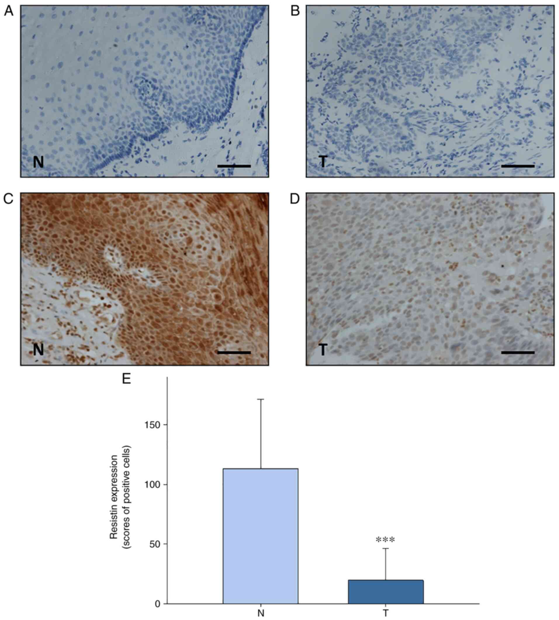
IHC analysis revealed that resistin expression was
significantly downreglated in esophageal tumor tissues compared
with paired adjacent normal esophageal tissues from patients with
ESCC (n=73) (P<0.001; Fig. 1).
The present study assessed the association between resistin
expression in ESCC tissues and the clinicopathological
characteristics of the patients, who were stratified into high and
low resistin expression groups determined by receiver-operating
characteristic curve analysis (Fig.
S1). As presented in Table I,
resistin expression was significantly associated with body mass
index (BMI) (P=0.042). However, no significant associations were
observed between resistin expression and lifestyle factors (alcohol
consumption, betal quid chewing or smoking), tumor characteristics
(histopathological grade, tumor size or lymph node metastasis),
treatment modalities (chemotherapy or radiotherapy) or patient
outcomes (recurrence or death) (Table
I).
 | Table I.Association between resistin
expression and the clinicopathological characteristics of patients
with esophageal squamous cell carcinoma (n=73). |
Table I.
Association between resistin
expression and the clinicopathological characteristics of patients
with esophageal squamous cell carcinoma (n=73).
|
| Resistin
expression |
|
|---|
|
|
|
|
|---|
| Characteristic | Low, n (%) | High, n (%) | P-value |
|---|
| Sex, male | 55 (75.3) | 18 (24.7) |
|
| Alcohol
consumption |
|
| 0.670 |
| No | 7 (87.5) | 1 (12.5) |
|
|
Yes | 48 (73.8) | 17 (26.2) |
|
| Betel quid
chewing |
|
| 0.287 |
| No | 30 (81.1) | 7 (18.9) |
|
|
Yes | 25 (69.4) | 11 (30.6) |
|
| Cigarette
smoking |
|
| 0.182 |
| No | 7 (100.0) | 0 (0.0) |
|
|
Yes | 48 (72.7) | 18 (27.3) |
|
| BMIb |
|
| 0.042a |
|
Low | 25 (92.6) | 2 (7.4) |
|
|
High | 20 (69.0) | 9 (31.0) |
|
| Histopathological
grade |
|
| >0.999 |
| I | 9 (75.0) | 3 (25.0) |
|
|
II+III | 46 (75.4) | 15 (24.6) |
|
| Tumor size, cm |
|
| 0.393 |
|
T1+T2 | 19 (82.6) | 4 (17.4) |
|
|
T3+T4 | 36 (72.0) | 14 (28.0) |
|
| Lymph node
metastasis |
|
| 0.277 |
| No | 28 (82.4) | 6 (17.6) |
|
|
Yes | 27 (69.2) | 12 (30.8) |
|
| Chemotherapy |
|
| 0.242 |
| No | 15 (65.2) | 8 (34.8) |
|
|
Yes | 40 (80.0) | 10 (20.0) |
|
| Radiotherapy |
|
| 0.233 |
| No | 13 (65.0) | 7 (35.0) |
|
|
Yes | 42 (79.3) | 11 (20.7) |
|
| Recurrence |
|
| 0.772 |
| No | 39 (76.5) | 12 (23.5) |
|
|
Yes | 16 (72.7) | 6 (27.3) |
|
| Death |
|
| 0.758 |
| No | 13 (72.2) | 5 (27.8) |
|
|
Yes | 42 (76.4) | 13 (23.6) |
|
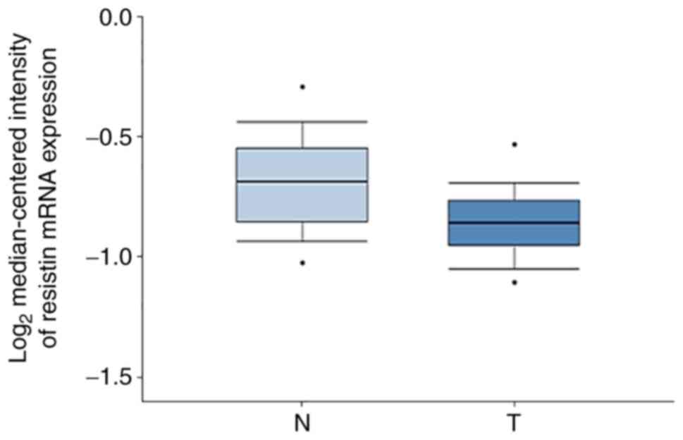
The present study also analyzed resistin mRNA
expression in ESCC tissues and normal esophageal tissues using the
Oncomine database. The results demonstrated that resistin
expression was higher in normal esophageal tissues compared with
ESCC tissues (Fig. 2). These results
suggest that the reduced resistin expression in ESCC tissues may
serve as a diagnotic marker for clinical use.
Serum resistin levels in patients with
ESCC
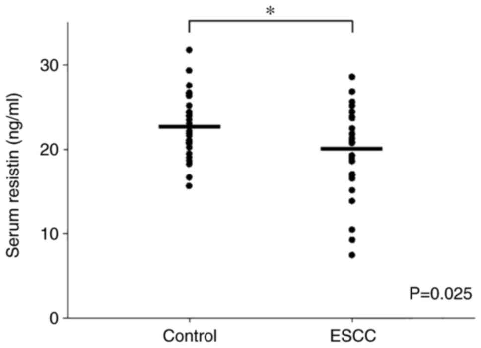
Given that circulating resistin is released by
adipose tissues or the associated tumor tissues (19), the present study investigated serum
resistin levels in patients with ESCC compared with healthy
individuals via ELISA. Despite the heterogeneous expression of
serum resistin between individuals, the results demonstrated that
the average serum resistin level was significantly lower in
patients with ESCC (n=26) compared with the healthy individuals
(n=26) (P=0.025; Fig. 3 and Table SI). Notably, the serum resistin
levels observed in the present study were within a similar range as
previously reported (26). These
results support the IHC findings of differential resistin
expression between ESCC tissues and adjacent normal esophageal
tissues. In addition, the detection of serum resistin level may
have the potential to be developed into a non-invasive liquid
biopsy test for ESCC.
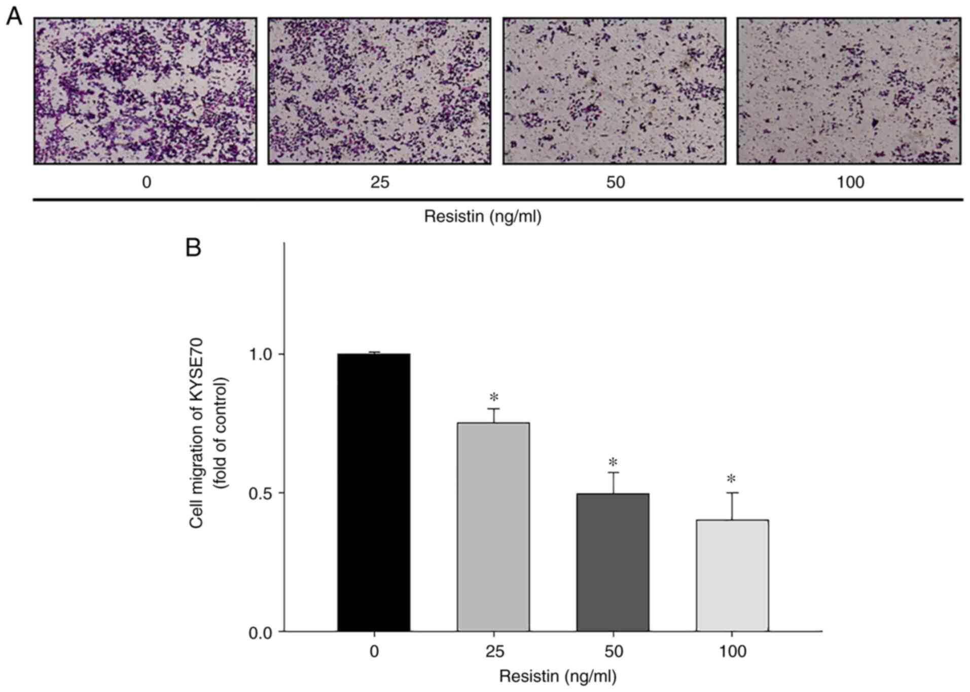
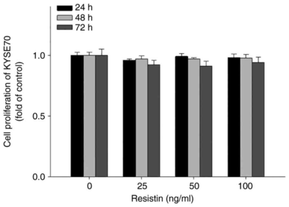
Resistin inhibits in vitro migration
but not proliferation of ESCC cells
To investigate the biological effects of resistin on
ESCC cell migration and proliferation, in vitro Transwell
and XTT assays were performed, respectively. The results
demonstrated that treatment with different concentrations of
resistin significantly inhibited the migratory ability of KYSE70
cells in a dose-dependent manner (25 ng/ml vs. control, P=0.023; 50
ng/ml vs. control, P<0.001; 100 ng/ml vs. control, P<0.001;
Fig. 4), while treatment with
resistin had no significant effect on KYSE70 cell proliferation
(Fig. 5), suggesting that resistin
may have a unique role in cell migration other than affecting
proliferation of ESCC.
Similar results of the effect of resistin on the
migration (Fig. S2A and B) and
proliferation (Fig. S2C) of TE8
cells were observed. However, whether the phenomena of
resistin-reduced in vitro cell migration can be translated
into the cancer progression of ESCC in clinical settings is yet to
be investigated. These results also suggest that the effect of
resistin on ESCC cell migration may result from a signaling pathway
that is distinctive from the signaling for ESCC proliferation.
Discussion
To the best of our knowledge, the present study was
the first to demonstrate that resistin expression is downregulated
in both ESCC tissues and serum samples of patients with ESCC
compared with the controls. Previous studies have reported that
resistin expression is upregulated in breast cancer (23,27) and
other types of cancer (28–31). However, the results of the present
study may indicate a unique role for resistin in the diagnosis of
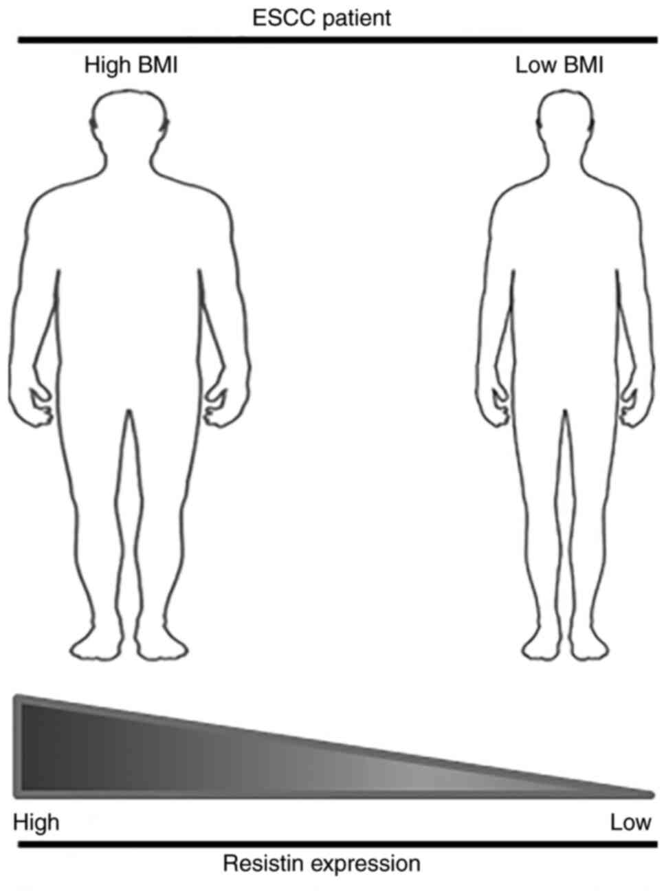
esophageal cancer. The results demonstrated that tissue resistin
expression was positively associated with the BMI of patients with
ESCC. As presented in Fig. 6, this
association may partly explain the discrepant resistin expression
between the results of the present study and previous studies on
different types of cancer (19–22), as
elevated resistin expression has been associated with obesity
(10,32), but patients with esophageal cancer
are usually on the opposite end of the body weight spectrum due to
the occurrence of dysphagia (33,34). A
meta-analysis also revealed that a higher BMI (≥25
kg/m2) is associated with lower risk of mortality in
patients with ESCC (35), suggesting
that obesity may have differential influence on ESCC compared with
other types of cancer, such as breast cancer.
Previous studies have reported an association
between increased resistin expression and ESCC (36,37). The
molecular mechanism underlying these paradoxical findings remains
elusive; however, factors that may affect the interpretation of
clinical studies include selection criteria of patients and
determination of high vs. low resistin expression levels via IHC
analysis (19). The latter may be
overcome with the advance of digital tissue image analysis, in
which the automated scoring system for IHC can be more objective
and consistent than manual assignment of the scores by different
pathologists or trained examiners across laboratories (38,39). The
results of the present study demonstrated a significant association
between BMI and resistin expression; thus, prospective clinical
studies may consider performing a primary stratification of
patients with ESCC, based on their BMIs for further analysis of
resistin expression in clinical samples.
Although resistin was originally found to be
associated with obesity and insulin resistance in rodents (10), subsequent studies have reported a
more complex role for resistin in humans, including its involvement
in a number of inflammatory diseases and cancers (40,41). The
biological effects of resistin may occur via activation of its
endogenous receptors (10). Adenylyl
cyclase-associated protein 1 (CAP1) was the first receptor
identified for human resistin in monocytes (42). In the study, overexpression of CAP1
enhanced resistin-mediated inflammatory responses, whereas these
responses were abrogated by suppressing CAP1 expression (42). Subsequent studies revealed that
elevated CAP1 expression is associated with breast tumor malignancy
and poor patient survival (43). Our
group recently demonstrated that toll-like receptor 4 (TLR4) is
also a functional receptor for human resistin, and activation of
the resistin/TLR4 signaling pathway is a critical step for the
enhanced epithelial-to-mesenchymal transition and cancer stemness
of breast cancer cells (23).
Decorin and the receptor tyrosine kinase-like orphan receptor 1 are
another two resistin receptors derived from mouse adipocyte
progenitor cells (44,45). Notably, increased expression of human
decorin is associated with the transformation of oral epithelial
cells into severe oral dysplasia-like phenotypes, although the
particular role of resistin in this paradigm has not been assessed
(46). Nevertheless, whether ESCC
cells express these resistin receptors and whether they
functionally affect resistin-associated cancer progression in
patients with ESCC remain unknown. The results of the present study
demonstrated that the migration ability of ESCC cells was reduced
following treatment with resistin. In addition, the preliminary
screening suggested that resistin-treated TE8 cells may have
increased protein expression of epithelial markers, such as ZO-1,
claudin-1 and E-cadherin, while decreasing the expression of
mesenchymal markers, such as twist and slug (unpublished data).
These suggest that the effect of resistin on ESCC cells may result
from a different signaling pathway compared with those identified
in breast cancer cells (19). As a
matter of fact, the diverse signaling pathways of resistin
receptors, such as STAT3, PI3K/AKT, mTOR, MAPK and NFκB, have been
reported to participate in the regulation of tumor growth and
metastasis in different types of cancer (19,47).
Furthermore, different expression levels of resistin receptors in
ESCC may play an important role in cancer development. For example,
the expression of apelin, one of the obesity-linked adipocytokines
(48), is upregulated during adverse
ESCC progression, whereas the expression of apelin receptor is
downregulated in patients with advanced tumor stage and lymph node
metastasis (37). Thus, further
studies are required to investigate the functional expression of
resistin receptors in ESCC, along with the detection of resistin
expression.
In conclusion, the results of the present study
suggest that resistin has the potential to be developed as a unique
diagnostic marker for ESCC. However, due to the limitation of the
cohort size in the present study, further studies with a larger
sample size are required to validate the use of resistin as a
prognostic marker for patients with ESCC. In addition,
investigations into the expression of resistin receptors and
associated signaling pathways in ESCC cells are required to provide
a biological foundation for future development of targeted
therapies for patients with resistin-mediated ESCC.
Supplementary Material
Supporting Data
Acknowledgements
Not applicable.
Funding
The present study was financially supported by the
Ministry of Health and Welfare of Taiwan (grant no.
MOHW110-TDU-B-212-144016, Health and Welfare Surcharge of Tobacco
Products), Kaohsiung Medical University Hospital (grant nos.
KMUH105-5R32, KMUH106-6R41, KMUH106-6R83, KMUH107-7R36 and
KMUH108-8R42) and Kaohsiung Medical University (Research Center
grant no. KMU-DK108005; Center for Cancer Research grant nos.
KMU-TC108A04-0 and KMU-TC108A04-1).
Availability of data and materials
The datasets used and/or analyzed during the current
study are available from the corresponding author on reasonable
request.
Authors' contributions
ACH, YYW, KTL, HHC, YKC, JKD, CMC, MYC, KJC, SCSH
and SSFY conceived and designed the experiments. YYW, HHC, YKC, JKD
and CMC performed the experiments. ACH, YYW, KTL, YKC, JKD, CMC and
SSFY analyzed the data. YYW, KTL, YKC, JKD, CMC and SSFY provided
experimental reagents and tools. ACH, YYW, SCSH and SSFY drafted
the initial manuscript and revised the manuscript for important
intellectual content. ACH, YYW and SSFY confirm the authenticity of
all the raw data. All authors have read and approved the final
manuscript.
Ethics approval and consent to
participate
The present study was approved by the Institutional
Review Board of Kaohsiung Medical University Hospital (approval no.
KMUH-IRB-20130627; Kaohsiung, Taiwan) and written informed consent
was obtained prior to this study.
Patient consent for publication
Not applicable.
Competing interests
The authors declare that they have no competing
interests.
References
|
1
|
Global Burden of Disease Cancer
Collaboration, ; Fitzmaurice C, Dicker D, Pain A, Hamavid H,
Moradi-Lakeh M, MacIntyre MF, Allen C, Hansen G, Woodbrook R, et
al: The global burden of cancer 2013. JAMA Oncol. 1:505–527. 2015.
View Article : Google Scholar : PubMed/NCBI
|
|
2
|
Global Burden of Disease Cancer
Collaboration, ; Fitzmaurice C, Abate D, Abbasi N, Abbastabar H,
Abd-Allah F, Abdel-Rahman O, Abdelalim A, Abdoli A, Abdollahpour I,
et al: Global, regional, and national cancer incidence, mortality,
years of life lost, years lived with disability, and
disability-adjusted life-years for 29 cancer groups, 1990 to 2017:
A systematic analysis for the global burden of disease study. JAMA
Oncol. 5:1749–1768. 2019. View Article : Google Scholar : PubMed/NCBI
|
|
3
|
Sung H, Ferlay J, Siegel RL, Laversanne M,
Soerjomataram I, Jemal A and Bray F: Global cancer statistics 2020:
GLOBOCAN estimates of incidence and mortality worldwide for 36
cancers in 185 countries. CA Cancer J Clin. 71:209–249. 2021.
View Article : Google Scholar : PubMed/NCBI
|
|
4
|
Lao-Sirieix P and Fitzgerald RC: Screening
for oesophageal cancer. Nat Rev Clin Oncol. 9:278–287. 2012.
View Article : Google Scholar : PubMed/NCBI
|
|
5
|
Edgren G, Adami HO, Weiderpass E and Nyren
O: A global assessment of the oesophageal adenocarcinoma epidemic.
Gut. 62:1406–1414. 2013. View Article : Google Scholar : PubMed/NCBI
|
|
6
|
Lagergren J, Smyth E, Cunningham D and
Lagergren P: Oesophageal cancer. Lancet. 390:2383–2396. 2017.
View Article : Google Scholar : PubMed/NCBI
|
|
7
|
Hajizadeh B, Jessri M, Moasheri SM, Rad AH
and Rashidkhani B: Fruits and vegetables consumption and esophageal
squamous cell carcinoma: A case-control study. Nutr Cancer.
63:707–713. 2011. View Article : Google Scholar : PubMed/NCBI
|
|
8
|
Liu J, Wang J, Leng Y and Lv C: Intake of
fruit and vegetables and risk of esophageal squamous cell
carcinoma: A meta-analysis of observational studies. Int J Cancer.
133:473–485. 2013. View Article : Google Scholar : PubMed/NCBI
|
|
9
|
Bosetti C, Franceschi S, Levi F, Negri E,
Talamini R and La Vecchia C: Smoking and drinking cessation and the
risk of oesophageal cancer. Br J Cancer. 83:689–691. 2000.
View Article : Google Scholar : PubMed/NCBI
|
|
10
|
Steppan CM, Bailey ST, Bhat S, Brown EJ,
Banerjee RR, Wright CM, Patel HR, Ahima RS and Lazar MA: The
hormone resistin links obesity to diabetes. Nature. 409:307–312.
2001. View
Article : Google Scholar : PubMed/NCBI
|
|
11
|
Steppan CM, Brown EJ, Wright CM, Bhat S,
Banerjee RR, Dai CY, Enders GH, Silberg DG, Wen X, Wu GD and Lazar
MA: A family of tissue-specific resistin-like molecules. Proc Natl
Acad Sci USA. 98:502–506. 2001. View Article : Google Scholar : PubMed/NCBI
|
|
12
|
Banerjee RR and Lazar MA: Dimerization of
resistin and resistin-like molecules is determined by a single
cysteine. J Biol Chem. 276:25970–25973. 2001. View Article : Google Scholar : PubMed/NCBI
|
|
13
|
McTernan PG, McTernan CL, Chetty R, Jenner
K, Fisher FM, Lauer MN, Crocker J, Barnett AH and Kumar S:
Increased resistin gene and protein expression in human abdominal
adipose tissue. J Clin Endocrinol Metab. 87:24072002. View Article : Google Scholar : PubMed/NCBI
|
|
14
|
Patel L, Buckels AC, Kinghorn IJ, Murdock
PR, Holbrook JD, Plumpton C, Macphee CH and Smith SA: Resistin is
expressed in human macrophages and directly regulated by PPAR gamma
activators. Biochem Biophys Res Commun. 300:472–476. 2003.
View Article : Google Scholar : PubMed/NCBI
|
|
15
|
Bokarewa M, Nagaev I, Dahlberg L, Smith U
and Tarkowski A: Resistin, an adipokine with potent proinflammatory
properties. J Immunol. 174:5789–5795. 2005. View Article : Google Scholar : PubMed/NCBI
|
|
16
|
Curat CA, Wegner V, Sengenès C, Miranville
A, Tonus C, Busse R and Bouloumié A: Macrophages in human visceral
adipose tissue: Increased accumulation in obesity and a source of
resistin and visfatin. Diabetologia. 49:744–747. 2006. View Article : Google Scholar : PubMed/NCBI
|
|
17
|
Lehrke M, Reilly MP, Millington SC, Iqbal
N, Rader DJ and Lazar MA: An inflammatory cascade leading to
hyperresistinemia in humans. PLoS Med. 1:e452004. View Article : Google Scholar : PubMed/NCBI
|
|
18
|
Reilly MP, Lehrke M, Wolfe ML, Rohatgi A,
Lazar MA and Rader DJ: Resistin is an inflammatory marker of
atherosclerosis in humans. Circulation. 111:932–939. 2005.
View Article : Google Scholar : PubMed/NCBI
|
|
19
|
Wang YY, Hung AC, Lo S and Yuan SF:
Adipocytokines visfatin and resistin in breast cancer: Clinical
relevance, biological mechanisms, and therapeutic potential. Cancer
Lett. 498:229–239. 2021. View Article : Google Scholar : PubMed/NCBI
|
|
20
|
Hlavna M, Kohut L, Lipkova J,
Bienertova-Vasku J, Dostalova Z, Chovanec J and Vasku A:
Relationship of resistin levels with endometrial cancer risk.
Neoplasma. 58:124–128. 2011. View Article : Google Scholar : PubMed/NCBI
|
|
21
|
Danese E, Montagnana M, Minicozzi AM,
Bonafini S, Ruzzenente O, Gelati M, De Manzoni G, Lippi G and Guidi
GC: The role of resistin in colorectal cancer. Clin Chim Acta.
413:760–764. 2012. View Article : Google Scholar : PubMed/NCBI
|
|
22
|
Tzanavari T, Tasoulas J, Vakaki C,
Mihailidou C, Tsourouflis G and Theocharis S: The role of
adipokines in the establishment and progression of head and neck
neoplasms. Curr Med Chem. 26:4726–4748. 2019. View Article : Google Scholar : PubMed/NCBI
|
|
23
|
Wang CH, Wang PJ, Hsieh YC, Lo S, Lee YC,
Chen YC, Tsai CH, Chiu WC, Chu-Sung Hu S, Lu CW, et al: Resistin
facilitates breast cancer progression via TLR4-mediated induction
of mesenchymal phenotypes and stemness properties. Oncogene.
37:589–600. 2018. View Article : Google Scholar : PubMed/NCBI
|
|
24
|
Detre S, Saclani Jotti G and Dowsett M: A
‘quickscore’ method for immunohistochemical semiquantitation:
Validation for oestrogen receptor in breast carcinomas. J Clin
Pathol. 48:876–878. 1995. View Article : Google Scholar : PubMed/NCBI
|
|
25
|
Rhodes DR, Yu J, Shanker K, Deshpande N,
Varambally R, Ghosh D, Barrette T, Pandey A and Chinnaiyan AM:
ONCOMINE: A cancer microarray database and integrated data-mining
platform. Neoplasia. 6:1–6. 2004. View Article : Google Scholar : PubMed/NCBI
|
|
26
|
Assiri AM and Kamel HF: Evaluation of
diagnostic and predictive value of serum adipokines: Leptin,
resistin and visfatin in postmenopausal breast cancer. Obes Res
Clin Pract. 10:442–453. 2016. View Article : Google Scholar : PubMed/NCBI
|
|
27
|
Lee YC, Chen YJ, Wu CC, Lo S, Hou MF and
Yuan SS: Resistin expression in breast cancer tissue as a marker of
prognosis and hormone therapy stratification. Gynecol Oncol.
125:742–750. 2012. View Article : Google Scholar : PubMed/NCBI
|
|
28
|
Karapanagiotou EM, Tsochatzis EA, Dilana
KD, Tourkantonis I, Gratsias I and Syrigos KN: The significance of
leptin, adiponectin, and resistin serum levels in non-small cell
lung cancer (NSCLC). Lung Cancer. 61:391–397. 2008. View Article : Google Scholar : PubMed/NCBI
|
|
29
|
Kerem M, Ferahkose Z, Yilmaz UT, Pasaoglu
H, Ofluoglu E, Bedirli A, Salman B, Sahin TT and Akin M: Adipokines
and ghrelin in gastric cancer cachexia. World J Gastroenterol.
14:3633–3641. 2008. View Article : Google Scholar : PubMed/NCBI
|
|
30
|
Nakajima TE, Yamada Y, Hamano T, Furuta K,
Gotoda T, Katai H, Kato K, Hamaguchi T and Shimada Y: Adipocytokine
levels in gastric cancer patients: Resistin and visfatin as
biomarkers of gastric cancer. J Gastroenterol. 44:685–690. 2009.
View Article : Google Scholar : PubMed/NCBI
|
|
31
|
Gonullu G, Kahraman H, Bedir A, Bektas A
and Yucel I: Association between adiponectin, resistin, insulin
resistance, and colorectal tumors. Int J Colorectal Dis.
25:205–212. 2010. View Article : Google Scholar : PubMed/NCBI
|
|
32
|
Tripathi D, Kant S, Pandey S and Ehtesham
NZ: Resistin in metabolism, inflammation, and disease. FEBS J.
287:3141–3149. 2020. View Article : Google Scholar : PubMed/NCBI
|
|
33
|
Nitenberg G and Raynard B: Nutritional
support of the cancer patient: Issues and dilemmas. Crit Rev Oncol
Hematol. 34:137–168. 2000. View Article : Google Scholar : PubMed/NCBI
|
|
34
|
Enzinger PC and Mayer RJ: Esophageal
cancer. N Engl J Med. 349:2241–2252. 2003. View Article : Google Scholar : PubMed/NCBI
|
|
35
|
Fahey PP, Mallitt KA, Astell-Burt T, Stone
G and Whiteman DC: Impact of pre-diagnosis behavior on risk of
death from esophageal cancer: A systematic review and
meta-analysis. Cancer Causes Control. 26:1365–1373. 2015.
View Article : Google Scholar : PubMed/NCBI
|
|
36
|
Nakajima TE, Yamada Y, Hamano T, Furuta K,
Oda I, Kato H, Kato K, Hamaguchi T and Shimada Y: Adipocytokines
and squamous cell carcinoma of the esophagus. J Cancer Res Clin
Oncol. 136:261–266. 2010. View Article : Google Scholar : PubMed/NCBI
|
|
37
|
Diakowska D, Markocka-Maczka K,
Nienartowicz M, Rosinczuk J and Krzystek-Korpacka M: Assessment of
apelin, apelin receptor, resistin, and adiponectin levels in the
primary tumor and serum of patients with esophageal squamous cell
carcinoma. Adv Clin Exp Med. 28:671–678. 2019. View Article : Google Scholar : PubMed/NCBI
|
|
38
|
Aeffner F, Wilson K, Martin NT, Black JC,
Hendriks CLL, Bolon B, Rudmann DG, Gianani R, Koegler SR, Krueger J
and Young GD: The gold standard paradox in digital image analysis:
Manual versus automated scoring as ground truth. Arch Pathol Lab
Med. 141:1267–1275. 2017. View Article : Google Scholar : PubMed/NCBI
|
|
39
|
Bankhead P, Fernández JA, McArt DG, Boyle
DP, Li G, Loughrey MB, Irwin GW, Harkin DP, James JA, McQuaid S, et
al: Integrated tumor identification and automated scoring minimizes
pathologist involvement and provides new insights to key biomarkers
in breast cancer. Lab Invest. 98:15–26. 2018. View Article : Google Scholar : PubMed/NCBI
|
|
40
|
Tilg H and Moschen AR: Adipocytokines:
Mediators linking adipose tissue, inflammation and immunity. Nat
Rev Immunol. 6:772–783. 2006. View Article : Google Scholar : PubMed/NCBI
|
|
41
|
Codoner-Franch P and Alonso-Iglesias E:
Resistin: Insulin resistance to malignancy. Clin Chim Acta.
438:46–54. 2015. View Article : Google Scholar : PubMed/NCBI
|
|
42
|
Lee S, Lee HC, Kwon YW, Lee SE, Cho Y, Kim
J, Lee S, Kim JY, Lee J, Yang HM, et al: Adenylyl
cyclase-associated protein 1 is a receptor for human resistin and
mediates inflammatory actions of human monocytes. Cell Metab.
19:484–497. 2014. View Article : Google Scholar : PubMed/NCBI
|
|
43
|
Rosendahl AH, Bergqvist M, Lettiero B,
Kimbung S and Borgquist S: Adipocytes and obesity-related
conditions jointly promote breast cancer cell growth and motility:
Associations with CAP1 for prognosis. Front Endocrinol (Lausanne).
9:6892018. View Article : Google Scholar : PubMed/NCBI
|
|
44
|
Daquinag AC, Zhang Y, Amaya-Manzanares F,
Simmons PJ and Kolonin MG: An isoform of decorin is a resistin
receptor on the surface of adipose progenitor cells. Cell Stem
Cell. 9:74–86. 2011. View Article : Google Scholar : PubMed/NCBI
|
|
45
|
Sanchez-Solana B, Laborda J and Baladron
V: Mouse resistin modulates adipogenesis and glucose uptake in
3T3-L1 preadipocytes through the ROR1 receptor. Mol Endocrinol.
26:110–127. 2012. View Article : Google Scholar : PubMed/NCBI
|
|
46
|
Banerjee AG, Bhattacharyya I, Lydiatt WM
and Vishwanatha JK: Aberrant expression and localization of decorin
in human oral dysplasia and squamous cell carcinoma. Cancer Res.
63:7769–7776. 2003.PubMed/NCBI
|
|
47
|
Sudan SK, Deshmukh SK, Poosarla T,
Holliday NP, Dyess DL, Singh AP and Singh S: Resistin: An
inflammatory cytokine with multi-faceted roles in cancer. Biochim
Biophys Acta Rev Cancer. 1874:1884192020. View Article : Google Scholar : PubMed/NCBI
|
|
48
|
Wysocka MB, Pietraszek-Gremplewicz K and
Nowak D: The role of apelin in cardiovascular diseases, obesity and
cancer. Front Physiol. 9:5572018. View Article : Google Scholar : PubMed/NCBI
|