Introduction
Anaplastic lymphoma kinase (ALK) is a receptor
protein tyrosine kinase that possesses a single transmembrane
domain and 1,620 amino acid residues (1,2). Among
normal tissues, ALK protein expression is prominent in the brain
and peripheral nervous system of developing embryos and decreases
rapidly after birth (3). In adults,
the ALK protein is expressed at low levels and only in the central
nervous system, while it has not been detected in other tissues
(3). Thus, expression of this
protein is considered to be a characteristic of abnormal cells.
Abnormalities in ALK at both the gene and protein
levels play an important role in the pathogenesis of various
cancers (4–13) and serve as important therapeutic
targets (4). ALK gene
translocations and oncogenic fusion proteins have been reported in
a variety of human malignancies, including anaplastic large cell
lymphoma (ALCL) (13), non-small
cell lung cancer (NSCLC) (4,5),
inflammatory myofibroblastic tumors (IMT) (6,7), and
neuroblastomas (8–12). Other genetic alterations have also
been detected, such as germ line or somatic mutations in
neuroblastomas (8), gene insertions
or amplifications in neuroblastomas and NSCLC (5,8). In
addition to IMT, rhabdomyosarcomas are another soft tissue tumors
for which there have been several studies of ALK protein expression
and gene amplification (14,15).
On the other hand, ALK gene abnormalities and aberrant
protein expression in other soft tissue tumors have not been well
investigated.
Previously, Takeuchi et al improved the
immunohistochemical sensitivity of the ALK protein through the
development of the intercalated antibody-enhanced polymer (iAEP)
method, which incorporates an intercalating antibody between the
primary antibody and the dextran polymer-based detection reagents
(16). In the case of lung cancer,
sensitivity of the ALK protein using the iAEP method was higher
than that using conventional procedures (16). ALK protein expression in soft tissue
tumors has not been evaluated previously by means of the iAEP
method.
ALK gene alterations and protein expression have the
potential to activate the STAT3 (17,18),
AKT/PI3K (19), and RAS/ERK
(20) pathways, which are involved
in oncogenic processes, including cell proliferation, migration,
and survival. Activation of those oncogenic pathways requires not
only gene mutations or abnormal protein expression but also
phosphorylation of ALK, which alters its function. Previous in
vitro studies indicated an association among ALK
phosphorylation, its oncogenic function, and poor prognosis
(21). However, the mechanism by
which ALK phosphorylation affects the clinical course of cancer
patients has not been investigated previously.
The present study investigated ALK protein
expression using the iAEP method, underlying ALK genetic
aberrations using fluorescence in situ hybridization (FISH),
and ALK protein phosphorylation using immunohistochemical staining
(IHS) in soft tissue tumors, as well as their relationship with the
clinical course of cancer, such as prognosis and metastasis.
Materials and methods
Tissue samples
We retrieved 81 soft tissue tumor specimens from the
Department of Orthopedic Surgery of Kurume University from 2001 to
2011. Paraffin-embedded tissues were used for diagnosis,
histopathology, and IHS analyses. The antibodies used for
immunostaining included those against CAM5.2 (BD Biosciences, San
Jose, CA, USA), CD3, MyoD1, Bcl6 and CD34 (all from Leica Ltd., New
Castle, UK), Bcl2 (Ventana Medical Systems, Inc., Tuscon, AZ, USA),
vimentin, SMA, desmin, myogenin, S100, KP-1, AE1+AE3, EMA, CD20,
and CD99 (all from Dako, Tokyo, Japan). All cases were diagnosed
according to the World Health Organization (WHO) classification
system (22). For IHS and FISH
analyses, a tissue microarray was generated from 3-mm diameter
cores derived from neoplastic lesions of each tumor case. Clinical
information was obtained from patient medical charts. The use of
clinical information and materials was approved by the Research
Ethics Committee of Kurume University and was in accordance with
the Declaration of Helsinki.
Immunohistochemical detection of the ALK
protein using the iAEP method
We applied the iAEP method for immunohistochemical
detection of ALK. The ALK Detection Kit (Nichirei Biosciences Inc.,
Tokyo, Japan) was used according to the manufacturer’s protocol.
Briefly, after deparaffinization, the slides were heated for 40 min
at 95°C in antigen retrieval solution and subsequently incubated at
room temperature with peroxidase blocking reagent for 5 min
followed by incubation with the ALK antibody (clone, 5A4; dilution,
1:50) for 30 min. The Bridge reagent and the Peroxidase-Labeled
Empower reagent were applied for 15 and 30 min, respectively, at
room temperature. The immunohistochemical staining (IHS) results
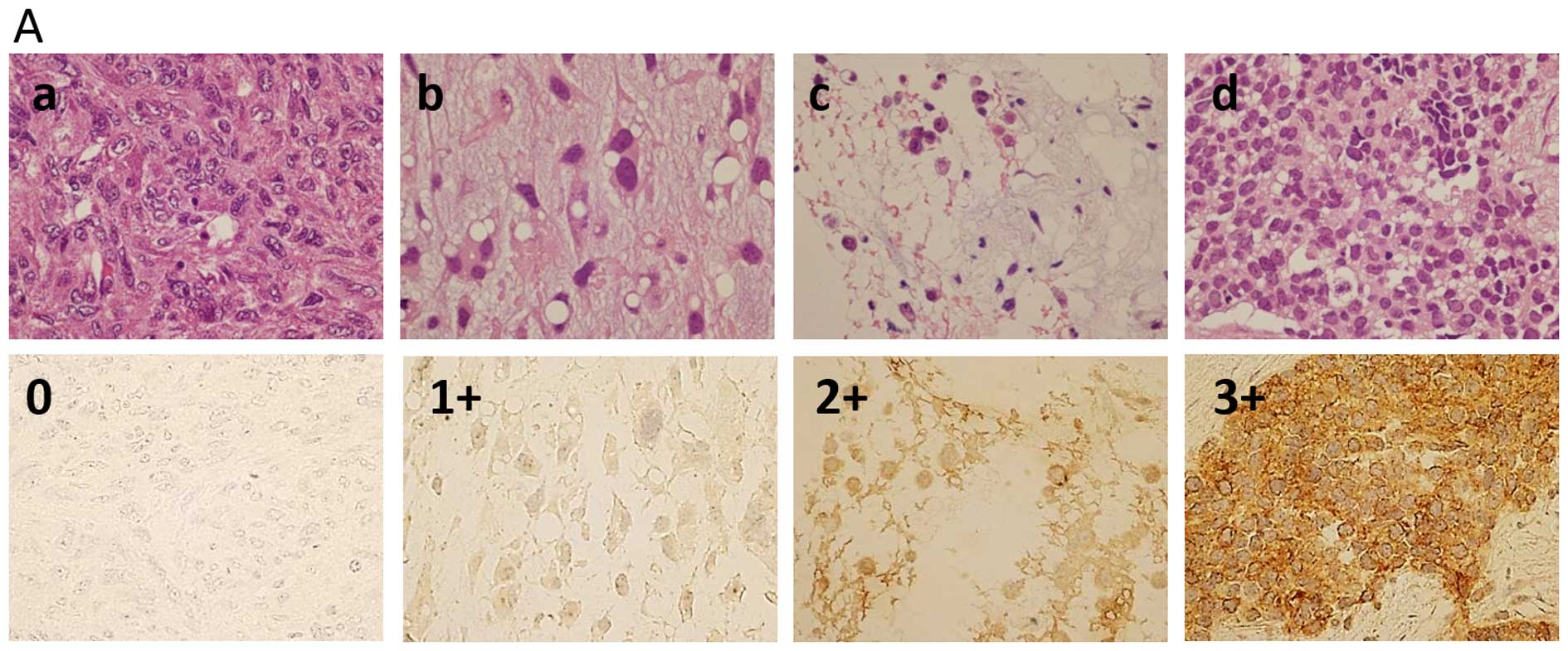
were categorized as either negative (−), weakly positive (1+),
moderately positive (2+), or strongly positive (3+) (Fig. 1A). If a neoplastic cell was at least
weakly positive, it was considered positive for ALK expression. We
defined a tumor sample with >30% positive neoplastic cells as a
positive case.
Determination of ALK gene status by FISH
analysis
FISH analysis was performed using an ALK (2p23)
split-signal FISH DNA Probe (Dako) to detect chromosomal
translocation of the ALK gene and gene signal gain (copy
no.) (Fig. 1B). If the split signal
was detected in at least one out of 30 neoplastic cells, the tumor
sample was defined as translocation-positive. If 3 or more gene
copies per sample were detected in at least one out of 30
neoplastic cells, the tumor specimen was categorized as gene signal
gain-positive.
Immunohistochemical analysis of
phospho-Y1604 ALK expression
To evaluate phospho-Y1604 ALK expression, we
performed immunostaining using the anti-ALK (phospho-Y1604)
antibody (clone, EP661Y) (Abcam, Cambridge, UK). If a neoplastic
cell was at least weakly positive (1+), it was considered positive
for ALK expression, and any tumor specimen with >30% positive
cells was considered a positive case (Fig. 1C).
Immunohistochemical detection of the MIB1
index
Sections (4 μm) were deparaffinized in xylol and
washed in distilled water, followed by treatment with an antibody
against Ki-67 (clone M7240; Dako) in 0.001 M EDTA buffer, pH 8.0,
for 20 min at 95–99°C in a microwave oven.
Using an optical microscope, Ki-67-positive cells
were counted in 5 fields of a high power view randomly selected
from each section. A case with >10% Ki-67-positive cells was
considered positive.
Statistical analysis
The Chi-square test and Fisher’s exact test
(two-sided test), which is appropriate for small values, were used
to assess the associations among the IHS, FISH, and protein
phosphorylation analyses. Event-free survival (EFS) was defined as
the period from the time of histological diagnosis to that of
detection of metastasis or the most recent follow-up evaluation.
EFS and overall survival (OS) were determined using the
Kaplan-Meier method, and the log-rank test was used to compare
survival curves between the favorable and unfavorable risk groups.
A P-value <0.05 in both Chi-square and Fisher’s exact tests was
indicative of statistical significance.
Results
Clinical characteristics
Table I presents the
clinical information of all patients from whom the tumor samples
used in this study originated. The 81 tissue samples were derived
from 44 males and 37 females, who ranged in age from 2 to 88 years
(mean 52.0 years). All patients underwent surgical resection. In
addition to resection, 17 patients also received chemotherapy, 6
received radiation therapy, and 3 received chemoradiotherapy.
Twenty-one of the subjects showed metastasis during the follow-up
period, which ranged from 0 to 245 months (mean, 44.1 months).
 | Table IClinical characteristics of the
patients with soft tissue tumors. |
Table I
Clinical characteristics of the
patients with soft tissue tumors.
| Disease | No. of cases | Gender | Age (years) | Additional
treatment | No. of metastatic
cases | Follow-up period
(months) |
|---|
|
|
|
|
|---|
| M/F | Mean | Range | Chemotherapy | Radiation |
Chemoradiotherapy | Resection only | Mean | Range |
|---|
| All | 81 | 44/37 | 52.0 | 2–88 | 17 | 6 | 3 | 55 | 21/81 | 44.1 | 0–245 |
| IMT | 1 | 1/0 | 6.0 | 6 | 0 | 0 | 0 | 1 | 0/1 | 16.0 | 16 |
| Alveolar soft part
sarcoma | 2 | 1/1 | 32.0 | 14–50 | 0 | 0 | 0 | 2 | 1/2 | 85.5 | 65–106 |
|
Leiomyosarcoma | 10 | 5/5 | 61.7 | 49–87 | 3 | 1 | 0 | 6 | 3/10 | 26.6 | 5–60 |
|
Well-differentiated liposarcoma | 7 | 4/3 | 70.1 | 44–88 | 0 | 0 | 0 | 7 | 0/7 | 16.9 | 5–30 |
| Pleomorphic
liposarcoma | 2 | 2/0 | 62.0 | 61–63 | 0 | 1 | 0 | 1 | 2/2 | 56.0 | 51–61 |
| Extraskeletal
osteosarcoma | 1 | 0/1 | 80.0 | 80 | 0 | 0 | 0 | 1 | 1/1 | 76.0 | 76 |
| Epithelioid
sarcoma | 1 | 0/1 | 19.0 | 19 | 1 | 0 | 0 | 0 | 1/1 | 5.0 | 5 |
| Synovial
sarcoma | 4 | 3/1 | 35.5 | 23–57 | 4 | 0 | 0 | 0 | 2/4 | 24.8 | 11–40 |
| MPNST | 4 | 3/1 | 25.1 | 14–43 | 1 | 0 | 0 | 3 | 2/4 | 36.7 | 13–127 |
| UPS | 19 | 11/8 | 61.6 | 22–77 | 2 | 3 | 0 | 14 | 5/19 | 45.1 | 1–124 |
|
Rhabdomyosarcoma | 6 | 3/3 | 16.2 | 2–57 | 3 | 0 | 1 | 2 | 1/6 | 36.2 | 0–108 |
|
Myxofibrosarcoma | 8 | 4/4 | 67.3 | 39–83 | 0 | 0 | 0 | 8 | 0/8 | 47.6 | 24–81 |
| Myxoid
liposarcoma | 11 | 5/6 | 49.8 | 24–78 | 2 | 0 | 2 | 7 | 2/11 | 65.7 | 0–245 |
| Fibrosarcoma | 4 | 2/2 | 46.0 | 24–71 | 1 | 0 | 0 | 3 | 1/4 | 71.5 | 27–120 |
| Desmoid-type
fibromatosis | 1 | 0/1 | 23.0 | 23 | 0 | 1 | 0 | 0 | 0/1 | 10.0 | 10 |
Analysis of ALK protein expression, gene
signal gain, and phosphorylation
Table II shows the
characteristics and proportions of tumor cases that were positive
for ALK protein expression, gene signal gain, and phosphorylation.
The ALK protein was expressed in 33 samples (40.7%), all of which
showed tumor cells with diffusely stained cytoplasms but no nuclear
staining (Fig. 1A). ALK gene
translocation was not observed in any of the tumors, but ALK
gene signal gain was detected in 55 cases (67.9%) (Fig. 1B). Thirty cases (37.0%) were
positive for phospho-Y1604 ALK expression, which had a diffuse
membrane staining pattern (Fig.
1C). We were able to evaluate the MIB1 index in 67 of 81 cases,
of which 39 samples (58.2%) were positive.
| Table IIStatuses of ALK protein expression,
gene signal gain, phosphorylation, and the MIB1 ratio in soft
tissue tumors. |
Table II
Statuses of ALK protein expression,
gene signal gain, phosphorylation, and the MIB1 ratio in soft
tissue tumors.
| Disease | ALK protein
expression (+) n/total (%) | ALK gene
signal gain (+) n/total (%) | ALK protein
phosphorylation (+) n/total (%) | MIB1>10% n/total
(%) |
|---|
| All | 33/81 (40.7) | 55/81 (67.9) | 30/81 (37.0) | 39/67 (58.2) |
| IMT | 1/1 (100) | 1/1 (100) | 1/1 (100) | 1/1 (100) |
| Alveolar soft part
sarcoma | 2/2 (100) | 2/2 (100) | 2/2 (100) | 2/2 (100) |
|
Leiomyosarcoma | 0/10 (0) | 7/10 (70) | 2/10 (20) | 6/8 (75) |
|
Well-differentiated liposarcoma | 4/7 (57.1) | 2/7 ( 28.6) | 2/7 (28.6) | 2/7 (28.6) |
| Pleomorphic
liposarcoma | 2/2 (100) | 1/2 (50) | 2/2 (100) | 1/2 (50) |
| Extraskeletal
osteosarcoma | 0/1 (0) | 1/1 (100) | 1/1 (100) | 1/1 (100) |
| Epithelioid
sarcoma | 1/1 (100) | 1/1 (100) | 1/1 (100) | - |
| Synovial
sarcoma | 3/4 (75) | 3/4 (75) | 2/4 (50) | 1/3 (33.3) |
| MPNST | 2/4 (50) | 4/4 (100) | 1/4 (25) | 1/2 (50) |
| UPS | 4/19 (21.1) | 15/19 (78.9) | 10/19 (52.6) | 10/15 (66.7) |
|
Rhabdomyosarcoma | 3/6 (50) | 5/6 (83.3) | 2/6 (33.3) | 3/3 (100) |
|
Myxofibrosarcoma | 5/8 (62.5) | 5/8 (62.5) | 1/8 (12.5) | 5/8 (62.5) |
| Myxoid
liposarcoma | 6/11 (54.5) | 4/11 (36.4) | 3/11 (27.3) | 3/11 (27.3) |
| Fibrosarcoma | 0/4 (0) | 3/4 (75) | 0/4 (0) | 3/3 (100) |
| Desmoid-type
fibromatosis | 0/1 (0) | 1/1 (100) | 0/1 (0) | 0/1 (0) |
Statistical association between ALK
protein expression and phosphorylation
Table III
indicates the statistical associations between protein expression
and phosphorylation. Positive phosphorylation rates were
significantly higher in the ALK protein-expressing samples. The
reverse relationship was also true, in that positive ALK protein
expression rates were higher in the tumors that displayed ALK
phosphorylation (Chi-square test, P=0.0003; Fisher’s exact test,
P=0.0004). In the undifferentiated pleomorphic sarcoma (UPS and
myxoid liposarcoma samples), the associations between ALK protein
expression and phosphorylation were statistically significant by
Chi-square test (UPS: P=0.0135; myxoid liposarcoma: P=0.0325), but
no significance was detected by the Fisher’s exact test (UPS:
P=0.0867; myxoid liposarcoma: P=0.1818).
| Table IIIStatistical association between ALK
protein expression and phosphorylation. |
Table III
Statistical association between ALK
protein expression and phosphorylation.
| ALK protein
(−) | ALK protein
(+) | P-value |
|---|
|
|
|
|
|---|
| Disease | Phosphorylation (−)
n/total (%) | Phosphorylation (+)
n/total (%) | Phosphorylation (−)
n/total (%) | Phosphorylation (+)
n/total (%) | Chi-square
test | Fisher’s exact
test |
|---|
| All | 38/48 (79.2) | 10/48 (20.8) | 13/33 (39.4) | 20/33 (60.6) | 0.0003 | 0.0004 |
| IMT | 0/0 | 0/0 | 0/1 (0) | 1/1 (100) | - | - |
| Alveolar soft part
sarcoma | 0/0 | 0/0 | 0/2 (0) | 2/2 (100) | - | - |
|
Leiomyosarcoma | 8/10 (80) | 2/10 (20) | 0/0 | 0/0 | - | - |
|
Well-differentiated liposarcoma | 3/3 (100) | 0/3 (0) | 2/4 (50) | 2/4 (50) | 0.0925 | 0.4286 |
| Pleomorphic
liposarcoma | 0/0 | 0/0 | 0/2 (0) | 2/2 (100) | - | - |
| Extraskeletal
osteosarcoma | 0/1 (0) | 1/1 (100) | 0/0 | 0/0 | - | - |
| Epithelioid
sarcoma | 0/0 | 0/0 | 0/1 (0) | 1/1 (100) | - | - |
| Synovial
sarcoma | 1/1 (100) | 0/1 (0) | 1/3 (33.3) | 2/3 (66.7) | 0.1889 | 1.0000 |
| MPNST | 2/2 (100) | 0/2 (0) | 1/2 (50) | 1/2 (50) | 0.1889 | 1.0000 |
| UPS | 9/15 (60) | 6/15 (40) | 0/4 (0) | 4/4 (100) | 0.0135 | 0.0867 |
|
Rhabdomyosarcoma | 2/3 (66.7) | 1/3 (33.3) | 2/3 (66.7) | 1/3 (33.3) | 1.0000 | 1.0000 |
|
Myxofibrosarcoma | 3/3 (100) | 0/3 (0) | 4/5 (80) | 1/5 (20) | 0.3115 | 1.0000 |
| Myxoid
liposarcoma | 5/5 (100) | 0/5 (0) | 3/6 (50) | 3/6 (50) | 0.0325 | 0.1818 |
| Fibrosarcoma | 4/4 (100) | 0/4 (0) | 0/0 | 0/0 | - | - |
| Desmoid-type
fibromatosis | 1/1 (100) | 0/1 (0) | 0/0 | 0/0 | - | - |
Statistical associations among ALK gene
signal gain, protein expression, phosphorylation, and the MIB1
index
In all samples, positive ALK protein expression
rates were not associated with positive signal gain rates
(Chi-square test: P=0.8437; Fisher’s exact test: P=1.0000) or
phosphorylation rates (Chi-square test: P=0.1889; Fisher’s test:
P=0.2261). In contrast, positive ALK gene signal gain rates
were in general positively associated with the MIB1 index
(Chi-square test: P<0.0001; Fisher’s test: P<0.0001)
(Table IV). In the same analysis
for leiomyosarcoma, well-differentiated liposarcoma, and UPS
samples, the associations were statistically significant
(Chi-square test: leiomyosarcoma, P=0.0027; well-differentiated
liposarcoma, P=0.0038 and UPS, P=0.0004; the Fisher’s exact test:
leiomyosarcoma, P=0.0357; well-differentiated liposarcoma, P=0.0476
and UPS, P=0.0037).
| Table IVStatistical association between ALK
gene signal gain and the MIB1 ratio. |
Table IV
Statistical association between ALK
gene signal gain and the MIB1 ratio.
| Gene signal gain
(−) | Gene signal gain
(+) | P-value |
|---|
|
|
|
|
|---|
| Disease | MIB1 (−) n/total
(%) | MIB1 (+) n/total
(%) | MIB1 (−) n/total
(%) | MIB1 (+) n/total
(%) | Chi-square
test | Fisher’s exact
test |
|---|
| All | 18/23 (78.3) | 5/23 (21.7) | 10/44 (22.7) | 34/44 (77.3) | <0.0001 | <0.0001 |
| IMT | 0/0 | 0/0 | 0/1 (0) | 1/1(100) | - | - |
| Alveolar soft part
sarcoma | 0/0 | 0/0 | 0/2 (0) | 2/2 (100) | - | - |
|
Leiomyosarcoma | 2/2 (100) | 0/2 (0) | 0/6 (0) | 6/6 (100) | 0.0027 | 0.0357 |
|
Well-differentiated liposarcoma | 5/5 (100) | 0/5 (0) | 0/2 (0) | 2/2 (100) | 0.0038 | 0.0476 |
| Pleomorphic
liposarcoma | 0/1 (0) | 1/1 (100) | 1/1 (100) | 0/1 (0) | 0.0959 | 1.0000 |
| Extraskeletal
osteosarcoma | 0/0 | 0/0 | 0/1 (0) | 1/1(100) | - | - |
| Epithelioid
sarcoma | - | - | - | - | - | - |
| Synovial
sarcoma | 0/0 | 0/0 | 2/3 (66.7) | 1/3 (33.3) | - | - |
| MPNST | 0/0 | 0/0 | 1/2 (50) | 1/2 (50) | - | - |
| UPS | 4/4 (100) | 0/4 (0) | 1/11 (9.1) | 10/11 (90.9) | 0.0004 | 0.0037 |
|
Rhabdomyosarcoma | 0/0 | 0/0 | 0/3 (0) | 3/3 (100) | - | - |
|
Myxofibrosarcoma | 1/3 (33.3) | 2/3 (66.7) | 2/5 (40) | 3/5 (60) | 0.8499 | 1.0000 |
| Myxoid
liposarcoma | 6/7 (85.7) | 1/7 (14.3) | 2/4 (50) | 2/4 (50) | 0.2053 | 0.4909 |
| Fibrosarcoma | 0/1 (0) | 1/1 (100) | 0/2 (0) | 2/2 (100) | - | - |
| Desmoid-type
fibromatosis | 0/0 | 0/0 | 1/1 (100) | 0/1 (0) | - | - |
Statistical association of the cancer
clinical course with ALK protein expression and
phosphorylation
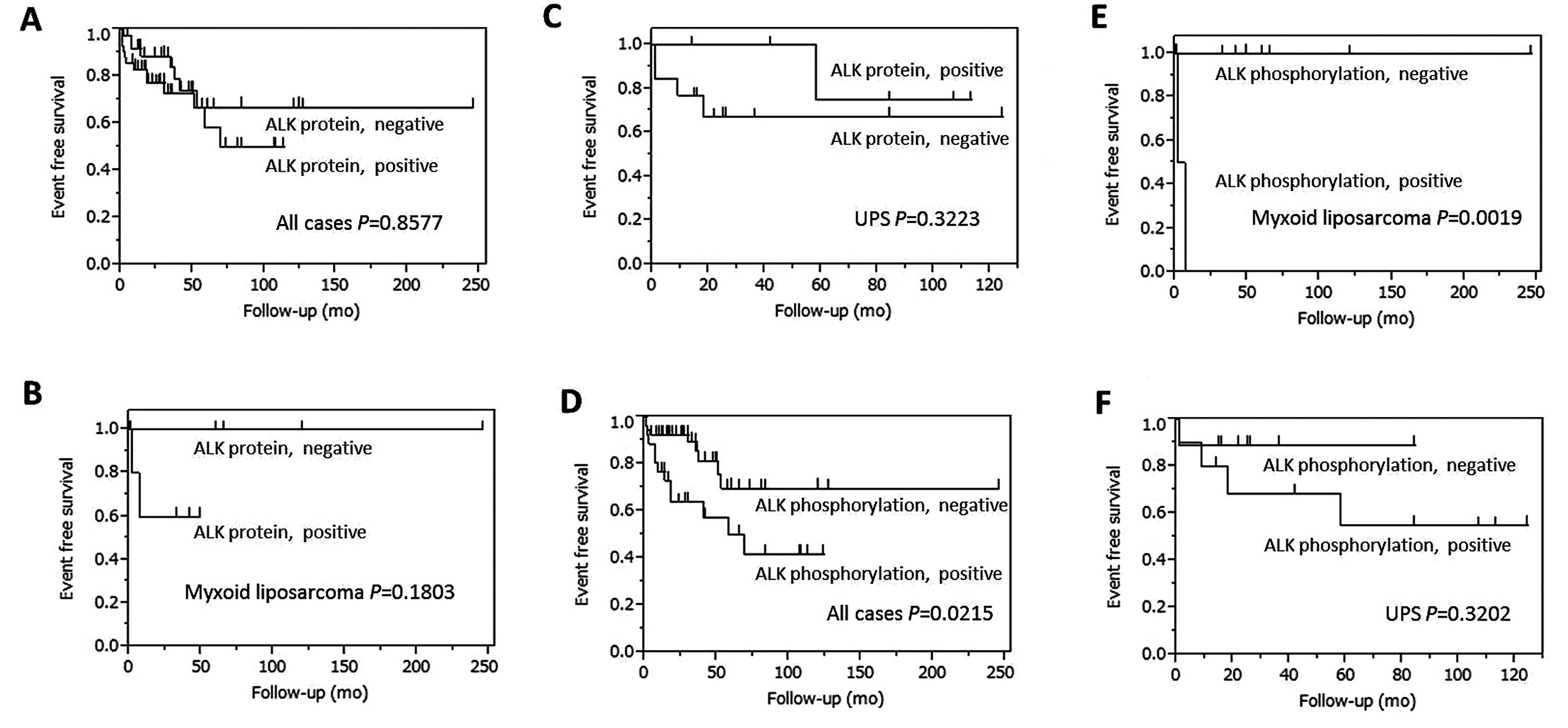
Table V and Fig. 2 show the survival curves for EFS
which are defined as the duration from the time of diagnosis to
metastatic detection. The EFS curves were not statistically
affected by ALK protein expression (P=0.8577).
| Table VP-values from the log-rank test
comparing event-free survival (EFS) curves in soft tissue
tumors. |
Table V
P-values from the log-rank test
comparing event-free survival (EFS) curves in soft tissue
tumors.
| EFS
(metastasis) |
|---|
|
|
|---|
| Disease | ALK protein
(+) | Phosphorylation
(+) |
|---|
| All | 0.8577 | 0.0215 |
|
Leiomyosarcoma | 0.2240 | 0.6495 |
| UPS | 0.3223 | 0.3202 |
| Myxoid
liposarcoma | 0.1803 | 0.0019 |
However, statistically worse EFS was noted for the
ALK phosphorylation-positive cases compared with the -negative
cases, (P=0.0215). In addition, we also investigated this
relationship for the sarcoma subtype, of which there were >10
cases. The phosphorylation of ALK was statistically associated with
worse EFS in the myxoid liposarcoma cases (P=0.0019).
On the other hand, the OS curves were not
statistically affected by either ALK protein expression or
phosphorylation (P=0.5261 and 0.8379, respectively).
Discussion
Our findings revealed that almost 40% of the soft
tissue tumor samples evaluated in the present study expressed the
ALK protein and its phosphorylated form, according to iAEP and IHS,
respectively, as well as ALK gene signal gain, according to
FISH. Statistically significant ratios were detected between ALK
protein expression and phosphorylation (Chi-square test, P=0.0003;
Fisher’s exact test: P=0.0004) and between gene signal gain and the
MIB1 index (Chi-square test, P<0.0001; Fisher’s test,
P<0.0001). The EFS was statistically worse for patients who were
positive for ALK phosphorylation than for those who were negative,
and this was also true for the subtype myxoid liposarcoma cases
(P=0.0019).
Li et al (23) evaluated the ALK protein in alveolar
soft part sarcomas (no. of positive cases/no. analyzed: n=1,
proportion: 0%), well-differentiated liposarcomas (n=19, 47.4%),
synovial sarcomas (n=13, 7.7%), malignant peripheral nerve sheath
tumor (MPNST) (n=12, 8.3%), pleomorphic malignant fibrous
histiocytoma (MFH) (n=27, 22.2%), myxoid liposarcomas (n=17,
70.6%), and myxofibrosarcoma (n=27, 22.2%), among others. In
comparison with those results, our study indicated equal or
slightly higher rates of ALK protein expression in the following
tumor types: alveolar soft part sarcomas (n=2, proportion: 100%),
well-differentiated liposarcomas (n=7, 57.1%), synovial sarcomas
(n=4, 75%), MPNST (n=4, 75%), pleomorphic MFH (n=19, 21.1%), myxoid
liposarcomas (n=11, 54.5%), and myxofibrosarcomas (n=8, 62.5%). A
reason for the discrepancies could be attributed to our use of the
iAEP method, which increased ALK detection sensitivity in
comparison with traditional immunohistochemical methods. This
method has been reported to be more efficacious for detecting the
ALK protein in soft tissue tumors as well as in lung cancer
(16).
Van Gaal et al (15), reported that 62.9% of
rhabdomyosarcoma cases had an ALK copy no. gain, but without
ALK translocation, and this was positively correlated with
ALK protein expression. In the present study, we also detected no
ALK translocation but positive ALK gene signal gain. Gene
signal gain was not positively correlated with ALK protein
expression but was positively correlated with the MIB1 index.
ALK gene signal gain seemed to have no association with
ALK specifically, but rather with mitosis and DNA polyploidy
patterns.
ALK activation may cause cell proliferation
depending on the position of phosphorylation within its
intracellular domain (24–26). Phosphorylation of ALK is followed by
activation of downstream signaling pathways, including PI3K/AKT,
STAT3 and RAS/RAF/ERK, which trigger oncogenic changes (24). The anti-phospho-Y1604 antibody
(clone, EP661Y), used in the clinical study, recognized the
phosphorylated 1,604th tyrosine residue as the epitope (25). Wang et al reported that
expression of phospho-Y 1604 ALK was associated with oncogenic
signaling (21). Other studies have
shown that ALK inhibitors, including crizotinib, suppressed
expression of phospho-Y1604 ALK (26). In a study on crizotinib therapy for
lung cancer, 125 patients (94%) with an ALK fusion event
experienced tumor shrinkage (27).
Our findings revealed that phospho-Y1604 ALK was expressed in
almost 40% of the soft tissue tumor cases and is associated with
poor clinical course. These results suggest that the expression of
phospho-Y1604 ALK in soft tissue tumors is a potential therapeutic
target of medication and, thus, can achieve more favorable
prognoses. Further studies on the effect of ALK inhibitors on
phospho-Y1604 ALK expression are needed.
The suppression of metastasis by ALK inhibitors was
reported in a mouse model, of pulmonary metastasis and survival,
using an in vivo subcutaneous ALK positive xenograft mouse
model (21). The present study
showed a statistical association of phospho-Y1604 ALK expression
with metastasis in soft tissue tumors and with ALK protein
expression. These results potentially suggest that activation of
ALK, indicated by its phosphorylation, is a more important factor
in neoplastic cells than is its protein expression. ALK inhibitors
may prevent progression of metastatic lesions in phospho-Y1604
ALK-positive tumors.
In conclusion, the iAEP method is considered to be
more appropriate than other methods for detection of the ALK
protein in soft tissue tumors. Aberrations of ALK protein
expression and phosphorylation have been observed frequently in
soft tissue tumors, and phosphorylation of ALK was associated with
poor progression of metastasis. ALK plays an important role in
tumor biology and provides potential therapeutic targets for soft
tissue tumors. Future research is needed to explore the oncogenic
function of ALK and the potential effect of ALK inhibitors.
Acknowledgements
The authors thank Mayumi Miura for her technical
contribution.
References
|
1
|
Morris SW, Naeve C, Mathew P, et al: ALK,
the chromosome 2 gene locus altered by the t(2;5) in non-Hodgkin’s
lymphoma, encodes a novel neural receptor tyrosine kinase that is
highly related to leukocyte tyrosine kinase (LTK). Oncogene.
14:2175–2188. 1997. View Article : Google Scholar : PubMed/NCBI
|
|
2
|
Iwahara T, Fujimoto J, Wen D, et al:
Molecular characterization of ALK, a receptor tyrosine kinase
expressed specifically in the nervous system. Oncogene. 14:439–449.
1997. View Article : Google Scholar
|
|
3
|
Pulford K, Lamant L, Morris SW, et al:
Detection of anaplastic lymphoma kinase (ALK) and nucleolar protein
nucleophosmin (NPM)-ALK proteins in normal and neoplastic cells
with the monoclonal antibody ALK1. Blood. 89:1394–1404.
1997.PubMed/NCBI
|
|
4
|
Zhang X, Zhang S, Yang X, et al: Fusion of
EML4 and ALK is associated with development of lung adenocarcinomas
lacking EGFR and KRAS mutations and is correlated with ALK
expression. Mol Cancer. 13:1882010. View Article : Google Scholar
|
|
5
|
Salido M, Pijuan L, Martinez-Aviles L, et
al: Increased ALK gene copy number and amplification are frequent
in non-small cell lung cancer. J Thorac Oncol. 6:21–27. 2011.
View Article : Google Scholar
|
|
6
|
Coffin CM, Hornick JL and Fletcher CD:
Inflammatory myofibroblastic tumor: comparison of
clinicopathologic, histologic, and immunohistochemical features
including ALK expression in atypical and aggressive cases. Am J
Surg Pathol. 31:509–520. 2007. View Article : Google Scholar : PubMed/NCBI
|
|
7
|
Cessna MH, Zhou H, Sanger WG, et al:
Expression of ALK1 and p80 in inflammatory myofibroblastic tumor
and its mesenchymal mimics: a study of 135 cases. Mod Pathol.
15:931–938. 2002. View Article : Google Scholar : PubMed/NCBI
|
|
8
|
De Brouwer BS, De Preter K, Kumps C, et
al: Meta-analysis of neuroblastomas reveals a skewed ALK mutation
spectrum in tumors with MYCN amplification. Clin Cancer Res.
16:4353–4362. 2010. View Article : Google Scholar : PubMed/NCBI
|
|
9
|
Chen Y, Takita J, Choi YL, et al:
Oncogenic mutations of ALK kinase in neuroblastoma. Nature.
455:971–974. 2008. View Article : Google Scholar : PubMed/NCBI
|
|
10
|
George RE, Sanda T, Hanna M, et al:
Activating mutations in ALK provide a therapeutic target in
neuroblastoma. Nature. 455:975–978. 2008. View Article : Google Scholar : PubMed/NCBI
|
|
11
|
Mossé YP, Laudenslager M, Longo L, et al:
Identification of ALK as a major familial neuroblastoma
predisposition gene. Nature. 455:930–935. 2008. View Article : Google Scholar : PubMed/NCBI
|
|
12
|
Janoueix-Lerosey I, Lequin D, Brugieres L,
et al: Somatic and germline activating mutations of the ALK kinase
receptor in neuroblastoma. Nature. 455:967–970. 2008. View Article : Google Scholar : PubMed/NCBI
|
|
13
|
Morris SW, Kirstein MN, Valentine MB, et
al: Fusion of a kinase gene, ALK, to a nucleolar protein gene, NPM,
in non-Hodgkin’s lymphoma. Science. 263:1281–1284. 1994. View Article : Google Scholar : PubMed/NCBI
|
|
14
|
Yoshida A, Shibata T, Wakai S, et al:
Anaplastic lymphoma kinase status in rhabdomyosarcomas. Mod Pathol.
26:772–781. 2013. View Article : Google Scholar : PubMed/NCBI
|
|
15
|
van Gaal JC, Flucke UE, Roeffen MH, et al:
Anaplastic lymphoma kinase aberrations in rhabdomyosarcoma:
clinical and prognostic implications. J Clin Oncol. 30:308–315.
2012. View Article : Google Scholar
|
|
16
|
Takeuchi K, Choi YL, Togashi Y, et al:
KIF5B-ALK, a novel fusion oncokinase identified by an
immunohistochemistry-based diagnostic system for ALK-positive lung
cancer. Clin Cancer Res. 15:3143–3149. 2009. View Article : Google Scholar : PubMed/NCBI
|
|
17
|
Kasprzycka M, Marzec M, Liu X, et al:
Nucleophosmin/anaplastic lymphoma kinase (NPM/ALK) oncoprotein
induces the T regulatory cell phenotype by activating STAT3. Proc
Natl Acad Sci. 103:9964–9969. 2006. View Article : Google Scholar : PubMed/NCBI
|
|
18
|
Chiarle R, Simmons WJ, Cai H, et al: Stat3
is required for ALK-mediated lymphomagenesis and provides a
possible therapeutic target. Nat Med. 11:623–629. 2005. View Article : Google Scholar : PubMed/NCBI
|
|
19
|
Bai RY, Ouyang T, Miething C, et al:
Nucleophosmin-anaplastic lymphoma kinase associated with anaplastic
large-cell lymphoma activates the phosphatidylinositol 3-kinase/Akt
antiapoptotic signaling pathway. Blood. 96:4319–4327.
2000.PubMed/NCBI
|
|
20
|
Zou HY, Li Q, Lee JH, et al: An orally
available small-molecule inhibitor of c-Met, PF-2341066, exhibits
cytoreductive antitumor efficacy through antiproliferative and
antiangiogenic mechanisms. Cancer Res. 67:4408–4417. 2007.
View Article : Google Scholar : PubMed/NCBI
|
|
21
|
Wang YW, Tu PH, Lin KT, et al:
Identification of oncogenic point mutations and
hyperphosphorylation of anaplastic lymphoma kinase in lung cancer.
Neoplasia. 13:704–715. 2011.PubMed/NCBI
|
|
22
|
Fletcher CDM, Bridge JA, Hogendoorn PCW
and Mertens F: World Health Organization Classification of Tumours
of Soft Tissue and Bone. IARC Press; Lyon: pp. 333–365. 2013
|
|
23
|
Li XQ, Hisaoka M, Shi DR, et al:
Expression of anaplastic lymphoma kinase in soft tissue tumors: an
immunohistochemical and molecular study of 249 cases. Hum Pathol.
35:711–721. 2004. View Article : Google Scholar : PubMed/NCBI
|
|
24
|
Duijkers FA, Gaal J, Meijerink JP, et al:
Anaplastic lymphoma kinase (ALK) inhibitor response in
neuroblastoma is highly correlated with ALK mutation status, ALK
mRNA and protein levels. Cell Oncol. 34:409–417. 2011. View Article : Google Scholar
|
|
25
|
Katayama R, Shaw AT, Khan TM, et al:
Mechanisms of acquired crizotinib resistance in ALK-rearranged lung
cancers. Sci Transl Med. 4:120ra172012.PubMed/NCBI
|
|
26
|
Palmer RH, Vernersson E, Grabbe C and
Hallberg B: Anaplastic lymphoma kinase: signalling in development
and disease. Biochem J. 420:345–361. 2009. View Article : Google Scholar : PubMed/NCBI
|
|
27
|
Camidge DR, Bang YJ, Kwak EL, et al:
Activity and safety of crizotinib in patients with ALK-positive
non-small-cell lung cancer: updated results from a phase 1 study.
Lancet Oncol. 13:1011–1019. 2012. View Article : Google Scholar : PubMed/NCBI
|