Introduction
Colorectal cancer (CRC) is the most common
malignancy of the colon and rectum and the third most common cause
of cancer-related death among men and women worldwide (1). Outcome prediction based on tumor stage
reflected by the tumor node metastasis (TNM) system of the Union
for International Cancer Control (UICC) is currently regarded as
the standard prognostic parameter (2,3).
Venous and lymphatic vessel invasion are also important malignant
factors of CRC (3–5). Both lymphangiogenesis and angiogenesis
also play important roles as poor prognostic factors in
tumorigenesis (6–8). In addition, the extracellular matrix
(ECM) influences cancer proliferation, activities of invasion and
metastasis by stimulating angiogenesis and lymphangiogenesis
(9,10).
In contrast, the relationship between CRC and
myofibroblasts in the tumor microenvironment has recently attracted
considerable attention. Myofibroblasts are not only known as a
principal cellular component in the granulation tissue of healing
wounds but are also one of the cancer stromal cells that constitute
the ECM (11,12). The myofibroblasts in the stroma of
CRC serve an important function in promoting the desmoplastic
reaction and influencing tumor invasion, microvessel density around
the invasive lesion and metastatic carcinomas (13–15).
Moreover, myofibroblast activation in tumor metastatic lymph nodes
influences the microenvironment supporting CRC metastasis (16).
With regard to both the tumor growth and spreading
of CRC, three histological layers of the colorectum, the submucosa
(SM), muscularis propria (MP) and subserosa (SS), may play
important functions in the mechanical and physiological protection
against invasive growth. MP is exclusively composed of smooth
muscle bundles and comprises tight connective tissue, whereas SM
and SS are mainly composed of loose connective tissue (17,18).
However, it is unclear how myofibroblasts are distributed around
the CRC invasive border of these three layers as well as how the
distribution is related to the malignant potential of CRC.
In the present study, we measured the myofibroblast
density of each colorectal layer using imaging analysis and
investigated the association between myofibroblast distribution and
clinicopathological factors such as lymph node metastasis and
venous invasion. Furthermore, we showed the relationship between
the myofibroblast distribution and overall survival of patients
with CRC.
Materials and methods
Patients
One hundred and twenty-one patients with advanced
CRC, defined as adenocarcinoma, which had invaded the SS layer of
the colorectal wall (pT3), underwent surgical resection from
January 2008 to December 2009 at Hirosaki University Hospital. The
clinical stages of these patients were stage II or III according to
the TNM classification of the UICC (2). Survival data were obtained from
hospital medical charts. Cancer-specific survival was measured from
the date of surgery until the date of death from CRC. None of the
patients were treated with neoadjuvant chemotherapy, and none of
them had synchronous multiple CRCs or synchronous metastasis to
other organs.
Pathological analysis
We used surgically resected specimens that were
fixed with 10% formalin, embedded in paraffin and stained with
hematoxylin and eosin (H&E) for pathological evaluation.
Degrees of lymphatic vessel invasion were classified as 0, no
invasion; 1, mild invasion; 2, moderate invasion and 3, severe
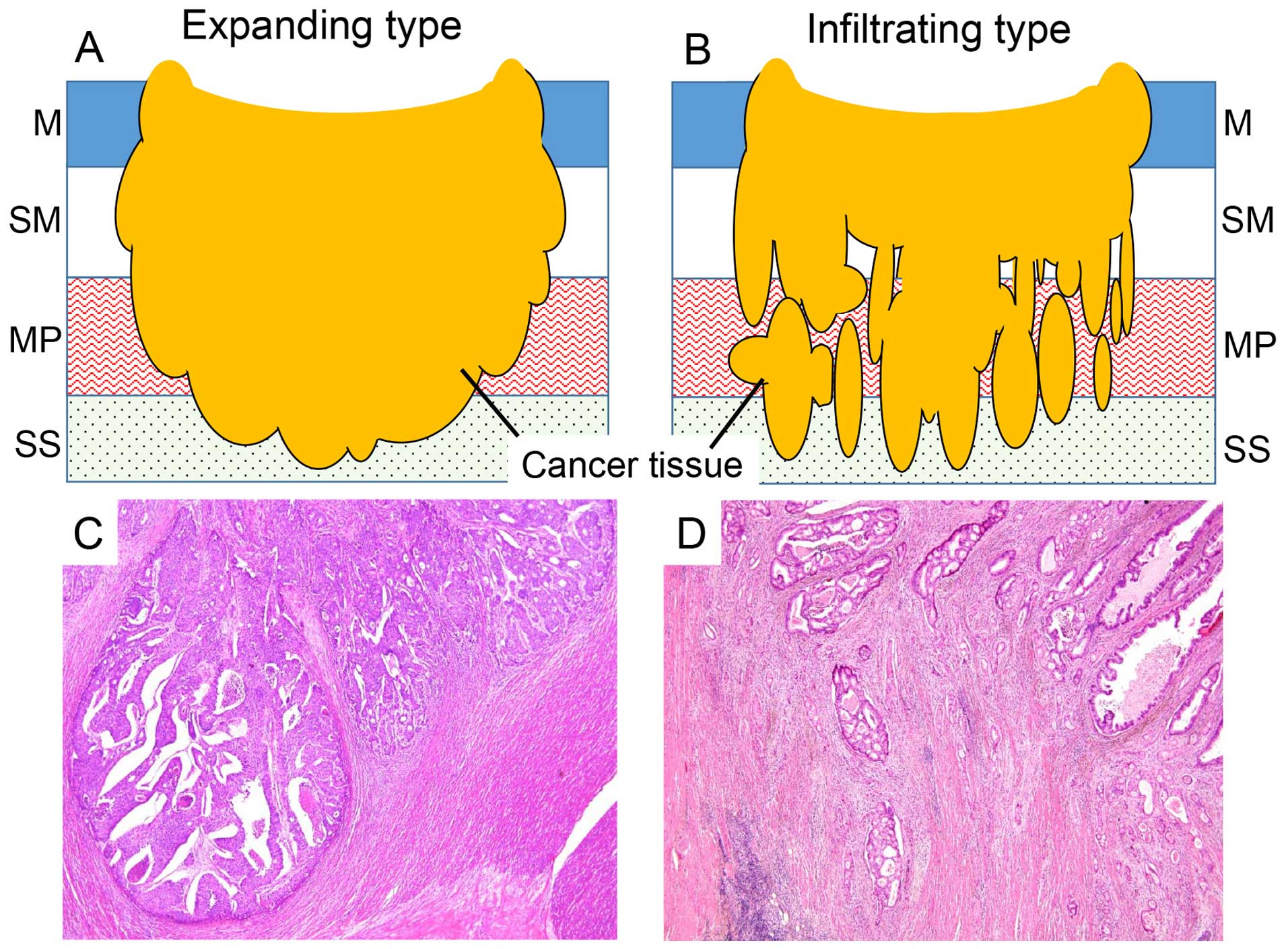
invasion. The modes of invasive growth pattern were classified into
two groups, namely expanding type, the overall pushing growth type
of adenocarcinoma with a clear invasive margin; and infiltrating
type, a widespread streaming form of adenocarcinoma with an unclear
borderline of the invasive front (Fig.
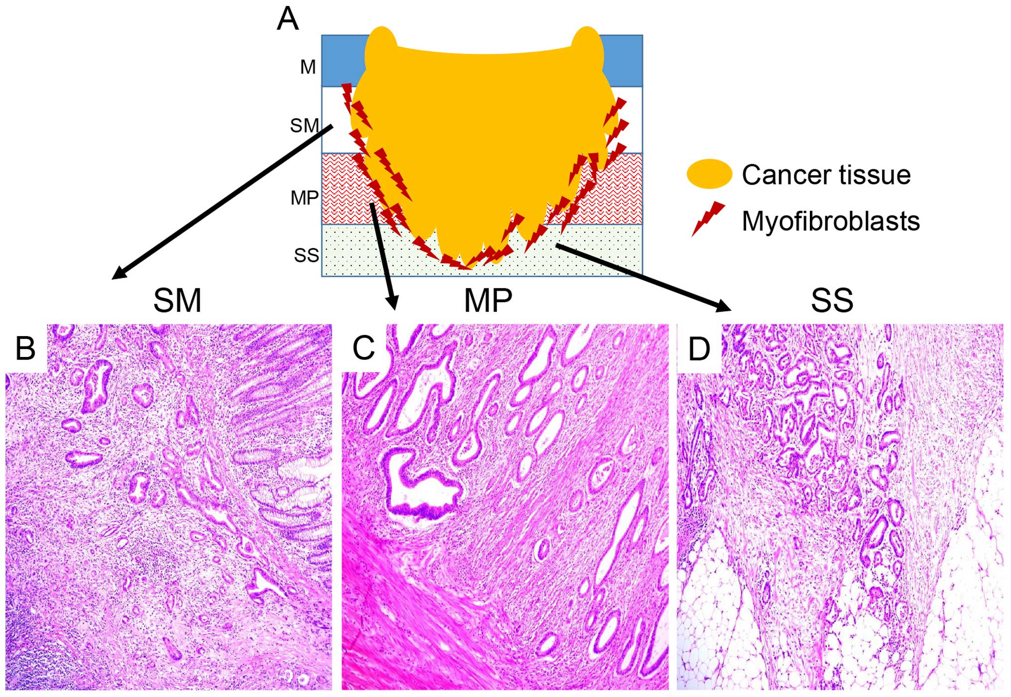
1). To evaluate the myofibroblast distribution of each case, we
selected the paraffin-embedded specimen that showed three invasive
lesions in each histological layer (SM, MP and SS) as diagnosed by
H&E staining (Fig. 2).
Immunohistochemistry
For immunohistochemical examination regarding the
myofibroblast distribution in each case, the paraffin-embedded
specimen which was described in 'Pathological analysis' was a
representative specimen of each case, and we used serial
4-µm sections for the immunohistochemical analysis. The
sections were mounted on saline-coated glass slides. The antibodies
used included α-smooth muscle actin (α-SMA, 1:100, clone 1A4) and
desmin (1:100, clone D-33) (both from Dako, Glostrup, Denmark).
Immunostaining for α-SMA and desmin was performed using the
standard avidin-biotin-peroxidase complex method with an automated
immunostainer (Benchmark XT; Ventana Medical System, Tucson, AZ,
USA). The signature characteristic of myofibroblasts is an
α-SMA-positive and desmin-negative pattern, whereas that of smooth
muscle is an α-SMA-positive and desmin-positive pattern.
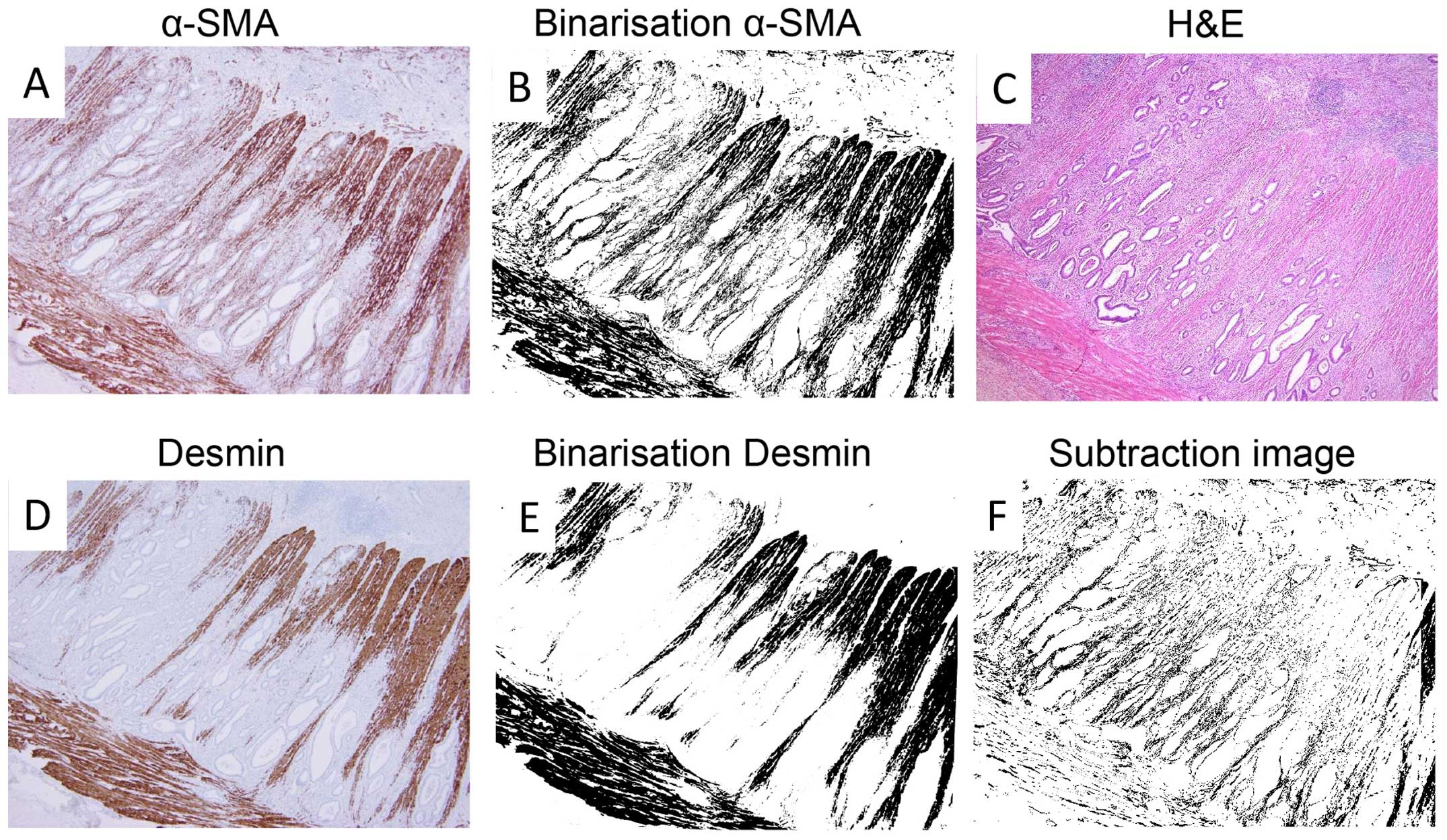
Image analysis
We used imaging analysis to investigate the
myofibroblast density. All cases had an invasive lesion of the
three colorectal walls: SM, MP and SS. To obtain the images, we
used an Olympus microscope BX50 with a U PlanApo objective lens (×4
magnification), DP Control software and a DP-70 digital camera (all
from Olympus, Tokyo, Japan). We applied ImageJ software (National
Institutes of Health, Bethesda, MD, USA) to view and analyze our
obtained images (19). We captured
images of α-SMA and desmin (Fig. 3A and
D), and these images were binarised (Fig. 3B and E). The binarised images showed
that the positively and negatively immunostained lesions were black
and white, respectively. We made a subtraction image by pasting the
binarised images of desmin onto the binarised images of α-SMA using
the subtraction mode in ImageJ software (Fig. 3F). The subtraction images were shown
as the value of α-SMA minus that of desmin, and we could interpret
the subtraction images showing myofibroblasts in the representative
sections of each case. From all 121 cases, we obtained subtraction
images of the three colorectal wall layers (SM, MP and SS) and
measured the myofibroblast density in 1×1 mm2 areas in
the invasive border of each layer. We selected a hot spot
myofibroblast density area from each invasive layer.
Statistical analysis
All values are presented as the means ± standard
error of the mean. Chi-square tests were performed for
non-continuous variables, while the Mann-Whitney test and Welch
t-tests were used for comparing continuous variables. Survival
curves were constructed using the Kaplan-Meier method, and
differences in survival were evaluated using the log-rank test. The
relative prognostic factors were analysed with a Cox proportional
hazards regression model. Differences were considered as
statistically significant if the P-value was <0.05. Statistical
analysis was performed with R (http://www.r-project.org) and Microsoft Excel software
(Microsoft Corporation, Redmond, WA, USA).
Results
Clinicopathological characteristics
The clinicopathological characteristics of the 121
CRC cases are summarised in Table
I. The series consisted of 66 men and 55 women, with a median
age of 67.5 years (range, 26–93 years). The carcinomas were located
in the colon (77 cases) and rectum (44 cases). One hundred and ten
carcinomas were diagnosed as well and moderately differentiated
adenocarcinoma, and 11 carcinomas were diagnosed as poorly
differentiated and mucinous adenocarcinoma. In terms of the CRC
invasive pattern, 57 cases were the expanding type, and 64 cases
were the infiltrating type. Eighty cases and 41 cases had low and
high degrees of lymphatic invasion, respectively. In contrast, the
numbers of cases with low and high degrees of venous invasion were
90 and 31 cases, respectively. Furthermore, the numbers of cases
with negative and positive lymph nodes were 64 and 57 cases,
respectively.
 | Table IHistopathological characteristics of
the 121 cases. |
Table I
Histopathological characteristics of
the 121 cases.
| Variables | No. of patients |
|---|
| Age in years, median
(range) | 67.4 (26–93) |
| Gender |
| Male | 66 |
| Female | 55 |
| Location |
| Colon | 77 |
| Rectum | 44 |
| Histological
type |
| Well, mod | 110 |
| Por, muc | 11 |
| Invasive type |
| Expanding | 57 |
| Infiltrating | 64 |
| Lymphatic
invasion |
| Low (ly0 or
ly1) | 80 |
| High (ly2 or
ly3) | 41 |
| Venous invasion |
| Low (v0 or v1) | 90 |
| High (v2 or v3) | 31 |
| Lymph node
metastasis |
| Negative | 64 |
| Positive | 57 |
Myofibroblast distribution in the
invasive lesion at each colorectal wall stratified by expanding
type vs. infiltrating type
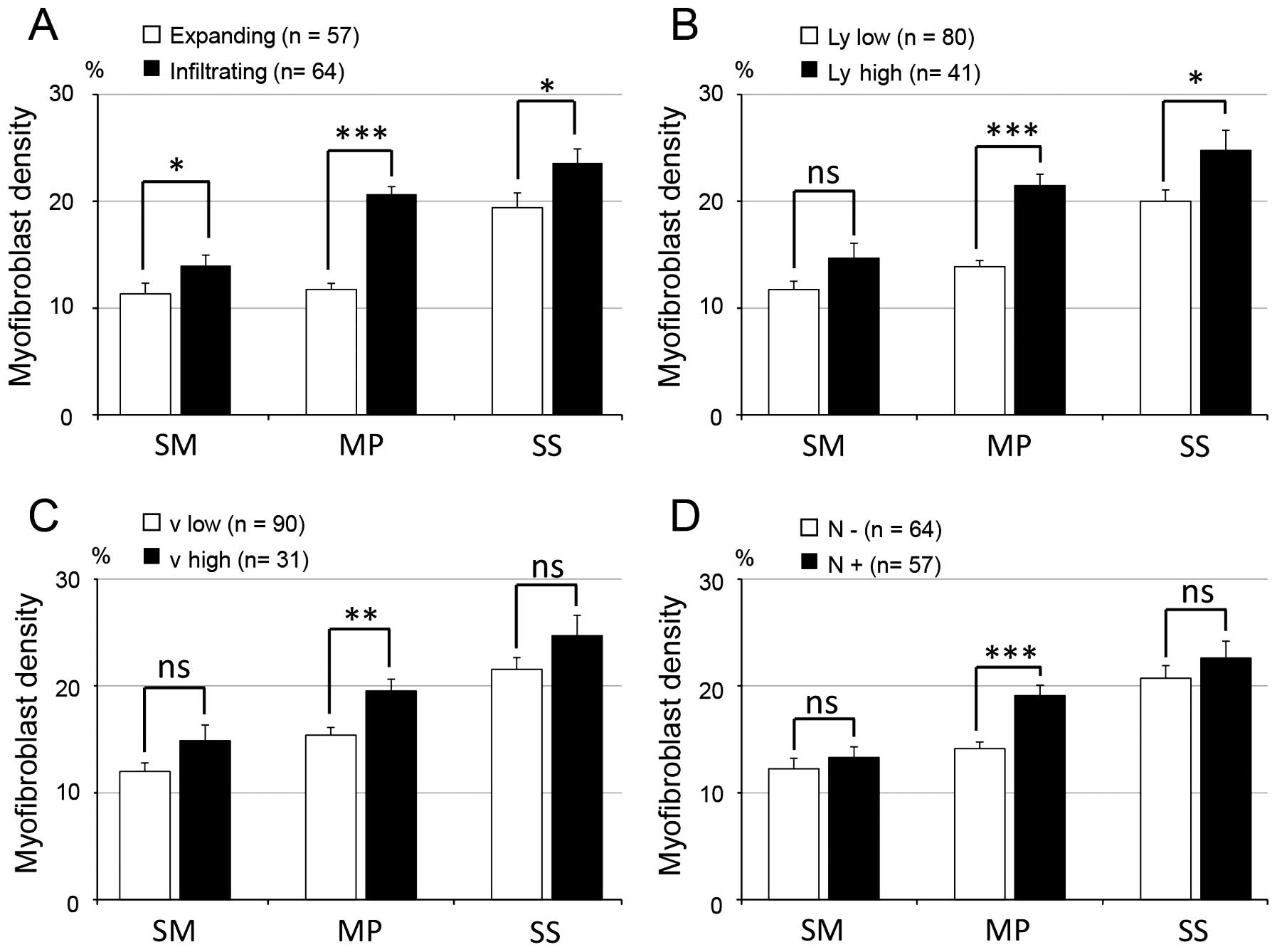
We measured the myofibroblast density around the
invasive front of each layer (SM, MP and SS) for the expanding and
infiltrating types (Fig. 4A). In 57
cases of the expanding type, the mean myofibroblast densities for
each wall of the invasive lesion were 11.03±0.88% (SM), 11.62±0.50%
(MP) and 19.24±1.34% (SS). Meanwhile, in 64 cases of the
infiltrating type, the mean myofibroblast densities were
13.60±0.79% (SM), 20.52±0.62% (MP) and 22.40±1.07% (SS).
Significantly more myofibroblasts were located around these three
invasive layers in the infiltrating type than the expanding type
(P<0.05).
Association between the distributions of
myofibroblast density and lymphatic vessel invasion
To investigate the association between the
myofibroblast distribution and the degree of lymphatic vessel
invasion, we stratified the 121 cases of CRC into either a low
lymphatic vessel invasion (ly0 and ly1) group or a high lymphatic
vessel invasion (ly2 and ly3) group. We analysed the myofibroblast
distribution around the invasive front of each layer (Fig. 4B). The mean myofibroblast densities
in the three layers within the low lymphatic vessel invasion group
(n=80) were 11.73±0.77% (SM), 13.89±0.57% (MP) and 19.99±1.06%
(SS). In contrast, the mean myofibroblast densities in the high
lymphatic vessel invasion group (n=41) were 14.68±1.39% (SM),
21.48±1.05% (MP) and 24.76±1.86% (SS). The myofibroblast density
was significantly higher in the group with high degree lymphatic
vessel invasion than that noted in the group with low degree
lymphatic vessel invasion in the MP (P<0.001) and SS layer
(P=0.04), respectively. On the other hand, there was no significant
difference between the low and high lymphatic vessel invasion group
in regards to the myofibroblast density of the SM layer
(P=0.103).
Association between the myofibroblast
density distribution and venous vessel invasion
To investigate the association between the
myofibroblast distribution and the degree of venous vessel
invasion, we stratified the 121 cases into a low venous vessel
invasion (v1 and v2) group and a high venous vessel invasion (v2
and v3) group and analysed the myofibroblast distribution around
the invasive front of each layer (Fig.
4C). The mean myofibroblast densities in the low venous
invasion group (n=90) were 11.99±0.79% (SM), 15.40±0.70% (MP) and
21.54±1.10% (SS), while the mean myofibroblast densities in the
high venous invasion group (n=31) were 14.85±1.47% (SM),
19.54±1.09% (MP) and 24.70±1.91% (SS). There was a significant
difference in the myofibroblast density of the MP layer between the
low and high venous invasion groups (P<0.01). There were not
significant differences between the two groups in regards to the
myofibroblast density of the SM layer (P=0.07) and SS layer
(P=0.06).
Association between the myofibroblast
distribution and lymph node metastasis
We stratified the 121 CRC cases into a lymph node
metastasis-negative group and -positive group and investigated the
myofibroblast distribution of the three invasive walls (Fig. 4D). The mean myofibroblast densities
of the three walls within the lymph node metastasis-negative group
(n=64) were 12.24±0.98% (SM), 14.12±0.63% (MP) and 20.73±1.16%
(SS). The mean myofibroblast densities in the lymph node
metastasis-positive group (n=57) were 13.28±1.01% (SM), 19.01±0.97%
(MP) and 22.61±1.56% (SS). The lymph node-positive group had higher
myofibroblast densities for all of the invasive layers than the
lymph node-negative group. Furthermore, there was a significant
difference between the lymph node metastasis-positive and -negative
groups relating to the myofibroblast density of the MP layer
(P<0.001). There were no significant differences between the two
groups in regards to the myofibroblast density of the SM (P=0.33)
and SS layer (P=0.35).
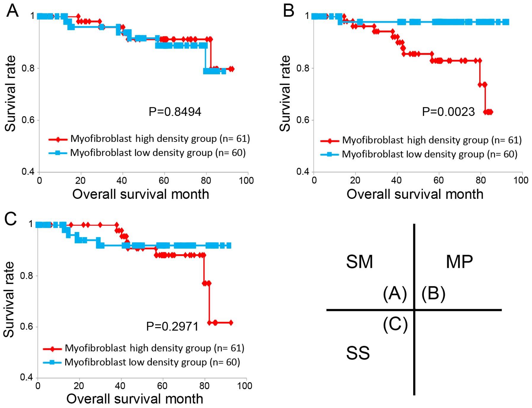
Association between the myofibroblast
density distribution and patient overall survival
To investigate the association between the
myofibroblast distribution and overall survival, we stratified the
121 cases of CRC into either a low myofibroblast density group or a
high density group in each invasive layer and compared the high and
low groups regarding the overall survival of the patients. The
cut-off point between the two groups was set at the median value of
the myofibroblast density in each invasive layer; the median values
of the myofibroblast density were 11.52% in the SM layer, 16.19% in
the MP layer and 21.48% in the SS layer. In only the MP level, but
not the SM and SS layers, patients with high myofibroblast
densities showed a significantly reduced overall survival
(P<0.003; Fig. 5). To clarify
the potential indicators, we analysed various pathological factors
that were recorded in this study (Table II). Univariate analysis revealed
that the following factors were correlated with poor prognosis:
myofibroblast density of MP [relative risk (RR), 10.504; 95%
confidence interval (CI), 1.344–82.09; P=0.025], invasive type (RR,
10.190; 95% CI, 1.302–79.75; P=0.027) and lymphatic invasion (RR,
4.4291; 95% CI, 1.175–16.7; P=0.028). In the multivariate analysis,
there was no significant difference among the myofibroblast density
of the MP layer, the invasive type and lymphatic invasion.
| Table IIUnivariate and multivariate analyses
of prognostic factors of survival. |
Table II
Univariate and multivariate analyses
of prognostic factors of survival.
| Variables | n (%) | Univariate analysis
P-value | Multivariate analysis
P-value |
|---|
| SM myofibroblast
density | | 0.728 | – |
| Low | 61 (50.5) | | |
| High | 60 (49.5) | | |
| MP myofibroblast
density | | 0.025 | 0.332 |
| Low | 61 (50.5) | | |
| High | 60 (49.5) | | |
| SS myofibroblast
density | | 0.303 | – |
| Low | 61 (50.5) | | |
| High | 60 (49.5) | | |
| Histological
type | | 0.998 | – |
| Well, mod | 110 (90.9) | | |
| Por, muc | 11 (9.1) | | |
| Invasive type | | 0.027 | 0.488 |
| Expanding | 57 (47.1) | | |
| Infiltrating | 64 (52.9) | | |
| Lymphatic
invasion | | 0.028 | 0.258 |
| Low (ly0 or
ly1) | 80 (66.1) | | |
| High (ly2 or
ly3) | 41 (33.9) | | |
| Venous
invasion | | 0.392 | – |
| Low (v0 or
v1) | 90 (74.4) | | |
| High (v2 or
v3) | 31 (25.6) | | |
| Lymph node
metastasis | | 0.319 | – |
| Negative | 64 (52.9) | | |
| Positive | 57 (47.1) | | |
Discussion
In the present study, we evaluated the association
between clinicopathological characteristics of CRC and the
myofibroblast distribution of three invasive layers using image
analysis. We revealed that the myofibroblast density of MP plays an
important role in CRC malignant behaviors, such as lymphatic
invasion, venous invasion and lymph node metastasis, which can
result in short overall survival of the patients.
We found that as the invasion of the CRC became
deeper, the number of myofibroblasts increased around the invasive
lesions, and the infiltrating growth type had a significantly
higher density of myofibroblasts than that noted in the expanding
type. Previous studies identified that the infiltrating type of CRC
carries a high risk of liver metastasis and a worse prognosis
compared with the expanding type (20–22).
Myofibroblasts are a type of cancer-associated fibroblasts (CAFs)
and are involved in desmoplastic reactions (23). CAFs actively associate with
neoplastic cells and form the ECM of cancer lesions that promote
cancer growth, angiogenesis and survival (24). CAFs interact with adjacent cancer
cells through soluble factors or direct cell-cell adhesion to
promote cancer cell invasion (25).
In malignancy of CRC, myofibroblasts also promote CRC invasion and
metastasis as they proliferate around the invasive lesion and alter
the adhesive and migratory properties of CRC cells (15,26). A
previous study showed that myofibroblasts co-cultured with CRC
cells may be involved in the invasiveness of CRC, even when the
expression of E-cadherin, which is understood to be an adhesion
molecule, prevents tumor cell invasiveness in vitro
(27). Therefore, we suggest that
it is possible that the large quantity of myofibroblasts which play
a role as CAFs may alter both the adhesive and migratory properties
of CRC cells and consequently aid CRC invasion into the deep
colorectal layers. Moreover, our study indicated that the
association between the infiltrating type, which is regarded as a
malignant factor and myofibroblasts is stronger than the
association between the expanding type and myofibroblasts.
Our results showed that the myofibroblast density of
the MP layer was significantly higher in the group with a high
frequency of lymphatic vessel and venous invasion compared with
that in the group with a low frequency of lymphatic vessel and
venous invasion. Furthermore, the lymph node-positive group had a
significantly higher myofibroblast density in the MP layer than
that of the lymph node-negative group. The lymphatic and venous
vessels exist in three colorectal layers (SM, MP and SS), despite
the differences in their histological structures. The distribution
of lymphatic and venous vessels in normal colonic tissue tends to
increase in frequency with depth throughout the wall (28). The functions of α-SMA-positive
myofibroblasts may be associated with promoting the ECM of tumor
cells and lymphogenesis of the metastatic microenvironment in oral
tongue squamous cell carcinoma (29). With respect to CRC, proliferation of
myofibroblasts in the peritumoral areas was predicted to play an
important role in lymphangiogenesis and was also found to be
associated with lymph node metastasis (15). A previous study indicated that the
CRC-invading MP layer may result in a greater ability to induce
angiogenesis in adjacent normal tissue (30). Another study showed that the
morphological mode of tumor invasion in the MP layer was associated
with hematogenous metastasis of CRC (31). Our study predicted that compared to
myofibroblasts of the other layers, myofibroblasts of the MP layer
change the morphological mode of tumor invasion in CRC and increase
the number of lymphatic and venous vessels that are invaded by CRC
cells. Therefore, myofibroblasts of the MP layer are associated
with the malignant potential of CRC, including lymph node
metastasis.
The results of the univariate analysis revealed that
myofibroblasts in the MP layer were significantly correlated with
poor patient prognosis; however, the multivariate analysis using
Cox proportional hazards model showed that a high myofibroblast
density of MP was not an independent prognostic factor for overall
survival. We suspected that the reason for this was that
myofibroblasts of the MP layer may be strongly associated with the
invasive growth pattern and lymphatic invasion.
In conclusion, we revealed that the myofibroblast
distribution contributes to the malignant potential of CRC.
Furthermore, we showed that myofibroblasts around the MP layer play
an important role in the malignant potential and poor prognosis of
CRC patients.
Acknowledgments
This study was supported by Grants-in Aid for
Science from the Ministry of Education, Culture, Sports, Science,
and Technology in Japan and a grant for Hirosaki University
Institutional Research.
Abbreviations:
References
|
1
|
Ferlay JS, Ervik M, Diskshit R, Eser S,
Mathers C, Rebelo M, Parkin D, Forman D and Baray F: GLOBOCAN 2012
v1.0, Cancer Incidence and Mortality Worldwide. IARC; 2012
|
|
2
|
Sobin LH, Gospodarowicz MK and Wittekind
C: TNM Classification of Malignant Tumours. 7th edition.
Wiley-Blackwell; 2009
|
|
3
|
Seefeld PH and Bargen JA: The spread of
carcinoma of the rectum: Invasion of lymphatics, veins and nerves.
Ann Surg. 118:76–90. 1943. View Article : Google Scholar : PubMed/NCBI
|
|
4
|
Chapuis PH, Dent OF, Fisher R, Newland RC,
Pheils MT, Smyth E and Colquhoun K: A multivariate analysis of
clinical and pathological variables in prognosis after resection of
large bowel cancer. Br J Surg. 72:698–702. 1985. View Article : Google Scholar : PubMed/NCBI
|
|
5
|
Shirouzu K, Isomoto H, Morodomi T, Araki Y
and Kakegawa T: Lymphatic permeation of colorectal cancer -
evaluation as a prognostic factor by prospective studies. Nihon
Geka Gakkai Zasshi. 92:1686–1693. 1991.In Japanese. PubMed/NCBI
|
|
6
|
Royston D and Jackson DG: Mechanisms of
lymphatic metastasis in human colorectal adenocarcinoma. J Pathol.
217:608–619. 2009. View Article : Google Scholar : PubMed/NCBI
|
|
7
|
van Netten JP, Cann SA and van der
Westhuizen NG: Angiogenesis and tumor growth. N Engl J Med.
334:920–921; author reply 921. 1996. View Article : Google Scholar : PubMed/NCBI
|
|
8
|
Kim ER and Kim YH: Clinical application of
genetics in management of colorectal cancer. Intest Res.
12:184–193. 2014. View Article : Google Scholar : PubMed/NCBI
|
|
9
|
Ou J, Deng J, Wei X, Xie G, Zhou R, Yu L
and Liang H: Fibronectin extra domain A (EDA) sustains
CD133(+)/CD44(+) subpopulation of colorectal cancer cells. Stem
Cell Res (Amst). 11:820–833. 2013. View Article : Google Scholar
|
|
10
|
Guan X: Cancer metastases: Challenges and
opportunities. Acta Pharm Sin B. 5:402–418. 2015. View Article : Google Scholar : PubMed/NCBI
|
|
11
|
Seemayer TA, Schürch W and Lagacé R:
Myofibroblasts in human pathology. Hum Pathol. 12:491–492. 1981.
View Article : Google Scholar : PubMed/NCBI
|
|
12
|
Bissell MJ and Radisky D: Putting tumours
in context. Nat Rev Cancer. 1:46–54. 2001. View Article : Google Scholar
|
|
13
|
Okamoto Y, Fujimori T, Ohkura Y, Sugai T,
Arai T, Watanabe G, Wada R, Ueno H, Togashi K, Yao T, et al:
Histological assessment of intra- and inter-institutional
reliabilities in detection of desmoplastic reaction in biopsy
specimens of early colorectal carcinomas. Pathol Int. 63:539–545.
2013. View Article : Google Scholar : PubMed/NCBI
|
|
14
|
Tsujino T, Seshimo I, Yamamoto H, Ngan CY,
Ezumi K, Takemasa I, Ikeda M, Sekimoto M, Matsuura N and Monden M:
Stromal myofibroblasts predict disease recurrence for colorectal
cancer. Clin Cancer Res. 13:2082–2090. 2007. View Article : Google Scholar : PubMed/NCBI
|
|
15
|
Liang P, Hong JW, Ubukata H, Liu G, Katano
M, Motohashi G, Kasuga T, Watanabe Y, Nakada I and Tabuchi T:
Myofibroblasts correlate with lymphatic microvessel density and
lymph node metastasis in early-stage invasive colorectal carcinoma.
Anticancer Res. 25:2705–2712. 2005.PubMed/NCBI
|
|
16
|
Yeung TM, Buskens C, Wang LM, Mortensen NJ
and Bodmer WF: Myofibroblast activation in colorectal cancer lymph
node metastases. Br J Cancer. 108:2106–2115. 2013. View Article : Google Scholar : PubMed/NCBI
|
|
17
|
Nakayama H, Enzan H, Miyazaki E, Naruse K,
Kiyoku H and Hiroi M: The role of myofibroblasts at the tumor
border of invasive colorectal adenocarcinomas. Jpn J Clin Oncol.
28:615–620. 1998. View Article : Google Scholar : PubMed/NCBI
|
|
18
|
Ueno H, Hase K, Hashiguchi Y, Ishiguro M,
Kajiwara Y, Shimazaki H and Mochizuki H: Growth pattern in the
muscular layer reflects the biological behaviour of colorectal
cancer. Colorectal Dis. 11:951–959. 2009. View Article : Google Scholar : PubMed/NCBI
|
|
19
|
Schneider CA, Rasband WS and Eliceiri KW:
NIH Image to ImageJ: 25 years of image analysis. Nat Methods.
9:671–675. 2012. View Article : Google Scholar : PubMed/NCBI
|
|
20
|
Morikawa T, Kuchiba A, Qian ZR,
Mino-Kenudson M, Hornick JL, Yamauchi M, Imamura Y, Liao X,
Nishihara R, Meyerhardt JA, et al: Prognostic significance and
molecular associations of tumor growth pattern in colorectal
cancer. Ann Surg Oncol. 19:1944–1953. 2012. View Article : Google Scholar :
|
|
21
|
Rajaganeshan R, Prasad R, Guillou PJ,
Chalmers CR, Scott N, Sarkar R, Poston G and Jayne DG: The
influence of invasive growth pattern and microvessel density on
prognosis in colorectal cancer and colorectal liver metastases. Br
J Cancer. 96:1112–1117. 2007. View Article : Google Scholar : PubMed/NCBI
|
|
22
|
Jass JR, Love SB and Northover JM: A new
prognostic classification of rectal cancer. Lancet. 1:1303–1306.
1987. View Article : Google Scholar : PubMed/NCBI
|
|
23
|
Karagiannis GS, Petraki C, Prassas I,
Saraon P, Musrap N, Dimitromanolakis A and Diamandis EP: Proteomic
signatures of the desmoplastic invasion front reveal collagen type
XII as a marker of myofibroblastic differentiation during
colorectal cancer metastasis. Oncotarget. 3:267–285. 2012.
View Article : Google Scholar : PubMed/NCBI
|
|
24
|
Karagiannis GS, Poutahidis T, Erdman SE,
Kirsch R, Riddell RH and Diamandis EP: Cancer-associated
fibroblasts drive the progression of metastasis through both
paracrine and mechanical pressure on cancer tissue. Mol Cancer Res.
10:1403–1418. 2012. View Article : Google Scholar : PubMed/NCBI
|
|
25
|
Yamaguchi H and Sakai R: Direct
interaction between carcinoma cells and cancer associated
fibroblasts for the regulation of cancer invasion. Cancers (Basel).
7:2054–2062. 2015. View Article : Google Scholar
|
|
26
|
Martin M, Pujuguet P and Martin F: Role of
stromal myofibroblasts infiltrating colon cancer in tumor invasion.
Pathol Res Pract. 192:712–717. 1996. View Article : Google Scholar : PubMed/NCBI
|
|
27
|
Dimanche-Boitrel MT, Vakaet L Jr, Pujuguet
P, Chauffert B, Martin MS, Hammann A, Van Roy F, Mareel M and
Martin F: In vivo and in vitro invasiveness of a rat colon-cancer
cell line maintaining E-cadherin expression: An enhancing role of
tumor-associated myofibroblasts. Int J Cancer. 56:512–521. 1994.
View Article : Google Scholar : PubMed/NCBI
|
|
28
|
Duff SE, Jeziorska M, Kumar S, Haboubi N,
Sherlock D, O'Dwyer ST and Jayson GC: Lymphatic vessel density,
microvessel density and lymphangiogenic growth factor expression in
colorectal cancer. Colorectal Dis. 9:793–800. 2007. View Article : Google Scholar : PubMed/NCBI
|
|
29
|
Ding L, Zhang Z, Shang D, Cheng J, Yuan H,
Wu Y, Song X and Jiang H: α-Smooth muscle actin-positive
myofibroblasts, in association with epithelial-mesenchymal
transition and lymphogenesis, is a critical prognostic parameter in
patients with oral tongue squamous cell carcinoma. J Oral Pathol
Med. 43:335–343. 2014. View Article : Google Scholar
|
|
30
|
Fox SH, Whalen GF, Sanders MM, Burleson
JA, Jennings K, Kurtzman S and Kreutzer D: Angiogenesis in normal
tissue adjacent to colon cancer. J Surg Oncol. 69:230–234. 1998.
View Article : Google Scholar
|
|
31
|
Kanno M, Kurosaka Y, Kosaka T, Yamaguchi
A, Yonemura Y, Miwa K and Miyazaki I: Study on correlation of
hematogenous metastasis in advanced colorectal cancer with the
morphological mode of tumor invasion in the pm layer. Nihon Geka
Gakkai Zasshi. 93:139–143. 1992.In Japanese. PubMed/NCBI
|