Reactive oxygen species (ROS) is a general term used
to describe molecules with high oxidative reactivity. They are
mainly produced by the electron transport chain during aerobic
respiration in the mitochondria or as a byproduct of the activity
of several metabolic enzymes, including xanthine oxidase,
lipoxygenase and cytochrome P450 (1).
In addition, exogenous stimuli, such as stress, ultraviolet
radiation, tumor chemotherapy and radiotherapy (RT), can stimulate
ROS production (2). Under
physiological conditions, cells can scavenge intracellular ROS
using antioxidants, including catalase, glutathione and ascorbic
acid, to maintain the dynamic redox balance (3). Once the level of ROS exceed the
tolerance threshold of cells, a variety of pathological disorders
occur. Previous studies have shown that abnormal ROS levels are
closely associated with the occurrence of tumors and
neurodegenerative diseases (4,5). During
the moderate redox state, ROS can induce tumorigenesis by
activating the MAPK and ERK signaling pathway or promoting
mutations in the genomic DNA (6).
However, previous studies have demonstrated that ROS production is
actually inhibited during breast and colon tumor progression, where
tumor cells attempt to reduce or eliminate the adverse effects of
ROS by potently activating their antioxidant systems (7,8). This
leads to resistance to treatments, including chemotherapy, RT and
immunotherapy (9,10). In addition, an elevation in ROS levels
in breast cancer and human multiple myeloma has been found to
promote tumor cell death in different signaling pathways and
increase sensitivity to anti-tumor therapy (11,12).
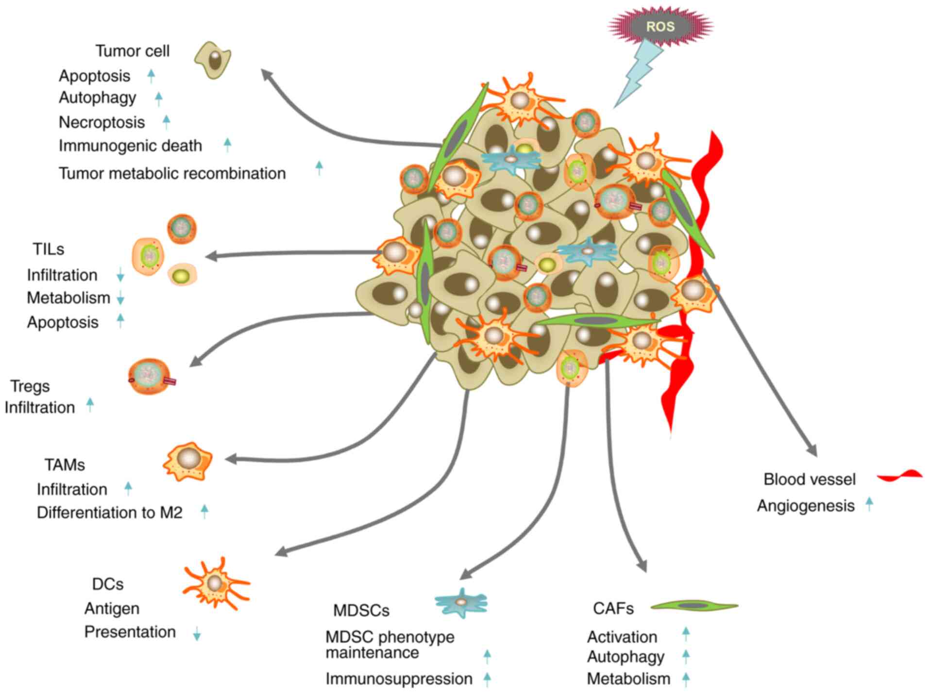
The tumor microenvironment (TME) mainly includes
tumor cells and their surrounding immune cells, cancer-associated
fibroblasts (CAFs) and vascular endothelial cells (13). It is characterized by hypoxia, low pH,
high interstitial pressure, overexpression of glutathione, redox
imbalance and immunosuppression (14,15). Paget
first proposed the hypothesis of ‘seed and soil’ in 1989, where
tumor cells were known as ‘seeds’ and the surrounding
microenvironment were known as ‘soil’ (16). Langley and Fidler (17) then revisited this theory and reviewed
the close relationship between tumor and angiogenesis in organ
metastasis. They found that angiogenesis can promote the metastasis
of tumor organs, which provides a theoretical basis for
antiangiogenic therapy. Recent studies in stomach and lung cancer
have found that in the TME, immune and metabolic reorganization can
also promote the occurrence, development, invasion, metastasis of
tumors (Fig. 1) (18,19).
Manipulating the TME may therefore be more beneficial for
controlling the progression of tumors and reverse the drug
resistance of tumors. Over the past decade, an increasing number of
studies have revealed that regulation of the levels of ROS can
exert anti-tumor effects by acting on the TME (20,21). These
effects include promoting tumor cell apoptosis, inhibiting
angiogenesis, inhibiting immune escape, regulating tumor metabolic
reorganization and reversing drug resistance (20,22). The
present review analyzes the complex role of ROS in anti-tumor
therapy in relation to the TME.
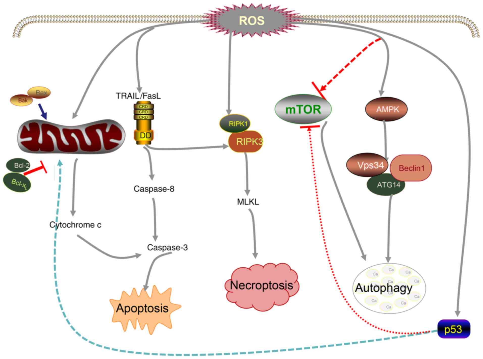
Apoptosis is also termed type I programmed cell
death and is a form of programmed cell death that serves to clear
damaged cells in an orderly manner, which are mainly divided into
two types, namely exogenous and endogenous apoptosis (25). The former primarily involves the
FasL/FasR, TNF-α/TNF receptor 1 (TNF-α/TNFR1), TNF ligand
superfamily member 12/death receptor (DR)3, TNF-related
apoptosis-inducing ligand (Apo2L)/DR4, and Apo2L/DR5 signaling
pathways (26–29). By contrast, the endogenous signaling
pathway is also termed the mitochondrial pathway and mainly entails
increasing the permeability of mitochondria, which elevates the
concentration of intracellular Ca2+ and regulates the
activity of the Bcl-2 family of proteins (30). This effect is accompanied by the
release of cytochrome c (Cyt c), apoptosis-inducing
factor and endonuclease G, leading to tumor cell apoptosis
(31,32). The final node of apoptosis is mainly
initiated by the activation of enzymes in the caspase family
(33,34). Under normal conditions, the production
and clearance of intracellular ROS are maintained in a dynamic
balance. Previous studies have shown that low levels of ROS can
promote cell proliferation, whereas excessive accumulation of ROS
will lead to colon cancer cell apoptosis (35,36). Tumor
cells proliferate at a high rate and are frequently in a state of
high oxidative stress (23).
Therefore, they tend to be more sensitive to internal and external
oxidative stimuli (24). A potential
anti-cancer strategy is to aggravate oxidative stress in the cancer
cells further by increasing the intracellular ROS levels or by
inhibiting the antioxidant capacity of cells (37,38). It
has been demonstrated that oxidation of the mitochondrial membrane
by ROS can release Cyt c into the cytosol more easily to
promote apoptosis (11).
Bcl-2 is a key regulator of apoptosis and as such,
ROS levels can influence the functionality of Bcl-2. In a previous
study with lung cancer, it was found that excessive ROS production
in H460 lung cancer cells inhibit the expression of Bcl-2, whilst
increasing the expression Bcl-2 served the effect of inhibiting the
increase of ROS (39). In addition,
ROS can also regulate a number of exogenous signaling pathways. As
the product of the FasL/FasR pathway by the NADPH oxidase system,
ROS can activate protein tyrosine kinase, which further promotes
Fas-mediated apoptosis (40). A
functional relationship between ROS and several signaling pathways
has also been found. Zhu et al (41) found that overproduction of ROS in
gastric cancer cells can effectively increase the expression of
JNK, which then participate in apoptosis mediated by the MAPK
pathway. In another study with breast cancer, Zang et al
(42) found that ROS can activate the
NF-κB and STAT3 signaling pathways to mediate tumor cell apoptosis.
Many chemotherapeutic drugs function by increasing the production
of ROS, which leads to irreversible apoptosis. Sulindac is a
nonsteroidal anti-inflammatory drug. In the treatment of lung
cancer, it has been shown to increase ROS production and subsequent
mitochondrial membrane damage, which promoted tumor cell apoptosis
(43). Doxorubicin can also increase
the production of ROS and activate the tumor suppressor p53,
resulting in tumor cell death (44).
In addition, photodynamic therapy, RT and emerging sonodynamic
therapy, chemodynamic therapy, enzyme dynamic therapy and ROS-based
nanomedicine therapy have all been documented to serve anti-tumor
roles by increasing the levels of cellular ROS (22,45,46).
Autophagy is termed type II programmed cell death
and is a process in which cells remove intracellular damage,
senescent organelles and structural and biological macromolecules,
such as proteins and lipids, by lysosome-mediated degradation
(47). Autophagy is highly conserved
and is regulated by the autophagy-related (ATG) family of proteins
(47). It is now considered to be not
only a mechanism of cell survival, but also an inhibitory mechanism
that can induce the death of transformed cancer cells (47). ROS is a classical autophagy inducer
and a key component for the interaction between apoptosis and
autophagy (48). In general,
autophagy induced by moderate levels of ROS can reduce the damage
caused by oxidative stress and protect cells (49). By contrast, high levels of ROS can
activate autophagic cell death and have destructive effects on
cells (49). Wu et al
(50) previously showed that
H2O2 pretreatment triggered autophagy in
hepatocellular carcinoma (HCC) cells, where high concentration of
H2O2 could stimulate autophagic apoptosis in
HCC cell lines. Another study has also shown that excessive ROS may
induce autophagic cell death in human oral cancer CAL 27 cells by
promoting Unc51-like kinase 1 protein ubiquitination and
upregulating the expression of the autophagy-related protein
Beclin-1 (51). In addition, ROS can
also alter the activity of signaling pathways that regulate
autophagy. Activation of mTOR kinase, an enzyme in the autophagic
pathway, is inhibited by the AKT and MAPK signaling pathways
(52). AKT induces protective
autophagy whilst sustaining the degradation of p53 and the
expression of NF-κB in HCC cells (53). Therefore, pathways that negatively
regulate mTOR, including the protein kinase 5AMP-activated protein
kinase and p53, which are sensitive to oxidative stress, can
promote autophagy (53,54).
Serving as the ‘soil’ for the growth of cancer
cells, the TME must provide sufficient nutrition for them.
Neovascularization is the main method used for the transport of
nutrients during tumor occurrence and metastasis (63). It has been shown that ROS can regulate
tumor angiogenesis and promote angiogenesis by targeting
transcription factors or tumor suppressors, such as activating
protein 1, hypoxia inducible factor-1α (HIF-1α), NF-κB, and p53
(64). However, in recent years, it
has been found that the increase of ROS production in the TME can
reduce neovascularization and inhibit tumor progression (65). Inducing apoptosis in vascular
endothelial cells is generally considered to be the core strategy
for inhibiting angiogenesis and treating related diseases,
including cancer, neovascular age-related macular degeneration and
diabetic retinopathy (66). ROS is a
promoter of vascular and endothelial cell death in colon and breast
tumors (67,68). Owing to the atypical metabolic
environment, vascular endothelial cells in the TME generally
exhibit higher levels of ROS compared with those in normal vascular
endothelial cells and are more vulnerable to cytotoxicity caused by
a further increment in ROS levels (69). Therefore, increasing the ROS levels
further is more likely to aggravate cell death (69). Topalovski et al (70) found that fibulin-5 can promote ROS
production in vascular endothelial cells by acting on the
fibronectin receptor β1 to exert its anti-angiogenic effects in
pancreatic cancer cells. In addition,
N-benzyl-2-nitro-1-imidazole-acetamide, a therapeutic agent for
Chagas disease, has been found to exert anti-tumor effects in
Ehrlich tumor cells by increasing ROS levels and inhibiting
angiogenesis (71). Synthesis of
redox regulators using silver nanoframe technology has been shown
to induce excessive ROS generation and enhance cytotoxicity in the
vascular endothelium (72). This
prevented formation of the tubular network in the endothelial cells
and poly-ADP ribose modification of VEGF, thereby inhibiting
angiogenesis (72). Furthermore, Cao
et al (73) found that
decylubiquinone can increase ROS to inhibit the formation of the
tubular structure through the ROS/p53/brain-specific angiogenesis
inhibitor 1 signaling pathway in the chicken embryo chorioallantoic
membrane model.
CAFs are particularly abundant in the matrix and can
be derived from a variety of sources (74). They serve an important role in tumor
growth, metastasis and drug resistance (74,75), by
secreting a variety of cytokines and growth factors. The specific
mechanisms involved in these effects include the maintenance of
cancer stem cell (CSC) stemness, promotion of
epithelial-mesenchymal transformation (EMT), remodeling of the
vascular system and regulation of tumor immunity (76–79). In
previous years, it was found that the autophagy of CAFs can also
serve an important role in the occurrence and development of tumors
(80,81). Activation of CAFs is largely dependent
on the stimulation of TME by local hypoxia, oxidative stress,
growth factors released by adjacent tumor cells and infiltrating
immune cells (82). A previous study
with ovarian cancer has shown that ROS produced by tumors can
induce the expression of chloride intracellular channel 4,
chemokine (C-C motif) ligand 2, TGF-β1, NF-κB and STAT3 in CAFs to
induce their activation (83). In
addition, ROS can also induce myofibroblast differentiation by
upregulating the expression of chemokine (C-X-C motif) ligand-12
whilst downregulating that of caveolin 1 (CAV1) (84–86). ROS
can also play an important role in the regulation of CAF autophagy.
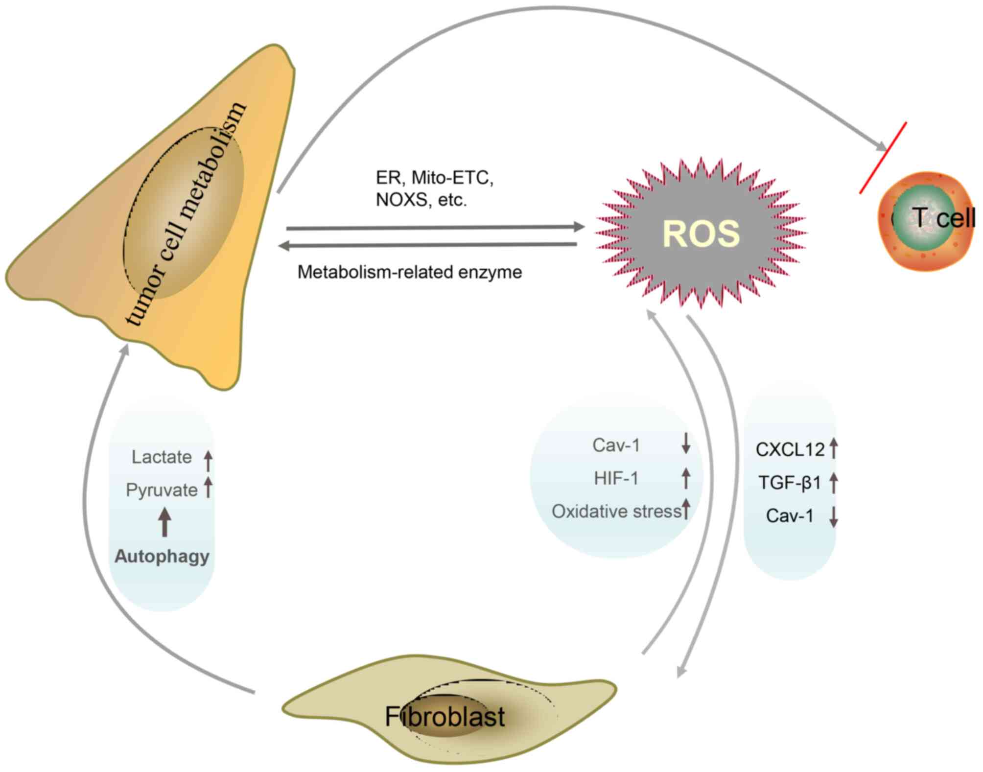
Tumor cells have been observed to induce oxidative stress in
adjacent CAFs (87). In breast
cancer, an increase in the levels of ROS can activate the
expression of HIF-1α and NF-κB in CAFs to subsequently induce CAF
autophagy, which may lead to a decrease in ROS-dependent CAV1
expression (88). The downregulation
of CAV1 promotes mitochondrial dysfunction and oxidative stress
further in CAFs, thereby forming a positive feedback loop (88,89). In
addition, ROS can also regulate the metabolism of CAFs. In tumor
cells, even under aerobic conditions, glycolysis is highly active,
which is characterized by increased glucose uptake and increased
lactic acid secretion (90). This is
known as the ‘Warburg effect’ (90).
CAFs will also produce a similar phenomenon of aerobic glycolysis
under the influence of tumor cells, which is called the ‘reverse
Warburg effect’ (86). During this
phenomenon, oxidative phosphorylation of tumor cells produces a
large quantity of ROS, which specifically triggers oxidative stress
in CAFs and disrupts the oxidative phosphorylation system (86). This results in the additive
accumulation of ROS and induces a chain reaction of oxidative
stress (86). The mode of glucose
metabolism in CAFs changes from oxidative phosphorylation to
glycolysis, which produces high-energy raw materials, such as
lactic acid and ketone, which supply tumors to promote cell
division and support Krebs cycle (86). Additionally, an acidic
microenvironment with high lactate content is also created during
this event, which inhibits the growth of normal cells whilst
promoting tumor cell proliferation and metastasis (86). Inhibition of CAV1 by ROS is known to
be one of the driving factors for changing in the metabolic pattern
of CAFs (91). Martinez-Outschoorn
et al (92) previously found
that when the MCF7 breast cancer cells were co-cultured with
fibroblasts, tumor cell apoptosis occurred after the removal of
H2O2. This effect was proposed to be caused
by the lack of lactic acid produced by CAFs in tumor cells
(92).
T cells (also known as T lymphocytes) consist of a
heterogeneous group of lymphocytes in the tumor matrix, which
includes cytotoxic T lymphocytes, helper T lymphocytes and Tregs
(109). They mediate immune
surveillance and the killing of cancer cells (109). ROS participate in the regulation of
T cells in the TME. Murphy and Siegel (110) previously reported that following
reduction in mitochondrial ROS produced by complex III, T cells
could no longer be continuously activated even after stimulation by
CD3 or CD28. Activation of T cells requires stimulation of the
T-cell receptor (TCR) through the induction of the MAPK signal
transduction pathway and transcription factors such as nuclear
factor of activated T cells (NFAT), NF-κB and activator protein-1
(111). Studies (112,113)
have shown that mitochondrial ROS can be transferred to T cell
immune synapses. After stimulation by antigens,
H2O2 in the mitochondria can enhance the MAPK
signaling pathway, leading to T cell activation and proliferation
(112). However, a number studies
have also shown that high levels of ROS in the TME can inhibit the
activation, proliferation and anti-tumor function of T cells
(114–117). H2O2-mediated
activation of TCR can promote the production of mitochondrial
superoxides, which can enhance the expression of FasL in T cells
and contribute to T cell activation-induced cell death (114,115).
Recent studies have shown that chronic oxidative stress can cause T
cell weakness or failure (116,117).
PD-1 is an immunosuppressive receptor that is mainly
expressed in activated T cells and can exert negative
immunoregulatory effects after activation by the PD-L1 ligand on
the surfaces of antigen-presenting cells (118). Kumar et al (119) and Chamoto et al (120) previously found that ROS and
mitochondrial activation serve an important role in T cell immunity
induced by PD-1 blockade. PD-1 blocking treatment can increase the
ROS content in T cells (119). Using
a ‘bilateral tumor model’, it was found that boosting mitochondrial
activity of T cells by the addition of bezafibrate, a
pan-peroxisome proliferator-activated receptor agonist, can
partially improve the efficacy of PD-1 blockade in a lung cancer
model with systemic immunosuppressive properties (119). These findings suggest that
regulation of mitochondria-derived ROS in T cells may have an
impact on PD-1 blocking therapy. Another study (121) found that use of the phenothiazine
calmodulin inhibitor trifluoperazine could increase the levels of
ROS and stimulate the expression of PD-L1 in colorectal cancer
cells, tumor-infiltrating CD4+ and CD8+ T
cells. However, other studies have shown that enhanced ROS levels
in neck squamous carcinoma cells can also reduce the expression of
PD-L1 (122,123). Therefore, ROS can exert different
biological effects in a manner that is dependent on its quantity,
where different levels of ROS can mediate different immune cell
responses.
Treg cells belong to a typical class of
immunosuppressive cells. In particular, the CD4+ subset
of forkhead box P3 (FOXP3) Tregs can play an important role in
mediating tumor immune tolerance (124). It has been shown that the levels of
ROS in the microenvironment are associated with immune tolerance
mediated by Treg cells (125). By
contrast, ROS can promote the differentiation of Treg cells
(126). It was found that bile acid
can promote the differentiation of Treg cells by increasing the
levels of mitochondrial ROS, which subsequently increased the
acetylation of H3K27 in the Foxp3 promoter (126). Kunisada et al (127) demonstrated that metformin blocked
the differentiation of immature CD4+ T cells into Treg
cells by inhibiting mitochondrial ROS, which subsequently
downregulated the expression of FOXP3 and reduced the number of
tumor-infiltrating Treg cells. Conversely, ROS can also maintain
the function of Treg cells. Yu et al (128) found that ROS induced by TCR
signaling specifically inhibited the protein degradation of
deubiquitin-like enzyme SUMO-specific peptidase 3 (SENP3), which
preserved the immunosuppressive activity of Treg cells. Interfering
with the levels of ROS can specifically inhibit the expression of
SENP3, resulting in the weakening of Treg cell function and
consequently improve the tumor immune response. Maj et al
(129) revealed the relationship
between ROS and immunosuppression by Treg cells in the TME of
ovarian cancer. The results of this previous study showed that ROS
in the TME may cause the apoptosis of Treg cells, where the
apoptotic cells can subsequently release large quantities of
adenosine triphosphate (ATP) (129).
Although ATP is beneficial to body function under normal
circumstances, early apoptotic Treg cells can rapidly convert ATP
to adenosine by CD39 and CD73 (129). These adenosines are specific to T
cells and can bind to their cell surface adenosine A2A receptors to
inhibit T cell activation (129). In
conclusion, ROS can serve an important role in the function of T
cells, whereby high levels of ROS in T cells may confer anti-tumor
effects, whereas ROS in Treg cells appear to be associated with
immunosuppression.
NK cells are a type of effector lymphocytes that
play an important role in the anti-tumor process and are profoundly
influenced by hypoxia and oxidative stress in the TME (130). Zheng et al (131) previously found that hypoxia in the
TME led to an increase in the levels of ROS, which could
continuously activate the mTOR/dynamin-related protein 1 pathway in
NK cells, resulting in excessive mitochondrial fission. After
mitochondrial fragmentation, the production of ROS was accelerated
and a positive feedback loop was established (131). This process ultimately leaded to
apoptosis and mitochondrial autophagy, which decreased the activity
and tumor killing ability of NK cells (131). Therefore, the aberrant increase in
ROS levels in the TME may be one of the mechanisms underlying the
failure of NKs. Supporting this, reducing ROS levels in the TME or
improving the tolerance of NK cells to ROS have been reported to
prevent this failure (132–134).
DCs are professional antigen-presenting cells that
play an important role in both innate and adaptive immunity.
Immature DCs have strong migratory ability, whilst mature DCs can
effectively activate initial T cells to initiate, regulate and
maintain the immune response (135).
The relationship between ROS and DCs is complex, which involve both
metabolic and transcriptional changes (136). Previous studies have found that an
increase in the environmental redox potential can hinder cross
presentation (137,138). Excessive ROS can lead to the chronic
activation of the endoplasmic reticulum stress response and
oxidative damage to intracellular lipids, which inhibit the ability
of DCs to present local antigens to T cells (137,138).
These effects aforementioned can impede the development of an
effective anti-tumor immune response. However, low levels of ROS
can act as a key signaling component to promote the maturation of
antigen-presenting cells through the activation of signaling
pathways, including NF-κB, mTOR and ERK, in addition to the
activation of intracellular Ca2+ channels (139). It has been demonstrated that ROS can
promote cytoplasmic antigen transmission in DCs by lysosome escape
and antigen protection, resulting in effective antigen cross
presentation and strong CD8+ T cell responses (140,141).
Macrophages are mainly derived from myeloprogenitor
cells in the bone marrow and serve the innate immune system
(142). Tumor-associated macrophages
(TAMs) have been frequently observed to infiltrate the tumor
tissue, which serve an ‘accomplice’ role in tumor development and
metastasis (142). They can be
divided into the M1 and M2 subtypes, which
are thought to inhibit and promote cancer progression, respectively
(142). Reprogramming or
repolarization of TAMs to an anti-tumor phenotype may be an
effective method for enhancing the efficacy of immunotherapy
(143). Previous studies have shown
that continuously increasing the levels of ROS in the TME can
contribute to the differentiation of TAMs into the M2
subtype (144,145). TAMs that were isolated from melanoma
after high ROS treatment appeared to show a more aggressive
phenotype, which may be associated with the secretion of
ROS-dependent TNF-α in mouse melanoma B16F1 and B16F10 cell lines
(146). Griess et al
(147) found that ROS elimination
can selectively inhibit the polarization and tumor-promoting
function of M2 macrophages through the STAT3 signaling
pathway. In addition, it was found that M2 TAMs can
express PD-1, but ROS clearance can polarize the TAM balance
towards the M1 phenotype and reduced the expression of
PD-L1 (148). However, the effects
of ROS on TAM differentiation and regulation of the PD-1 immune
checkpoint pathway are worthy of further study.
B cells are derived from bone marrow and are
specialized antigen-presenting cells that can also mediate the
humoral immune response by producing antibodies (149). A number of previous studies found a
positive correlation between B lymphocyte infiltration and patient
response to immunotherapy in various types of tumors, such as
sarcoma, melanoma and renal cell carcinoma, which highlights the
important role of B cells in anti-tumor immunity (150–152). A
study has also found that increasing ROS levels in the
microenvironment can promote the expression of HIF-1α, nuclear
factor erythroid 2-related factor 2 (NRF2) and C-X-C chemokine
receptor type 4, which in turn regulate the multiple stages of B
cell development (153). Feng et
al (154) previously found that
B-cell receptor (BCR)-induced B cell activation also required ROS
(154), similar to T cells.
Mechanistically, ROS mediates the activation and proliferation of B
cells by activating the NF-κB and PI3K signaling pathways (154). After treatment with N-acetylcysteine
for 3 h, the proliferation of B cells was significantly inhibited
(154). ROS has also been found to
determine cell fate after B-cell activation. B cells treated with
high levels of ROS can undergo class switch recombination, whereas
low levels of ROS can induce differentiation into plasma cells
(155). In addition, ROS can
regulate apoptosis and autophagy in B cells (156,157).
The p66SHC protein not only antagonized BCR survival signals and
promoted apoptosis, but also prevented B cell survival through
selective autophagy/mitochondrial autophagy, by increasing ROS
production (156,157). In conclusion, ROS can be considered
to be involved in multiple stages of B cell development, including
activation, differentiation and death.
MDSCs are heterogeneous cell groups that consist of
myeloid progenitor cells and immature bone marrow cells (IMCs).
They form an important part of the TME and possess potent
immunosuppressive activity (158).
ROS serve an important role in maintaining the undifferentiated
state of MDSCs (159). In mice
transplanted with colon cancer and sarcoma, scavenging of
H2O2 induced the differentiation of immature
myelocytes into macrophages (160).
Of note, in the absence of NADPH oxidase (NOX) activity, MDSCs
differentiated into macrophages and DCs (160). Therefore, endogenous oxidative
stress may be a mechanism of MDSC inhibition to suppress its
differentiation in tumors. MDSCs can act on other immune cells
through ROS. It has been previously found that ROS produced by
MDSCs can permanently inactivate T cells and destroy their ability
to initiate the immune response (161). Inhibition of ROS in MDSCs can
reverse immunosuppression and exert anti-tumor effects (161). In addition, not only the T cell
response is a target of ROS-mediated MDSC inhibition. MDSCs can
also inhibit the response of NK cells to adenovirus vectors and
vaccinia virus infection by releasing ROS (162,163).
Recent studies have shown that MDSCs can also negatively regulate B
cell-mediated immune response through ROS (164,165).
Lelis et al (166) found that
MDSCs can inhibit B cell proliferation and antibody production by
cell contact through argininase, nitric oxide and ROS. In addition
to playing a role in MDSC-mediated immunosuppression, ROS has also
been found to be intrinsically involved with the activation of
transcription factors, including NRF2 and HIF-1α (167). This process induces the
transcriptional and metabolic reprogramming of MDSCs, which affects
their differentiation and maintenance (167). Therefore, in the TME, ROS act as
inducers of oxidative stress and a medium of immune regulation,
which is an important process in the formation of cancer cells
(159).
Tumors maintain the microenvironment of immune
suppression by displaying low immunogenicity and secreting
immunosuppressive cytokines, including IL-10, TGF-β, and VEGF
(168). ICD can be triggered by
various treatments, such as chemotherapy, RT and photodynamic
therapy (169). Various cell
death-related molecules, such as damage-associated molecular
patterns, are released to enhance the immunogenicity of tumor cells
and the initial immune response, which is an innovative measure in
immunotherapy (169). At present,
the prevailing notion is that there is a positive association
between ROS production and ICD induction in anti-tumor therapy
(170). Excessive levels of ROS are
frequently used for the oxidative killing of tumors and induction
of ICD. These processes can provide potential antigen stimulation
to the immune system. A nano-study based on sonodynamic therapy
found that enhanced continuous ultrasonic-triggered inertial
cavitation increased ROS production and induced strong ICD
(107). This was characterized by
increased antigen exposure and presentation, enhanced maturation of
DCs and increased infiltration of active-effector CD8+ T
cells (107). Li et al
(171) previously explored the
potential use of a fluorine-assembled nanocluster to reverse
immunosuppression and reawaken the immune system. Following the
production of sufficient ROS levels by fluorine
assembly@ photodynamic immunotherapy for tumor (PMPt) to
break ROS-sensitive connectors under laser irradiation,
cisplatin-coupled PMPt is released to penetrate the tumor and kill
Treg cells and MDSCs (171).
Additionally, ROS can strongly induce ICD by increasing
infiltration by DCs and T cells to turn a cold tumor into a hot
tumor and stimulate an effective anti-tumor immune response
(171). However, a number of studies
have shown that the increase of ROS in the TME can markedly reduce
ICD and the number of tumor-infiltrating T lymphocytes (172,173).
In a previous study using a breast cancer model, Deng et al
(172) found that the elimination of
ROS from the TME using nano-scavengers could alleviate
immunosuppressive ICD induced by oleandrin anticancer drug and
prolong the survival time of T cells in breast cancer. Elimination
of ROS also lead to an increase in anti-tumor immunity and T
lymphocyte infiltration, resulting in a potent anti-tumor effect
(172). It is hypothesized that
these contradictory results may be related to the different levels
of ROS. Therefore, further studies are warranted to confirm these
findings aforementioned.
The prognosis of cancer has markedly improved due
to the advent of targeted therapy and immunotherapy (185). However, drug resistance remains to
be a challenge for the treatment of cancer. The mechanisms of drug
resistance include: i) Heterogeneity of tumor cells; ii) the TME,
including hypoxia, abnormal angiogenesis, EMT, tumor metabolic
recombination and the immunosuppressive microenvironment; iii)
tumor stem cells; iv) mutation of the drug target gene or signaling
compensation; v) detoxification mechanism; vi) pharmacological
changes, such as drug inactivation, decreased drug absorption,
enhanced drug metabolic activity and increased expression of drug
efflux transporter; vii) reduction in the sensitivity of apoptosis;
and 8) increase in the ability to repair DNA damage (185). Previous studies have confirmed that
a high concentration of ROS is one of the characteristics of
drug-resistance in cancer cells (186,187).
ROS can promote drug resistance in tumors through a variety of
mechanisms. As such, ROS can promote the formation of an
immunosuppressive microenvironment and mediate resistance to
immunotherapy by promoting the phenotypes of MDSCs, DCs and TAMs
(106). ROS can also promote EMT,
where cells undergoing EMT typically exhibit characteristics of
cancer stem cells, with high rates of self-renewal and resistance
to drugs and radiation (188). In
addition, ROS can regulate the expression of multidrug resistance
genes, such as the transmembrane drug efflux protein P-glycoprotein
(P-gp), and ATP-dependent substrate transport on both mRNA and
protein levels (186). Therefore,
blocking ROS has been proposed to overcome resistance to
chemotherapy. However, ROS has also been found to promote the
sensitivity of tumor cells to drug treatment (11,12). The
levels of ROS in tumors are generally higher than those observed in
normal cells obtained from the same tissue source (24). Therefore, once the levels of ROS
exceed the threshold through continuous accumulation, the cells
will undergo apoptosis (35). RT and
a number of chemotherapeutic drugs, including cisplatin,
5-fluorouracil and oxaliplatin can kill tumor cells by promoting
the excessive accumulation of ROS. However, tumor cells can
initiate the mechanism for the inhibition of the excessive
accumulation of ROS, to develop drug resistance (189). Wang et al (190) found that inhibition of solute
carrier family 7 member 11 using vorinostat, an inhibitor of
histone deacetylase, can lead to an increase in ROS in
drug-resistant melanoma cells to lethal levels, which lead to
apoptosis only in drug-resistant cells. CSCs consist of a
subpopulation of tumor cells that is resistant to chemotherapy and
are characterized by high invasiveness and metastasis (191). Choi et al (192) previously demonstrated that CSCs can
maintain low ROS levels by coupling forkhead box M1-dependent
peroxiredoxin 3 expression and fatty acid oxidation-mediated NADPH
regeneration, both of which are essential for maintaining the
biological characteristics of CSCs. The accumulation of ROS in
vivo and in vitro can render CSCs sensitive to RT and
chemotherapy (193,194). In addition, the production of ROS,
especially mitochondrial-derived ROS, is essential for the
induction of apoptosis, autophagy and ICD of tumor cells.
Therefore, ROS-based nanotechnology can increase the sensitivity of
tumor cells to RT, chemotherapy, targeted therapy and immunotherapy
(195–198). The relationship between ROS and
drug-resistant tumors is highly complex, where various studies
yielded conflicting findings. For example, P-gp was found to be
overexpressed in MCF-7 cells after treatment with a low
concentration of H2O2 (1 M) (199). However, a high concentration of
H2O2 (10 M) downregulated the expression of
P-gp in human myelogenous leukemia K562/DOX cells (200). These studies were conducted in
different cell lines, such that some conclusions remained
contradictory. Therefore, the relationship between ROS and
multidrug resistance warrants further comprehensive investigation
(Fig. 4).
If a malignant tumor is known as a ‘seed’, the TME
can be termed as the ‘soil’ that allows the ‘seed’ to grow. The TME
serves a key role in several steps of tumor development, including
local drug resistance, immune escape and distant metastasis
(15,18,19). The
combination of immune checkpoint inhibitors or cell therapy with
microenvironment-targeted therapy is expected to improve the
prognosis of patients with cancer in the future (201). Tumor cells are characterized by
indefinite proliferative potential, which is frequently accompanied
with local tissue hypoxia, abnormal angiogenesis and metabolic
reprogramming (202). In addition,
persistent endoplasmic reticulum stress appears to be a new feature
of tumors, which allows tumor cells to adapt to carcinogenic and
environmental challenges to coordinate different immunomodulatory
mechanisms and promote tumor progression (203,204).
ROS production caused by hypoxia, metabolic reprogramming and
endoplasmic reticulum stress altogether serve an important role in
the cross-dialogue between the tumor and the surrounding
microenvironment. ROS plays a dual role in intracellular signal
transduction and cell fate regulation in this process (Fig. 4), the levels of which in cancer cells
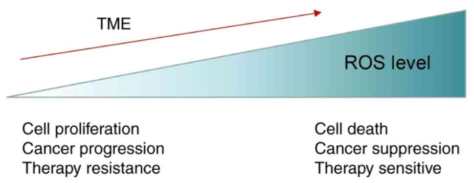
are largely dependent on the antioxidant defense system (186). Therefore, increasing the levels ROS
to accurately break the redox balance is the key prerequisite for
the effective treatment of cancer. However, ROS can also promote
tumor cell proliferation, vascular proliferation, CAF
differentiation, immune escape and drug resistance. It has been
suggested that there are different pools of ROS in cancer cells
with differing functions (205). ROS
derived from NADPH oxidase can promote the proliferation of small
intestinal crypt cells, whereas ROS induced by p53-induced
glycolysis and apoptosis regulator (TIGAR) deletion can exert the
opposite effect (206). NADPH
oxidase produces extracellular superoxides, while TIGAR protects
against intracellular ROS damage by supporting the pentose
phosphate pathway (206). The
beneficial or harmful effects of ROS in cells are not necessarily
mutually exclusive. Further studies on the different tumor cell
types, ROS levels and even the level-effect relationship between
ROS and tumor cells are required. Collectively, these findings
indicate that the combination of ROS-based redox regulators with
standard RT and chemotherapy or even immunotherapy may be of great
significance in tumor therapy.
Not applicable.
Not applicable.
Not applicable.
YCS conceptualized the design of the present
review. WL and XYH performed the literature search wrote the paper.
JQB and TTH made several revisions of the text, making crucial
contributions to the scientific analysis and discussion of the
thesis presented in the review. All authors have read and agreed to
the published version of the manuscript.
Not applicable.
Not applicable.
The authors declare that they have no competing
interests.
|
1
|
Nosaka Y and Nosaka AY: Generation and
detection of reactive oxygen species in photocatalysis. Chem Rev.
117:11302–11336. 2017. View Article : Google Scholar : PubMed/NCBI
|
|
2
|
Kumari S, Badana AK, G MM GS and Malla RR:
Reactive oxygen species: A key constituent in cancer survival.
Biomark Insights. 13:11772719187553912018. View Article : Google Scholar : PubMed/NCBI
|
|
3
|
Valko M, Leibfritz D, Moncol J, Cronin MT,
Mazur M and Telser J: Free radicals and antioxidants in normal
physiological functions and human disease. Int J Biochem Cell Biol.
39:44–84. 2007. View Article : Google Scholar : PubMed/NCBI
|
|
4
|
Yang B, Chen Y and Shi J: Reactive oxygen
species (ROS)-based nanomedicine. Chem Rev. 119:4881–4985. 2019.
View Article : Google Scholar : PubMed/NCBI
|
|
5
|
Cruces-Sande A, Rodríguez-Pérez AI,
Herbello-Hermelo P, Bermejo-Barrera P, Méndez-Álvarez E,
Labandeira-García JL and Soto-Otero R: Copper increases brain
oxidative stress and enhances the ability of 6-hydroxydopamine to
cause dopaminergic degeneration in a rat model of parkinsons
disease. Mol Neurobiol. 56:2845–2854. 2019. View Article : Google Scholar : PubMed/NCBI
|
|
6
|
Gorrini C, Harris IS and Mak TW:
Modulation of oxidative stress as an anticancer strategy. Nat Rev
Drug Discov. 12:931–947. 2013. View Article : Google Scholar : PubMed/NCBI
|
|
7
|
Chatterjee R and Chatterjee J: ROS and
oncogenesis with special reference to EMT and stemness. Eur J Cell
Biol. 99:1510732020. View Article : Google Scholar : PubMed/NCBI
|
|
8
|
Okon IS and Zou MH: Mitochondrial ROS and
cancer drug resistance: Implications for therapy. Pharmacol Res.
100:170–174. 2015. View Article : Google Scholar : PubMed/NCBI
|
|
9
|
Birben E, Sahiner UM, Sackesen C, Erzurum
S and Kalayci O: Oxidative stress and antioxidant defense. World
Allergy Organ J. 5:9–19. 2012. View Article : Google Scholar : PubMed/NCBI
|
|
10
|
Parekh A, Das S, Parida S, Das CK, Dutta
D, Mallick SK, Wu PH, Kumar BNP, Bharti R, Dey G, et al:
Multi-nucleated cells use ROS to induce breast cancer
chemo-resistance in vitro and in vivo. Oncogene. 37:4546–4561.
2018. View Article : Google Scholar : PubMed/NCBI
|
|
11
|
Li Z, Guo D, Yin X, Ding S, Shen M, Zhang
R, Wang Y and Xu R: Zinc oxide nanoparticles induce human multiple
myeloma cell death via reactive oxygen species and
Cyt-C/Apaf-1/Caspase-9/Caspase-3 signaling pathway in vitro. Biomed
Pharmacother. 122:1097122020. View Article : Google Scholar : PubMed/NCBI
|
|
12
|
Xia B and Wang J: Effects of adenosine on
apoptosis of ovarian cancer a2780 cells via ROS and caspase
pathways. Onco Targets Ther. 12:9473–9480. 2019. View Article : Google Scholar : PubMed/NCBI
|
|
13
|
Hanahan D and Coussens LM: Accessories to
the crime: Functions of cells recruited to the tumor
microenvironment. Cancer Cell. 21:309–322. 2012. View Article : Google Scholar : PubMed/NCBI
|
|
14
|
Guo X, Cheng Y, Zhao X, Luo Y, Chen J and
Yuan WE: Advances in redox-responsive drug delivery systems of
tumor microenvironment. J Nanobiotechnology. 16:742018. View Article : Google Scholar : PubMed/NCBI
|
|
15
|
Zheng J and Gao P: Toward normalization of
the tumor microenvironment for cancer therapy. Integr Cancer Ther.
18:15347354198623522019. View Article : Google Scholar : PubMed/NCBI
|
|
16
|
Paget S: The distribution of secondary
growths in cancer of the breast. 1889. Cancer Metastasis Rev.
8:98–101. 1989.PubMed/NCBI
|
|
17
|
Langley RR and Fidler IJ: The seed and
soil hypothesis revisited-the role of tumor-stroma interactions in
metastasis to different organs. Int J Cancer. 128:2527–2535. 2011.
View Article : Google Scholar : PubMed/NCBI
|
|
18
|
Akhtar M, Haider A, Rashid S and Al-Nabet
ADMH: Pagets ‘seed and soil’ theory of cancer metastasis: An idea
whose time has come. Adv Anat Patho. 26:69–74. 2019. View Article : Google Scholar : PubMed/NCBI
|
|
19
|
Zhao Y, Li J, Li D, Wang Z, Zhao J, Wu X,
Sun Q, Lin PP, Plum P, Damanakis A, et al: Tumor biology and
multidisciplinary strategies of oligometastasis in gastrointestinal
cancers. Semin Cancer Biol. 60:334–343. 2020. View Article : Google Scholar : PubMed/NCBI
|
|
20
|
Malla R, Surepalli N, Farran B, Malhotra
SV and Nagaraju GP: Reactive oxygen species (ROS): Critical roles
in breast tumor microenvironment. Crit Rev Oncol Hematol.
160:1032852021. View Article : Google Scholar : PubMed/NCBI
|
|
21
|
Kuo CL, Chou HY, Chiu YC, Cheng AN, Fan
CC, Chang YN, Chen CH, Jiang SS, Chen NJ and Lee AY: Mitochondrial
oxidative stress by Lon-PYCR1 maintains an immunosuppressive tumor
microenvironment that promotes cancer progression and metastasis.
Cancer Lett. 474:138–150. 2020. View Article : Google Scholar : PubMed/NCBI
|
|
22
|
An J, Hu YG, Cheng K, Li C, Hou XL, Wang
GL, Zhang XS, Liu B, Zhao YD and Zhang MZ: ROS-augmented and
tumor-microenvironment responsive biodegradable nanoplatform for
enhancing chemo-sonodynamic therapy. Biomaterials. 234:1197612020.
View Article : Google Scholar : PubMed/NCBI
|
|
23
|
Arfin S, Jha NK, Jha SK, Kesari KK,
Ruokolainen J, Roychoudhury S, Rathi B and Kumar D: Oxidative
stress in cancer cell metabolism. Antioxidants (Basel). 10:6422021.
View Article : Google Scholar : PubMed/NCBI
|
|
24
|
Mirzaei S, Hushmandi K, Zabolian A, Saleki
H, Torabi SMR, Ranjbar A, SeyedSaleh S, Sharifzadeh SO, Khan H,
Ashrafizadeh M, et al: Elucidating role of reactive oxygen species
(ROS) in cisplatin chemotherapy: A focus on molecular pathways and
possible therapeutic strategies. Molecules. 26:23822021. View Article : Google Scholar : PubMed/NCBI
|
|
25
|
Igney FH and Krammer PH: Death and
anti-death: Tumour resistance to apoptosis. Nat Rev Cancer.
2:277–288. 2002. View
Article : Google Scholar : PubMed/NCBI
|
|
26
|
Saxena N, Yadav P and Kumar O: The Fas/Fas
ligand apoptotic pathway is involved in abrin-induced apoptosis.
Toxicol Sci. 135:103–118. 2013. View Article : Google Scholar : PubMed/NCBI
|
|
27
|
Jo E, Jang HJ, Yang KE, Jang MS, Huh YH,
Yoo HS, Park JS, Jang IS and Park SJ: Cordyceps militaris induces
apoptosis in ovarian cancer cells through TNF-α/TNFR1-mediated
inhibition of NF-κB phosphorylation. BMC Complement Med Ther.
20:12020. View Article : Google Scholar : PubMed/NCBI
|
|
28
|
Zhang P, Wang H, Chen Y, Lodhi A, Sun C,
Sun F, Yan L, Deng Y and Ma H: DR5 related autophagy can promote
apoptosis in gliomas after irradiation. Biochem Biophys Res Commun.
522:910–916. 2020. View Article : Google Scholar : PubMed/NCBI
|
|
29
|
Bergeron S, Beauchemin M and Bertrand R:
Camptothecin- and etoposide-induced apoptosis in human leukemia
cells is independent of cell death receptor-3 and −4 aggregation
but accelerates tumor necrosis factor-related apoptosis-inducing
ligand-mediated cell death. Mol Cancer Ther. 3:1659–1669.
2004.PubMed/NCBI
|
|
30
|
Brenner C, Cadiou H, Vieira HL, Zamzami N,
Marzo I, Xie Z, Leber B, Andrews D, Duclohier H, Reed JC and
Kroemer G: Bcl-2 and Bax regulate the channel activity of the
mitochondrial adenine nucleotide translocator. Oncogene.
19:329–336. 2000. View Article : Google Scholar : PubMed/NCBI
|
|
31
|
Sun KX and Xia HW: Pachymic acid inhibits
growth and induces cell cycle arrest and apoptosis in gastric
cancer SGC-7901 cells. Oncol Lett. 16:2517–2524. 2018.PubMed/NCBI
|
|
32
|
Haque M and Islam M: Pleurotus
mushroom induces apoptosis by altering the balance of
proapoptotic and antiapoptotic genes in breast cancer cells and
inhibits tumor sphere formation. Medicina (Kaunas). 55:7162019.
View Article : Google Scholar : PubMed/NCBI
|
|
33
|
Kim JS, Cho IA, Kang KR, Lim H, Kim TH, Yu
SK, Kim HJ, Lee SA, Moon SM, Chun HS, et al: Reversine induces
caspase-dependent apoptosis of human osteosarcoma cells through
extrinsic and intrinsic apoptotic signaling pathways. Genes
Genomics. 41:657–665. 2019. View Article : Google Scholar : PubMed/NCBI
|
|
34
|
Kuranaga E: Beyond apoptosis: Caspase
regulatory mechanisms and functions in vivo. Genes Cells. 17:83–97.
2012. View Article : Google Scholar : PubMed/NCBI
|
|
35
|
Moloney JN and Cotter TG: ROS signalling
in the biology of cancer. Semin Cell Dev Biol. 80:50–64. 2018.
View Article : Google Scholar : PubMed/NCBI
|
|
36
|
Lin S, Li Y, Zamyatnin AA Jr, Werner J and
Bazhin AV: Reactive oxygen species and colorectal cancer. J Cell
Physiol. 233:5119–5132. 2018. View Article : Google Scholar : PubMed/NCBI
|
|
37
|
Lin B, Chen H, Liang D, Lin W, Qi X, Liu H
and Deng X: Acidic pH and high-H2O2 dual
tumor microenvironment-responsive nanocatalytic graphene oxide for
cancer selective therapy and recognition. ACS Appl Mater
Interfaces. 11:11157–11166. 2019. View Article : Google Scholar : PubMed/NCBI
|
|
38
|
Choi EJ and Jeon SM: NRF2-driven redox
metabolism takes center stage in cancer metabolism from an
outside-in perspective. Arch Pharm Res. 43:321–336. 2020.
View Article : Google Scholar : PubMed/NCBI
|
|
39
|
Um HD: Bcl-2 family proteins as regulators
of cancer cell invasion and metastasis: A review focusing on
mitochondrial respiration and reactive oxygen species. Oncotarget.
7:5193–5203. 2016. View Article : Google Scholar : PubMed/NCBI
|
|
40
|
You L, Dong X, Ni B, Fu J, Yang C, Yin X,
Leng X and Ni J: Triptolide induces apoptosis through fas death and
mitochondrial pathways in HepaRG cell line. Front Pharmacol.
9:8132018. View Article : Google Scholar : PubMed/NCBI
|
|
41
|
Zhu Q, Guo Y, Chen S, Fu D, Li Y, Li Z and
Ni C: Irinotecan induces autophagy-dependent apoptosis and
positively regulates ROS-related JNK- and p38-MAPK pathways in
gastric cancer cells. Onco Targets Ther. 13:2807–2817. 2020.
View Article : Google Scholar : PubMed/NCBI
|
|
42
|
Zang YQ, Feng YY, Luo YH, Zhai YQ, Ju XY,
Feng YC, Sheng YN, Wang JR, Yu CQ and Jin CH: Quinalizarin induces
ROS-mediated apoptosis via the MAPK, STAT3 and NF-κB signaling
pathways in human breast cancer cells. Mol Med Rep. 20:4576–4586.
2019.PubMed/NCBI
|
|
43
|
Hwang KE, Park C, Kwon SJ, Kim YS, Park
DS, Lee MK, Kim BR, Park SH, Yoon KH, Jeong ET, et al: Synergistic
induction of apoptosis by sulindac and simvastatin in A549 human
lung cancer cells via reactive oxygen species-dependent
mitochondrial dysfunction. Int J Oncol. 43:262–270. 2013.
View Article : Google Scholar : PubMed/NCBI
|
|
44
|
Zhang T, He WH, Feng LL and Huang HG:
Effect of doxorubicin-induced ovarian toxicity on mouse ovarian
granulosa cells. Regul Toxicol Pharmacol. 86:1–10. 2017. View Article : Google Scholar : PubMed/NCBI
|
|
45
|
Liu H, Jiang W, Wang Q, Hang L and Wang Y
and Wang Y: ROS-sensitive biomimetic nanocarriers modulate tumor
hypoxia for synergistic photodynamic chemotherapy. Biomater Sci.
7:3706–3716. 2019. View Article : Google Scholar : PubMed/NCBI
|
|
46
|
Lopes TZ, de Moraes FR, Tedesco AC, Arni
RK, Rahal P and Calmon MF: Berberine associated photodynamic
therapy promotes autophagy and apoptosis via ROS generation in
renal carcinoma cells. Biomed Pharmacother. 123:1097942020.
View Article : Google Scholar : PubMed/NCBI
|
|
47
|
Mowers EE, Sharifi MN and Macleod KF:
Functions of autophagy in the tumor microenvironment and cancer
metastasis. FEBS J. 285:1751–1766. 2018. View Article : Google Scholar : PubMed/NCBI
|
|
48
|
Gao L, Loveless J, Shay C and Teng Y:
Targeting ROS-mediated crosstalk between autophagy and apoptosis in
cancer. Adv Exp Med Biol. 1260:1–12. 2020. View Article : Google Scholar : PubMed/NCBI
|
|
49
|
Li L, Tan J, Miao Y, Lei P and Zhang Q:
ROS and autophagy: Interactions and molecular regulatory
mechanisms. Cell Mol Neurobiol. 35:615–621. 2015. View Article : Google Scholar : PubMed/NCBI
|
|
50
|
Wu Z, Wang H, Fang S and Xu C: Roles of
endoplasmic reticulum stress and autophagy on H2O2-induced
oxidative stress injury in HepG2 cells. Mol Med Rep. 18:4163–4174.
2018.PubMed/NCBI
|
|
51
|
Lien JC, Lin MW, Chang SJ, Lai KC, Huang
AC, Yu FS and Chung JG: Tetrandrine induces programmed cell death
in human oral cancer CAL 27 cells through the reactive oxygen
species production and caspase-dependent pathways and associated
with beclin-1-induced cell autophagy. Environ Toxicol. 32:329–343.
2017. View Article : Google Scholar : PubMed/NCBI
|
|
52
|
Kim KY, Park KI, Kim SH, Yu SN, Park SG,
Kim YW, Seo YK, Ma JY and Ahn SC: Inhibition of autophagy promotes
salinomycin-induced apoptosis via reactive oxygen species-mediated
PI3K/AKT/mTOR and ERK/p38 MAPK-dependent signaling in human
prostate cancer cells. Int J Mol Sci. 18:10882017. View Article : Google Scholar : PubMed/NCBI
|
|
53
|
Wei B, Huang Q, Huang S, Mai W and Zhong
X: Trichosanthin-induced autophagy in gastric cancer cell MKN-45 is
dependent on reactive oxygen species (ROS) and NF-κB/p53 pathway. J
Pharmacol Sci. 131:77–83. 2016. View Article : Google Scholar : PubMed/NCBI
|
|
54
|
Li L, Chen Y and Gibson SB:
Starvation-induced autophagy is regulated by mitochondrial reactive
oxygen species leading to AMPK activation. Cell Signa. 25:50–65.
2013. View Article : Google Scholar : PubMed/NCBI
|
|
55
|
Zhang DW, Shao J, Lin J, Zhang N, Lu BJ,
Lin SC, Dong MQ and Han J: RIP3, an energy metabolism regulator
that switches TNF-induced cell death from apoptosis to necrosis.
Science. 325:332–336. 2009. View Article : Google Scholar : PubMed/NCBI
|
|
56
|
Schenk B and Fulda S: Reactive oxygen
species regulate Smac mimetic/TNFα-induced necroptotic signaling
and cell death. Oncogene. 34:5796–5806. 2015. View Article : Google Scholar : PubMed/NCBI
|
|
57
|
Li Y, Gong P, Kong C and Tian X: Bufalin
engages in RIP1-dependent and ROS-dependent programmed necroptosis
in breast cancer cells by targeting the RIP1/RIP3/PGAM5 pathway.
Anticancer Drugs. 30:e07702019. View Article : Google Scholar : PubMed/NCBI
|
|
58
|
Zhang Y, Su SS, Zhao S, Yang Z, Zhong CQ,
Chen X, Cai Q, Yang Z, Huang D, Wu R and Han J: RIP1
autophosphorylation is promoted by mitochondrial ROS and is
essential for RIP3 recruitment into necrosome. Nat Commun.
8:143292017. View Article : Google Scholar : PubMed/NCBI
|
|
59
|
Pawlikowska M, Piotrowski J, Jędrzejewski
T, Kozak W, Slominski AT and Brożyna AA: Coriolus
versicolor-derived protein-bound polysaccharides trigger the
caspase-independent cell death pathway in amelanotic but not
melanotic melanoma cells. Phytother Res. 34:173–183. 2020.
View Article : Google Scholar : PubMed/NCBI
|
|
60
|
Yang Z, Wang Y, Zhang Y, He X, Zhong CQ,
Ni H, Chen X, Liang Y, Wu J, Zhao S, et al: RIP3 targets pyruvate
dehydrogenase complex to increase aerobic respiration in
TNF-induced necroptosis. Nat Cell Biol. 20:186–197. 2018.
View Article : Google Scholar : PubMed/NCBI
|
|
61
|
Tu HC, Ren D, Wang GX, Chen DY, Westergard
TD, Kim H, Sasagawa S, Hsieh JJ and Cheng EH: The p53-cathepsin
axis cooperates with ROS to activate programmed necrotic death upon
DNA damage. Proc Natl Acad Sci USA. 106:1093–1098. 2009. View Article : Google Scholar : PubMed/NCBI
|
|
62
|
Ying Y and Padanilam BJ: Regulation of
necrotic cell death: p53, PARP1 and cyclophilin D-overlapping
pathways of regulated necrosis? Cell Mol Life Sci. 73:2309–2324.
2016. View Article : Google Scholar : PubMed/NCBI
|
|
63
|
Zheng Q and Hou W: Regulation of
angiogenesis by microRNAs in cancer. Mol Med Rep. 24:5832021.
View Article : Google Scholar : PubMed/NCBI
|
|
64
|
Aggarwal V, Tuli HS, Varol A, Thakral F,
Yerer MB, Sak K, Varol M, Jain A, Khan MA and Sethi G: Role of
reactive oxygen species in cancer progression: Molecular mechanisms
and recent advancements. Biomolecules. 9:7352019. View Article : Google Scholar : PubMed/NCBI
|
|
65
|
Liu B, Cui LS, Zhou B, Zhang LL, Liu ZH
and Zhang L: Monocarbonyl curcumin analog A2 potently inhibits
angiogenesis by inducing ROS-dependent endothelial cell death. Acta
Pharmacol Sin. 40:1412–1423. 2019. View Article : Google Scholar : PubMed/NCBI
|
|
66
|
Watson EC, Grant ZL and Coultas L:
Endothelial cell apoptosis in angiogenesis and vessel regression.
Cell Mol Life Sci. 74:4387–4403. 2017. View Article : Google Scholar : PubMed/NCBI
|
|
67
|
Sakamaki K: Regulation of endothelial cell
death and its role in angiogenesis and vascular regression. Curr
Neurovasc Res. 1:305–315. 2004. View Article : Google Scholar : PubMed/NCBI
|
|
68
|
Miao Y, Cui L, Chen Z and Zhang L: Gene
expression profiling of DMU-212-induced apoptosis and
anti-angiogenesis in vascular endothelial cells. Pharm Biol.
54:660–666. 2016. View Article : Google Scholar : PubMed/NCBI
|
|
69
|
Li GH, Lin XL, Zhang H, Li S, He XL, Zhang
K, Peng J, Tang YL, Zeng JF, Zhao Y, et al: Ox-Lp(a) transiently
induces HUVEC autophagy via an ROS-dependent PAPR-1-LKB1-AMPK-mTOR
pathway. Atherosclerosis. 243:223–235. 2015.Corrigendum in:
Atherosclerosis 250: 189, 2016. View Article : Google Scholar : PubMed/NCBI
|
|
70
|
Topalovski M, Hagopian M, Wang M and
Brekken RA: Hypoxia and transforming growth factor β cooperate to
induce fibulin-5 expression in pancreatic cancer. J Biol Chem.
291:22244–22252. 2016. View Article : Google Scholar : PubMed/NCBI
|
|
71
|
Zeferino RC, Mota NSRS, Grinevicius VMAS,
Filipe KB, Sulis PM, Silva FRMB, Filho DW, Pich CT and Pedrosa RC:
Targeting ROS overgeneration by
N-benzyl-2-nitro-1-imidazole-acetamide as a potential therapeutic
reposition approach for cancer therapy. Invest New Drugs.
38:785–799. 2020. View Article : Google Scholar : PubMed/NCBI
|
|
72
|
Duraipandy N, Dharunya G, Lakra R,
Korapatti PS and Syamala Kiran M: Fabrication of plumbagin on
silver nanoframework for tunable redox modulation: Implications for
therapeutic angiogenesis. J Cell Physiol. 234:13110–13127. 2019.
View Article : Google Scholar : PubMed/NCBI
|
|
73
|
Cao J, Liu X, Yang Y, Wei B, Li Q, Mao G,
He Y, Li Y, Zheng L, Zhang Q, et al: Decylubiquinone suppresses
breast cancer growth and metastasis by inhibiting angiogenesis via
the ROS/p53/ BAI1 signaling pathway. Angiogenesis. 23:325–338.
2020. View Article : Google Scholar : PubMed/NCBI
|
|
74
|
Nurmik M, Ullmann P, Rodriguez F, Haan S
and Letellier E: In search of definitions: Cancer-associated
fibroblasts and their markers. Int J Cancer. 146:895–905. 2020.
View Article : Google Scholar : PubMed/NCBI
|
|
75
|
Liao Z, Tan ZW, Zhu P and Tan NS:
Cancer-associated fibroblasts in tumor microenvironment-Accomplices
in tumor malignancy. Cell Immunol. 343:1037292019. View Article : Google Scholar : PubMed/NCBI
|
|
76
|
Pereira BA, Vennin C, Papanicolaou M,
Chambers CR, Herrmann D, Morton JP, Cox TR and Timpson P: CAF
Subpopulations: A new reservoir of stromal targets in pancreatic
cancer. Trends Cancer. 5:724–741. 2019. View Article : Google Scholar : PubMed/NCBI
|
|
77
|
Kim BG, Sung JS, Jang Y, Cha YJ, Kang S,
Han HH, Lee JH and Cho NH: Compression-induced expression of
glycolysis genes in CAFs correlates with EMT and angiogenesis gene
expression in breast cancer. Commun Biol. 2:3132019. View Article : Google Scholar : PubMed/NCBI
|
|
78
|
Eiro N, González L, Martínez-Ordoñez A,
Fernandez-Garcia B, González LO, Cid S, Dominguez F,
Perez-Fernandez R and Vizoso FJ: Cancer-associated fibroblasts
affect breast cancer cell gene expression, invasion and
angiogenesis. Cell Oncol (Dordr). 41:369–378. 2018. View Article : Google Scholar : PubMed/NCBI
|
|
79
|
Takahashi H, Sakakura K, Kudo T, Toyoda M,
Kaira K, Oyama T and Chikamatsu K: Cancer-associated fibroblasts
promote an immunosuppressive microenvironment through the induction
and accumulation of protumoral macrophages. Oncotarget.
8:8633–8647. 2017. View Article : Google Scholar : PubMed/NCBI
|
|
80
|
Yan Y, Chen X, Wang X, Zhao Z, Hu W, Zeng
S, Wei J, Yang X, Qian L, Zhou S, et al: The effects and the
mechanisms of autophagy on the cancer-associated fibroblasts in
cancer. J Exp Clin Cancer Res. 38:1712019. View Article : Google Scholar : PubMed/NCBI
|
|
81
|
Zhang X, Schönrogge M, Eichberg J, Wendt
EHU, Kumstel S, Stenzel J, Lindner T, Jaster R, Krause B, Vollmar B
and Zechner D: Blocking autophagy in cancer-associated fibroblasts
supports chemotherapy of pancreatic cancer cells. Front Oncol.
8:5902018. View Article : Google Scholar : PubMed/NCBI
|
|
82
|
Attieh Y and Vignjevic D: The hallmarks of
CAFs in cancer invasion. Eur J Cell Biol. 95:493–502. 2016.
View Article : Google Scholar : PubMed/NCBI
|
|
83
|
Yao Q, Qu X, Yang Q, Wei M and Kong B:
CLIC4 mediates TGF-beta1-induced fibroblast-to-myofibroblast
transdifferentiation in ovarian cancer. Oncol Rep. 22:541–548.
2009.PubMed/NCBI
|
|
84
|
Sampson N, Brunner E, Weber A, Puhr M,
Schäfer G, Szyndralewiez C and Klocker H: Inhibition of
Nox4-dependent ROS signaling attenuates prostate fibroblast
activation and abrogates stromal-mediated protumorigenic
interactions. Int J Cancer. 143:383–395. 2018. View Article : Google Scholar : PubMed/NCBI
|
|
85
|
Toullec A, Gerald D, Despouy G, Bourachot
B, Cardon M, Lefort S, Richardson M, Rigaill G, Parrini MC,
Lucchesi C, et al: Oxidative stress promotes myofibroblast
differentiation and tumour spreading. EMBO Mol Med. 2:211–230.
2010. View Article : Google Scholar : PubMed/NCBI
|
|
86
|
Martinez-Outschoorn UE, Lisanti MP and
Sotgia F: Catabolic cancer-associated fibroblasts transfer energy
and biomass to anabolic cancer cells, fueling tumor growth. Semin
Cancer Biol. 25:47–60. 2014. View Article : Google Scholar : PubMed/NCBI
|
|
87
|
Lisanti MP, Martinez-Outschoorn UE,
Chiavarina B, Pavlides S, Whitaker-Menezes D, Tsirigos A,
Witkiewicz A, Lin Z, Balliet R, Howell A and Sotgia F:
Understanding the ‘lethal’ drivers of tumor-stroma co-evolution:
Emerging role(s) for hypoxia, oxidative stress and
autophagy/mitophagy in the tumor micro-environment. Cancer Biol
Ther. 10:537–542. 2010. View Article : Google Scholar : PubMed/NCBI
|
|
88
|
Martinez-Outschoorn UE, Trimmer C, Lin Z,
Whitaker-Menezes D, Chiavarina B, Zhou J, Wang C, Pavlides S,
Martinez-Cantarin MP, Capozza F, et al: Autophagy in cancer
associated fibroblasts promotes tumor cell survival: Role of
hypoxia, HIF1 induction and NFκB activation in the tumor stromal
microenvironment. Cell Cycle. 9:3515–3533. 2010. View Article : Google Scholar : PubMed/NCBI
|
|
89
|
Bernard M, Yang B, Migneault F, Turgeon J,
Dieudé M, Olivier MA, Cardin GB, El-Diwany M, Underwood K, Rodier F
and Hébert MJ: Autophagy drives fibroblast senescence through
MTORC2 regulation. Autophagy. 16:2004–2016. 2020. View Article : Google Scholar : PubMed/NCBI
|
|
90
|
Urbano AM: Otto Warburg: The journey
towards the seminal discovery of tumor cell bioenergetic
reprogramming. Biochim Biophys Acta Mol Basis Dis. 1867:1659652021.
View Article : Google Scholar : PubMed/NCBI
|
|
91
|
Zhang D, Wang Y, Shi Z, Liu J, Sun P, Hou
X, Zhang J, Zhao S, Zhou BP and Mi J: Metabolic reprogramming of
cancer-associated fibroblasts by IDH3α downregulation. Cell Rep.
10:1335–1348. 2015. View Article : Google Scholar : PubMed/NCBI
|
|
92
|
Martinez-Outschoorn UE, Goldberg A, Lin Z,
Ko YH, Flomenberg N, Wang C, Pavlides S, Pestell RG, Howell A,
Sotgia F and Lisanti MP: Anti-estrogen resistance in breast cancer
is induced by the tumor microenvironment and can be overcome by
inhibiting mitochondrial function in epithelial cancer cells.
Cancer Biol Ther. 12:924–938. 2011. View Article : Google Scholar : PubMed/NCBI
|
|
93
|
Feng X, Xu W, Li Z, Song W, Ding J and
Chen X: Immunomodulatory nanosystems. Adv Sci (Weinh).
6:19001012019. View Article : Google Scholar : PubMed/NCBI
|
|
94
|
Hamieh M, Dobrin A, Cabriolu A, van der
Stegen SJC, Giavridis T, Mansilla-Soto J, Eyquem J, Zhao Z,
Whitlock BM, Miele MM, et al: CAR T cell trogocytosis and
cooperative killing regulate tumour antigen escape. Nature.
568:112–116. 2019. View Article : Google Scholar : PubMed/NCBI
|
|
95
|
Strickler JH, Hanks BA and Khasraw M:
Tumor mutational burden as a predictor of immunotherapy response:
Is more always better? Clin Cancer Res. 27:1236–1241. 2021.
View Article : Google Scholar : PubMed/NCBI
|
|
96
|
Carreau N and Pavlick A: Revolutionizing
treatment of advanced melanoma with immunotherapy. Surg Oncol. Jan
12–2019.(Epub ahead of print). View Article : Google Scholar : PubMed/NCBI
|
|
97
|
Boyero L, Sánchez-Gastaldo A, Alonso M,
Noguera-Uclés JF, Molina-Pinelo S and Bernabé-Caro R: Primary and
acquired resistance to immunotherapy in lung cancer: Unveiling the
mechanisms underlying of immune checkpoint blockade therapy.
Cancers (Basel). 12:37292020. View Article : Google Scholar : PubMed/NCBI
|
|
98
|
Anichini A, Perotti VE, Sgambelluri F and
Mortarini R: Immune escape mechanisms in non small cell lung
cancer. Cancers (Basel). 12:36052020. View Article : Google Scholar : PubMed/NCBI
|
|
99
|
Marshall LA, Marubayashi S, Jorapur A,
Jacobson S, Zibinsky M, Robles O, Hu DX, Jackson JJ, Pookot D,
Sanchez J, et al: Tumors establish resistance to immunotherapy by
regulating Treg recruitment via CCR4. J Immunother Cancer.
8:e0007642020. View Article : Google Scholar : PubMed/NCBI
|
|
100
|
Mima K, Kosumi K, Baba Y, Hamada T, Baba H
and Ogino S: The microbiome, genetics, and gastrointestinal
neoplasms: The evolving field of molecular pathological
epidemiology to analyze the tumor-immune-microbiome interaction.
Hum Genet. 140:725–746. 2021. View Article : Google Scholar : PubMed/NCBI
|
|
101
|
Ali AMR, Tsai JW, Leung CH, Lin H, Ravi V,
Conley AP, Lazar AJ, Wang WL and Nathenson MJ: The immune
microenvironment of uterine adenosarcomas. Clin Sarcoma Res.
10:52020. View Article : Google Scholar : PubMed/NCBI
|
|
102
|
Kosmaczewska A, Ciszak L, Potoczek S and
Frydecka I: The significance of Treg cells in defective tumor
immunity. Arch Immunol Ther Exp (Warsz). 56:181–191. 2008.
View Article : Google Scholar : PubMed/NCBI
|
|
103
|
Lindau D, Gielen P, Kroesen M, Wesseling P
and Adema GJ: The immunosuppressive tumour network: Myeloid-derived
suppressor cells, regulatory T cells and natural killer T cells.
Immunology. 138:105–115. 2013. View Article : Google Scholar : PubMed/NCBI
|
|
104
|
Augustin RC, Delgoffe GM and Najjar YG:
Characteristics of the tumor microenvironment that influence immune
cell functions: Hypoxia, oxidative stress, metabolic alterations.
Cancers (Basel). 12:38022020. View Article : Google Scholar : PubMed/NCBI
|
|
105
|
Lötscher J and Balmer ML: Sensing between
reactions-how the metabolic microenvironment shapes immunity. Clin
Exp Immunol. 197:161–169. 2019. View Article : Google Scholar : PubMed/NCBI
|
|
106
|
Kotsafti A and Scarpa M, Castagliuolo I
and Scarpa M: Reactive oxygen species and antitumor immunity-from
surveillance to evasion. Cancers (Basel). 12:17482020. View Article : Google Scholar : PubMed/NCBI
|
|
107
|
Yin Y, Jiang X, Sun L, Li H, Su C, Zhang
Y, Xu G, Li X, Zhao C, Chen Y, Xu H and Zhang K: Continuous
inertial cavitation evokes massive ROS for reinforcing sonodynamic
therapy and immunogenic cell death against breast carcinoma. Nano
Today. 36:1010092021. View Article : Google Scholar : PubMed/NCBI
|
|
108
|
Yang J, Ma S, Xu R, Wei Y, Zhang J, Zuo T,
Wang Z, Deng H, Yang N and Shen Q: Smart biomimetic metal organic
frameworks based on ROS-ferroptosis-glycolysis regulation for
enhanced tumor chemo-immunotherapy. J Control Release. 334:21–33.
2021. View Article : Google Scholar : PubMed/NCBI
|
|
109
|
Nakamura Y, Zhenjie Z, Oya K, Tanaka R,
Ishitsuka Y, Okiyama N, Watanabe R and Fujisawa Y: Poor lymphocyte
infiltration to primary tumors in acral lentiginous melanoma and
mucosal melanoma compared to cutaneous melanoma. Front Oncol.
10:5247002020. View Article : Google Scholar : PubMed/NCBI
|
|
110
|
Murphy MP and Siegel RM: Mitochondrial ROS
fire up T cell activation. Immunity. 38:201–202. 2013. View Article : Google Scholar : PubMed/NCBI
|
|
111
|
Kaminski MM, Sauer SW, Klemke CD, Süss D,
Okun JG, Krammer PH and Gülow K: Mitochondrial reactive oxygen
species control T cell activation by regulating IL-2 and IL-4
expression: Mechanism of ciprofloxacin-mediated immunosuppression.
J Immunol. 184:4827–4841. 2010. View Article : Google Scholar : PubMed/NCBI
|
|
112
|
Li Y, Liang R, Zhang X, Wang J, Shan C,
Liu S, Li L and Zhang S: Copper chaperone for superoxide dismutase
promotes breast cancer cell proliferation and migration
ROS-Mediated MAPK/ERK signaling. Front Pharmacol. 10:3562019.
View Article : Google Scholar : PubMed/NCBI
|
|
113
|
Ball JA, Vlisidou I, Blunt MD, Wood W and
Ward SG: Hydrogen peroxide triggers a dual signaling axis to
selectively suppress activated human T lymphocyte migration. J
Immunol. 198:3679–3689. 2017. View Article : Google Scholar : PubMed/NCBI
|
|
114
|
Wang L, Kuang Z, Zhang D, Gao Y, Ying M
and Wang T: Reactive oxygen species in immune cells: A new
antitumor target. Biomed Pharmacother. 133:1109782021. View Article : Google Scholar : PubMed/NCBI
|
|
115
|
Belikov AV, Schraven B and Simeoni L: T
cells and reactive oxygen species. J Biomed Sci. 22:852015.
View Article : Google Scholar : PubMed/NCBI
|
|
116
|
Scharping NE, Rivadeneira DB, Menk AV,
Vignali PDA, Ford BR, Rittenhouse NL, Peralta R, Wang Y, Wang Y,
DePeaux K, et al: Mitochondrial stress induced by continuous
stimulation under hypoxia rapidly drives T cell exhaustion. Nat
Immunol. 22:205–215. 2021. View Article : Google Scholar : PubMed/NCBI
|
|
117
|
Franco F, Jaccard A, Romero P, Yu YR and
Ho PC: Metabolic and epigenetic regulation of T-cell exhaustion.
Nat Metab. 2:1001–1012. 2020. View Article : Google Scholar : PubMed/NCBI
|
|
118
|
Salas-Benito D, Conde E, Tamayo-Uria I,
Mancheño U, Elizalde E, Garcia-Ros D, Aramendia JM, Muruzabal JC,
Alcaide J, Guillen-Grima F, et al: The mutational load and a T-cell
inflamed tumour phenotype identify ovarian cancer patients
rendering tumour-reactive T cells from PD-1+tumour-infiltrating
lymphocytes. Br J Cancer. 124:1138–1149. 2021. View Article : Google Scholar : PubMed/NCBI
|
|
119
|
Kumar A, Chamoto K, Chowdhury PS and Honjo
T: Tumors attenuating the mitochondrial activity in T cells escape
from PD-1 blockade therapy. Elife. 9:e523302020. View Article : Google Scholar : PubMed/NCBI
|
|
120
|
Chamoto K, Chowdhury PS, Kumar A, Sonomura
K, Matsuda F, Fagarasan S and Honjo T: Mitochondrial activation
chemicals synergize with surface receptor PD-1 blockade for T
cell-dependent antitumor activity. Proc Natl Acad Sci USA.
114:E761–E770. 2017. View Article : Google Scholar : PubMed/NCBI
|
|
121
|
Xia Y, Jia C, Xue Q, Jiang J, Xie Y, Wang
R, Ran Z, Xu F, Zhang Y and Ye T: Antipsychotic drug
trifluoperazine suppresses colorectal cancer by inducing G0/G1
arrest and apoptosis. Front Pharmacol. 10:10292019. View Article : Google Scholar : PubMed/NCBI
|
|
122
|
Bailly C: Regulation of PD-L1 expression
on cancer cells with ROS-modulating drugs. Life Sci.
246:1174032020. View Article : Google Scholar : PubMed/NCBI
|
|
123
|
Liu K, Du S, Gao P and Zheng J:
Verteporfin suppresses the proliferation, epithelial-mesenchymal
transition and stemness of head and neck squamous carcinoma cells
via inhibiting YAP1. J Cancer. 10:4196–4207. 2019. View Article : Google Scholar : PubMed/NCBI
|
|
124
|
Marangoni F, Zhakyp A, Corsini M, Geels
SN, Carrizosa E, Thelen M, Mani V, Prüßmann JN, Warner RD, Ozga AJ,
et al: Expansion of tumor-associated Treg cells upon disruption of
a CTLA-4-dependent feedback loop. Cell. Jun 21–2021.(Epub ahead of
print). View Article : Google Scholar : PubMed/NCBI
|
|
125
|
Ni D, Tang T, Lu Y, Xu K, Shao Y, Saaoud
F, Saredy J, Liu L, Drummer C 4th, Sun Y, et al: Canonical
secretomes, innate immune caspase-1-, 4/11-gasdermin D
non-canonical secretomes and exosomes may contribute to maintain
treg-ness for treg immunosuppression, tissue repair and modulate
anti-tumor immunity via ROS pathways. Front Immunol. 12:6782012021.
View Article : Google Scholar : PubMed/NCBI
|
|
126
|
Hang S, Paik D, Yao L, Kim E, Trinath J,
Lu J, Ha S, Nelson BN, Kelly SP, Wu L, et al: Bile acid metabolites
control T(H)17 and T(reg) cell differentiation. Nature.
576:143–148. 2019. View Article : Google Scholar : PubMed/NCBI
|
|
127
|
Kunisada Y, Eikawa S, Tomonobu N, Domae S,
Uehara T, Hori S, Furusawa Y, Hase K, Sasaki A and Udono H:
Attenuation of CD4 + CD25 + regulatory T cells in the tumor
microenvironment by metformin, a type 2 diabetes drug.
EBioMedicine. 25:154–164. 2017. View Article : Google Scholar : PubMed/NCBI
|
|
128
|
Yu X, Lao Y, Teng XL, Li S, Zhou Y, Wang
F, Guo X, Deng S, Chang Y, Wu X, et al: SENP3 maintains the
stability and function of regulatory T cells via BACH2
deSUMOylation. Nat Commun. 9:31572018. View Article : Google Scholar : PubMed/NCBI
|
|
129
|
Maj T, Wang W, Crespo J, Zhang H, Wang W,
Wei S, Zhao L, Vatan L, Shao I, Szeliga W, et al: Oxidative stress
controls regulatory T cell apoptosis and suppressor activity and
PD-L1-blockade resistance in tumor. Nat Immunol. 18:1332–1341.
2017. View Article : Google Scholar : PubMed/NCBI
|
|
130
|
Betten A, Dahlgren C, Mellqvist UH,
Hermodsson S and Hellstrand K: Oxygen radical-induced natural
killer cell dysfunction: Role of myeloperoxidase and regulation by
serotonin. J Leukoc Biol. 75:1111–1115. 2004. View Article : Google Scholar : PubMed/NCBI
|
|
131
|
Zheng X, Qian Y, Fu B, Jiao D, Jiang Y,
Chen P, Shen Y, Zhang H, Sun R, Tian Z and Wei H: Mitochondrial
fragmentation limits NK cell-based tumor immunosurveillance. Nat
Immunol. 20:1656–1667. 2019. View Article : Google Scholar : PubMed/NCBI
|
|
132
|
Mimura K, Kua LF, Shimasaki N, Shiraishi
K, Nakajima S, Siang LK, Shabbir A, So J, Yong WP and Kono K:
Upregulation of thioredoxin-1 in activated human NK cells confers
increased tolerance to oxidative stress. Cancer Immunol Immunother.
66:605–613. 2017. View Article : Google Scholar : PubMed/NCBI
|
|
133
|
Aydin E, Johansson J, Nazir FH, Hellstrand
K and Martner A: Role of NOX2-derived reactive oxygen species in NK
cell-mediated control of murine melanoma metastasis. Cancer Immunol
Res. 5:804–811. 2017. View Article : Google Scholar : PubMed/NCBI
|
|
134
|
Aurelius J, Martner A, Riise RE, Romero
AI, Palmqvist L, Brune M, Hellstrand K and Thorén FB: Chronic
myeloid leukemic cells trigger poly(ADP-ribose)
polymerase-dependent inactivation and cell death in lymphocytes. J
Leukoc Biol. 93:155–160. 2013. View Article : Google Scholar : PubMed/NCBI
|
|
135
|
Gu FF, Zhang K, Ma LL, Liu YY, Li C, Hu Y,
Yang QF, Liang JY, Zeng YL, Wang Y and Liu L: The superior ability
of human BDCA3 + (CD141 +) dendritic cells (DCs) to cross-present
antigens derived from necrotic lung cancer cells. Front Immunol.
11:12672020. View Article : Google Scholar : PubMed/NCBI
|
|
136
|
Paardekooper LM, Vos W and van den Bogaart
G: Oxygen in the tumor microenvironment: Effects on dendritic cell
function. Oncotarget. 10:883–896. 2019. View Article : Google Scholar : PubMed/NCBI
|
|
137
|
Giovanelli P, Sandoval TA and
Cubillos-Ruiz JR: Dendritic cell metabolism and function in tumors.
Trends Immunol. 40:699–718. 2019. View Article : Google Scholar : PubMed/NCBI
|
|
138
|
Chougnet CA, Thacker RI, Shehata HM,
Hennies CM, Lehn MA, Lages CS and Janssen EM: Loss of phagocytic
and antigen cross-presenting capacity in aging dendritic cells is
associated with mitochondrial dysfunction. J Immunol.
195:2624–2632. 2015. View Article : Google Scholar : PubMed/NCBI
|
|
139
|
Mao D, Hu F, Yi Z, Kenry, Xu S, Yan S, Luo
Z, Wu W, Wang Z, Kong D, et al: AIEgen-coupled upconversion
nanoparticles eradicate solid tumors through dual-mode ROS
activation. Sci Adv. 6:eabb27122020. View Article : Google Scholar : PubMed/NCBI
|
|
140
|
Wang C, Li P, Liu L, Pan H, Li H, Cai L
and Ma Y: Self-adjuvanted nanovaccine for cancer immunotherapy:
Role of lysosomal rupture-induced ROS in MHC class I antigen
presentation. Biomaterials. 79:88–100. 2016. View Article : Google Scholar : PubMed/NCBI
|
|
141
|
Oberkampf M, Guillerey C, Mouriès J,
Rosenbaum P, Fayolle C, Bobard A, Savina A, Ogier-Denis E, Enninga
J, Amigorena S, et al: Mitochondrial reactive oxygen species
regulate the induction of CD8 T cells by plasmacytoid dendritic
cells. Nature Commun. 9:22412018. View Article : Google Scholar : PubMed/NCBI
|
|
142
|
DeNardo D and Ruffell B: Macrophages as
regulators of tumour immunity and immunotherapy. Nat Rev Immunol.
19:369–382. 2019. View Article : Google Scholar : PubMed/NCBI
|
|
143
|
Allavena P, Anfray C, Ummarino A and Andón
FT: Therapeutic manipulation of tumor-associated macrophages: Facts
and hopes from a clinical and translational perspective. Clin
Cancer Res. 27:3291–3297. 2021. View Article : Google Scholar : PubMed/NCBI
|
|
144
|
Zhang J, Li H, Wu Q, Chen Y, Deng Y, Yang
Z, Zhang L and Liu B: Tumoral NOX4 recruits M2 tumor-associated
macrophages via ROS/PI3K signaling-dependent various cytokine
production to promote NSCLC growth. Redox Biol. 22:1011162019.
View Article : Google Scholar : PubMed/NCBI
|
|
145
|
Li L, Sun F, Han L, Liu X, Xiao Y, Gregory
AD, Shapiro SD, Xiao G and Qu Z: PDLIM2 repression by ROS in
alveolar macrophages promotes lung tumorigenesis. JCI Insight.
6:e1443942021. View Article : Google Scholar : PubMed/NCBI
|
|
146
|
Lin X, Zheng W, Liu J, Zhang Y, Qin H, Wu
H, Xue B, Lu Y and Shen P: Oxidative stress in malignant melanoma
enhances tumor necrosis factor-α secretion of tumor-associated
macrophages that promote cancer cell invasion. Antioxid Redox
Signal. 19:1337–1355. 2013. View Article : Google Scholar : PubMed/NCBI
|
|
147
|
Griess B, Mir S, Datta K and
Teoh-Fitzgerald M: Scavenging reactive oxygen species selectively
inhibits M2 macrophage polarization and their pro-tumorigenic
function in part, via Stat3 suppression. Free Radic Biol Med.
147:48–60. 2020. View Article : Google Scholar : PubMed/NCBI
|
|
148
|
Ruan J, Ouyang M, Zhang W, Luo Y and Zhou
D: The effect of PD-1 expression on tumor-associated macrophage in
T cell lymphoma. Clin Transl Oncol. 23:1134–1141. 2021. View Article : Google Scholar : PubMed/NCBI
|
|
149
|
Wei Y, Huang CX, Xiao X, Chen DP, Shan H,
He H and Kuang DM: B cell heterogeneity, plasticity, and functional
diversity in cancer microenvironments. Oncogene. Jun 29–2021.(Epub
ahead of print). View Article : Google Scholar
|
|
150
|
Helmink BA, Reddy SM, Gao J, Zhang S,
Basar R, Thakur R, Yizhak K, Sade-Feldman M, Blando J, Han G, et
al: B cells and tertiary lymphoid structures promote immunotherapy
response. Nature. 577:549–555. 2020. View Article : Google Scholar : PubMed/NCBI
|
|
151
|
Cabrita R, Lauss M, Sanna A, Donia M,
Skaarup Larsen M, Mitra S, Johansson I, Phung B, Harbst K, et al:
Tertiary lymphoid structures improve immunotherapy and survival in
melanoma. Nature. 577:561–565. 2020. View Article : Google Scholar : PubMed/NCBI
|
|
152
|
Petitprez F, de Reyniès A, Keung EZ, Chen
TW, Sun CM, Calderaro J, Jeng YM, Hsiao LP, Lacroix L, Bougoüin A,
et al: B cells are associated with survival and immunotherapy
response in sarcoma. Nature. 577:556–560. 2020. View Article : Google Scholar : PubMed/NCBI
|
|
153
|
Jang JW, Thuy PX, Lee JW and Moon EY:
CXCR4 promotes B cell viability by the cooperation of nuclear
factor (erythroid-derived 2)-like 2 and hypoxia-inducible factor-1α
under hypoxic conditions. Cell Death Dis. 12:3302021. View Article : Google Scholar : PubMed/NCBI
|
|
154
|
Feng YY, Tang M, Suzuki M, Gunasekara C,
Anbe Y, Hiraoka Y, Liu J, Grasberger H, Ohkita M, Matsumura Y, et
al: Essential role of NADPH oxidase-dependent production of
reactive oxygen species in maintenance of sustained B Cell receptor
signaling and b cell proliferation. J Immunol. 202:2546–2557. 2019.
View Article : Google Scholar : PubMed/NCBI
|
|
155
|
Jang KJ, Mano H, Aoki K, Hayashi T, Muto
A, Nambu Y, Takahashi K, Itoh K, Taketani S, Nutt SL, et al:
Mitochondrial function provides instructive signals for
activation-induced B-cell fates. Nat Commun. 6:67502015. View Article : Google Scholar : PubMed/NCBI
|
|
156
|
Onnis A, Cianfanelli V, Cassioli C,
Samardzic D, Pelicci PG, Cecconi F and Baldari CT: The pro-oxidant
adaptor p66SHC promotes B cell mitophagy by disrupting
mitochondrial integrity and recruiting LC3-II. Autophagy.
14:2117–2138. 2018. View Article : Google Scholar : PubMed/NCBI
|
|
157
|
Onnis A, Cassioli C, Finetti F and Baldari
CT: Regulation of selective B cell autophagy by the pro-oxidant
adaptor p66SHC. Front Cell Dev Biol. 8:1932020. View Article : Google Scholar : PubMed/NCBI
|
|
158
|
Yin K, Xia X, Rui K, Wang T and Wang S:
Myeloid-derived suppressor cells: A new and pivotal player in
colorectal cancer progression. Front Oncol. 10:6101042020.
View Article : Google Scholar : PubMed/NCBI
|
|
159
|
Ohl K and Tenbrock K: Reactive oxygen
species as regulators of MDSC-mediated immune suppression. Front
Immunol. 9:24992018. View Article : Google Scholar : PubMed/NCBI
|
|
160
|
Kusmartsev S and Gabrilovich DI:
Inhibition of myeloid cell differentiation in cancer: The role of
reactive oxygen species. J Leukoc Biol. 74:186–196. 2003.
View Article : Google Scholar : PubMed/NCBI
|
|
161
|
Park MJ, Lee SH, Kim EK, Lee EJ, Baek JA,
Park SH, Kwok SK and Cho ML: Interleukin-10 produced by
myeloid-derived suppressor cells is critical for the induction of
Tregs and attenuation of rheumatoid inflammation in mice. Sci Rep.
8:37532018. View Article : Google Scholar : PubMed/NCBI
|
|
162
|
Fortin C, Yang Y and Huang X: Monocytic
myeloid-derived suppressor cells regulate T-cell responses against
vaccinia virus. Eur J Immunol. 47:1022–1031. 2017. View Article : Google Scholar : PubMed/NCBI
|
|
163
|
Zhu J, Huang X and Yang Y: Myeloid-derived
suppressor cells regulate natural killer cell response to
adenovirus-mediated gene transfer. J Virol. 86:13689–13696. 2012.
View Article : Google Scholar : PubMed/NCBI
|
|
164
|
Dong G, Yang Y, Li X, Yao X, Zhu Y, Zhang
H, Wang H, Ma Q, Zhang J, Shi H, et al: Granulocytic
myeloid-derived suppressor cells contribute to IFN-I signaling
activation of B cells and disease progression through the lncRNA
NEAT1-BAFF axis in systemic lupus erythematosus. Biochim Biophys
Acta Mol Basis Dis. 1866:1655542020. View Article : Google Scholar : PubMed/NCBI
|
|
165
|
Jaufmann J, Lelis FJN, Teschner AC, Fromm
K, Rieber N, Hartl D and Beer-Hammer S: Human monocytic
myeloid-derived suppressor cells impair B-cell phenotype and
function in vitro. Eur J Immunol. 50:33–47. 2020. View Article : Google Scholar : PubMed/NCBI
|
|
166
|
Lelis FJN, Jaufmann J, Singh A, Fromm K,
Teschner AC, Pöschel S, Schäfer I, Beer-Hammer S, Rieber N and
Hartl D: Myeloid-derived suppressor cells modulate B-cell
responses. Immunol Lett. 188:108–115. 2017. View Article : Google Scholar : PubMed/NCBI
|
|
167
|
Satoh H, Moriguchi T, Taguchi K, Takai J,
Maher JM, Suzuki T, Winnard PT Jr, Raman V, Ebina M, Nukiwa T and
Yamamoto M: Nrf2-deficiency creates a responsive microenvironment
for metastasis to the lung. Carcinogenesis. 31:1833–1843. 2010.
View Article : Google Scholar : PubMed/NCBI
|
|
168
|
Saleh R and Elkord E: Acquired resistance
to cancer immunotherapy: Role of tumor-mediated immunosuppression.
Semin Cancer Biol. 65:13–27. 2020. View Article : Google Scholar : PubMed/NCBI
|
|
169
|
Hernández ÁP, Juanes-Velasco P,
Landeira-Viñuela A, Bareke H, Montalvillo E, Góngora R and Fuentes
M: Restoring the immunity in the tumor microenvironment: Insights
into immunogenic cell death in onco-therapies. Cancers (Basel).
13:28212021. View Article : Google Scholar : PubMed/NCBI
|
|
170
|
Galluzzi L, Buqué A, Kepp O, Zitvogel L
and Kroemer G: Immunogenic cell death in cancer and infectious
disease. Nat Rev Immunol. 17:97–111. 2017. View Article : Google Scholar : PubMed/NCBI
|
|
171
|
Li Z, Zhu L, Sun H, Shen Y, Hu D, Wu W,
Wang Y, Qian C and Sun M: Fluorine assembly nanocluster breaks the
shackles of immunosuppression to turn the cold tumor hot. Proc Natl
Acad Sci USA. 117:32962–32969. 2020. View Article : Google Scholar : PubMed/NCBI
|
|
172
|
Deng H, Yang W, Zhou Z, Tian R, Lin L, Ma
Y, Song J and Chen X: Targeted scavenging of extracellular ROS
relieves suppressive immunogenic cell death. Nat Commun.
11:49512020. View Article : Google Scholar : PubMed/NCBI
|
|
173
|
Garg AD, Dudek AM, Ferreira GB, Verfaillie
T, Vandenabeele P, Krysko DV, Mathieu C and Agostinis P:
ROS-induced autophagy in cancer cells assists in evasion from
determinants of immunogenic cell death. Autophagy. 9:1292–1307.
2013. View Article : Google Scholar : PubMed/NCBI
|
|
174
|
Buono R and Longo VD: Starvation, stress
resistance, and cancer. Trends Endocrinol Metab. 29:271–280. 2018.
View Article : Google Scholar : PubMed/NCBI
|
|
175
|
Li XX, Wang ZJ, Zheng Y, Guan YF, Yang PB,
Chen X, Peng C, He JP, Ai YL, Wu SF, et al: Nuclear receptor Nur77
facilitates melanoma cell survival under metabolic stress by
protecting fatty acid oxidation. Mol Cell. 69:480–492 e7. 2018.
View Article : Google Scholar : PubMed/NCBI
|
|
176
|
Sullivan LB and Chandel NS: Mitochondrial
reactive oxygen species and cancer. Cancer Metab. 2:172014.
View Article : Google Scholar : PubMed/NCBI
|
|
177
|
Wu Z, Zuo M, Zeng L, Cui K, Liu B, Yan C,
Chen L, Dong J, Shangguan F, Hu W, et al: OMA1 reprograms
metabolism under hypoxia to promote colorectal cancer development.
EMBO Rep. 22:e508272020.PubMed/NCBI
|
|
178
|
Wang YP, Zhou W, Wang J, Huang X, Zuo Y,
Wang TS, Gao X, Xu YY, Zou SW, Liu YB, et al: Arginine Methylation
of MDH1 by CARM1 inhibits glutamine metabolism and suppresses
pancreatic cancer. Mol Cell. 64:673–687. 2016. View Article : Google Scholar : PubMed/NCBI
|
|
179
|
Panieri E, Telkoparan-Akillilar P, Suzen S
and Saso L: The NRF2/KEAP1 axis in the regulation of tumor
metabolism: Mechanisms and therapeutic perspectives. Biomolecules.
10:7912020. View Article : Google Scholar : PubMed/NCBI
|
|
180
|
Shao S, Qin T, Qian W, Yue Y, Xiao Y, Li
X, Zhang D, Wang Z, Ma Q and Lei J: Positive feedback in Cav-1-ROS
signalling in PSCs mediates metabolic coupling between PSCs and
tumour cells. J Cell Mol Med. 24:9397–9408. 2020. View Article : Google Scholar : PubMed/NCBI
|
|
181
|
Ilkhani K, Bastami M, Delgir S, Safi A,
Talebian S and Alivand MR: The engaged role of tumor
microenvironment in cancer metabolism: Focusing on
cancer-associated fibroblast and exosome mediators. Anticancer
Agents Med Chem. 21:254–266. 2021. View Article : Google Scholar : PubMed/NCBI
|
|
182
|
Zhai Y, Chai L and Chen J: The
relationship between the expressions of tumor associated
fibroblasts Cav-1 and MCT4 and the prognosis of papillary carcinoma
of breast. Pak J Pharm Sci. 30 (Suppl 1):S263–S372. 2017.
|
|
183
|
Ngwa VM, Edwards DN, Philip M and Chen J:
Microenvironmental metabolism regulates antitumor immunity. Cancer
Res. 79:4003–4008. 2019. View Article : Google Scholar : PubMed/NCBI
|
|
184
|
Song M, Sandoval TA, Chae CS, Chopra S,
Tan C, Rutkowski MR, Raundhal M, Chaurio RA, Payne KK, Konrad C, et
al: IRE1α-XBP1 controls T cell function in ovarian cancer by
regulating mitochondrial activity. Nature. 562:423–428. 2018.
View Article : Google Scholar : PubMed/NCBI
|
|
185
|
Gottesman MM, Lavi O, Hall MD and Gillet
JP: Toward a better understanding of the complexity of cancer drug
resistance. Annu Rev Pharmacol Toxicol. 56:85–102. 2016. View Article : Google Scholar : PubMed/NCBI
|
|
186
|
Cui Q, Wang JQ, Assaraf YG, Ren L, Gupta
P, Wei L, Ashby CR Jr, Yang DH and Chen ZS: Modulating ROS to
overcome multidrug resistance in cancer. Drug Resist Updat.
41:1–25. 2018. View Article : Google Scholar : PubMed/NCBI
|
|
187
|
Jadhao M, Tsai EM, Yang HC, Chen YF, Liang
SS, Wang TN, Teng YN, Huang HW, Wang LF and Chiu CC: The long-term
DEHP exposure confers multidrug resistance of triple-negative
breast cancer cells through ABC transporters and intracellular ROS.
Antioxidants (Basel). 10:9492021. View Article : Google Scholar : PubMed/NCBI
|
|
188
|
Lee SY, Jeong EK, Ju MK, Jeon HM, Kim MY,
Kim CH, Park HG, Han SI and Kang HS: Induction of metastasis,
cancer stem cell phenotype, and oncogenic metabolism in cancer
cells by ionizing radiation. Mol Cancer. 16:102017. View Article : Google Scholar : PubMed/NCBI
|
|
189
|
Ge W, Zhao K, Wang X, Li H, Yu M, He M,
Xue X, Zhu Y, Zhang C, Cheng Y, et al: iASPP is an antioxidative
factor and drives cancer growth and drug resistance by competing
with Nrf2 for Keap1 Binding. Cancer Cell. 32:561–573.e6. 2017.
View Article : Google Scholar : PubMed/NCBI
|
|
190
|
Wang L, Leite de Oliveira R, Huijberts S,
Bosdriesz E, Pencheva N, Brunen D, Bosma A, Song JY, Zevenhoven J,
Los-de Vries GT, et al: An acquired vulnerability of drug-resistant
melanoma with therapeutic potential. Cell. 173:1413–1425.e14. 2018.
View Article : Google Scholar : PubMed/NCBI
|
|
191
|
Menéndez ST, Gallego B, Murillo D,
Rodríguez A and Rodríguez R: Cancer stem cells as a source of drug
resistance in bone sarcomas. J Clin Med. 10:26212021. View Article : Google Scholar : PubMed/NCBI
|
|
192
|
Choi HJ, Jhe YL, Kim J, Lim JY, Lee JE,
Shin MK and Cheong JH: FoxM1-dependent and fatty acid
oxidation-mediated ROS modulation is a cell-intrinsic drug
resistance mechanism in cancer stem-like cells. Redox Biol.
36:1015892020. View Article : Google Scholar : PubMed/NCBI
|
|
193
|
Banerjee S, Mukherjee S, Bhattacharya A,
Basak U, Chakraborty S, Paul S, Khan P, Jana K, Hazra TK and Das T:
Pyridoxine enhances chemo-responsiveness of breast cancer stem
cells via redox reconditioning. Free Radic Biol Med. 152:152–165.
2020. View Article : Google Scholar : PubMed/NCBI
|
|
194
|
Li D, Fu Z, Chen R, Zhao X, Zhou Y, Zeng
B, Yu M, Zhou Q, Lin Q, Gao W, et al: Inhibition of glutamine
metabolism counteracts pancreatic cancer stem cell features and
sensitizes cells to radiotherapy. Oncotarget. 6:31151–31163. 2015.
View Article : Google Scholar : PubMed/NCBI
|
|
195
|
Tsai TL, Lai YH, Hw Chen H and Su WC:
Overcoming radiation resistance by iron-platinum metal alloy
nanoparticles in human copper transport 1-overexpressing cancer
cells via mitochondrial disturbance. Int J Nanomedicine.
16:2071–2085. 2021. View Article : Google Scholar : PubMed/NCBI
|
|
196
|
Li Q, Zhang J, Li J, Ye H, Li M, Hou W, Li
H and Wang Z: Glutathione-activated NO-/ROS-generation
nanoparticles to modulate the tumor hypoxic microenvironment for
enhancing the effect of HIFU-combined chemotherapy. ACS Appl Mater
Interfaces. 13:26808–26823. 2021. View Article : Google Scholar : PubMed/NCBI
|
|
197
|
Chen W, Yu D, Sun SY and Li F:
Nanoparticles for co-delivery of osimertinib and selumetinib to
overcome osimertinib-acquired resistance in non-small cell lung
cancer. Acta Biomater. 29:258–268. 2021. View Article : Google Scholar : PubMed/NCBI
|
|
198
|
Banstola A, Poudel K, Pathak S, Shrestha
P, Kim JO, Jeong JH and Yook S: Hypoxia-mediated ROS amplification
triggers mitochondria-mediated apoptotic cell death via
PD-L1/ROS-responsive, dual-targeted, drug-laden thioketal
nanoparticles. ACS Appl Mater Interfaces. 13:22955–22969. 2021.
View Article : Google Scholar : PubMed/NCBI
|
|
199
|
Cen J, Zhang L, Liu F, Zhang F and Ji BS:
Long-term alteration of reactive oxygen species led to multidrug
resistance in MCF-7 cells. Oxid Med Cell Longev. 2016:70534512016.
View Article : Google Scholar : PubMed/NCBI
|
|
200
|
Wang J, Liu L, Cen J and Ji B: BME, a
novel compound of anthraquinone, down regulated P-glycoprotein
expression in doxorubicin-resistant human myelogenous leukemia
(K562/DOX) cells via generation of reactive oxygen species. Chem
Biol Interact. 239:139–145. 2015. View Article : Google Scholar : PubMed/NCBI
|
|
201
|
Murciano-Goroff YR, Warner AB and Wolchok
JD: The future of cancer immunotherapy: Microenvironment-targeting
combinations. Cell Res. 30:507–519. 2020. View Article : Google Scholar : PubMed/NCBI
|
|
202
|
Reina-Campos M, Moscat J and Diaz-Meco M:
Metabolism shapes the tumor microenvironment. Curr Opin Cell Biol.
48:47–53. 2017. View Article : Google Scholar : PubMed/NCBI
|
|
203
|
Chen X and Cubillos-Ruiz J: Endoplasmic
reticulum stress signals in the tumour and its microenvironment.
Nat Rev Cancer. 21:71–88. 2021. View Article : Google Scholar : PubMed/NCBI
|
|
204
|
Andrews AM, Tennant MD and Thaxton JE:
Stress relief for cancer immunotherapy: Implications for the ER
stress response in tumor immunity. Cancer Immunol Immunother.
70:1165–1175. 2020. View Article : Google Scholar : PubMed/NCBI
|
|
205
|
Harris IS and DeNicola GM: The Complex
Interplay between Antioxidants and ROS in Cancer. Trends Cell Biol.
30:440–451. 2020. View Article : Google Scholar : PubMed/NCBI
|
|
206
|
Cheung EC, Lee P, Ceteci F, Nixon C, Blyth
K, Sansom OJ and Vousden KH: Opposing effects of TIGAR- and
RAC1-derived ROS on Wnt-driven proliferation in the mouse
intestine. Genes Dev. 30:52–63. 2016. View Article : Google Scholar : PubMed/NCBI
|