Introduction
Myocardial ischemia is a result of impaired coronary
blood flow to the heart, which may be caused by various factors,
including atherosclerosis, thrombosis or vascular spasm. Ischemia
causes the demand for oxygen and the amount of oxygen supplied to
the heart to become imbalanced, and this imbalance may lead to
damage and necrosis in cardiac tissues. Coronary heart diseases and
ischemic heart diseases (IHDs), particularly acute myocardial
infarction, are among the leading causes of morbidity and mortality
worldwide. According to the World Health Organization,
cardiovascular disease is set to become the leading cause of death
globally (1,2). Increasing evidence indicates that
cardiomyocyte apoptosis occurs during the first few h of myocardial
ischemia, and this leads to widespread myocardial cell damage,
cardiac dysfunction and ultimately heart failure. Despite recent
advances in research and clinical improvements to combat myocardial
ischemia, there have been limited or no fundamental breakthroughs
in drug treatment (3,4).
Shexiang Tongxin Dropping Pill (STP) is a
traditional China Food and Drug Administration (CFDA)-approved
prescription medicine (approval no. Z20080018), which consists of a
combination of seven medicinal components, including moschus,
Radix et Rhizoma Ginseng, Calculus Bovis, bear gall, Venenum
Bufonis, borneol and salvia miltiorrhiza (5,6). In
a previous study, we analyzed and identified the major constituents
of STP by high-performance liquid chromatography-quadrupole
time-of-flight mass spectrometry, and these constituents included
triterpene saponins, bufadienolides, bile acids and phenyl allyl
compounds. Of the identified constituents, 13 were subsequently
quantified by ultra-performance liquid
chromatography-triple-quadrupole tandem mass spectrometry (5). STP has been widely used for the
clinical treatment of cardiovascular diseases, particularly
coronary heart disease in China and Southeast Asia (7–10). A
previous pharmacological study indicated that STP may protect
endothelial cells against atherosclerotic lesions by reducing the
expression of endothelin-1, C-reactive protein and tumor necrosis
factor-α, while increasing nitrogen oxide levels in the blood
(11). A separate study
demonstrated that STP attenuates atherosclerotic lesions in ApoE
deficient mice, indicating potential effects on various
pathological, biochemical and molecular aspects of atherosclerosis,
including lipid regulation, fibrosis, inflammation and oxidative
stress (6). However, the
cardioprotective effects of STP on myocardial ischemia and its
underlying mechanisms remain largely unknown. The present study
investigated the cardioprotective effects of STP in a pituitrin
(PTT)-induced rat model of acute myocardial ischemic injury. In
addition, the mechanism of action of STP was investigated in order
to provide a theoretical basis for the clinical treatment of
IHDs.
Materials and methods
Materials
The present study was approved by the Center of
Laboratory Animals, Fujian University of Traditional Chinese
Medicine [Fuzhou, China; certified no. SYXK (Min) 2009-0001].
Healthy male Sprague-Dawley rats (n=30; age, ~10 weeks; weight,
200–220 g) were purchased from Shanghai Laboratory Animal Center
Laboratory Animal Co., Ltd. (Shanghai, China). The animals were
kept in a temperature-controlled room at 20.1–23.1°C and 40–50%
humidity, under a 12-h light/dark cycle and free access to food and
water.
STP was provided by Inner Mongolia Conba
Pharmaceutical Co., Ltd. (Shanghai, China). Sodium pentobarbital
was purchased from Merck KGaA (Darmstadt, Germany). PTT injection
was purchased from Ningbo Second Hormone Factory (Ningbo, China).
Isosorbide mononitrate (IM) was purchased from Xi'an Lijun
Pharmaceutical Co., Ltd. (Xi'an, China). Antibodies against B-cell
lymphoma-2 (Bcl-2; cat no. ab32124), Bcl-2-associated X protein
(Bax; cat no. ab32503) and GAPDH (cat no. ab9485) were obtained
from Abcam (Cambridge, UK). Unless indicated otherwise, all
chemicals used were purchased from Sigma-Aldrich (Merck KGaA).
Rat model of acute myocardial
ischemia
The rats were randomly assigned into five groups,
each containing six rats. Rats in the control group and
experimental model group received saline pretreatment. Rats in the
STP-low and -high dose groups were pretreated with STP (20 and 40
mg/kg, respectively). Rats in the IM group were pretreated with IM
(4 mg/kg) as the positive control. All pretreatments involved oral
administration for seven days, at a volume of 5 ml/kg/day.
Myocardial ischemia was established by PTT injection
as previously described (12–14).
Briefly, after 1 h of the final pretreatment, the rats were
anesthetized with sodium pentobarbital (30 mg/kg injected
intraperitoneally). Rats then received PTT by subcutaneous
injection (40 U/kg, except for the control group) to record
electrocardiosignals immediately. After 30 min, 8-ml blood samples
were collected from the abdominal aorta and separated for serum
enzyme assays. Hearts were excised, rinsed in ice-cold isotonic
saline and blotted with filters for biochemical assays. Blood and
heart samples were stored at −80°C prior to further analysis.
Determination of ST-segment
elevation
Normal electrocardiograms were recorded by the
placement of subcutaneous electrodes connected to an
electrocardiograph (BL-420S; Chengdu Taimeng Software Co., Ltd.,
Chengdu, China). Electrocardiograms (ECGs) recorded ST-segment
elevation immediately following PTT injection.
Determination of blood rheology
The viscosity of whole blood (mPa·s) was determined
at different shear rates (SRs) in reciprocal sec (10, 60 and 150
s−1) using an automatic blood viscosity meter (LBY-N6B;
Beijing Precil Instrument Co., Ltd., Beijing, China).
Determination of creatine kinase-MB
(CK-MB) and lactate dehydrogenase (LDH) in the serum
The serum was obtained from whole blood by
centrifugation at 1,200 × g at 4°C for 20 min. The serum levels of
LDH (cat no. F16113) and CK-MB (cat no. F15213) in the rat blood
were measured by the corresponding rat ELISA kits (Shanghai Westang
Bio-Tech Co., Ltd., Shanghai, China).
Histological examination of myocardium
using hematoxylin and eosin staining
Heart sections were excised and fixed in 4%
paraformaldehyde solution for 24 h at room temperature. Heart
tissues were then processed for paraffin-embedding, sectioning and
staining by standard histological methods. The tissues were cut
into 5 µm sections using a rotary microtome (RM 2235; Leica
Microsystems GmbH, Wetzlar, Germany). Slides were stained with
hematoxylin for 20 min and eosin for 2 min at room temperature and
observed under a light microscope (Nikon Corporation, Tokyo, Japan)
at ×200 magnification.
Determination of myocardial apoptosis
by terminal deoxynucleotidyl transferase (TdT) dUTP nick end
labeling (TUNEL)
Heart sections were excised and fixed in 4%
paraformaldehyde solution for 24 h at room temperature. Heart
tissues were then processed for paraffin-embedding, sectioning and
staining by standard histological methods. The tissues were cut
into 5 µm sections using a rotary microtome. Myocardial apoptosis
of the tissues sections was analyzed by TUNEL assay using the TdT
in situ apoptosis detection kit (R&D Systems, Inc.,
Minneapolis, MN, USA), according to the manufacturer's protocol.
Individual nuclei were visualized at a magnification of ×400 under
a fluorescent microscope (Leica Microsystems GmbH) for quantitative
analysis. TUNEL-positive cells exhibited apoptosis characteristics,
including condensed chromatin and cellular shrinkage. Apoptotic
cells were counted as 3,3′-diaminobenzidine (DAB)-positive cells
(stained brown) in five randomly selected fields for each slide.
The percentage of apoptotic cells was calculated as the ratio of
the number of TUNEL-positive cells to the total number of cells, as
the mean of the five randomly selected fields.
Reverse transcription-quantitative
polymerase chain reaction (RT-qPCR)
Total RNA from heart samples was extracted using
TRIzol reagent according to the manufacturer's protocol
(Invitrogen; Thermo Fisher Scientific, Inc.). Oligo (dT)-primed
(Thermo Fisher Scientific, Inc.) RNA (1 µg) was reverse transcribed
with the PrimerScript RT reagent Kit with gDNA Eraser (cat. no.
RR047; Takara Bio, Inc., Otsu, Japan), according to the
manufacturer's protocol. The temperature protocol used was as
follows: At 37°C for 15 min, at 85°C for 5 sec, followed by
annealing at 4°C for 15 min. cDNA was used to determine the
expression of Bcl-2, Bax and GAPDH mRNA using SYBR Green Master Mix
(Thermo Fisher Scientific, Inc., Waltham, MA, USA) and the 7500
Fast Real Time PCR System (Applied Biosystems; Thermo Fisher
Scientific, Inc.). GAPDH was used as the housekeeping gene was used
to normalize the expression of Bcl-2 and Bax. The primers used in
the present study were as follows: Bax, forward
5′-GCTGATGGCAACTTCAACTGGG-3′, reverse 5′-TTCTTCCAGATGGTGAGCGAGG-3′;
Bcl-2, forward 5′-TACCGTCGTGACTTCGCAGAGAT-3′, reverse
5′-AGGAGAAATCAAACAGAGGTCGC-3′; and GAPDH, forward
5′-CTGCCTTCTCTTGTGACA-3′ and reverse 5′-TGTAGACCATGTAGTTGAGG-3′.
The thermocycling conditions were as follows: Pre-denaturation at
95°C for 30 sec, followed by 40 cycles of denaturation at 95°C for
3 sec, annealing at 60°C for 30 sec and extension at 72°C for 15
sec. Relative gene expression was calculated according to the
2−ΔΔCq method (15) and
were presented as the fold change compared with the control. The
experiment was repeated three times.
Immunohistochemistry (IHC)
Myocardial tissues were fixed with 4%
paraformaldehyde for 24 h at room temperature, and the samples were
then processed for paraffin-embedding and sectioning using standard
histological methods. The tissues were cut into 5 µm sections using
a rotary microtome. The slides were rehydrated in gradient ethanol
(100, 95, 90, 80 and 70%) and distilled water. Subsequently, the
slides were subjected to antigen retrieval at 37°C for 10 min and
washed with PBS. The endogenous peroxidase activity was quenched
using 3% hydrogen peroxide at room temperature for 10 min.
Following blocking of non-specific binding with 5% bovine serum
albumin (Thermo Fisher Scientific, Inc.) in PBS containing 0.1%
Tween-20 at room temperature for 20 min, the slides were incubated
with rabbit polyclonal antibodies against Bcl-2 (1:200), Bax
(1:200) and GAPDH (1:300) at 37°C overnight. After washing with
PBS, slides were incubated with biotinylated secondary antibody
(1:400; cat no. sc-516142; Santa Cruz Biotechnology, Inc., Dallas,
TX, USA) for 20 min at 37°C, followed by incubation with 1 mg/ml
horseradish peroxidase (HRP)-labeled streptavidin (cat no. ab7403;
Abcam) for 20 min at 37°C, and then washed with PBS. The slides
were subsequently incubated with DAB as the chromogen for 20 min at
room temperature, followed by counterstaining with diluted Harris
hematoxylin (1:10) for 2 min at room temperature. Following
staining, five high-power fields (×400) were randomly selected from
each slide under a light microscope (Nikon Corporation, Tokyo,
Japan). The mean number of positive cells in each field was counted
using Image-Pro Plus software version 6.0 (Media Cybernetics, Inc.,
Rockville, MD, USA). To rule out any non-specific staining, PBS was
used to replace the primary antibody as a negative control.
Statistical analysis
All experiments were repeated at least three times.
Data are presented as the mean ± standard deviation. One-way
analysis of variance followed by Dunnett's post hoc test was used
for statistical comparisons. Analyses were conducted with SPSS
version 18.0 statistical software (SPSS, Inc., Chicago, IL, USA).
P<0.05 was considered to indicate a statistically significant
difference.
Results
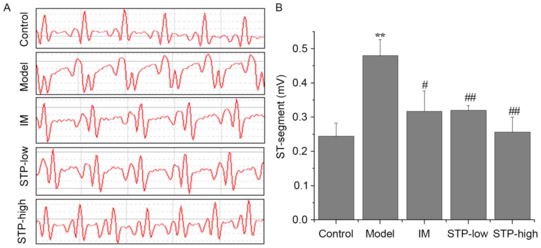
Effect of STP on ST-segment
elevation
Electrocardiograms indicating ST segment elevation
were used to demonstrate successful myocardial ischemia and
establish the experimental model. At 1 min after PTT
administration, ST-segment elevation remained evident in the
experimental model group, whereas mice pretreated with STP (20 and
40 mg/kg dose groups) exhibited significantly reduced ST-segment
elevation compared with the model group. The results for the
STP-low and high dose groups were similar to those in the IM
positive control group (Fig.
1).
Effects of STP on hemorheology
The whole blood viscosities at different SRs in the
experimental model group were significantly higher compared with
the control group (P<0.01). However, whole blood viscosities in
the STP-low and -high dose treatment groups (20 and 40 mg/kg,
respectively) exhibited significant dose-dependent reductions at
different SRs compared with the experimental model group.
Similarly, the IM group also exhibited significantly reduced whole
blood viscosities compared with the experimental model group
(Table I). Notably, the whole
blood viscosities in the STP-high dose group were lower compared
with the IM and STP-low dose groups (Table I).
 | Table I.Variations in hemorheology between
groups. |
Table I.
Variations in hemorheology between
groups.
|
| Viscosity at
different SRs, mPas·s |
|---|
|
|
|
|---|
| Groups | SR 10
s−1 | SR 60
s−1 | SR 150
s−1 |
|---|
| Control |
8.78±0.75 | 4.93±0.24 | 3.89±0.17 |
| Model |
12.09±0.89a |
5.91±0.22a |
4.53±0.24a |
| IM |
11.05±0.46b |
5.39±0.61b |
4.24±0.20b |
| STP-low |
10.73±1.30b | 5.55±0.50 |
4.18±0.19c |
| STP-high |
10.22±0.72c |
5.16±0.18c |
3.94±0.20c |
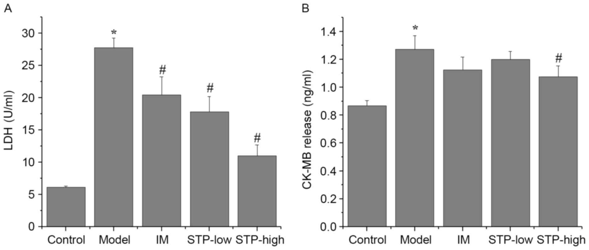
Effect of STP on CK-MB and LDH serum
levels
Levels of the myocardial injury marker enzymes CK-MB
and LDH were significantly increased in the experimental model
group compared with the control group (P<0.05; Fig. 2). Pretreatment with STP
significantly reduced serum levels of LDH in a dose-dependent
manner compared with the experimental model group (P<0.05;
Fig. 2). Levels of CK-MB were also
reduced by pretreatment with STP compared with the experimental
model group, however, this reduction was only significant in the
high dose group (P<0.05; Fig.
2).
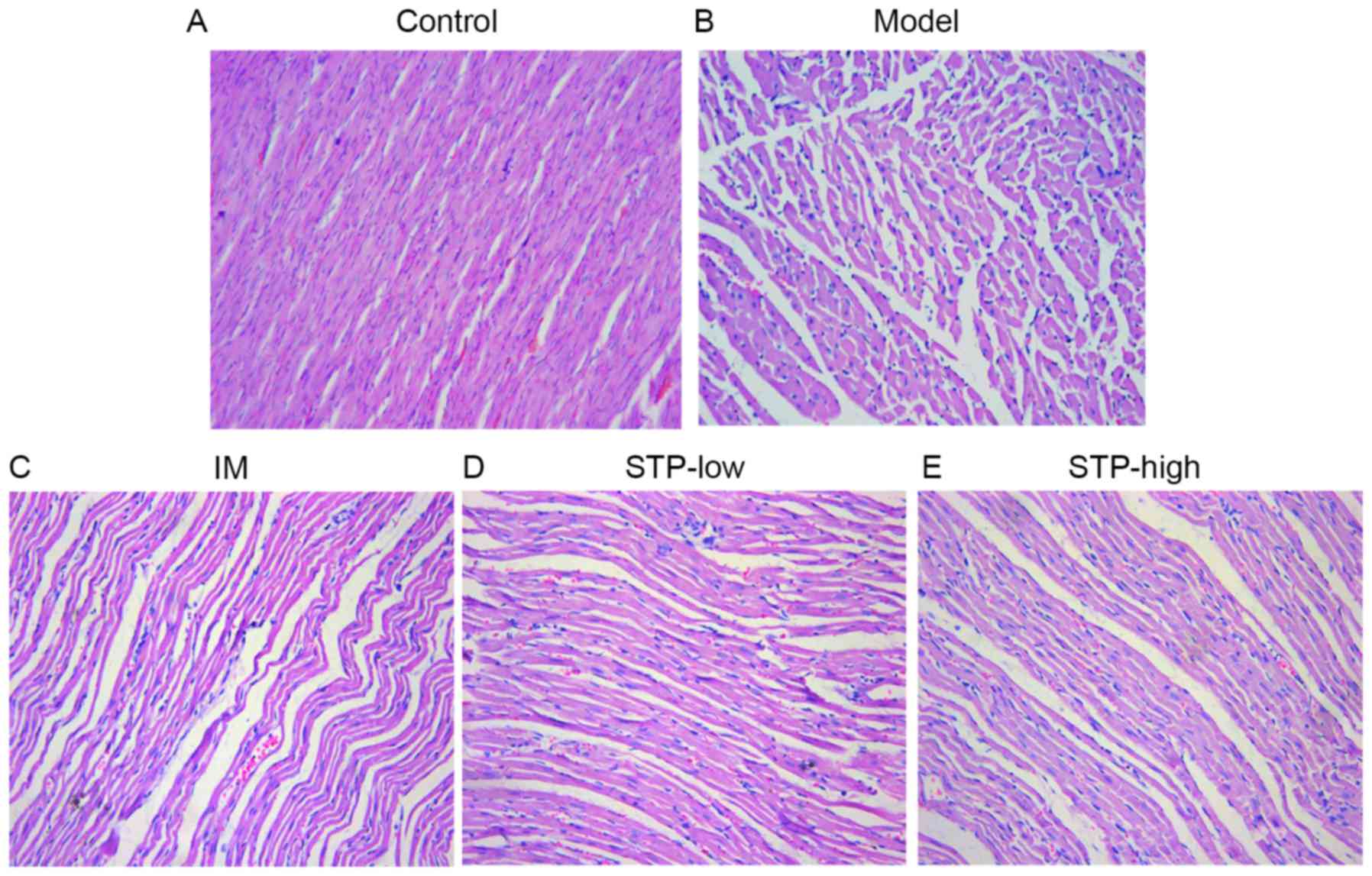
Effect of STP on myocardial
histology
Histological analysis demonstrated that the
myocardium of control group rats displayed a normal myofibrillar
structure with striations, branched appearance and continuity with
adjacent myofibrils. By contrast, tissues from the experimental
model rats revealed obvious swelling of myocardial cells,
degeneration and loss of transverse striations. Tissues from both
STP treatment groups exhibited normal, well-preserved myocardial
cell morphology. Tissue sections from the IM group also revealed
normal myofibrillar structure with obvious transverse striations.
These results indicate that STP treatment may protect against
tissue necrosis following myocardial ischemia (Fig. 3).
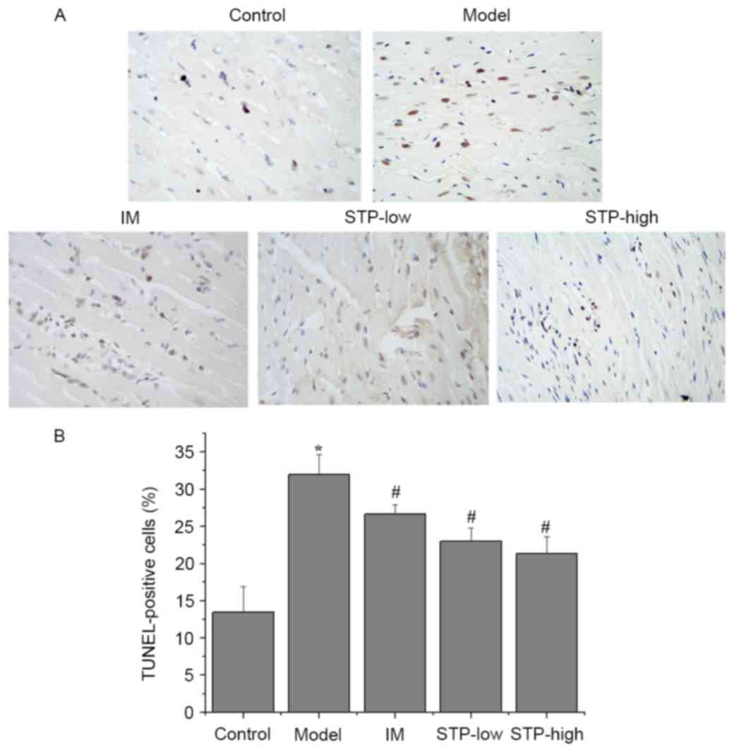
Effect of STP on myocardial apoptosis
using TUNEL staining
A significantly higher percentage of TUNEL-positive
cells were observed in the experimental model group compared with
the control group (P<0.05; Fig.
4). However, pretreatment with low and high doses of STP
significantly reduced the percentage of TUNEL-positive cells,
compared with the experimental model group (P<0.05; Fig. 4). The IM group also exhibited a
significant decrease in the percentage of TUNEL-positive cells
compared with the experimental model group (P<0.05; Fig. 4).
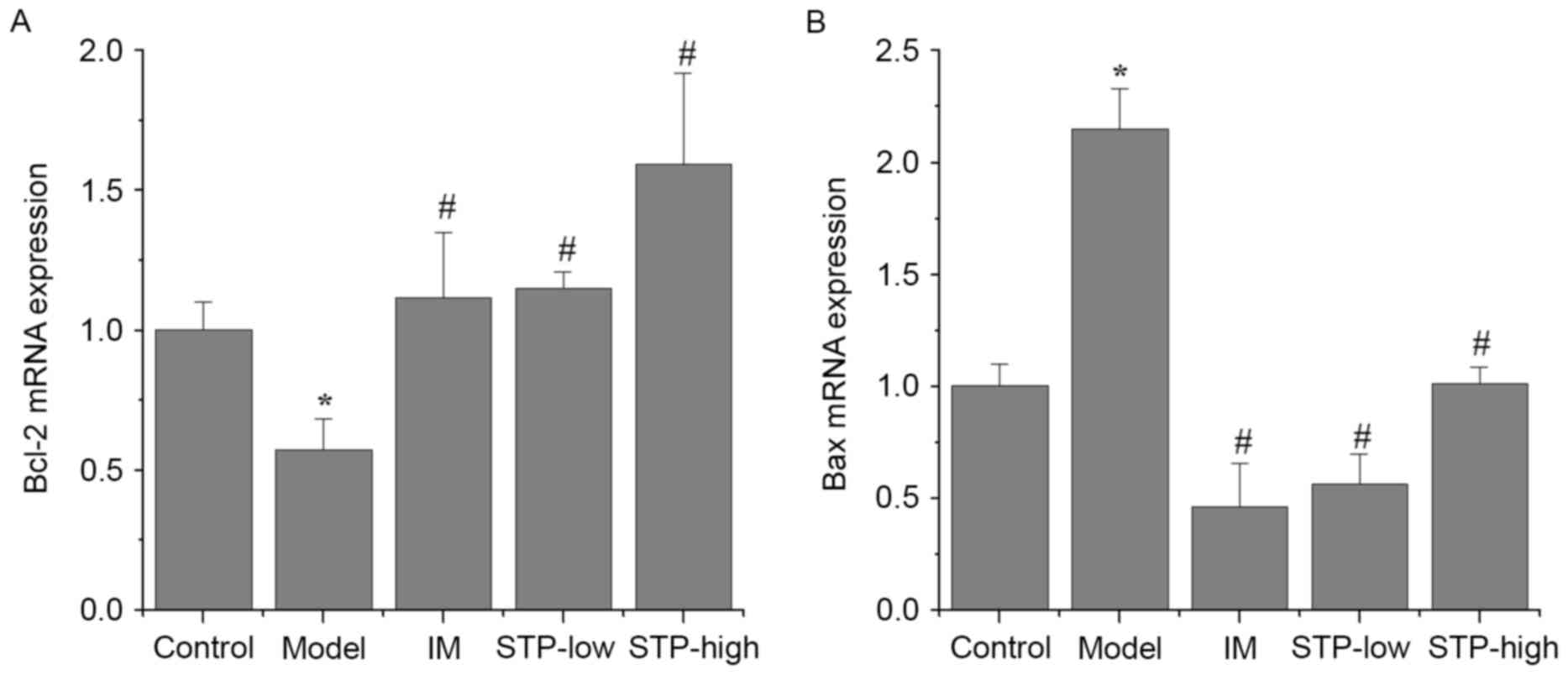
Effect of STP on Bcl-2 and Bax mRNA
expression in cardiomyocytes using RT-qPCR
There was a significant decrease in Bcl-2 mRNA
expression and a significant increase in Bax mRNA expression in the
experimental model group, compared with the control group
(P<0.05; Fig. 5). By contrast,
STP pretreatment groups exhibited significantly increased Bcl-2
expression and decreased Bax expression, compared with the
experimental model group (P<0.05; Fig. 5). In particular, STP pretreatment
progressively increased the expression of Bcl-2 in a dose-dependent
manner (Fig. 5A), whilst the same
dose-dependent effect was not observed for Bax expression (Fig. 5B).
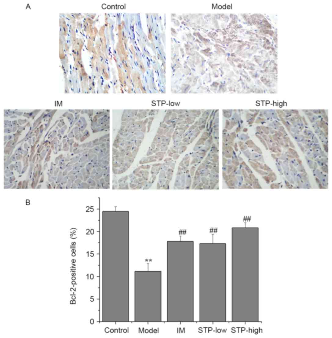
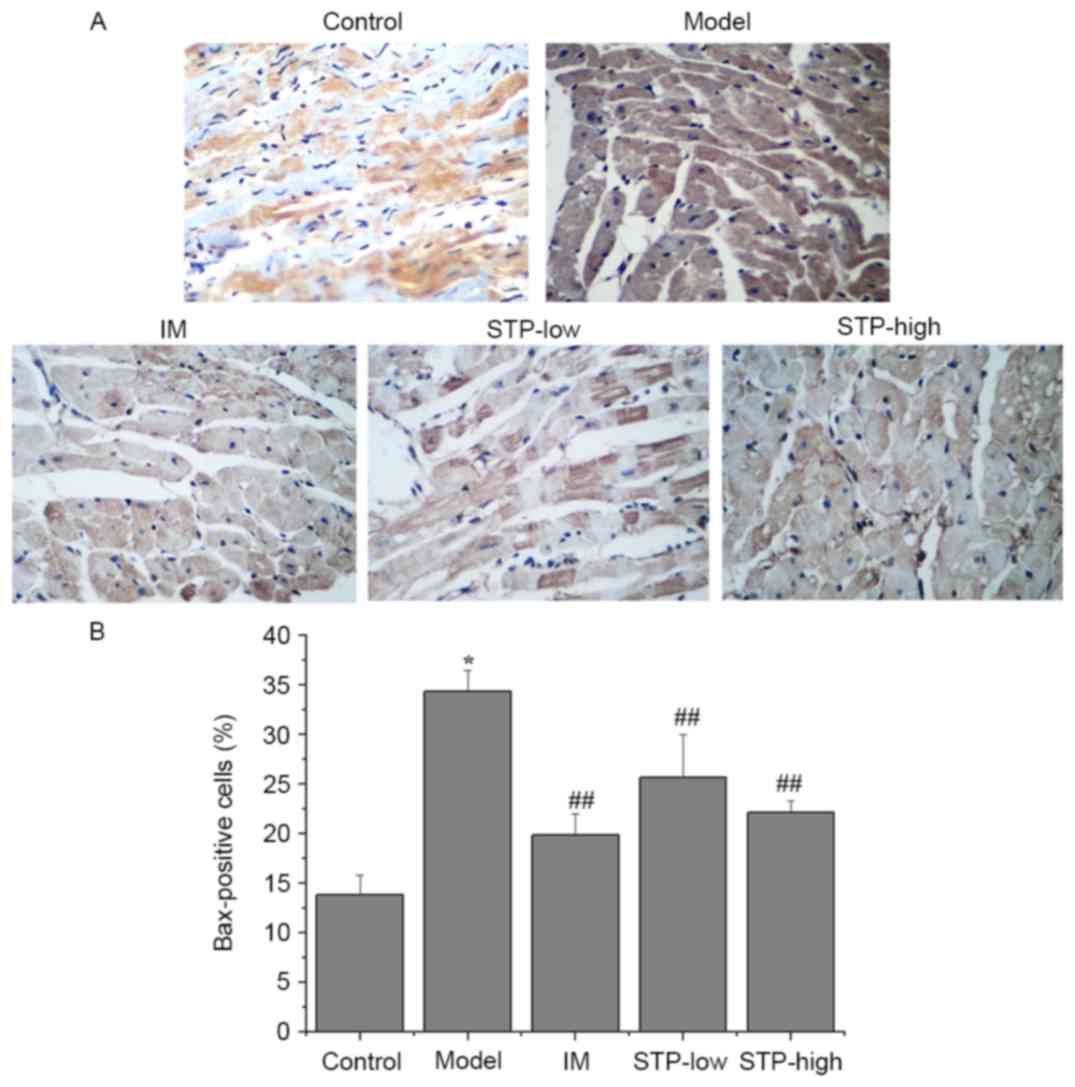
Effect of STP on Bcl-2 and Bax protein
expression in cardiomyocytes using IHC
There was a significant decrease in the level of
cytoplasmic Bcl-2 protein in the experimental model group compared
with the control group (P<0.01; Fig. 6). However, STP pretreatment groups
exhibited significantly increased Bcl-2 protein expression levels
compared with the experimental model group, in a dose-dependent
manner (P<0.01; Fig. 6). In
addition, the protein expression of Bax in the experimental model
group was significantly higher compared with the control group
(P<0.05; Fig. 7), and STP
pretreatment groups exhibited significantly decreased Bax
expression compared with the experimental model group (P<0.01;
Fig. 7).
Discussion
PTT is an extract of hormones that are released from
the posterior lobe of the pituitary gland, which consists of
vasopressin and oxytocin hormones. These hormones are synthesized
in the neuronal cells of hypothalamic nuclei and stored in nerve
cell endings in the posterior pituitary gland (16). In 1960, Black (17) used PTT to successfully establish an
animal model of acute myocardial ischemia, which was subsequently
used in numerous studies investigating myocardial ischemic
antagonists (13,16).
The present study demonstrated that STP pretreatment
reduced ST-segment elevation, whole blood viscosity, and the mRNA
expression of CK-MB and LDH in rats with PTT-induced acute
myocardial ischemic injury. These results indicate that STP may
have cardioprotective effects that alleviate myocardial ischemic
injury.
Electrocardiograms indicating ST-segment elevation
were used to demonstrate that myocardial ischemia was successfully
established. Observing alterations in the ECG pattern, particularly
ST-segment elevation, is the standard method for accurately
diagnosing myocardial ischemic injury in animals (18). In the current study, the
experimental model group exhibited significant ST-segment elevation
following PTT treatment, compared with the control group.
Hemorheological parameters are one of the primary
risk factors involved in IHD. Clinical experiments have
demonstrated that patients with IHD often exhibited an increase in
blood viscosity (19,20). In the present study, STP
pretreatment significantly reduced whole blood viscosity in rats
following PTT-induced myocardial ischemia.
LDH and CK-MB are established markers of cellular
necrosis, and CK-MB has also been established as a key biomarker of
cardiomyocyte injury (21). Serum
CK-MB and LDH levels are often employed to monitor the extent of
myocardial damage (22).
PTT-induced myocardial ischemia resulted in significant leakage of
CK-MB and LDH from ruptured myocardial membranes into the serum.
This cardiotoxic effect was demonstrated using histopathological
examination, which revealed a higher degree of myocardial cell
swelling, degeneration and loss of transverse striation in
PTT-induced rats. In the current study, pretreatment with STP
restored these histopathological changes to a certain degree.
In addition, TUNEL staining in STP pretreatment
groups demonstrated decreased cardiomyocyte apoptosis compared with
the experimental model group. Furthermore, this myocardial
apoptosis may initiate extensive loss of cardiomyocytes, contribute
to the pathogenesis of underlying ischemic injury and ultimately
result in the deterioration of cardiac function (23).
Cardiomyocyte apoptosis is implicated in various
myocardial diseases, including myocardial hypertrophy, heart
failure and myocardial ischemia (24,25).
Previous studies have demonstrated that a loss of cardiomyocytes as
a result of apoptosis is a key factor in myocardial ischemia injury
and inevitably leads to heart failure. Cardiomyocyte apoptosis is
the predominant form of post-ischemic cardiomyocyte death in the
heart and contributes substantially to the impairment of cardiac
performance. Reducing cardiomyocyte loss by suppression of cell
death is therefore an important strategy in protecting against
myocardial diseases (26,27).
During apoptosis, the Bcl-2 family of proteins is
the key regulator of mitochondrial-mediated apoptosis. Members of
this family include Bcl-2 and Bax, which are anti-apoptotic and
proapoptotic proteins, respectively, and have a key role in
triggering cell death via the outer mitochondrial membrane
(28,29). Typically, the balance between pro-
and anti-apoptotic proteins determines whether cells undergo
apoptosis or survive under pathophysiological stress following
injury (30,31). In the current study, STP
pretreatment upregulated the mRNA and protein expression of Bcl-2,
while the expression of Bax was downregulated. This indicates that
STP may prevent myocardial cells from undergoing apoptosis
following ischemic injury, due to its anti-apoptotic and
cardioprotective properties.
Additional studies are required to investigate the
signaling pathways involved in STP-induced cardioprotection. In
addition, the effects and mechanisms of STP on ischemia/reperfusion
injury in cultured cardiomyocytes should be investigated.
The present study demonstrated that STP may exert
cardioprotective effects on myocardial ischemia by alleviating
myocardial ischemic injury and preventing cardiomyocyte apoptosis,
based on hemodynamic results, biochemical data, myocardial
pathology, and the expression of Bcl-2 and Bax. Therefore, the
results of the current study provide evidence that STP may be an
effective and promising drug for the clinical prevention and
treatment of IHDs.
Acknowledgements
This study was sponsored by the Foundation of Fujian
University of Traditional Chinese Medicine (grant no. X2013026),
the Developmental Fund of Chen Keji Integrative Medicine (grant no.
CKJ2013016) and the Education Department of Fujian Province (grant
no. JA14163). The authors also thank Inner Mongolia Conba
Pharmaceutical Co., Ltd. for their support. Y.Z. is an employee of
the Inner Mongolia Conba Pharmaceutical Co., Ltd.
References
|
1
|
Fan J, Li GQ, Liu J, Wang W, Wang M, Qi Y,
Xie WX, Liu J, Zhao F, Li Y and Zhao D: Impact of cardiovascular
disease deaths on life expectancy in Chinese population. Biomed
Environ Sci. 27:162–168. 2014.PubMed/NCBI
|
|
2
|
Sivaraman V and Yellon DM: Pharmacologic
therapy that simulates conditioning for cardiac
ischemic/reperfusion injury. J Cardiovasc Pharmacol Ther. 19:83–96.
2014. View Article : Google Scholar : PubMed/NCBI
|
|
3
|
Guo X, Cao W, Yao J, Yuan Y, Hong Y, Wang
X and Xing J: Cardioprotective effects of tilianin in rat
myocardial ischemia-reperfusion injury. Mol Med Rep. 11:2227–2233.
2015.PubMed/NCBI
|
|
4
|
Panda S, Kar A and Ramamurthy V:
Cardioprotective effect of vincristine on isoproterenol-induced
myocardial necrosis in rats. Eur J Pharmacol. 723:451–458. 2014.
View Article : Google Scholar : PubMed/NCBI
|
|
5
|
Chen D, Lin S, Xu W, Huang M, Chu J, Xiao
F, Lin J and Peng J: Qualitative and quantitative analysis of the
major constituents in shexiang tongxin dropping pill by
HPLC-Q-TOF-MS/MS and UPLC-QqQ-MS/MS. Molecules. 20:18597–18619.
2015. View Article : Google Scholar : PubMed/NCBI
|
|
6
|
Xiong M, Jia C, Cui J, Wang P, Du X, Yang
Q, Zhu Y, Wang W, Zhang T and Chen Y: Shexiang Tongxin dropping
pill attenuates atherosclerotic lesions in ApoE deficient mouse
model. J Ethnopharmacol. 159:84–92. 2015. View Article : Google Scholar : PubMed/NCBI
|
|
7
|
Hua X, Zhan Y and Li Z: Effect of Shexiang
Tongxin Dropping Pill on Cardiac Function in Patients with Chronic
Heart Failure. Chin J Integr Med Cardio-/Cerebrovasc Dis. 2011.
|
|
8
|
Ning H: The analysis of Shexiang Tongxin
dropping pill on treating coronary heart disease angina pectoris
curative effect. Chin J Geriatr Care. 2012.
|
|
9
|
Sui W: Clinical observation of Shexiang
Tongxin dropping pill in treating senile unstable agina pectoris.
Chin Community Doctors. 9:22011.
|
|
10
|
Zhang JXX and Wang W: Shexiang Tongxin
dropping pill Evaluate the Efficacy of the Treatment of Unstable
Angina. Chin J Integr Med Cardio-/Cerebrovasc Dis. 9:22011.
|
|
11
|
Liu ZH QXD-LYLYL-YJJ-GSA-JXX-GWW-M: The
Protective Function of Shexiang on Endothelial Injury Induced by
AngiotensinIIOsmotic Pump in the Rat. Chin J Clin Med. 2009.
|
|
12
|
Jiang C: Protective Efect of
Qi-dan-hua-tan Decoction on Acute Myocardial Ischemia Injury of
Rat. J Chengdu Univ Traditional Chin Med. 2011.
|
|
13
|
Liu X, Liu H, Zeng Z, Zhou W, Liu J and He
Z: Pharmacokinetics of ligustrazine ethosome patch in rats and
anti-myocardial ischemia and anti-ischemic reperfusion injury
effect. Int J Nanomedicine. 6:1391–1398. 2011. View Article : Google Scholar : PubMed/NCBI
|
|
14
|
Zhang M and Cao H: Research on the Animal
Model of Coronary Heart Disease Due to Heart Yang Deficiency. China
J Basic Med Traditional Chin Med. 2002.
|
|
15
|
Livak KJ and Schmittgen TD: Analysis of
relative gene expression data using real-time quantitative PCR and
the 2(−Delta Delta C(T)) Method. Methods. 25:402–408. 2001.
View Article : Google Scholar : PubMed/NCBI
|
|
16
|
Qian Y, Wang S, Xie Y, Wang J, Li H, Zhou
X and Liu W: Effect of salvianolic Acid b and paeonol on blood
lipid metabolism and hemorrheology in myocardial ischemia rabbits
induced by pituitruin. Int J Mol Sci. 11:3696–3704. 2010.
View Article : Google Scholar : PubMed/NCBI
|
|
17
|
Black JW: Electrocardiographic changes
produced in rabbits by vasopressin (pitressin) and their alteration
by prolonged treatment with a commercial heart extract. J Pharm
Pharmacol. 12:87–94. 1960. View Article : Google Scholar : PubMed/NCBI
|
|
18
|
Jr JH and Rikkers LF: Success of medical
and surgical management of acute variceal hemorrhage. Am J Surg.
140:816–820. 1980. View Article : Google Scholar : PubMed/NCBI
|
|
19
|
Simpson LO: Angina, ischemic heart disease
and blood viscosity. BMJ. 339:464. 2015.
|
|
20
|
Sokolov EI, Zykova AA, Sushchik VV and
Goncharov IN: Blood viscosity in patients with ischemic heart
disease. Kardiologiia. 54:9–14. 2014.(In Russian). View Article : Google Scholar : PubMed/NCBI
|
|
21
|
Deng C, Sun Z, Tong G, Yi W, Ma L, Zhao B,
Cheng L, Zhang J, Cao F and Yi D: α-Lipoic acid reduces infarct
size and preserves cardiac function in rat myocardial
ischemia/reperfusion injury through activation of PI3K/Akt/Nrf2
pathway. PLoS One. 8:e583712013. View Article : Google Scholar : PubMed/NCBI
|
|
22
|
Gottlieb RA and Engler RL: Apoptosis in
myocardial ischemia-reperfusion. Ann N Y Acad Sci. 874:412–426.
1999. View Article : Google Scholar : PubMed/NCBI
|
|
23
|
Veinot JP, Gattinger DA and Fliss H: Early
apoptosis in human myocardial infarcts. Hum Pathol. 28:485–892.
1997. View Article : Google Scholar : PubMed/NCBI
|
|
24
|
Shen M, Wu RX, Zhao L, Li J, Guo HT, Fan
R, Cui Y, Wang YM, Yue SQ and Pei JM: Resveratrol attenuates
ischemia/reperfusion injury in neonatal cardiomyocytes and its
underlying mechanism. PLoS One. 7:e512232012. View Article : Google Scholar : PubMed/NCBI
|
|
25
|
Song M, Huang L, Zhao G and Song Y:
Beneficial effects of a polysaccharide from Salvia miltiorrhiza on
myocardial ischemia-reperfusion injury in rats. Carbohydr Polym.
98:1631–1636. 2013. View Article : Google Scholar : PubMed/NCBI
|
|
26
|
Caroppi P, Sinibaldi F, Fiorucci L and
Santucci R: Apoptosis and human diseases: Mitochondrion damage and
lethal role of released cytochrome C as proapoptotic protein. Curr
Med Chem. 16:4058–4065. 2009. View Article : Google Scholar : PubMed/NCBI
|
|
27
|
Foadoddini M, Esmailidehaj M, Mehrani H,
Sadraei SH, Golmanesh L, Wahhabaghai H, Valen G and Khoshbaten A:
Pretreatment with hyperoxia reduces in vivo infarct size and cell
death by apoptosis with an early and delayed phase of protection.
Eur J Cardiothorac Surg. 39:233–240. 2011. View Article : Google Scholar : PubMed/NCBI
|
|
28
|
Wang YL, Wang CY, Zhang BJ and Zhang ZZ:
Shenfu injection suppresses apoptosis by regulation of Bcl-2 and
caspase-3 during hypoxia/reoxygenation in neonatal rat
cardiomyocytes in vitro. Mol Biol Rep. 36:365–370. 2009. View Article : Google Scholar : PubMed/NCBI
|
|
29
|
Xu J, Min Z, Jian O, Wang J, Zhang Q, Xu
Y, Xu Y, Zhang Q, Xu X and Zeng H: Gambogic acid induces
mitochondria-dependent apoptosis by modulation of Bcl-2 and Bax in
mantle cell lymphoma JeKo-1 cells. Chin J Cancer Res. 25:183–191.
2013.PubMed/NCBI
|
|
30
|
Ji L, Fu FL, Liu W, Cai X, Zhang L, Zheng
Q, Zhang H and Gao F: Insulin attenuates myocardial
ischemia/reperfusion injury via reducing oxidative/nitrative
stress. Am J Physiol Endocrinol Metab. 298:E871–E880. 2010.
View Article : Google Scholar : PubMed/NCBI
|
|
31
|
Liao YH, Xia N, Zhou SF, Tang TT, Yan XX,
Lv BJ, Nie SF, Wang J, Iwakura Y, Xiao H, et al: Interleukin-17A
contributes to myocardial ischemia/reperfusion injury by regulating
cardiomyocyte apoptosis and neutrophil infiltration. J Am Coll
Cardiol. 59:420–429. 2012. View Article : Google Scholar : PubMed/NCBI
|