Introduction
Acute lung injury (ALI) is a serious respiratory
condition with high morbidity (1)
and mortality (~35%) rates (2). It
is characterized by damage to alveolar capillary membranes,
atelectasis of lung airspaces, and infiltration of neutrophils and
protein-rich fluid into the alveolar space (3–5). The
mechanisms underlying ALI have not been fully determined. Potential
mechanisms include inflammatory reactions (6), apoptosis (7,8) and
redox imbalance (9). As a result,
the majority of studies have focused on the imbalance of
proinflammatory/anti- inflammatory mediators and
oxidation/reduction. Neutrophils have important roles in
inflammatory processes; neutrophil transepithelial migration is an
important pathological feature of ALI (10). Excessive or prolonged activation of
neutrophils results in increased permeability of the
alveolar/vascular barrier (10).
Furthermore, neutrophils release proinflammatory and proapoptotic
factors, damaging adjacent cells (10). In addition to neutrophils, free
radicals also can damage lung structures (9). Massive production of free radicals
leads to lipid peroxidation in lung tissues, promoting the
apoptosis of alveolar epithelial cells and vascular endothelial
cells (9). During the process,
apoptosis induces oxidative stress (7,8).
This repeated cycle eventually leads to lung injury (9).
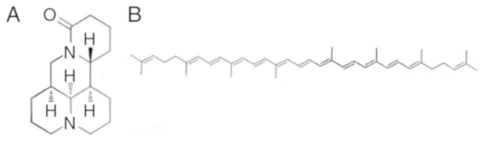
Matrine (MAT; Fig.
1A), a major active component extracted from the traditional
Chinese herb Sophora flavescens Ait, is frequently used to
treat diseases such as hepatitis, enteritis and atopic dermatitis
in China (11). MAT exhibits
various biological properties, of which immune modulation and
anti-inflammation are the most prominent (12,13).
The carotene family is known to suppress oxidative damage by
activating antioxidant enzymes and the stimulating the immune
system (14). Lycopene (LY;
Fig. 1B) is a member of the
carotene family (15). As it has
been reported that ALI involves superoxide radicals and
inflammatory processes (16,17),
it was hypothesized that combined treatment with MAT and LY may
protect the lungs from ALI. Therefore, in the present study, the
effects of MAT, LY and MAT + LY on lipopolysaccharide (LPS)-induced
ALI in mice were determined, and the mechanisms were preliminarily
investigated. In addition, the efficacy of MAT, LY and MAT + LY
were compared with dexamethasone (DEX), to explore the potential
use of these compounds as alternatives to glucocorticoid
therapy.
Materials and methods
Chemicals
MAT and LY (>98% purity) were obtained from the
pharmaceutics laboratory of the Logistics University of Chinese
People's Armed Police Force (PAPF; Tianjin, China). DEX was
provided by the Affiliated Hospital of Logistics University of
Chinese PAPF. LPS (055:B5) was purchased from Sigma-Aldrich (Merck
KGaA). Antibodies used during the study included rabbit anti-IκBα
(cat. no. CY5026; Abways Technology, Inc.), rabbit
anti-phosphorylated (p)-IκBα (cat. no. 2859; Cell Signaling
Technology, Inc.), rabbit anti-NF-κB p65 (cat. no. 8242; Cell
Signaling Technology, Inc.), rabbit anti-p-NF-κB p65 (cat. no.
3033; Cell Signaling Technology, Inc.), and horseradish peroxidase
(HRP)-conjugated goat anti-rabbit IgG (cat. no. S0001; Affinity
Biosciences). ELISA kits for the detection of interleukin-6 (IL-6;
cat. no. E-EL-M0044c) and tumor necrosis factor-α (TNF-α; cat. no.
E-EL-M0049c) were purchased from Elabscience Biotechnology Co.,
Ltd. Kits for detecting the activity of malondialdehyde (MDA; cat.
no. A003-1), glutathione (GSH; cat. no. A006-2) and myeloperoxidase
(MPO; cat. no. A044) were purchased from Nanjing Jiancheng
Bio-Engineering Institute Co., Ltd. TRIzol reagent and SuperRT One
Step RT-PCR Kit (CW0742) for reverse transcription-polymerase chain
reaction (RT-PCR) were purchased from Beijing CoWin Biotech Co.,
Ltd. Primers were purchased from Integrated DNA Technologies, Inc.
BCA Protein Assay kit (cat. no. PC0020) and SDS-PAGE Gel Kit (cat.
no. P1200) were purchased from Solarbio Co., Ltd. SPlink Detection
kit (cat. no. SP-9001) for immunohistochemistry (IHC) was purchased
from OriGene Technologies, Inc. All other chemicals were purchased
from Beijing Dingguo Changsheng Biotechnology Co., Ltd.
Animals
Adult male BALB/c mice (18–22 g) were obtained from
Vital River Laboratory Animal Technology Co., Ltd. The mice (aged 7
weeks old) were maintained in a 12-h light/dark cycle at 22±2°C
with 60±10% humidity, and provided with sufficient food and water
ad libitum. All animal experiments were approved by the
Ethics Committee of Affiliated Hospital of Logistic University of
Chinese People's Armed Police Force (permit no. AF-PJHEC-017-02.0),
and were conducted in accordance with the guidelines set by the
committee. A total of 36 mice were randomly divided into 6 groups:
Control; 6 h LPS; DEX (5 mg/kg) treatment + 6 h LPS; MAT (25 mg/kg)
treatment + 6 h LPS; LY (100 mg/kg) treatment + LPS; and MAT (25
mg/kg) + LY (100 mg/kg) treatment + 6 h LPS. DEX was administered
via intraperitoneal injection for 7 consecutive days; MAT, LY, and
MAT + LY combination treatments were administered orally for 7
consecutive days. The animals in the control group and LPS group
received isovolumetric normal saline orally for the same period. At
30 min following the final administrations, the mice in all groups
except the control group received an intratracheal instillation of
LPS (5 mg/kg), whereas the animals in the control group received an
intratracheal instillation of saline. At 6 h after LPS induction,
the mice were euthanized via cervical dislocation following
anesthetization with diethyl ether. Lung tissues were obtained and
stored for further experiments.
Hematoxylin and eosin (H&E)
staining
The lower lobe of right lungs were fixed with 4%
paraformaldehyde at room temperature (23±2°C) for 1 day, then
dehydrated and embedded in paraffin. All samples in paraffin were
cut into 5-µm sections. For H&E staining, sections were
immersed in hematoxylin for 10 min, followed by eosin for 3 min at
room temperature. Following mounting using neutral balsam, the
stained slides were observed under a light microscope (Olympus CHK;
Olympus Corporation) and histopathology images were captured
(C-4040 Zoom; Olympus Corporation). The lung injury score was
assessed based on the method reported by Aeffner et al
(18): No injury, 0; injury in 25%
of the field, 1; injury in 50% of the field, 2; injury in 75% of
the field, 3; and injury throughout the field, 4. A total of 10
random microscopic images were acquired for each section, and the
fields were scored blindly by two pathologists and averaged. After
the lung injury scores were assessed, Q value was used to evaluate
the synergistic protective effect (19). Q value was calculated as
EA+B/(EA+EB-EA•EB).
EA represents the average lung injury score in MAT
group, EB represents the average lung injury score in LY
group, and EA+B represents the average lung injury score
in MAT + LY group. Synergy or antagonism were defined by Q value
>1 or <1 respectively, while Q=1 indicated no
interaction.
Lung wet-to-dry (W/D) weight
ratios
The degree of pulmonary edema can be determined
based on lung W/D weight ratios (20). Absorbent paper was used to absorb
exudate and blood on the surface of superior lobes of right lungs.
Then, the tissues were weighed and recorded as wet weights. The
tissues were then dried at 70°C for 72 h to constant weight and
recorded as dry weights. Lung W/D weight ratios=wet weight/dry
weight.
IHC
The following steps, if not described otherwise,
were all performed at room temperature (23±2°C). Lung sections (5
µm) were deparaffinized in xylene (20 min) and rehydrated in a
graded alcohol series (absolute ethyl alcohol for 10 min, 90%
alcohol for 5 min, 80% alcohol for 5 min, 70% alcohol for 5 min).
Antigens were retrieved in 0.01 M citric acid heated by microwave
for 20 min. Endogenous peroxidases were inactivated by 3%
H2O2 for 20 min. Goat serum was used to block
for 1 h at 37°C. Then, slides were incubated with diluted NF-κB p65
antibody (1:500) overnight at 4°C. The following day, the antibody
was removed and the sections were washed with PBS prior to
incubation with HRP-conjugated secondary antibody for 30 min at
37°C. 3,3′-Diaminobenzidine was used to visualize expression and
used to stain the slides for 5 min. Finally, the slides were
counterstained with hematoxylin for 30 sec and dehydrated prior to
observation under a light microscope. The results were evaluated
semi-quantitatively. Five randomly fields were imaged from each
section. Images were quantified using two different scores:
Intensity score and positive cell ratio score. For the intensity
score, negative, weakly positive, positive and strongly positive
were graded as 0, 1, 2 and 3, respectively; for the positive cell
ratio score, the positive cell area/the total area <1% was
scored 0; 1–10% scored 1; >10–50% scored 2; >50–80% scored 3;
and, >80% scored 4. The product of the two scores was used as
the IHC value for each field.
ELISA for IL-6 and TNF-α
Mouse lung tissues were homogenized and then
centrifuged at 12,000 × g at 4°C for 20 min. The supernatant was
collected and diluted. The standard ELISA solutions or samples
(diluent supernatant) were added to each well, then incubated for
90 min at 37°C. Diluted biotin-labeled antibodies (1:99) were added
to the wells, and the plates were covered and incubated for 2 h at
37°C. The plates were then washed with washing buffer three times.
HRP conjugate was then added to wells which allows binding with the
biotin-labeled antibodies, and plates were incubated for 1 h at
37°C. After washing the plates, the chemiluminescent substrate was
detected using colorimetric 3,3′,5,5′-tetramethylbenzidine
solution. Finally, the stop solution was added into each well, and
optical density of each well was read at 450 nm immediately by a
microplate reader (Tecan Sunrise). The absorbance of samples was
compared with the standard curve to calculate the
concentration.
Western blot analysis
Total lung proteins were prepared from lung
homogenates in RIPA buffer and protease/phosphatase inhibitor
cocktail (Solarbio Co., Ltd.) and the protein concentration was
determined by BCA method. Protein (30 µg/lane) was separated via
SDS-PAGE on 10% gels and transferred to PVDF membranes. PVDF
membranes were incubated with 5% non-fat milk at 25°C for 1 h with
gentle agitation for blocking. Then, the membranes were incubated
overnight with the aforementioned antibodies (1:1,000) at 4°C.
Following incubation with secondary antibodies (1:3,000) at 25°C
for 1 h, the blots were visualized using enhanced chemiluminescence
reagent (cat. no. PE0010; Solarbio Co., Ltd.). ImageJ version
2.1.4.7 (National Institutes of Health) was used for densitometric
quantification of expression.
Determination of MDA, GSH and MPO
Mouse lung tissues were homogenized, saline was
added (tissue weight/g: Saline volume/ml=1:9) and samples were
centrifuged at 10,000 × g at 4°C for 10 min. The supernatant was
collected and used for subsequent experiments. MDA, GSH and MPO
levels were detected using the respective kits, according to the
manufacturer's protocols. The absorbances were measured at 532 nm
for MDA, 405 nm for GSH, and 460 nm for MPO.
RT-PCR
Total RNA was isolated from lung tissues homogenates
using TRIzol reagent (21), and
RT-PCR was performed according to manufacturer's protocols. The kit
allows reverse transcription and PCR in one step. The protocol
includes 1 cycle at 45°C for 30 min followed by 95°C for 2 min;
then 40 cycles of 94°C for 30 sec, 60°C for 30 sec, 72°C for 30
sec; and, finally, 1 cycle at 72°C for 5 min. The following primers
were used: GAPDH, forward 5′-CCCAGCAAGGACACTGAGCAAG-3′, reverse
5′-GGTCTGGGATGGAAATTGTGAGGG-3′; IL-6, forward
5′-GGATACCACTCCCAACAGACC-3′, reverse 5′-TTCTGCAAGTGCATCATCGT-3′;
TNF-α, forward 5′-GGCCTCCCTCTCATCAGTTC-3′, reverse
5′-CTTGGTGGTTTGCTACGACG-3′; and NF-κB p65, forward
5′-TCCGGTTACGTAATGAGTGGT-3′ and reverse
5′-GATCTGGTTCTCTTTCCGAAGTC-3′. PCR products were resolved on 1.5%
agarose gels via electrophoresis. Results were visualized using
EtBr, and ImageJ version 2.1.4.7 (National Institutes of Health)
was used to quantify expression.
Statistical analysis
SPSS version 17.0 (SPSS, Inc.) was used for data
analysis. Data are expressed as the mean ± standard deviation. Data
were analyzed using one-way analysis of variance (ANOVA) followed
by a Bonferroni test for multiple comparisons. For the lung injury
scores and IHC values, the Kruskal-Wallis test was used, then all
data were transformed logarithmically to make them conform to the
normal distribution before using one-way ANOVA to compare specific
groups. P<0.05 was considered to indicate a statistically
significant difference.
Results
MAT + LY treatment attenuates lung
edema induced by LPS
As presented in Fig.
2A, LPS induction significantly increased the W/D ratio of lung
tissues compared with the control group. The W/D ratios of tissues
from the MAT-treated and LY-treated groups were similar, and were
not significantly different to those from the LPS group; however,
treatment with DEX or MAT + LY significantly decreased the W/D
ratio compared with the LPS group.
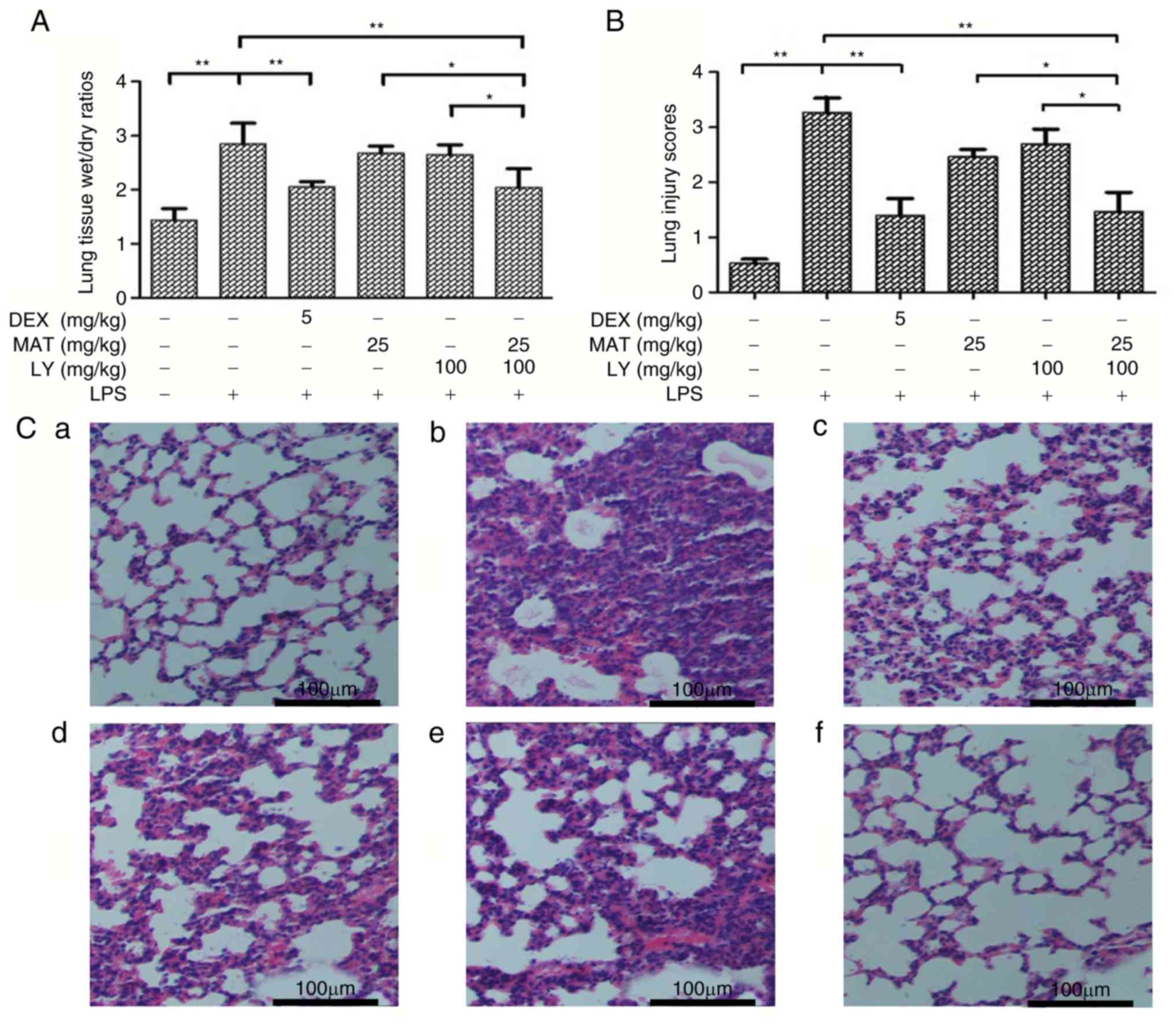 | Figure 2.Edema and morphopathological
alterations in lung tissues. (A) Superior lobes of right mouse
lungs were extracted and weighed. Lung tissue wet/dry ratios=wet
weight/dry weight. n=6. (B) Lung injury scores were determined by
two pathologists. (C) Hematoxylin and eosin staining of the
inferior lobes of right lungs: a, control; b, LPS; c, DEX + LPS; d,
MAT + LPS; e, LY + LPS; and f, MAT + LY + LPS. Scale bar, 100 µm.
Data are presented as the mean ± standard deviation. *P<0.05,
**P<0.01. DEX, dexamethasone; MAT, matrine; LY, lycopene; LPS,
lipopolysaccharide. |
MAT + LY treatment prevents
histopathological changes to the lungs induced by LPS
The synergistic protective effects of MAT + LY were
calculated using lung injury scores (Fig. 2B) and the Q value was 1.27,
indicating that MAT and LY exhibited synergy. As presented in
Fig. 2C, lung tissues from the
control group exhibited a normal lung structure, with the alveolar
wall mainly composed of single layer of epithelial cells and
possessing a complete structure. Conversely, hemorrhage, alveolar
wall thickening, alveolar collapse and inflammatory infiltration
were observed following LPS administration. The pathological
alterations to the lungs were significantly attenuated by MAT + LY
or DEX treatment (Fig. 2B and C),
but were not significantly reduced in the MAT or LY only-treated
groups.
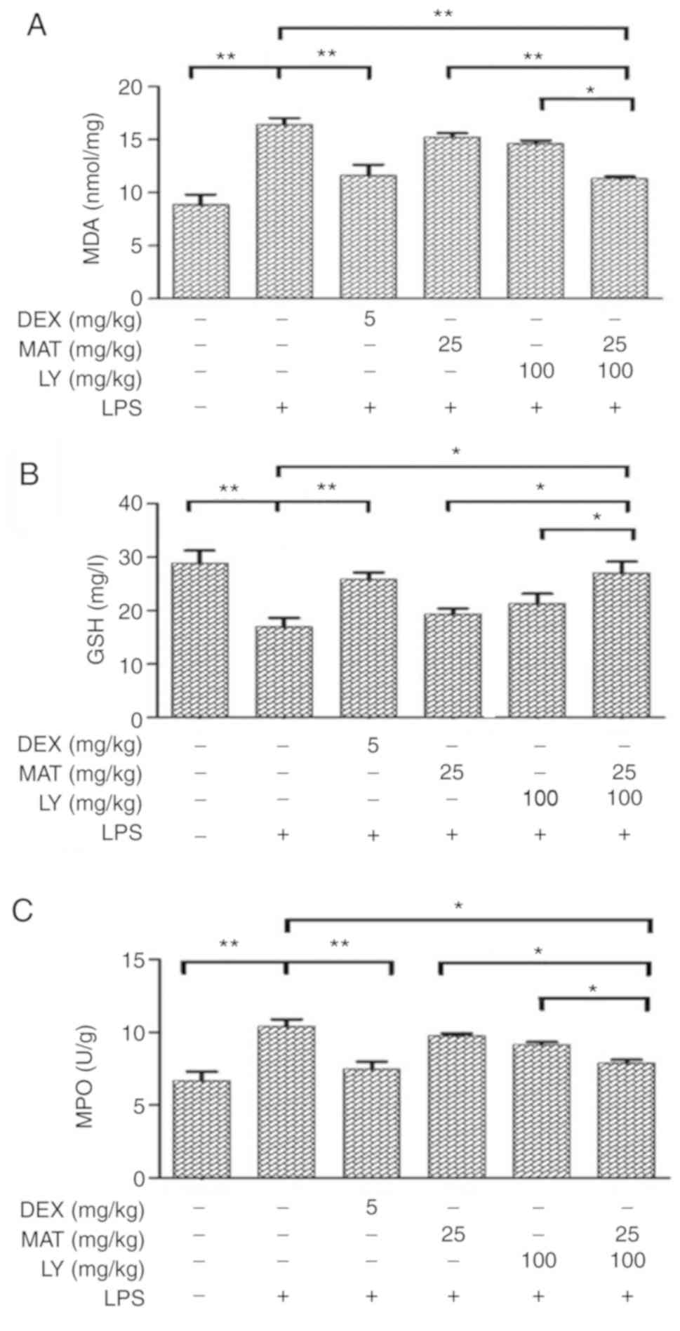
MAT + LY treatment decreases oxidative
stress in vivo
MDA is a product of lipid peroxidation and is
frequently used as biomarker for determining oxidative stress
(22). MDA levels were
significantly increased following LPS induction (Fig. 3A); however, treatments with MAT +
LY or DEX significantly attenuated LPS-induced MDA increases. GSH
is an important antioxidant used as a marker of oxidative stress
(23). Administration of LPS
induced a reduction in GSH (Fig.
3B). Treatment with MAT or LY alone increased GSH levels
compared with the LPS group, although the differences were not
statistically significant; however, combined treatment with MAT +
LY or DEX significantly increased GSH levels. Furthermore, to
determine the effects of MAT + LY on neutrophil accumulation, the
levels of MPO were measured. Following intratracheal instillation
of LPS, the levels of MPO in lung tissue were significantly
elevated (Fig. 3C). MAT or LY
treatment alone did not significantly affect MPO levels compared
with the LPS group; however, the combination of treatment with MAT
+ LY significantly attenuated this LPS-induced increase.
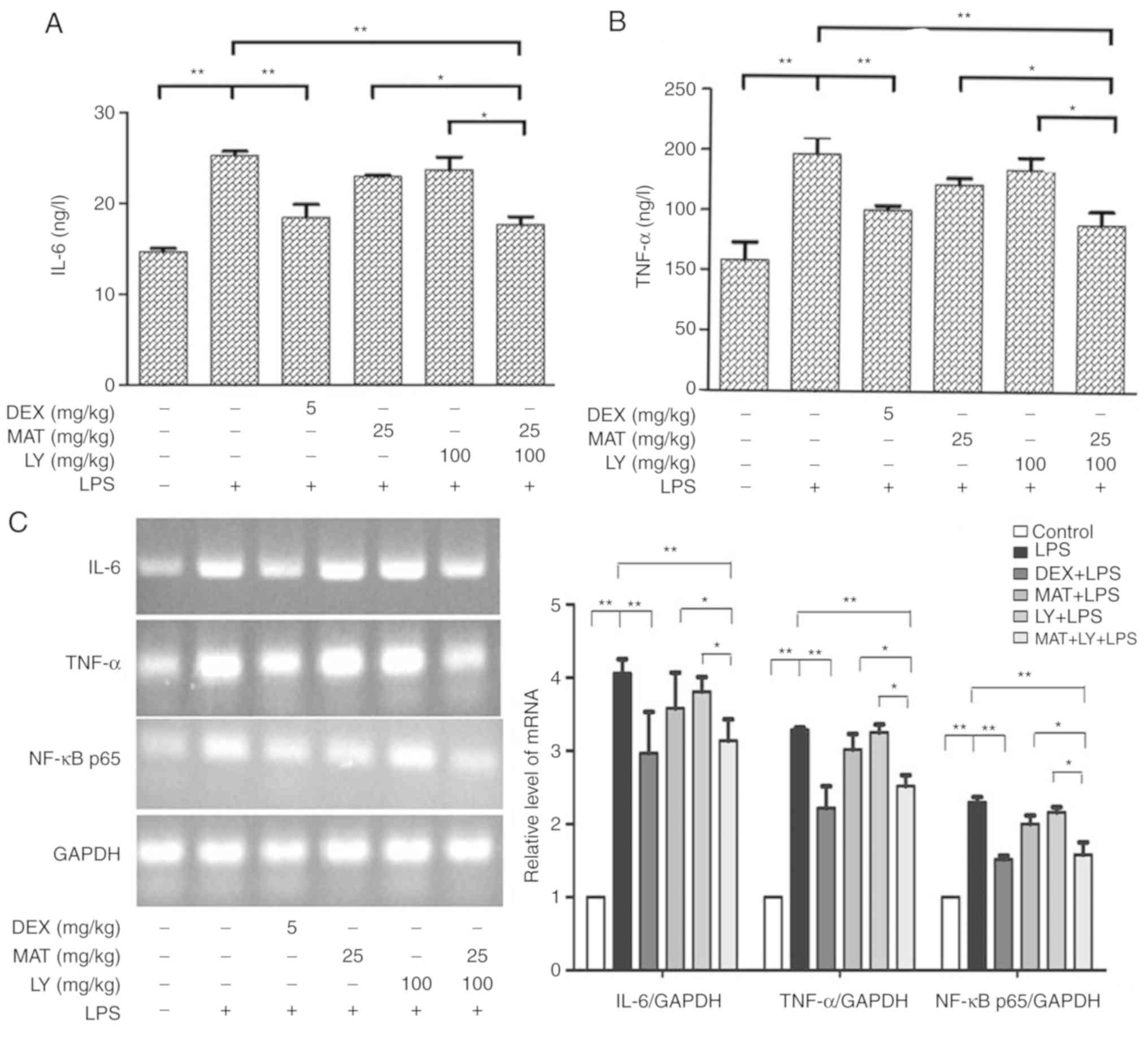
MAT + LY treatment reduces the
inflammatory response in vivo
The levels of IL-6 and TNF-α in lung tissues were
determined via ELISA. The results indicated that the levels of IL-6
(Fig. 4A) and TNF-α (Fig. 4B) were significantly increased
following LPS administration compared with the control group.
Treatment with MAT or LY did not significantly affect the
LPS-induced increased in IL-6 or TNF-α levels, whereas treatment
with MAT + LY or DEX significantly decreased the levels of IL-6 and
TNF-α compared with the LPS group. To further validate these
results, IL-6 and TNF-α mRNA levels were measured via RT-PCR
analysis, with the same trends observed (Fig. 4C).
MAT + LY treatment inhibits the NF-κB
signaling pathway
NF-κB signaling pathway is a classical inflammatory
signaling pathway. LPS activated the IκB/NF-κB signaling,
stimulating the expression of NF-κB p65 compared with the control
group (Figs. 4C and 5). Treatment with MAT or LY alone
significantly inhibited NF-κB signaling, while the effect of MAT +
LY was more pronounced. MAT + LY treatment downregulated the
expression of NF-κB p65 at the mRNA and protein levels, and the
phosphorylation of NF-κB p65 and IκBα compared with the LPS group,
inhibiting the activation of signaling induced by LPS (Fig. 5C).
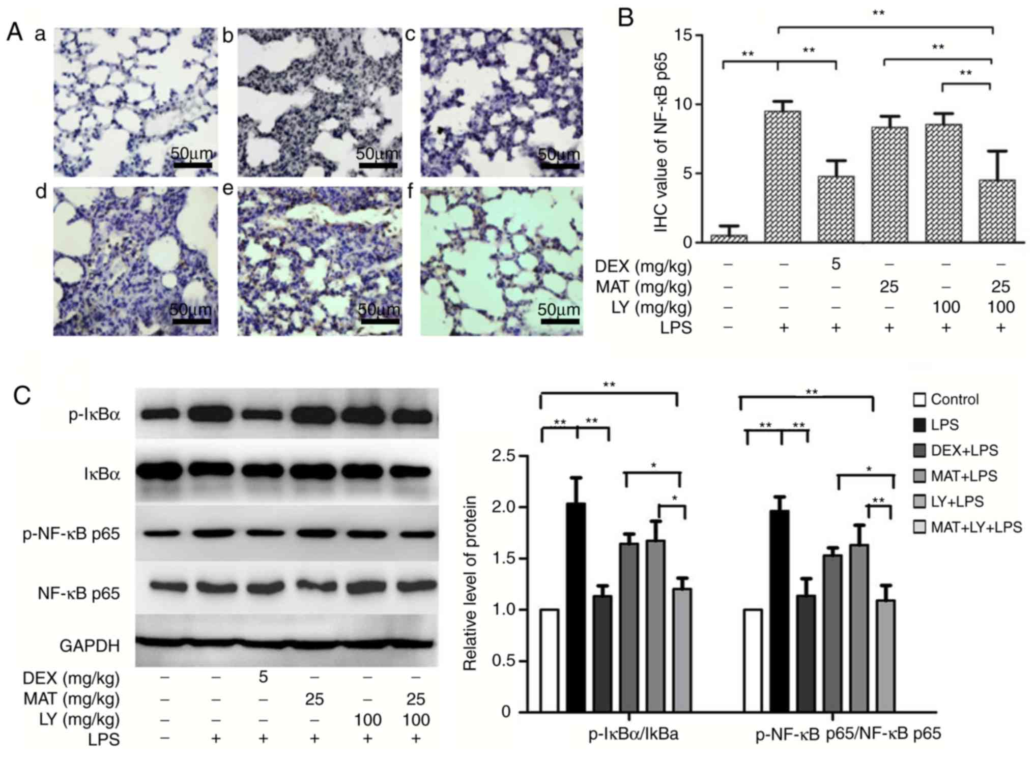 | Figure 5.Effects of MAT and/or LY on NF-κB
signaling. (A) Immunohistochemical staining for NF-κB p65: a,
control; b, LPS; c, DEX + LPS; d, MAT + LPS; e, LY + LPS; and f,
MAT + LY + LPS. Scale bar, 50 µm. (B) Protein expression of NF-κB
p65 as evaluated semi-quantitatively based on immunohistochemistry.
(C) Activation levels of IκBα and NF-κB p65 were analyzed via
western blotting and densitometry analysis. Data are presented as
the mean ± standard deviation. n=6. *P<0.05, **P<0.01. DEX,
dexamethasone; MAT, matrine; LY, lycopene; LPS,
lipopolysaccharide. |
Discussion
ALI is a serious disease characterized by bilateral
lung infiltration. Various therapeutic interventions are under
development; however, at present, there is no effective cure
(24). The mortality rate of ALI
is 30–40% (25). Glucocorticoids
are potentially useful as anti-inflammatory drugs for the treatment
of ALI (26); however, long-term
use of glucocorticoids at high doses may lead to immunosuppression,
resulting in infections that exacerbate the lung injury, or other
complications, including Cushing's syndrome or psychiatric symptoms
(27). It was previously reported
that treatment with high doses of glucocorticoid for short or long
periods of time did not improve the survival of patients with ALI
(28). As a result, it is
necessary to identify alternative treatments for ALI. Zhang et
al (11) reported that
intraperitoneal injection of MAT (100 mg/kg) protected against
LPS-induced ALI in a mouse model. Türkoğlu et al (15) observed that LY (at 200 times the
recommended daily intake in humans) exhibited protective effects in
a rat model of oleic acid-induced ALI. The high doses and
administration protocols in these studies restricted their clinical
usefulness. Therefore, in the present study, these issues were
amended; the two compounds were administered orally at a relatively
low dosage, and their effects in combination were investigated.
LPS-induced lung injury in rodents is a frequently
used ALI model, mimicking a number of the characteristics of ALI in
humans (29). In the present
study, combined treatment of MAT + LY achieved similar effects to
DEX in protecting the lungs in the LPS-induced mouse model. The
combination treatment attenuated lung injury and pulmonary edema,
potentially via anti-inflammatory and antioxidative mechanisms,
exhibiting synergistic effects.
The pathogenesis of ALI has not been fully
determined. Inflammatory responses (30) and oxidative stress imbalances
(31) have been identified to have
important roles during the development and progression of ALI.
These two mechanisms can affect the disease progression separately
and synchronously. TNF-α is an early endogenous mediator of
inflammatory responses produced by monocytes/macrophages (32). Excessive levels of TNF-α will
damage lysosomes, leading to injuries in pulmonary vascular
endothelial cells and epithelial cells (3). Furthermore, TNF-α induces the
secretion of cytokines such as IL-6 (33). ELISA and RT-PCR analyses in the
current study revealed that treatment with MAT alone markedly
affected TNF-α and IL-6 levels; however, when LY was added, a
significant decrease in the levels of the two cytokines was
observed compared with the LPS group.
Oxidative stress is increased during inflammation of
airways, in turn exacerbating the inflammation (31). This leads to the formation of a
positive feedback loop that promotes inflammatory responses,
leading to deterioration of the condition (31). The release of reactive oxygen
species (ROS), and subsequent induction of oxidative stress, has an
important role in this process. ROS mainly attack polyunsaturated
fatty acids in plasma membranes, inducing lipid peroxidation and
increasing the levels of MDA (34,35);
thus, the levels of MDA reflect whether cells are subjected to
attack by oxygen free radicals. In addition to interactions with
inflammatory factors, ROS induce cell damage via a series of other
mechanisms, such as the inactivation of antioxidant defense
systems. The system includes various antioxidant enzymes, including
superoxide dismutase and GSH peroxidase (31). These enzymes can reduce and
eliminate the production of ROS, thereby protecting against damage
caused by oxidative stress (36,37).
GSH is a member of the antioxidant defense system, and promotes the
decomposition of hydrogen peroxide and inhibits the production of
free radicals (38). GSH activity
indicates the degree of endogenous oxygen free radical scavenging.
LY is an antioxidant; however, 100 mg/kg LY did not notably protect
mice against ALI. It was observed that the recruitment of
neutrophils was decreased when the mice were treated with MAT + LY,
as determined by a significant decrease in MPO levels. MAT + LY
also decreased the levels of lipid peroxidation in the mouse model
of ALI, as determined by decreased MDA levels, and attenuated the
reduction in GSH in lung tissue. The increase in GSH level in mice
lung tissue may be a result of the upregulation of antioxidant
expression or a decrease of ROS production.
The potential mechanism underlying the synergistic
protective effect was investigated. NF-κB signaling has been
identified as a traditional immune and inflammatory pathway. The
NF-κB/Rel family members are dimeric transcription factors. In the
absence of stimulating signals, NF-κB dimers are bound to
inhibitory IκB proteins. In the canonical pathway, activators, such
as proinflammatory cytokines, activate an IκB kinase complex,
leading to phosphorylation of IκB (39). Thus, the NF-κB/Rel complexes are
freed and further phosphorylated prior to translocation into the
nucleus, where they induce gene expression (40). Treatment with MAT + LY
significantly suppressed the LPS-induced activation of NF-κB
activation compared with the single drug treatment groups. RT-PCR,
western blot and IHC analyses indicated that the two agents
inhibited NF-κB p65 transcription and translation, in turn
decreasing NF-κB signaling. Specifically, MAT + LY treatment
reduced the phosphorylation of IκBα, which allows NF-κB p65 to be
anchored in the cytoplasm rather than phosphorylate and transfer to
the nucleus to regulate proinflammatory gene transcription. The
findings suggested that reduced signaling decreased the expression
of proinflammatory factors, inhibiting the production of ROS and
other oxygen radicals.
In conclusion, the results of the present study
demonstrated that MAT or LY treatment alone exhibited limited
protection against LPS-induced ALI in mice; however, combined
treatment of MAT and LY significantly attenuated the LPS-induced
alterations in cytokine expression and oxidative stress, suggesting
that there may be a synergistic effect of the two chemicals. One of
the mechanisms underlying this potential synergy was the inhibition
of the NF-κB signaling pathway. The roles of other signaling
pathways requires further investigation. Additionally, it should be
noted that the pathology of ALI in clinical settings is more
complex that in the mouse model. In the present study, drugs were
administered for 7 days prior to LPS induction, an unlikely
situation in a clinical setting. Additional groups and time points
will be explored in future experiments. According to the present
findings, the combined administration of MAT and LY may be a
potential alternative to glucocorticoid therapy for the treatment
of ALI.
Acknowledgements
Not applicable.
Funding
The present study was supported by the Key
Laboratory for Occupational and Environmental Hazards Prevention of
Tianjin (grant no. WHKF201705) and the National Natural Science
Foundation of China (grant no. 81600051).
Availability of data and materials
The datasets used and/or analyzed during the present
study are available from the corresponding author on reasonable
request.
Authors' contributions
WL and TW were major contributors in writing the
manuscript. WL, YR and JW performed the experiments and analyzed
the data. TW, BC and BL contributed to the conception and design of
the study. FW and LW performed the histological examination. HC and
YL designed the present study and revised the manuscript. All
authors read and approved the final manuscript.
Ethics approval and consent to
participate
Animal experiments were approved by the Ethics
Committee of Affiliated Hospital of Logistic University of Chinese
People's Armed Police Force (permit number: AF-PJHEC-017-02.0), and
were conducted in accordance with the guidelines set by the
committee.
Patient consent for publication
Not applicable.
Competing interests
The authors declare that they have no competing
interests.
References
|
1
|
Bersten AD, Edibam C, Hunt T and Moran J;
Australian; New Zealand Intensive Care Society Clinical Trials
Group, : Incidence and mortality of acute lung injury and the acute
respiratory distress syndrome in three Australian States. Am J
Respir Crit Care Med. 165:443–448. 2002. View Article : Google Scholar : PubMed/NCBI
|
|
2
|
Erickson SE, Martin GS, Davis JL, Matthay
MA and Eisner MD; NIH NHLBI ARDS Network, : Recent trends in acute
lung injury mortality: 1996–2005. Crit Care Med. 37:1574–1579.
2009. View Article : Google Scholar : PubMed/NCBI
|
|
3
|
Zhong WT, Wu YC, Xie XX, Zhou X, Wei MM,
Soromou LW, Ci XX and Wang DC: Phillyrin attenuates LPS-induced
pulmonary inflammation via suppression of MAPK and NF-κB activation
in acute lung injury mice. Fitoterapia. 90:132–139. 2013.
View Article : Google Scholar : PubMed/NCBI
|
|
4
|
Zhang X, Li C, Li J, Xu Y, Guan S and Zhao
M: Protective effectsof protocatechuic acid on acute lung injury
induced by lipopolysaccharide in mice via p38MAPK and NF-κB
signalpathways. Int Immunopharmacol. 26:229–236. 2015. View Article : Google Scholar : PubMed/NCBI
|
|
5
|
Zhang Y, Liang D, Dong L, Ge X, Xu F, Chen
W, Dai Y, Li H, Zou P, Yang S and Liang G: Anti-inflammatory
effects of novel curcumin analogs in experimental acute lung
injury. Respir Res. 16:432015. View Article : Google Scholar : PubMed/NCBI
|
|
6
|
Menezes SL, Bozza PT, Neto HC, Laranjeira
AP, Negri EM, Capelozzi VL, Zin WA and Rocco PR: Pulmonary and
extrapulmonary acute lung injury: Inflammatory and ultrastructural
analyses. J Appl Physiol (1985). 98:1777–1783. 2005. View Article : Google Scholar : PubMed/NCBI
|
|
7
|
Chopra M, Reuben JS and Sharma AC: Acute
lung injury: Apoptosis and signaling mechanisms. Exp Biol Med
(Maywood). 234:361–371. 2009. View Article : Google Scholar : PubMed/NCBI
|
|
8
|
Z'graggen BR, Tornic J, Müller-Edenborn B,
Reyes L, Booy C and Beck-Schimmer B: Acute lung injury: Apoptosis
in effector and target cells of the upper and lower airway
compartment. Clin Exp Immunol. 161:324–331. 2010.PubMed/NCBI
|
|
9
|
Sarma JV and Ward PA: Oxidants and redox
signaling in acute lung injury. Compr Physiol. 1:1365–1381.
2011.PubMed/NCBI
|
|
10
|
Zemans RL, Colgan SP and Downey GP:
Transepithelial migration of neutrophils: Mechanisms and
implications for acute lung injury. Am J Respir Cell Mol Biol.
40:519–535. 2009. View Article : Google Scholar : PubMed/NCBI
|
|
11
|
Zhang B, Liu ZY, Li YY, Luo Y, Liu ML,
Dong HY, Wang YX, Liu Y, Zhao PT, Jin FG and Li ZC:
Antiinflammatory effects of matrine in LPS-induced acute lung
injury in mice. Eur J Pharm Sci. 44:573–579. 2011. View Article : Google Scholar : PubMed/NCBI
|
|
12
|
Niu Y, Dong Q and Li R: Matrine regulates
Th1/Th2 cytokine responses in rheumatoid arthritis by attenuating
the NF-κB signaling. Cell Biol Int. 41:611–621. 2017. View Article : Google Scholar : PubMed/NCBI
|
|
13
|
Huang WC, Chan CC, Wu SJ, Chen LC, Shen
JJ, Kuo ML, Chen MC and Liou CJ: Matrine attenuates allergic airway
inflammation and eosinophil infiltration by suppressing eotaxin and
Th2 cytokine production in asthmatic mice. J Ethnopharmacol.
151:470–477. 2014. View Article : Google Scholar : PubMed/NCBI
|
|
14
|
Prabhala RH, Braune LM, Garewal HS and
Watson RR: Influence of beta-carotene on immune functions. Ann N Y
Acad Sci. 691:262–263. 1993. View Article : Google Scholar : PubMed/NCBI
|
|
15
|
Türkoğlu S, Muz MH, Ozercan R, Gürsu F and
Kırkıl G: Effects of lycopene on the model of oleic acid-induced
acute lung injury. Tuberk Toraks. 60:101–107. 2012. View Article : Google Scholar : PubMed/NCBI
|
|
16
|
Allen TC and Kurdowska A: Interleukin 8
and acute lung injury. Arch Pathol Lab Med. 138:266–269. 2014.
View Article : Google Scholar : PubMed/NCBI
|
|
17
|
Mokra D and Kosutova P: Biomarkers in
acute lung injury. Respir Physiol Neurobiol. 209:52–58. 2015.
View Article : Google Scholar : PubMed/NCBI
|
|
18
|
Aeffner F, Bolon B and Davis IC: Mouse
models of acute respiratory distress syndrome: A review of
analytical approaches, pathologic features, and common
measurements. Toxicol Pathol. 43:1074–1092. 2015. View Article : Google Scholar : PubMed/NCBI
|
|
19
|
Zhang D, Liu B, Cao B, Wei F, Yu X, Li GF,
Chen H, Wei LQ and Wang PL: Synergistic protection of Schizandrin B
and Glycyrrhizic acid against bleomycin-induced pulmonary fibrosis
by inhibiting TGF-β1/Smad2 pathways and overexpression of NOX4. Int
Immunopharmacol. 48:67–75. 2017. View Article : Google Scholar : PubMed/NCBI
|
|
20
|
Parker JC and Townsley MI: Evaluation of
lung injury in rats and mice. Am J Physiol Lung Cell Mol Physiol.
286:L231–L246. 2004. View Article : Google Scholar : PubMed/NCBI
|
|
21
|
Chomczynski P and Sacchi N: Single-step
method of RNA isolation by acid guanidinium
thiocyanate-phenol-chloroform extraction. Anal Biochem.
162:156–159. 1987. View Article : Google Scholar : PubMed/NCBI
|
|
22
|
Gaweł S, Wardas M, Niedworok E and Wardas
P: Malondialdehyde (MDA) as a lipid peroxidation marker. Wiad Lek.
57:453–455. 2004.(In Polish). PubMed/NCBI
|
|
23
|
Mytilineou C, Kramer BC and Yabut JA:
Glutathione depletion and oxidative stress. Parkinsonism Relat
Disord. 8:385–387. 2002. View Article : Google Scholar : PubMed/NCBI
|
|
24
|
Del Sorbo L, Goffi A and Ranieri VM:
Mechanical ventilation during acute lung injury: Current
recommendations and new concepts. Presse Med. 40:e569–e583. 2011.
View Article : Google Scholar : PubMed/NCBI
|
|
25
|
Su ZQ, Mo ZZ, Liao JB, Feng XX, Liang YZ,
Zhang X, Liu YH, Chen XY, Chen ZW, Su ZR and Lai XP: Usnic acid
protects LPS-induced acute lung injury in mice through attenuating
inflammatory responses and oxidative stress. Int Immunopharmacol.
22:371–378. 2014. View Article : Google Scholar : PubMed/NCBI
|
|
26
|
Thompson BT: Glucocorticoids and acute
lung injury. Crit Care Med. 31 (Suppl 4):S253–S257. 2003.
View Article : Google Scholar : PubMed/NCBI
|
|
27
|
Oray M, Abu Samra K, Ebrahimiadib N, Meese
H and Foster CS: Long-term side effects of glucocorticoids. Expert
Opin Drug Saf. 15:457–465. 2016. View Article : Google Scholar : PubMed/NCBI
|
|
28
|
Steinberg KP, Hudson LD, Goodman RB, Hough
CL, Lanken PN, Hyzy R, Thompson BT and Ancukiewicz M; National
Heart Lung, Blood Institute Acute Respiratory Distress Syndrome
(ARDS) Clinical Trials Network, : Efficacy and safety of
corticosteroids for persistent acute respiratory distress syndrome.
N Engl J Med. 354:1671–1684. 2006. View Article : Google Scholar : PubMed/NCBI
|
|
29
|
Rittirsch D, Flierl MA, Day DE, Nadeau BA,
McGuire SR, Hoesel LM, Ipaktchi K, Zetoune FS, Sarma JV, Leng L, et
al: Acute lung injury induced by lipopolysaccharide is independent
of complement activation. J Immunol. 180:7664–7672. 2008.
View Article : Google Scholar : PubMed/NCBI
|
|
30
|
Ward PA: Acute lung injury: How the lung
inflammatory response works. Eur Respir J. (Suppl 44):S22–S23.
2003. View Article : Google Scholar
|
|
31
|
Ward PA: Oxidative stress: Acute and
progressive lung injury. Ann N Y Acad Sci. 1203:53–59. 2010.
View Article : Google Scholar : PubMed/NCBI
|
|
32
|
Zelová H and Hošek J: TNF-α signalling and
inflammation: Interactions between old acquaintances. Inflamm Res.
62:641–651. 2013. View Article : Google Scholar : PubMed/NCBI
|
|
33
|
Ohta K, Naruse T, Ishida Y, Shigeishi H,
Nakagawa T, Fukui A, Nishi H, Sasaki K, Ogawa I and Takechi M:
TNF-α-induced IL-6 and MMP-9 expression in immortalized
ameloblastoma cell line established by hTERT. Oral Dis. 23:199–209.
2017. View Article : Google Scholar : PubMed/NCBI
|
|
34
|
Faurschou M and Borregaard N: Neutrophil
granules and secretory vesicles in inflammation. Microbes Infect.
5:1317–1327. 2003. View Article : Google Scholar : PubMed/NCBI
|
|
35
|
Macdonald J, Galley HF and Webster NR:
Oxidative stress and gene expression in sepsis. Br J Anaesth.
90:221–232. 2003. View Article : Google Scholar : PubMed/NCBI
|
|
36
|
Victor VM, Rocha M and De la Fuente M:
Immune cells: Free radicals and antioxidants in sepsis. Int
Immunopharmacol. 4:327–347. 2004. View Article : Google Scholar : PubMed/NCBI
|
|
37
|
Del Rio D, Stewart AJ and Pellegrini N: A
review of recent studies on malondialdehyde as toxic molecule and
biological marker of oxidative stress. Nutr Metab Cardiovasc Dis.
15:316–328. 2005. View Article : Google Scholar : PubMed/NCBI
|
|
38
|
Schettler V, Wieland E, Methe H,
Schuff-Werner P and Müller GA: Oxidative stress during dialysis:
Effect on free radical scavenging enzyme (FRSE) activities and
glutathione (GSH) concentration in granulocytes. Nephrol Dial
Transplant. 13:2588–2593. 1998. View Article : Google Scholar : PubMed/NCBI
|
|
39
|
Hinz M and Scheidereit C: The IκB kinase
complex in NF-κB regulation and beyond. EMBO Rep. 15:46–61. 2014.
View Article : Google Scholar : PubMed/NCBI
|
|
40
|
Luo K: Signaling CROSS Talk between
TGF-β/Smad and other signaling pathways. Cold Spring Harb Perspect
Biol. 9:a0221372017. View Article : Google Scholar : PubMed/NCBI
|