Introduction
Pancreatic cancer is ranked fourth among
cancer-related deaths worldwide and has a 5-year survival rate ~5%
(1). This is likely due to an
inadequate response with the currently available treatments along
with late detection, to the aggressive pathogenesis course of the
disease, and to the development of extreme resistance to radiation,
chemotherapies, targeting agents or a combination of these
(1,2). Thus, there is a steady movement of
ongoing research on various promising therapies for pancreatic
cancer (3–5). Gemcitabine is a widely used therapy to
treat patients with pancreatic cancer (6,7).
Gemcitabine affects the S phase of cell cycle, preventing cell
division, and has proven to be effective for symptom management and
prolonged survival in advanced pancreatic cancer (8). Recent clinical trials of combination
therapy with gemcitabine and another agent had statistical
significance, but not significant enough for patients (9–11).
The use of traditional herbal medicines, such as
extracts of herbal mixtures, has been revisited since these
medicines often present anticancer activities with no or less
adverse side-effects (12). Herbal
mixture extracts have been studied in pancreatic cancer, as well as
other types of cancers (5,13,14).
Cocktail therapies of herbal mixture extracts and antitumor drugs
may have synergistic effects against tumor growth, allowing the use
of lower doses of anticancer drug (15). Thus, the combinatorial use of a
herbal mixture and an anticancer drug is of great interest to
enhance the beneficial effects of cancer chemotherapeutics.
The present study assesses the antitumor effect of
an herbal mixture extract (H3) in the pancreatic adenocarcinoma
cell line PANC1 and in a PANC1-induced heterotopic xenograft model.
These effects were compared to those of gemcitabine alone and to a
H3-gemcitabine combinatorial therapy.
Materials and methods
Preparation of H3
H3 was composed of 40% Meliae fructus
(China), 40% Cinnamon bark (Vietnam) and 20% Sparganium
rhizome (China) that were obtained from the Oriental Medical
Hospital, Dongguk University (Korea). H3 was prepared as follows:
the dried and pulverized medicinal herbs were mixed together in 8
kg batches and were soaked in 40% ethanol (80 l). The ethanol
extract was concentrated using a rotary evaporator, lyophilized and
reconstituted in distilled water.
In vitro experiment
The human pancreatic cancer cell line (PANC1; KCLB
#21469) was purchased from the Korean Cell Line Bank (KCLB; Seoul,
Korea). PANC1 was cultured in Dulbecco's modified Eagle's medium
(DMEM) supplemented with 10% fetal bovine serum (FBS) (both from
WelGENE Inc., Daegu, Korea), 100 U/ml penicillin and 100
µg/ml streptomycin. The cells were maintained in a
humidified incubator at 37°C containing 5% CO2. PANC1
cells were seeded in cell culture plates (Sarstedt Inc.,
Nuermbrecht, Germany) at a density of 5×10⁵ cells. The
cells were treated with H3 (0.07 mg/ml) or gemcitabine (10 nM;
Sigma-Aldrich, St. Louis, MO, USA). For co-treatment, the cells
were treated sequentially with 0.05 mg/ml of H3 and 3 nM of
gemcitabine, with a 2 h interval between the two treatments. After
a 72 h incubation, the cells were harvested using 0.05%
trypsin-EDTA.
Animal model
Five-week-old female BALB/c-nu/nu mice (Nara Biotech
Co., Seoul, Korea) with body weights between 18–20 g were used in
the present study. All animals were maintained in standard
conditions under 12 h light and 12 h dark cycle, and allowed to
acclimate for a week prior to the start of the study. All
procedures were approved by the Internal Animal Care and Use
Committee, Korea University (KUIACUC-2014-218).
The xenograft models were created by injection of
human tumor cells using the methods described by Kim et al
(16). The mice were randomly
divided into 4 groups, each experimental group contained 5 mice. H3
was administered orally every 6 days and gemcitabine hydrochloride
salt (Eli Lilly and Company, Indianapolis, IN, USA) was
administered by intraperitoneal (IP) injection every 3 days. Tumor
volumes and body weights were measured 3 times a week. Tumor
volumes were calculated by the formula of (α × 2b) × 0.5, where α
and b are the long and short dimensions, respectively. Inhibition
of tumor growth was expressed as the T/C ratio, the ratio of the
median tumor volume in the treated vs. the control group.
In vitro assays
Cell viability was determined by trypan blue dye
exclusion assay. Cell cycle and apoptosis analysis were performed
using the methods described by Jung et al (17). Analysis of the side population (SP)
has been previously described by Goodell et al (19). SP cells are a small population of
stem cell-like cells that stain faintly or not at all upon
treatment with Hoechst 33342 dye. Verapamil was used to confirm the
SP as it blocks the ABC transporters leading to a disappearance of
the SP phenotype. Briefly, after drug treatment, PANC1 cells were
incubated with Hoechst 33342 dye (5 µg/ml) in the presence
or absence of verapamil (50 µM/l) (both from Sigma-Aldrich).
The cells were analyzed and the SP was sorted using FACSAria (BD
Biosciences, San Jose, CA, USA).
The CytoSelect 24-Well Wound Healing assay (Cell
Biolabs, San Diego, CA, USA) was used to analyze PANC1 migration.
The assay was performed according to the manufacturer's
protocol.
For the analysis of transcriptional levels, total
RNA was isolated from cells using RNAeasy (Qiagen, Valencia, CA,
USA), followed by first strand cDNA synthesis using Reverse
Transcription System (Promega, Madison, WI, USA) as directed by the
manufacturer's instruction. Real-time PCR was performed using
TaqMan Gene expression master mix and TaqMan Gene expression assay
(both from Applied Biosystems, Foster City, CA, USA). GAPDH was
used as an internal control. The real-time PCR was performed using
CFX-96 (Bio-Rad, Hercules, CA, USA). The PCR cycle conditions were
95°C for 10 min followed by 50 cycles of 95°C for 15 sec and 60°C
for 1 min. Each analysis was performed in triplicate. Gene
expression was quantified using the 2−ΔΔCt method and
normalized vs. the expression of the housekeeping gene GAPDH.
In vivo assay
Tumors were excised and fixed with 4% formaldehyde.
Fixed tumors were embedded in paraffin. Paraffin-embedded sample
slides were deparaffinized, hydrated and stained with hematoxylin.
After rinsing, the slides were stained with eosin, rinsed and
sealed with coverslips using Canada balsam. Stained samples were
examined and photographed using an Olympus CK40 microscope
(Olympus, Tokyo, Japan).
Tumor sample protein extracts were separated by
sodium dodecyl sulfate-polyacrylamide gel electrophoresis
(SDS-PAGE) and transferred to a polyvinylidene fluoride (PVDF)
membrane (0.45 µm; EMD Millipore, Billerica, MA, USA). The
membrane was blocked with 5% skim milk and incubated overnight with
primary antibodies at 4°C. Primary antibodies against COX-2,
cytochrome c and β-actin were purchased from Cell Signaling
Technology (Danvers, MA, USA). After washing, horse-radish
peroxidase (HRP)-conjugated secondary antibodies (Santa Cruz
Biotechnology, Inc., Santa Cruz, CA, USA) were applied. The blots
were visualized using the enhanced chemiluminescence (ECL) Western
Blotting Detection Reagents (Thermo Fisher Scientific, Inc.,
Waltham, MA, USA).
Statistical analysis
Data are expressed as means ± standard error (SE) of
at least 3 independent experiments. Statistical analyses were
performed using the SPSS 20.0 software (IBM Corp., Armonk, NY,
USA). Statistical significance was determined using Student's
t-test or Mann-Whitney U test for comparisons between two means at
p-value <0.05 or 0.01.
Results
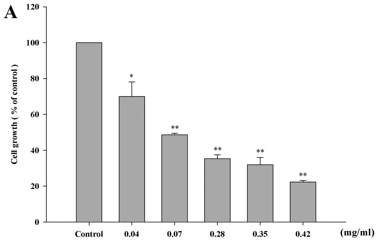
H3 inhibits PANC1 cell growth
H3 is the ethanol extract of a mixture of Meliae
fructus, Cinnamon bark and Sparganium rhizome
(Table I). To examine the cytotoxic
effects of this herbal mixture extracts (H3) in PANC1 cells, we
used the trypan blue exclusion assay. Both H3 and H3-gemcitabine
inhibited the growth rate in a dose-dependent manner (Fig. 1). We hypothesized that co-treatment
would allow the use of lower concentrations of anticancer drug
leading to less harmful side-effects. Thus, the concentration of
gemcitabine was varied after fixing the concentration of H3. The
half-maximal inhibitions (IC50) of proliferation were
0.07 mg/ml H3, 10 nM gemcitabine or 0.05 mg/ml H3 with 3 nM
gemcitabine (co-treatment). The IC50 value of
gemcitabine was obtained by Park et al (18). These data indicate that H3 inhibits
PANC1 cell growth at the relatively low concentration of 0.07
mg/ml.
 | Table IThe composition of H3. |
Table I
The composition of H3.
| Oriental name | Country of
origin | Grams of dried
materials | % |
|---|
| Meliae fructus | China | 3,200 | 40 |
| Cinnamon bark | Vietnam | 3,200 | 40 |
| Sparganium
rhizome | China | 1,600 | 20 |
| Total amount | | 8,000 | 100 |
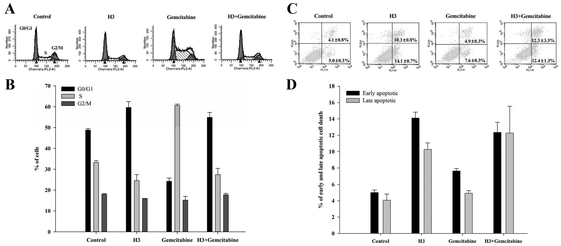
H3 affects cell cycle arrest and
apoptotic cell death
After H3 treatment, the PANC1 cells were fixed in
99% cold ethanol for cell cycle progression. Treatment with H3 led
PANC1 cells to cell cycle arrest at the G0/G1 phase (59.6±3.9%)
with a concomitant reduction in the percentage of cells in the S
and G2/M phase, compared with the values for the control
(48.8±1.1%; Fig. 2A and B). When
the cells were treated with H3 and gemcitabine (co-treatment), the
increase in the percentage of cells in the G0/G1 phase (54.9±3.4%)
was slightly lower than with H3 treatment. In contrast, gemcitabine
induced S phase arrest in 60.6±0.9% of cells, which is much higher
than the percentage seen in the control population (33.2±1.2%;
Fig. 2B).
We examined apoptotic cell death in cells treated
with H3 and/or gemcitabine. Cell death was evaluated by
double-staining with Annexin V-FITC and PI. The percentage of early
apoptotic cell death was 14.1±0.7% with H3 treatment, compared to
5.0±0.3% in the control group (Fig. 2C
and D). The percentage of early apoptotic cell death was
12.4±1.2% with co-treatment, which is slightly lower than with H3.
The percentage of late apoptotic cell death by co-treatment was
12.3±3.3%, which was slightly higher than with H3 only (10.3±0.8%)
or in the control (4.1±0.8%). However, with gemcitabine treatment,
the percentage of early and late apoptotic cell death was 7.6±0.3
and 4.9±0.3%, respectively, which was lower than with H3 only and
co-treatment.
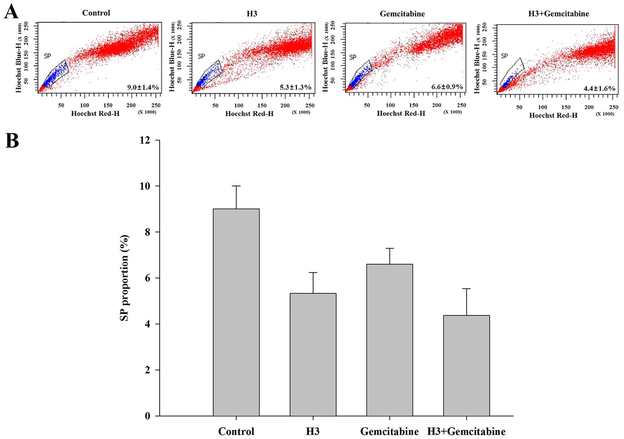
H3 suppresses SP cells
To examine the inhibitory effect of H3 and/or
gemcitabine in cancer stem cells, we performed an SP assay. The ABC
transporter inhibitor verapamil was administered in order to
confirm SP cells (19). The SP gate
was determined by the disappearance of the cell population in the
presence of verapamil. The difference in the percentage of SP cells
between the control (9.0±1.4%) and H3 (5.3±1.3%) treatment groups
was ~3.7%. With co-treatment (4.4±1.6%), the percentage of SP cells
decreased even further. However, with gemcitabine treatment
(6.6±0.9%), the percentage of SP cells was greater than with H3
only and co-treatment, indicating that gemcitabine is less
effective than the other treatments (Fig. 3). Thus, among the 3 treatments,
co-treatment with H3-gemcitabine is most effective at inhibiting SP
cells.
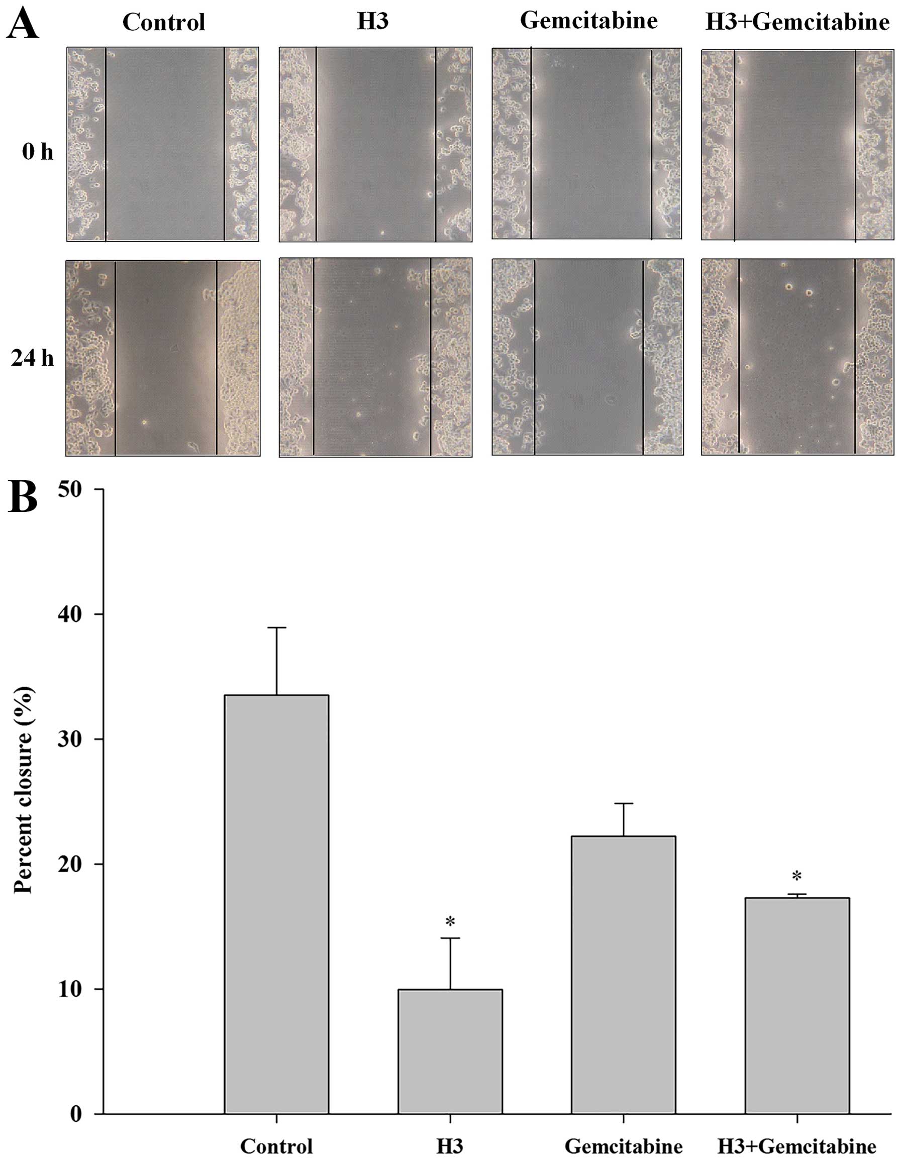
H3 inhibits migration ability
After treatment with H3 and/or gemcitabine, the
migration activity was measured using a wound healing assay. The
percent of closure was much lower in the H3-treated cells than in
the control (9.9 vs. 33.5%, respectively; Fig. 4). The percent of closure followed
the order: control (33.5%) > gemcitabine only (22.2%) >
co-treatment (17.3%) > H3 only (9.9%), indicating that H3
treatment is most effective at inhibiting the migration activity of
PANC1 cells.
H3 decreases the mRNA expression of
apoptosis-associated genes
As H3 induced apoptotic cell death, we investigated
the effect of H3 on the mRNA expression of apoptosis-associated
genes. After treatment with H3 and/or gemcitabine, the mRNA levels
of apoptosis-related genes was analyzed by RT-PCR. As shown in
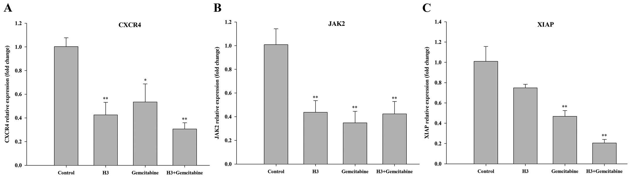
Fig. 5A, treatment with H3 and
co-treatment with H3-gemcitabine significantly decreased CXCR4 mRNA
levels by ~57 and 69%, respectively. JAK2 mRNA levels were reduced
by ~56 and 58% with H3 and co-treatment treatment, respectively
(Fig. 5B). With co-treatment XIAP
mRNA levels were markedly inhibited by ~80%, but the effects were
markedly less with H3 treatment only (Fig. 5C).
H3 decreases the mRNA levels of stem
cell-related genes
As H3 suppressed the percentage of SP cells, we
investigated the effect of H3 on the mRNA accumulation of stem
cell-related genes. Following H3 and/or gemcitabine treatment, the
mRNA levels of stem cell-associated genes were analyzed by RT-PCR.
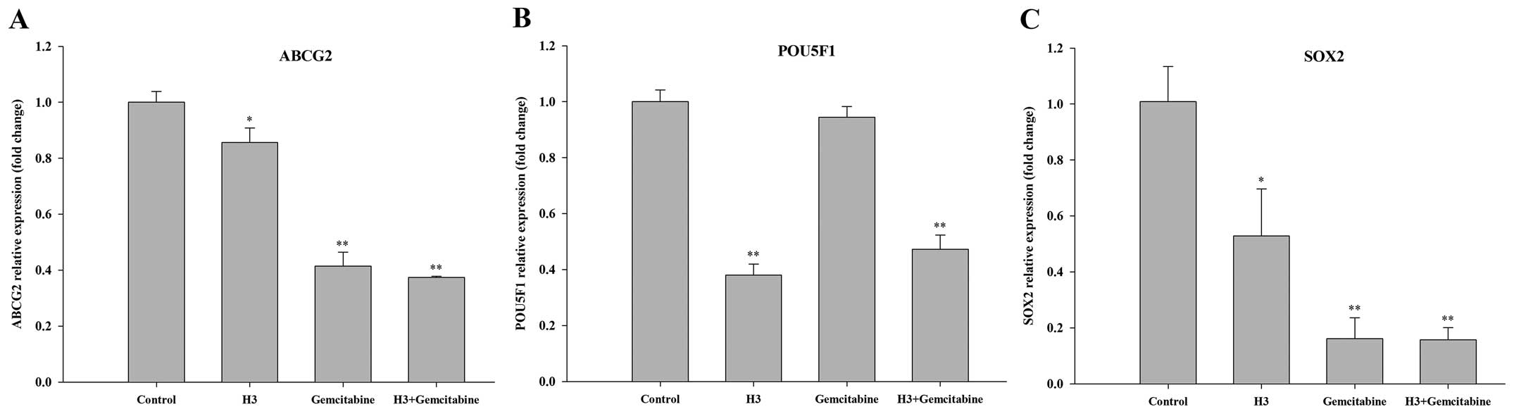
As shown in Fig. 6A, gemcitabine
treatment and co-treatment strongly decreased the levels of ABCG2
mRNA by ~56 and 58%, respectively. The level of ABCG2 mRNA with H3
was less significant (15%) than with gemcitabine treatment and
co-treatment. The extent of suppression was statistically lower
than that of the control. H3 treatment and co-treatment decreased
POU5F1 mRNA levels by ~62 and 53%, respectively (Fig. 6B). All the 3 treatments strongly
suppressed SOX2 mRNA levels, but the suppression was more
significant with gemcitabine treatment and co-treatment (~85% each)
than with H3 treatment (~48%; Fig.
6C).
H3 suppresses PANC1-induced tumor growth
in vivo
The tumor volumes of control, H3, gemcitabine and
co-treated mice at day 31 were ~160.5, 113.1, 109.6 and 267.4
mm3, respectively (Fig. 1A
and B). The tumor growth was less in H3 only- and
gemcitabine-treated mice than in the control group, and there was
not much difference between the H3 group or gemcitabine group.
However, the tumor growth was much greater in the co-treated mice
than in the other groups (Fig. 7A, B
and D). No significant differences in body weight were observed
between the groups (Fig. 7C). To
quantify the treatment effects in the tumor xenograft experiments,
the T/C ratio was used as an antitumor activity rating. It uses an
arbitrary cutoff point and typically has no formal statistical
inference (20). The tumor
inhibition rates of each treatment group were as follows: control
(T/C ratio=1.00), H3 (T/C ratio=0.54), gemcitabine (T/C
ratio=0.64), co-treatment (T/C ratio=2.00). Consequently, the tumor
inhibition rate of the H3 treatment was greater than all the other
groups (Fig. 7B).
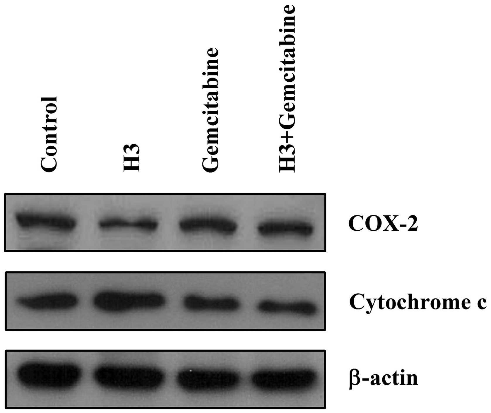
The antitumor effects of H3 occur through
COX-2 and cytochrome c-mediated apoptotic cell death
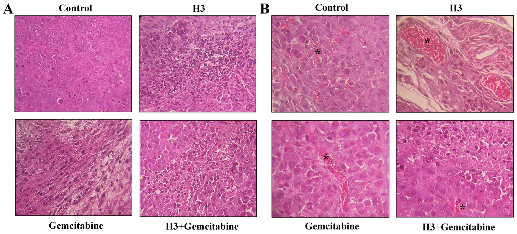
Induction of necrosis and erythrocytes in the tumor
tissue was examined using H&E staining. Significant necrotic
cell death was detected in H3-treated tumor tissue compared to that
in the control (Fig. 8A).
Erythrocyte-containing cavities lined by tumor cells were also
observed (Fig. 8B). These phenomena
were also seen in co-treated and gemcitabine-treated tumor tissues
but to a lesser degree. These data indicate that H3 treatment leads
to significant necrotic cell death and erythrocyte-containing
cavities in tumor tissue. Erythrocytes may enter apoptosis-like
suicidal death or eryptosis, which is characterized by cell
shrinkage and phosphatidylserine scrambling of the cell membrane.
Eryptosis may also be triggered by an increase in cytosolic
Ca2+, and the changes in Ca2+ mobilization
could be linked to altered levels of COX-2 (21). Thus, the levels of
apoptosis-associated factor (cytochrome c) and
anti-apoptosis-associated factor (COX-2) were investigated.
Proteins extracted from tumor tissue samples were analyzed by
western blotting. In H3-treated samples, the expression of COX-2
was significantly reduced and the expression of cytochrome c
was strongly increased compared to that in the other treatments
(Fig. 9).
Discussion
The use of current anticancer drugs can lead to many
harmful side-effects such as resistance, metastasis and even death
of normal cells (i.e., apoptosis). However, herbal mixture extracts
could be a complementary medicine for anticancer drugs (15). Whereas, Western science and medicine
focuses mainly on targeting specific malignant molecular
mechanisms, complementary medicine using herbal mixture extracts
employs a holistic approach that treats the entire human body
(22). There is some dispute
regarding the use of herbal mixture extracts since complex mixtures
of medicinal herbs are employed in contrast to isolated single
natural products. It is often suggested that herbal mixture
extracts work synergistically to increase the therapeutic effect,
while reducing the amount of adverse side-effects to healthy
tissues (22). However, scientific
data regarding the efficacy and safety of these complex herbal
mixture extracts are often insufficient, so further research into
this complementary medicine is needed (7,12,13).
The present study has shown that the herbal mixture extract H3 is a
candidate for novel cancer therapies, and performs better in the
selected tests than gemcitabine. There are some reports on each of
the individual medicinal herbs, but the present study is the first
to report the effect of this herbal mixture extract (23–25).
We investigated the effect of H3 as an alternative
to gemcitabine. We also expected that combinatorial treatment (i.e.
co-treatment) with H3-gemcitabine could have an enhanced effect
against PANC1 cells. Thus, our experiments were performed in the
presence of H3 only, gemcitabine only, or H3 and gemcitabine
(co-treatment). We found that H3 suppressed cell growth by inducing
G0/G1 cell cycle arrest (Figs. 1
and 2), unlike gemcitabine that is
an anticancer drug which delays DNA synthesis through inhibition of
the S phase of the cell cycle (8).
We also observed that H3 induced greater early and late apoptotic
cell death than gemcitabine, and that co-treatment resulted in a
similar extent of early and late apoptotic cell death as with H3
treatment (Fig. 2C and D). It is
known that most chemotherapeutic drugs kill cancer cells by
inducing a programmed form of cell death (i.e. apoptosis) (26). Therefore, we hypothesized that H3
may be a possible therapeutic agent for pancreatic cancer or an
adjuvant to gemcitabine treatment.
A subset of cancer cells called CSCs are oncogenic,
and these cells are more recuperative than normal tumor cells.
Thus, in their presence tumors relapse more easily after anticancer
drug treatment. CSCs could be one of the best targets for cancer
therapy (28). CSCs as therapeutic
targets have been studied in various types of cancer such as
breast, liver and prostate cancer (29–31).
To investigate the effect of H3 on the characteristics of
pancreatic CSCs, an SP analysis was conducted (Fig. 3). Most importantly, the average
percentage of SP cells was slightly lower with H3 treatment and
co-treatment than with gemcitabine treatment, indicating that the
presence of H3 helps suppress SP cells. Additionally, the migratory
ability of PANC1 cells was suppressed more greatly with H3
treatment and co-treatment than with gemcitabine only. This also
indicates that the presence of H3 is critical for suppression of SP
cells.
Since H3 induces apoptosis of PANC1 cells, we
examined the mRNA levels of several apoptosis-related genes in the
3 different cell treatment groups (Fig.
5). The accumulation of CXCR4 mRNA, a gene related to
metastasis and anti-apoptosis, and JAK2, another anti-apoptotic
gene, was significantly decreased with all 3 treatments, suggesting
that they each induce apoptosis efficiently. The mRNA levels of the
anti-apoptotic protein XIAP decreased markedly by co-treatment with
H3-gemcitabine, while levels decreased to a lesser degree with H3
or gemcitabine (data not shown). Due to their partial apoptotic
resistance, it is known that pancreatic cancer cells often respond
poorly to chemotherapy and/or radiotherapy (27). These results indicate that H3 may be
involved in the suppression of anti-apoptotic signaling, which
could help overcome this resistance. Further studies are required
to unveil the details of the relevant mechanistic pathway used by
H3 for this purpose.
Since H3 suppresses SP cells, the mRNA accumulation
of several stem cell-related genes was investigated (Fig. 6). The expression of ABCG2, which
plays a role in multi-drug resistance, significantly decreased with
co-treatment and gemcitabine treatment. The expression of POU5F1,
which is related to the self-renewal of undifferentiated embryonic
stem cells, was significantly reduced by H3 treatment and
co-treatment. The expression of SOX2, which is a transcription
factor that is essential for maintaining self-renewal or
pluripotency of undifferentiated embryonic stem cells, was
significantly decreased by co-treatment and gemcitabine treatment.
Although there is no specific trend for the suppression of the 3
stem cell-related genes by the 3 different treatments, most of stem
cell-related genes were suppressed to some extent by H3 as well as
gemcitabine. This suggest that H3 is as potent as gemcitabine at
suppressing pancreatic CSCs.
The in vivo study revealed that H3 and
gemcitabine effectively suppress tumor growth (Fig. 7). However, contrary to the in
vitro results, tumor growth in vivo was enhanced with
co-treatment with H3-gemcitabine. This suggests that co-treatment
with H3 may not be recommended for pancreatic cancer. Significant
increases in necrotic cell death and erythrocyte-containing
cavities were observed in H3-treated tumor tissue compared to the
findings in other treatments (Fig.
8). The effectiveness of H3 as an anticancer agent for
pancreatic cancer was also evidenced by western blot analysis which
revealed that the expression of apoptosis-associated factor
(cytochrome c) was upregulated and the expression of
anti-apoptosis-associated factor (COX-2) was downregulated in the
tumors of mice having received this treatment (Fig. 9).
In conclusion, our experimental results show the
remarkable effect of H3 in pancreatic cancer cells, cancer stem
cells, and animal models. H3 is another example of an herbal
mixture extracts which requires further studies to be developed
into an anticancer therapeutic agent. Further study may be needed
for detailed anticancer signaling pathway and, more importantly,
application of H3 to other types of cancers.
Acknowledgments
The present study was financially supported by a
Grant (B110053) from the Korean Health Technology R&D Project,
Ministry of Health & Welfare, Republic of Korea.
References
|
1
|
Hidalgo M: Pancreatic cancer. N Engl J
Med. 362:1605–1617. 2010. View Article : Google Scholar : PubMed/NCBI
|
|
2
|
Li D, Xie K, Wolff R and Abbruzzese JL:
Pancreatic cancer. Lancet. 363:1049–1057. 2004. View Article : Google Scholar : PubMed/NCBI
|
|
3
|
Hong SK, Yang SY, Yin SH and Yang KX:
RC-3095, a gastrin-releasing peptide receptor antagonist,
synergizes with gemcitabine to inhibit the growth of human
pancreatic cancer CFPAC-1 in vitro and in vivo. Pancreas. 43:15–21.
2014. View Article : Google Scholar
|
|
4
|
Karakhanova S, Mosl B, Harig S, von Ahn K,
Fritz J, Schmidt J, Jäger D, Werner J and Bazhin AV: Influence of
interferon-alpha combined with chemo (radio) therapy on
immunological parameters in pancreatic adenocarcinoma. Int J Mol
Sci. 15:4104–4125. 2014. View Article : Google Scholar : PubMed/NCBI
|
|
5
|
Qu C and Chen Z: Antitumor effect of water
decoctions of Taxus cuspidate on pancreatic cancer. Evid Based
Complement Alternat Med. 2014:2916752014. View Article : Google Scholar : PubMed/NCBI
|
|
6
|
Laquente B, Lacasa C, Ginestà MM,
Casanovas O, Figueras A, Galán M, Ribas IG, Germà JR, Capellà G and
Viñals F: Antiangiogenic effect of gemcitabine following metronomic
administration in a pancreas cancer model. Mol Cancer Ther.
7:638–647. 2008. View Article : Google Scholar : PubMed/NCBI
|
|
7
|
O'Reilly EM: Adjuvant therapy for pancreas
adenocarcinoma: Where are we going? Expert Rev Anticancer Ther.
11:173–177. 2011. View Article : Google Scholar : PubMed/NCBI
|
|
8
|
Burris HA III, Moore MJ, Andersen J, Green
MR, Rothenberg ML, Modiano MR, Cripps MC, Portenoy RK, Storniolo
AM, Tarassoff P, et al: Improvements in survival and clinical
benefit with gemcitabine as first-line therapy for patients with
advanced pancreas cancer: A randomized trial. J Clin Oncol.
15:2403–2413. 1997.PubMed/NCBI
|
|
9
|
Moore MJ, Goldstein D, Hamm J, Figer A,
Hecht JR, Gallinger S, Au HJ, Murawa P, Walde D, Wolff RA, et al
National Cancer Institute of Canada Clinical Trials Group:
Erlotinib plus gemcitabine compared with gemcitabine alone in
patients with advanced pancreatic cancer: A phase III trial of the
National Cancer Institute of Canada Clinical Trials Group. J Clin
Oncol. 25:1960–1966. 2007. View Article : Google Scholar : PubMed/NCBI
|
|
10
|
Isayama H, Nakai Y, Yamamoto K, Sasaki T,
Mizuno S, Yagioka H, Yashima Y, Kawakubo K, Kogure H, Arizumi T, et
al: Gemcitabine and oxaliplatin combination chemotherapy for
patients with refractory pancreatic cancer. Oncology. 80:97–101.
2011. View Article : Google Scholar : PubMed/NCBI
|
|
11
|
Khalil MA, Qiao W, Carlson P, George B,
Javle M, Overman M, Varadhachary G, Wolff RA, Abbruzzese JL and
Fogelman DR: The addition of erlotinib to gemcitabine and cisplatin
does not appear to improve median survival in metastatic pancreatic
cancer. Invest New Drugs. 31:1375–1383. 2013. View Article : Google Scholar : PubMed/NCBI
|
|
12
|
Wang S, Wu X, Tan M, Gong J, Tan W, Bian
B, Chen M and Wang Y: Fighting fire with fire: Poisonous Chinese
herbal medicine for cancer therapy. J Ethnopharmacol. 140:33–45.
2012. View Article : Google Scholar : PubMed/NCBI
|
|
13
|
Choi YK, Cho SG, Woo SM, Yun YJ, Park S,
Shin YC and Ko SG: Herbal extract SH003 suppresses tumor growth and
metastasis of MDA-MB-231 breast cancer cells by inhibiting
STAT3-IL-6 signaling. Mediators Inflamm. 2014:4921732014.
View Article : Google Scholar : PubMed/NCBI
|
|
14
|
Sikdar S, Mukherjee A, Ghosh S and
Khuda-Bukhsh AR: Condurango glycoside-rich components stimulate DNA
damage-induced cell cycle arrest and ROS-mediated caspase-3
dependent apoptosis through inhibition of cell-proliferation in
lung cancer, in vitro and in vivo. Environ Toxicol Pharmacol.
37:300–314. 2014. View Article : Google Scholar : PubMed/NCBI
|
|
15
|
Yu J and Chen Q: Antitumor activities of
Rauwolfia vomitoria extract and potentiation of gemcitabine effects
against pancreatic cancer. Integr Cancer Ther. 13:217–225. 2014.
View Article : Google Scholar : PubMed/NCBI
|
|
16
|
Kim MP, Evans DB, Wang H, Abbruzzese JL,
Fleming JB and Gallick GE: Generation of orthotopic and heterotopic
human pancreatic cancer xenografts in immunodeficient mice. Nat
Protoc. 4:1670–1680. 2009. View Article : Google Scholar : PubMed/NCBI
|
|
17
|
Jung HJ, Pak PJ, Park SH, Ju JE, Kim JS,
Lee HS and Chung N: Silver wire amplifies the signaling mechanism
for IL-1beta production more than silver submicroparticles in human
monocytic THP-1 cells. PLoS One. 9:e1122562014. View Article : Google Scholar : PubMed/NCBI
|
|
18
|
Park SH, Sung JH, Kim EJ and Chung N:
Berberine induces apoptosis via ROS generation in PANC-1 and
MIA-PaCa2 pancreatic cell lines. Braz J Med Biol Res. 48:111–119.
2015. View Article : Google Scholar :
|
|
19
|
Goodell MA, Brose K, Paradis G, Conner AS
and Mulligan RC: Isolation and functional properties of murine
hematopoietic stem cells that are replicating in vivo. J Exp Med.
183:1797–1806. 1996. View Article : Google Scholar : PubMed/NCBI
|
|
20
|
Wu J: Statistical inference for tumor
growth inhibition T/C ratio. J Biopharm Stat. 20:954–964. 2010.
View Article : Google Scholar : PubMed/NCBI
|
|
21
|
Lang F, Gulbins E, Lerche H, Huber SM,
Kempe DS and Foller M: Eryptosis, a window to systemic disease.
Cell Physiol Biochem. 22:373–380. 2008. View Article : Google Scholar : PubMed/NCBI
|
|
22
|
Efferth T, Li PC, Konkimalla VS and Kaina
B: From traditional Chinese medicine to rational cancer therapy.
Trends Mol Med. 13:353–361. 2007. View Article : Google Scholar : PubMed/NCBI
|
|
23
|
Koppikar SJ, Choudhari AS, Suryavanshi SA,
Kumari S, Chattopadhyay S and Kaul-Ghanekar R: Aqueous cinnamon
extract (ACE-c) from the bark of Cinnamomum cassia causes apoptosis
in human cervical cancer cell line (SiHa) through loss of
mitochondrial membrane potential. BMC Cancer. 10:2102010.
View Article : Google Scholar : PubMed/NCBI
|
|
24
|
Zhang JW and Wei YH: Anti-cancer effects
of Grailsine-Al-glycoside isolated from Rhizoma Sparganii. BMC
Complement Altern Med. 14:822014. View Article : Google Scholar : PubMed/NCBI
|
|
25
|
Xie F, Zhang M, Zhang CF, Wang ZT, Yu BY
and Kou JP: Anti-inflammatory and analgesic activities of ethanolic
extract and two limonoids from Melia toosendan fruit. J
Ethnopharmacol. 117:463–466. 2008. View Article : Google Scholar : PubMed/NCBI
|
|
26
|
Makin G and Hickman JA: Apoptosis and
cancer chemotherapy. Cell Tissue Res. 301:143–152. 2000. View Article : Google Scholar : PubMed/NCBI
|
|
27
|
Li L and Leung PS: Use of herbal medicines
and natural products: An alternative approach to overcoming the
apoptotic resistance of pancreatic cancer. Int J Biochem Cell Biol.
53:224–236. 2014. View Article : Google Scholar : PubMed/NCBI
|
|
28
|
Abel EV and Simeone DM: Biology and
clinical applications of pancreatic cancer stem cells.
Gastroenterology. 144:1241–1248. 2013. View Article : Google Scholar : PubMed/NCBI
|
|
29
|
Kumar D, Shankar S and Srivastava RK:
Rottlerin induces autophagy and apoptosis in prostate cancer stem
cells via PI3K/Akt/mTOR signaling pathway. Cancer Lett.
343:179–189. 2014. View Article : Google Scholar
|
|
30
|
Suntharalingam K, Lin W, Johnstone TC,
Bruno PM, Zheng YR, Hemann MT and Lippard SJ: A breast cancer stem
cell-selective, mammospheres-potent osmium(VI) nitrido complex. J
Am Chem Soc. 136:14413–14416. 2014. View Article : Google Scholar : PubMed/NCBI
|
|
31
|
Hashimoto N, Tsunedomi R, Yoshimura K,
Watanabe Y, Hazama S and Oka M: Cancer stem-like sphere cells
induced from de-differentiated hepatocellular carcinoma-derived
cell lines possess the resistance to anti-cancer drugs. BMC Cancer.
14:7222014. View Article : Google Scholar : PubMed/NCBI
|