Introduction
Gene therapy has a broad application prospect in the
treatment of cancer and genetic diseases, but its application is
limited by the potential danger of virus vectors or the low
transfection efficiency of non-virus vectors (1,2). To
enhance the transgenic expression of the non-viral gene transfer
system, the transgenic technology of ultrasound-mediated gene
transfection may be applied (3–6).
Microbubbles, which act as contrast agents for medical ultrasound
imaging, could improve the transfection efficiency (7–11).
Furthermore, microbubbles excited by ultrasound exposure may
temporarily ‘open’ the cell membranes of nearby cells allowing
plasmid DNA to be delivered into cells. Subsequently, the cells
could reseal themselves and maintain their vitality (12).
While liposomes have a very low immunogenicity,
liposome microbubbles (LMs) are a research hotspot that has
developed rapidly in recent years, which is capable of targeting
increasing specific drug treatment or gene transfection (7,13–17).
However, a number of factors could affect the transfection
efficiency, including the ultrasonic intensity, the duration of
ultrasound treatment and the microbubble concentration. Although
the application of sonoporation has been optimized in certain
studies, the delivery of drugs is often difficult to control and
has been observed in a number of cases to have numerous unwanted
side effects. Proteins with high wavelength emission spectra,
including red fluorescent protein (RFP), may be a better choice as
a reporter gene (18). However,
few cases exist in the literature regarding in vitro and
in vivo optimization using an RFP reporter gene. The effects
of LM on transgenic expression have not been systematically
assessed, and the applications of ultrasound-mediated liposome
microbubble destruction (UMLMD) have not yet been optimized.
However, we suggest this technique effectively transfers genes. In
previous experiments (19,20), a significant problem arose from the
application of the non-viral gene transfer system of UMLMD.
However, this UMLMD technique has not been studied thus far as a
delivery system.
For this reason, we established a method that is
cost-effective and safer than viral carrier gene therapy. The
present study utilized the method of UMLMD to examine the
transfection efficiency and safety of RFP in vivo and in
vitro, while assessing the safety and efficacy of UMLMD. Our
purpose was to obtain optimal transgenic expression with the lowest
cell or tissue injury, in order to lay the foundation for
efficient, targeted and non-invasive gene therapy.
Materials and methods
Cell culture
HeLa cells were obtained from the China Center for
Type Culture Collection (CCTCC) and incubated in Dulbecco’s
modified Eagle’s medium (DMEM, Gibco, Grand Island, NY, USA) with
10% fetal bovine serum (FBS, Gibco), 100 U/ml penicillin and 100
μg/ml streptomycin at 37°C in a humidified environment of 5%
CO2/95% air. The total cell count was determined using a
hemocytometer (Burker Turk, The Netherlands). The initial cell
viability was determined by exclusion with trypan blue dye
(Sigma-Aldrich, St. Louis, MO, USA). Exponentially growing cells
were used in all experiments.
Plasmid DNA
The expression vector for the RFP gene plasmid
(pDsRed-Express, Clontech, BD Biosciences, Billerica, MA, USA) had
an excitation and emission maxima occurring at 557 nm and 579 nm,
respectively. The plasmid DNA was obtained from DH5α Escherichia
coli (Invitrogen, Carlsbad, CA, USA) and was prepared using a
Qiaquick kit (Qiagen, Crawley, UK) according to the manufacturer’s
instructions. The absorbance ratio at the wavelength of 260–280 nm
for the plasmid DNA solution was measured to be between 1.8 and
2.0.
Preparation of LMs
LMs were kindly provided by the Department of
Ultrasonography of Xinqiao Hospital, The Third Military Medical
University, Chongqing, China. The LMs were prepared using
mechanical agitation. The activation device was refitted from an
amalgamator (ST-D, Beijing AT&M Biomaterials Co., Ltd, Beijing,
China) at a calibration rate of (4,500±100) oscillations/min for 45
sec, using a reciprocating motion and an oscillation amplitude of
(15±1) mm.
In vitro studies
Experimental apparatus
In vitro experiments were performed in an
exposure water tank. The tank contained deionized water with an
ultrasound transducer (Accusonic, Metron Medical Australia Pty.,
Ltd., Carrum Downs, Australia) fixed at the bottom. The culture
plate was placed on the center of the transducer. The culture plate
was covered with the lid to prevent it from being polluted by the
surrounding environment. The experiments were performed on a
sterile decontamination bench. To prevent the nearby wells from
being affected by ultrasound irradiation, cells were planted in
only six of 24-well culture plates. During ultrasound irradiation,
the plate was agitated slowly, while remaining close to the
transducer surface.
Experimental grouping and
processes
HeLa cells were conventionally harvested by
trypsinization and resuspended in a concentration of
5.0×10⁶ cells/ml in DMEM (300 μl, non-FBS). Firstly,
cells were irradiated using various ultrasound parameters and
microbubble concentrations. Prior to ultrasound irradiation, LMs
were mixed with 0.9% saline at various concentrations (v/v; 3, 6
and 10%). LM suspension (150 μl) and plasmid solution (final
concentration 1.0 μg/μl) were fully mixed to prepare the LM/plasmid
complexes. Ultrasound parameters were set as: ultrasonic frequency
of 1 MHz, pulse repetition frequency of 100 Hz, 20% duty cycle, and
ultrasound intensities of 0.8, 1.0 and 1.2 W/cm2 with
3-min exposure.
To investigate the effect of LMs on the transfection
and ultrasound exposure time, experiments were divided into the
following groups: plasmid DNA group (n=3); plasmid + ultrasound
group, 3 wells irradiated for 1 min and 3 wells irradiated for 3
min; LM + plasmid group (n=3); plasmid + LM + ultrasound group (P+
UMLMD), 3 wells irradiated for 1 min and 3 wells irradiated for 3
min. Plasmid solution (150 μl) (10 μg/well) or LM suspension (6%)
or LM/plasmid complexes (as described above, LM concentration of
6%) were added to the culture plate. The ultrasound intensity was
fixed at 1.0 W/cm2. When all the processes were
completed, the culture plates were removed from the tank, wiped dry
and returned to the incubator. A total of 8 h following ultrasound
irradiation, the medium was replaced by one containing 10% FBS for
continuous cultivation.
Cell viability and transgenic
expression
Forty-eight hours after ultrasound irradiation, the
RFP expression was observed using a fluorescence contrast phase
microscope (IX71, Olympus, Japan). The cells were then harvested to
assess the cell viability and the transgenic expression using
different samples of the same cell suspension.
Propidium iodide (20 μl) (PI, 40 μg/ml,
Sigma-Aldrich) was added into the cell suspension. Cells with PI
were considered to be dead and cells without PI were considered to
be alive. Following a 15 min incubation at room temperature, the
cells were stored at 4°C. Using flow cytometry (FACSCalibur, Becton
Dickinson, Franklin Lakes, NJ, USA), the ratios of cells stained
with or without PI were determined.
Approximately 1×10⁵ cells were obtained
from each sample for transgenic expression analysis, using a 550 nm
wavelength excitation light and a 585±42 nm wavelength emission
light to detect the red fluorescence. The transgenic expression was
assessed as the number of cells expressing RFP per total number of
survival cells. Data were analyzed using WinMDI software (version
2.8).
In vivo studies
Animal protocol
Animal handling and experimental procedures were
approved by the Medical College Animal Experiments Committee.
Four-to-six weeks old female Balb/c nu/nu mice, weighing 17–22 g,
were maintained in specified pathogen-free (SPF) conditions
throughout the experimental period.
Prior to animal modeling, HeLa cells were harvested,
collected and centrifuged, and then resuspended in 100 μl DMEM to
prepare a single cell suspension. The mice were fixed on a
superclean bench according to the principle of aseptic surgery, and
inoculated subcutaneously into the flank with 2×10⁶
cells per mouse following local sterilization. The mice were raised
under the SPF condition following surgery, and were observed once
every two days. Two weeks later, the experiments were initiated
when the tumors reached a size of 5–10 mm.
Experimental grouping
A total of 20 mice were randomly divided into 4
experimental groups with 5 mice in each group: Plasmid group,
injection of plasmid DNA (50 μg/200 μl) alone; plasmid + ultrasound
exposure group; LM + plasmid group, complexes of LM (30 μl, 6%) and
plasmid (50 μg) were gently agitated with PBS to a final volume of
200 μl and directly injected; plasmid + LM + ultrasound group (P +
UMLMD), complexes of plasmid DNA/LM were injected and followed by
local ultrasound irradiation.
The plasmid DNA or LM/plasmid complexes were
administered into the tail vein of the mice. The mice were
anesthetized by diethylether and fixed on the flats. The tumors
were subsequently sonicated using a transducer placed on the skin
with a contact gel (Aquasonic 100, Parker Laboratories Inc., WI,
USA). Ultrasound parameters were set at 3 MHz, 2 W/cm2,
2 min and 20% duty cycle. During the exposure, the ultrasound
transducer was moved in a circular motion to ensure the whole tumor
was exposed.
Analysis of transgenic expression in
vivo
Three days following ultrasound treatment, the mice
were sacrificed by cervical dislocation. The tumor specimens,
surrounding tissues, the skin around the tumors, the hearts, livers
and muscles, were immediately removed, embedded in optimal cutting
temperature compound (OCT, Tissue-Tek, Sakura Finetek, Torrance,
CA, USA), and stored at −80°C until further analyses. Cross
sections (10 μm) were cut with a cryostat (CM1900, Leica, Germany)
and affixed to glass slides. Fluorescence expression and
distribution patterns were observed with confocal laser microscopy
(Fluoview FV500, Olympus, Japan). The quantitative detection of
transfection was performed using flow cytometry.
Histology
The specimens were fixed using formaldehyde,
dehydrated with a graded alcohol series, and embedded in paraffin.
Hematoxylin and eosin (H&E) staining was performed on the
specimens for histopathological evaluation of hemorrhage, necrosis
and inflammation.
Statistical analysis
Statistical analyses were performed by the SPSS 13.0
software package (SPSS, Inc., Chicago, IL, USA). All values were
expressed as the mean ± SD. Analysis of variance with paired t-test
and factorial design analysis of variance (ANOVA) test were used to
determine the significance of the difference in a multiple
comparison. If the ANOVA was significant, the Tukey’s procedure was
used as a post hoc test. P<0.05 was considered to indicate a
statistically significant difference.
Results
Effects of UMLMD on the transgenic
expression in vitro
As shown in Table
I, when the LM concentration was 3 and 10%, there was no
significant difference in the transgenic expression among the
various ultrasound intensities (F=3.483, P=0.099; F=1.159,
P=0.375). When the LM concentration was 6%, RFP expression of 1.0
W/cm2 was higher than that of 0.8 W/cm2
(P=0.017); however, the difference was not significant as compared
with 1.2 W/cm2 (P=0.176). When the ultrasound intensity
was fixed, the differences of transgenic expression among the
various LM concentrations were significant (F=5.715, P=0.041;
F=10.238, P=0.012; F=28.631, P=0.001).
 | Table ITransgenic expression of different
ultrasound intensities and LM concentrations. |
Table I
Transgenic expression of different
ultrasound intensities and LM concentrations.
| Ultrasound
intensity (W/cm2) | LM concentration
(%) |
|---|
|
|---|
| 3 | 6 | 10 |
|---|
| 0.8 | 9.03±2.33 | 18.33±2.59a | 16.59±5.13 |
| 1.0 | 12.73±2.81 | 31.18±5.48b,d | 19.16±6.26 |
| 1.2 | 13.48±1.13 | 24.47±3.24b | 13.32±1.03b,c |
Cell injury of different ultrasound
intensities and LM concentrations
Ultrasound exposure alone did not damage cells and
the cell injury rate was <14%. When the ultrasound intensity was
0.8 or 1.0 W/cm2, the differences among the various LM
concentrations were not significant (F=1.072, P=0.414; F=0.376,
P=0.773). When the LM concentration was 3 or 6%, there was no
apparent cell damage in any experimental group. In addition, the
differences between the ultrasound intensities were not significant
(F=1.368, P=0.324; F=2.063, P=0.208). The injury ratio was the
highest with an ultrasound intensity of 1.2 W/cm2 and
10% LM (F=7.070, P=0.012; F=6.612, P=0.030; Table II).
| Table IICell injury of different ultrasound
intensities and LM concentrations. |
Table II
Cell injury of different ultrasound
intensities and LM concentrations.
| Ultrasound
intensity (W/cm2) | LM concentration
(%) |
|---|
|
|---|
| − | 3 | 6 | 10 |
|---|
| 0.8 | 10.57±1.37 | 12.06±1.99 | 12.82±1.95 | 12.64±1.44 |
| 1.0 | 12.59±1.84 | 13.50±1.93 | 13.99±1.23 | 14.71±4.08 |
| 1.2 | 13.95±1.86 | 14.60±1.72 | 15.85±2.21 | 20.31±1.63a,b,c |
The factorial design analysis of variance indicated
that the main effects analysis of 2 types of parameters were
statistically significant (both P<0.01), but there was no
existing interaction between them (P>0.05). RFP expression was
significantly higher in cells treated with an ultrasound intensity
of 1.0 W/cm2 and 6% LM, without causing any apparently
adverse effect. According to these results, the subsequent
experiments were performed under the optimal UMLMD conditions.
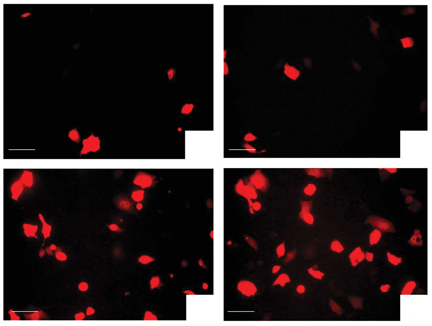
Augmentation of transgenic expression by
UMLMD in vitro
Table III shows
that without LM, the RFP expression of ultrasound irradiation alone
was not significantly higher than that of the plasmid group alone
(P=0.816). The transgenic expression exposure of 3 min was not
significantly higher than that of 1 min (paired t-test, t=−2.443,
P=0.135; Fig. 1A and B). LMs alone
were insufficient to transfect the gene into cells without
ultrasound irradiation. The differences had no statistical
significance as compared with the plasmid alone or the ultrasound
irradiation and plasmid group (P=0.095 and P=0.312).
| Table IIIEffects of LM and ultrasound
irradiation time on UMLMD. |
Table III
Effects of LM and ultrasound
irradiation time on UMLMD.
| Ultrasound
irradiation time (min) | LM (%) |
|---|
|
|---|
| − | + |
|---|
| − | 1.9±0.76 | 4.48±1.18 |
| 1 | 2.73±1.39 | 14.48±1.18a,c |
| 3 | 3.83±0.98 | 31.18±5.48a,b,c |
However, the cells that received an injection of
plasmid with LM followed by ultrasound irradiation (P + UMLMD)
demonstrated a significantly higher transgenic expression than any
other group (all P<0.01, Fig.
1C). Moreover, the transgenic expression for an exposure time
of 3 min was significantly higher than that of 1 min (paired
t-test, t=−5.714, P=0.029; Fig.
1D). These results demonstrated that LM had a markedly enhanced
effect on the transgenic expression of UMLMD.
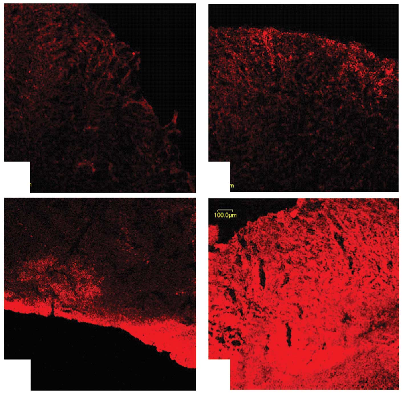
Targeted delivery and enhancement of
transgenic expression by UMLMD in vivo
Without ultrasound irradiation, limited RFP
expression in the plasmid injection alone group was detected
(1.83±1.21%), and the fluorescence signal was weak (Fig. 2A). The plasmid and LM injection
group also demonstrated a similar fluorescence expression rate
(2.33±1.39%, P=0.753; Fig. 2B).
The RFP expression rate of the plasmid injection and ultrasound
irradiation group was 3.48±0.18%; however, when compared with the
plasmid injection alone, the difference was not significant
(P=0.517). In addition, the majority of fluorescent protein
expression detected was distributed in the superficial area of the
tumors (Fig. 2C). However, when
the plasmid was injected with LM followed by ultrasound
irradiation, RFP expression increased significantly (23.96±2.13%),
with a stronger signal and a greater density (Fig. 2D). As compared with the other three
groups, the differences were significant (F=172.954, P<0.001,
all P<0.01).
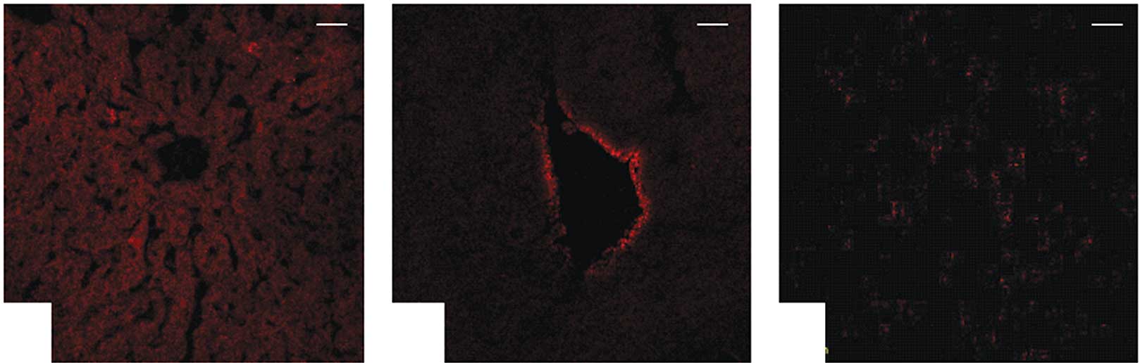
Tissue specificity by UMLMD in vivo
None of the nude mice died during the experiment.
Regardless of the ultrasound exposure, a weak RFP expression was
evident in certain livers (Fig.
3A), hearts (Fig. 3B) and
muscles (Fig. 3C), whereas other
organ tissues had no significant expression.
Histological observation
H&E staining revealed that tissue damage was not
observed in the tumors (Fig. 4A)
and other organs (Fig. 4B-D),
which remained intact. Moreover, the results demonstrated no
abnormalities, including inflammation or degeneration, in any
tissues.
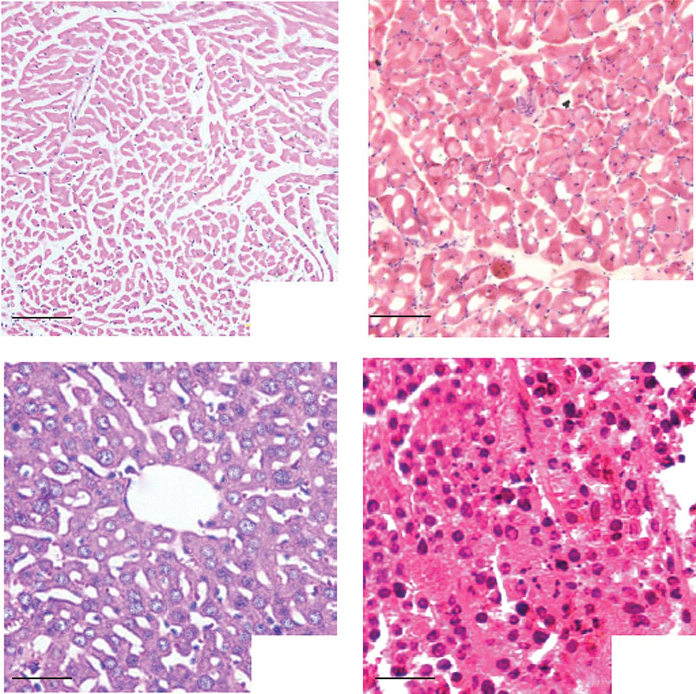 | Figure 4Histological observations; as
mentioned in Materials and methods in the in vivo
experimental grouping. (A) Heart, bar, 100 μm, (B) Muscle, bar, 100
μm, (C) Liver, bar, 50 μm, (D) Tumor, bar, 100 μm; H&E staining
demonstrated that tissue damage, inflammation and degeneration were
not observed in the representative sections. |
Discussion
For gene therapy, although the viral vector is an
effective transgenic method for gene transfection, its safety and
side-effects remain a concern for clinical application. In
non-viral transgenic technologies, UMLMD is a simple, non-invasive
method. It can focus on specific tissues or organs directly and it
is beneficial for the localization of the gene of interest. It is
safer than other methods, and may be used in clinical applications
in the near future. Cell permeability may be transiently changed by
sonoporation, in order that macromolecules, including the plasmid
DNA are able to enter the cells instantaneously (8,9,21,22).
Results of previous studies showed that (8–11,23)
the addition of microbubbles were capable of promoting transgenic
expression. However, if sonoporation was extremely strong, it would
lead to apparent cell death. The ideal concentration of
microbubbles should deliver the drug or gene into cells with the
greatest efficiency without lethal effect.
A significant problem from the application of the
non-viral gene transfer system of UMLMD occurred in our study. This
technology provided a new promising approach for gene delivery
in vitro and in vivo. However, this technique for the
delivery system of shRNA in vitro has yet to be optimized.
In the present study, parameters for the delivery system were
optimized. Fig. 1 shows that
transfection efficiency was affected by the transfection
parameters, including ultrasound intensity and exposure time. The
optimal conditions were capable of achieving the highest
transfection efficiency. Our results demonstrated that the RFP
transgenic expression was the highest when LMs were combined with
ultrasound exposure, while the cells were not markedly injured.
Moreover, the UMLMD was also an effective and tumor-specific
transgenic technology in vivo. Plasmid DNA was rapidly
transferred to transplanted tumors even under the conditions in the
bloodstream and serum, and transgenic expression depended on the
ultrasound irradiation site. In this study, the reporter gene was
RFP, whose provocation and emission wavelength were longer than
other genes such as GFP, and its emission peak was located beyond
the scope of the fluorescence background generated by the medium
and tissue culture equipment. RFP, which is expressed in mammalian
cells effectively, has a high signal-to-noise ratio, is not easy to
induce fluorescence quenching, and has a high conversion
efficiency. The far-red emissions may be preferred for certain
applications due to the lower background autofluorescence in
certain tissues (18).
Microbubbles are not only useful as contrast agents,
but are also widely used in therapeutic applications. Microbubbles
may amplify the cavitation effect of ultrasound, particularly in
targeting drug and gene delivery in vitro and in
vivo. However, the mean diameter of conventional microbubbles
is approximately 2.0–4.5 μm. Tsunoda et al (24) reported that several mice died
immediately following i.v. injection due to lethal embolisms in
certain organs. It is therefore necessary to develop novel
microbubbles and new techniques to improve the transfection
efficiency. LMs have been used in clinical ultrasound diagnosis and
treatment safely and stably, with low immunogenicity and
cytotoxicity. However, LM alone was insufficient to transfect the
gene into cells without ultrasound exposure. The transfection
efficiency was enhanced by the novel combination of LM and
ultrasound exposure via the mechanism of sonoporation. In this
study, LMs were composed of polyethyleneglicol (PEG) and liposome,
which independently have been used in clinical ultrasound diagnosis
and treatment safely and stably with a long intravascular half-life
(25). The mean diameter of LMs
was smaller than that of red blood cells (26), meaning that it was capable of
reaching peripheral tissues and passing through capillary vessels.
Therefore, the LMs were used repeatedly to further enhance and
maintain the gene expression. Moreover, under ultrasound
irradiation, LM significantly promoted transgenic expression.
However, when excessive LM was added, the transgenic expression
declined and caused cell damage (27). Similarly to another study (28), various ultrasound intensities and
irradiation times had a great impact on UMLMD. As the matching of
specific ultrasound parameters and transgenic technologies were
different, the optimal conditions of various cell types were also
different. The ultrasound response differences among various cell
lines also lead to different conclusions (29). Furthermore, LM could be developed
as a gene transfection tool and delivered into specific tissues or
organs. Significantly, the in vivo study concerning the
targeted delivery of LMs and the effects of ultrasound allowed for
further investigation into the transfection of transplantation
tumors in nude mice in our laboratory. For this type of gene
transfer system, modulating the biodistribution of the LMs and the
plasmid DNA in the systemic injection is crucial. Unlike the in
vitro study, ultrasound exposure of 3 MHz facilitated the
degree of tumor targeting, indicating the significance of
localizing ultrasonic energy in the tumor volume (30). Ultrasound irradiation had no
significant impact on other organs. Our results demonstrated that
significant transgenic expression and noninvasive, targeting gene
transfer were obtained by LM and plasmid DNA complex injections
with ultrasound irradiation.
However, there were certain limitations to our
study, and further studies are required to investigate the mode of
injection and the time point and dose-response correlation in
larger animal models. In addition, the optimal conditions in the
in vivo experiment, including ultrasound parameters,
concentration of LMs and plasmid DNA, are likely to be different
from the in vitro experiment. Furthermore, to enhance the
tissue specificity of the transgenic expression, several
modifications are required and should be studied. For example,
certain peptides or receptors on the surface of LMs may be
investigated and may specify the gene expression in certain
tissues.
In conclusion, in this study, UMLMD was found to
enhance the transgenic efficiency without causing any apparent
detrimental effect. If the appropriate target gene is added, this
transgenic technology is expected to become an effective strategy
for human gene therapy, and provide a non-invasive, safe and
promising technique for transgenic research.
Acknowledgements
This study was supported by the Medical Research
Foundation of Guangdong Province (No. A2010270), the Research
Projects of Guangzhou Education Bureau (No. 10A242), the Youth
Research Support Projects of the Third Affiliated Hospital of
Guangzhou Medical University (No. 2011Y02) and the Medical Research
Foundation of Liwan District (No. 20111213067).
References
|
1
|
S Mehier-HumbertRH GuyPhysical methods for
gene transfer: improving the kinetics of gene delivery into
cellsAdv Drug Deliv
Rev57733753200510.1016/j.addr.2004.12.00715757758
|
|
2
|
DL MillerSV PislarJE
GreenleafSonoporation: mechanical DNA delivery by ultrasonic
cavitationSomat Cell Mol
Genet27115134200210.1023/A:102298390722312774945
|
|
3
|
H AzumaN TomitaT SakamotoMarked regression
of liver metastasis by combined therapy of ultrasound mediated NF
κB-decoy transfer and transportal injection of paclitaxel, in
mouseInt J Cancer12216451656200818058816
|
|
4
|
D SheynN Kimelman-BleichG PelledY
ZilbermanD GazitZ GazitUltrasound-based nonviral gene delivery
induces bone formation in vivoGene
Ther15257266200810.1038/sj.gt.330307018033309
|
|
5
|
LB Feril JrR OgawaK TachibanaT
KondoOptimized ultrasound-mediated gene transfection in cancer
cellsCancer
Sci9711111114200610.1111/j.1349-7006.2006.00286.x16925580
|
|
6
|
A WatanabeR OtakeT NozakiEffects of
microbubbles on ultrasound-mediated gene transfer in human prostate
cancer PC3 cells: comparison among Levovist, YM454, and
MRX-815HCancer
Lett265107112200810.1016/j.canlet.2008.02.00418329166
|
|
7
|
T LiK TachibanaM KurokiM KurokiGene
transfer with echo-enhanced contrast agents: comparison between
Albunex, Optison, and Levovist in mice-initial
resultsRadiology229423428200310.1148/radiol.229202050014512507
|
|
8
|
Y TaniyamaK TachibanaK HiraokaDevelopment
of safe and efficient novel nonviral gene transfer using
ultrasound: enhancement of transfection efficiency of naked plasmid
DNA in skeletal muscleGene
Ther9372380200210.1038/sj.gt.330167811960313
|
|
9
|
M Duvshani-EshetD AdamM MachlufThe effects
of albumin-coated microbubbles in DNA delivery mediated by
therapeutic ultrasoundJ Control
Release112156166200610.1016/j.jconrel.2006.02.01316632040
|
|
10
|
Y ManomeN NakayamaK NakayamaH
FuruhataInsonation facilitates plasmid DNA transfection into the
central nervous system and microbubbles enhance the
effectUltrasound Med
Biol31693702200510.1016/j.ultrasmedbio.2005.01.01515866419
|
|
11
|
S HernotAL KlibanovMicrobubbles in
ultrasound-triggered drug and gene deliveryAdv Drug Deliv
Rev6011531166200810.1016/j.addr.2008.03.00518486268
|
|
12
|
F YangN GuD ChenExperimental study on cell
self-sealing during sonoporationJ Control
Release131205210200810.1016/j.jconrel.2008.07.03818727944
|
|
13
|
R SuzukiT TakizawaY NegishiN UtoguchiK
MaruyamaEffective gene delivery with novel liposomal bubbles and
ultrasonic destruction technologyInt J
Pharm3544955200810.1016/j.ijpharm.2007.10.03418082343
|
|
14
|
RE VandenbrouckeI LentackerJ DemeesterSC
De SmedtNN SandersUltrasound assisted siRNA delivery using
PEG-siPlex loaded microbubblesJ Control
Release126265273200810.1016/j.jconrel.2007.12.00118237813
|
|
15
|
H Leong-PoiMA KuliszewskiM
LekasTherapeutic arteriogenesis by ultrasound-mediated VEGF165
plasmid gene delivery to chronically ischemic skeletal muscleCirc
Res101295303200710.1161/CIRCRESAHA.107.14867617585071
|
|
16
|
EC UngerT PorterW CulpR LabellT MatsunagaR
ZutshiTherapeutic applications of lipid-coated microbubblesAdv Drug
Deliv Rev5612911314200410.1016/j.addr.2003.12.00615109770
|
|
17
|
YK LuoYZ ZhaoCT LuJ TangXK LiApplication
of ultrasonic gas-filled liposomes in enhancing transfer for breast
cancer-related antisense oligonucleotides: an experimental studyJ
Liposome Res18341351200810.1080/0363904080250986818985510
|
|
18
|
E YurchenkoH FriedmanV HayA PetersonCA
PiccirilloUbiquitous expression of mRFP-1 in vivo by site-directed
transgenesisTransgenic
Res162940200710.1007/s11248-006-9030-617077985
|
|
19
|
ZY ChenMX XieX WangQ LuEffects of lipid
shell microbubble on ultrasound mediated EGFP gene delivery to
transplanted tumors: initial experienceChin Ger J Clin
Oncol7424428200810.1007/s10330-008-0058-3
|
|
20
|
ZY ChenK LiangJH LiuEnhancement of
survivin gene downregulation and cell apoptosis by a novel
combination: liposome microbubble and ultrasound exposureMed
Oncol26491500200910.1007/s12032-008-9161-019137432
|
|
21
|
WG PittGA HusseiniBJ StaplesUltrasonic
drug delivery: a general reviewExpert Opin Drug
Deliv13756200410.1517/17425247.1.1.3716296719
|
|
22
|
CM NewmanT BettingerGene therapy progress
and prospects: ultrasound for gene transferGene
Ther14465475200710.1038/sj.gt.330292517339881
|
|
23
|
CS YoonHS JungTK KimComparison of the
efficiency and toxicity of sonoporation with branched
polyethylenimine-mediated gene transfection in various cultured
cell linesJ Drug
Target16773779200810.1080/1061186080247054919005939
|
|
24
|
S TsunodaO MazdaY OdaSonoporation using
microbubble BR14 promotes pDNA/siRNA transduction to murine
heartBiochem Biophys Res
Commun336118127200510.1016/j.bbrc.2005.08.05216125678
|
|
25
|
AN BianYH GaoKB TanPreparation of human
hepatocellular carcinoma-targeted liposome microbubbles and their
immunological propertiesWorld J Gastroenterol10342434272004
|
|
26
|
P LiuYH GaoKB TanZ LiuS ZuoGrey scale
enhancement of rabbit liver and kidney by intravenous injection of
a new lipid-coated ultrasound contrast agentWorld J
Gastroenterol1023692372200415285021
|
|
27
|
A RahimSL TaylorNL BushGR ter HaarJC
BamberCD PorterPhysical parameters affecting
ultrasound/microbubble-mediated gene delivery efficiency in
vitroUltrasound Med
Biol3212691279200610.1016/j.ultrasmedbio.2006.04.01416875960
|
|
28
|
HD LiangQL LuSA XueGR ter HaarJC BamberCD
PorterOptimisation of ultrasound-mediated gene transfer
(sonoporation) in skeletal muscle cellsUltrasound Med
Biol3015231529200410.1016/j.ultrasmedbio.2004.08.02115588963
|
|
29
|
IV LarinaBM EversRO EsenalievOptimal drug
and gene delivery in cancer cells by ultrasound-induced
cavitationAnticancer Res25149156200515816532
|
|
30
|
ZG GaoHD FainN RapoportControlled and
targeted tumor chemotherapy by micellar-encapsulated drug and
ultrasoundJ Control
Release102203222200510.1016/j.jconrel.2004.09.02115653146
|