Introduction
The cytosine deaminase/5-fluorocytosine (CD/5-FC)
and the thymidine kinase/ganciclovir (TK/GCV) are the most common
suicide gene therapy systems (1,2).
Several studies have adopted strategies making use of the CD/5-FC
and the herpes simplex virus thymidine kinase/ganciclovir
(HSV-TK/GCV) systems, which are more effective when combined as
compared to their use alone (3–5).
However, relatively few reports exist on the effect of the vector
combining the two systems on cancer cells. Therefore, we initiated
experiments to assess the effect of a recombinant adenovirus (Ad)
containing the CD/TK fusion gene on MCF-7 cells, in order to
contribute to the development of new strategies for the improvement
of the clinical application of current double suicide gene therapy
protocols.
Recombinant adenoviral (Ad) vectors have been widely
used as a gene delivery vehicle, since they can efficiently
transfer genes into a wide spectrum of cell types at a high
efficiency in vitro and in vivo (6–8). One
of the major goals of cancer gene therapy is to increase selective
death of cancer cells. To achieve this, a number of methods have
been adopted, based on either cell type-specific receptors allowing
targeted gene delivery, or tissue-specific promoters allowing
heterologous gene expression in specific organs (9–11);
these methods have in general proven satisfactory in vitro.
Based on the overexpression of the vascular endothelial growth
factor (VEGF) in breast cancer cells and the absence of its
expression in healthy breast tissues (12,13),
a recombinant Ad carrying the VEGF gene promoter and the
CD/TK fusion gene, named Ad-VEGFp-CD/TK, was previously
constructed by our group (14).
This vector is expected to allow specific expression of the fusion
suicide gene (CD/TK) via the VEGF promoter.
In the present study, we investigated whether the
suicide gene fusion CD/TK driven by the VEGF promoter
can achieve high-efficiency gene transfer and high-level expression
of the CD and TK proteins in MCF-7 breast cancer cells. Moreover,
we studied the effects of the recombinant Ad containing the
CD/TK gene fusion on the morphology and growth
characteristics of breast cancer cells. We investigated the
feasibility of the approach using the VEGF-driven CD
and TK genes to target breast cancer cells in vitro.
Since VEGF is overexpressed in numerous solid tumors, this
approach may improve selectivity towards cancer cells and may be
applicable on a wide range of tumors.
Materials and methods
Cell culture
The human embryonic kidney epithelial 293 (HEK-293)
and the breast cancer MCF-7 cell lines were obtained from the
American Type Culture Collection (Manassas, VA, USA). As previously
described (14), MCF-7 cells and
HEK-293 cells were cultured in Dulbecco’s modified Eagle’s medium
(DMEM; Invitrogen Life Technologies, Carlsbad, CA, USA) containing
10% fetal bovine serum (Invitrogen Life Technologies) in an
incubator at 5% CO2, at 37°C. When the cells had reached
90% confluence, they were digested into single cell suspensions
using trypsin (Amresco, Solon, OH, USA). Cells were harvested and
subcultured. Subsequently, cells in logarithmic growth phase were
selected for investigation.
Recombinant adenoviral vector
The recombinant Ad carrying the VEGF gene
promoter and the CD/TK fusion gene, named Ad-VEGFp-CD/TK,
was previously constructed and preserved by our group (14). The vector also contains the green
fluorescence protein reporter gene (GFP), which was used as
a marker for the delivery of target genes into MCF-7 cells. The
vector was repeatedly transfected into HEK-293 cells to allow
amplification. Next, the Ads were purified by caesium chloride
gradient ultracentrifugation at 32,000 × g, at 15°C, for 1 h.
Subsequently, the adenoviral titers were determined with the
endpoint dilution assay and the plaque forming units (pfu) were
assessed with a plaque assay, as described in (14,15),
respectively. The titer of Ad-VEGFp-CD/TK was 2.2×10¹¹
pfu/ml.
Adenoviral infection
Four million MCF-7 cells were inoculated in each
well of 6-well plates. Cultures were maintained for 12 h, and cells
were then infected with Ad-VEGFp-CD/TK at multiplicities of
infection (MOI) of 20, 40, 60, 80, 100 and 200 pfu/cell, for 24 h.
The number of GFP-positive cells was counted under an inverted
fluorescence microscope (Leica, Mannheim, Germany).
Microscopy and cellular morphology
The experimental group was infected for 24 h with
the adenoviral vector at MOI 100. The control group was cultured in
DMEM for 24 h. One day later, the cells in the two groups entered
the logarithmic growth phase. The morphological changes of MCF-7
cells were examined under a phase contrast light microscope (Leica,
Mannheim, Germany).
Transmission electron microscopy
(TEM)
MCF-7 cells were incubated overnight in a 75-ml cell
culture bottle. Then, the experimental group was incubated with the
adenoviral vector Ad-VEGFp-CD/TK for 72 h at 37°C with 5%
CO2. The control group was cultured in DMEM for 72 h.
The cultured cells were harvested using trypsin and centrifuged for
10 min at 2,000 × g at room temperature. The pellets were next
fixed overnight in 3% (v/v) glutaraldeyde at 4°C. The specimens
were washed in phosphate-buffered saline (PBS) and post-fixed in 1%
osmium tetraoxide for 20 min. Then, the specimens were dehydrated
in a graded series of acetone dilutions. The area of interest in
the resin block containing the embedded cells was selected using
toluidine blue staining (Polysciences, Warrington, PA, USA), and
later examined under a light microscope. Ultrathin sections of the
selected area were performed using a Leica EM UC7 ultramicrotome
(Leica). The stained samples were then observed using TEM (Philips,
Eindhoven, The Netherlands). The nucleus-to-cytoplasm ratio was
evaluated using the ratio of the volume size of the cell nucleus to
the volume size of the cell cytoplasm.
Western blot analysis
MCF-7 cells were inoculated on 90-mm dishes and
transduced with recombinant Ads for 24 h using the protocol of
adenovirus vector infection. Cells were then lysed using lysis
buffer (50 mM Tris/pH 8.0, 150 mM NaCl, 1% (w/v) Triton-X-100, 0.1%
(w/v) SDS, 1% (w/v) sodium deoxycholate) and proteins were
extracted from the MCF-7 cell lysate by centrifugation (Sigma,
Deisenhofen, Germany) at 14,000 × g for 10 min. The extracted
proteins were subjected to sodium dodecyl sulfate-polyacrylamide
gel electrophoresis, then transferred onto a polyvinylidene
fluoride membrane. Subsequently, the membrane was incubated with 30
g/l non-fat milk, and next, with sheep anti-CD (Biogenesis, Poole,
UK) or goat anti-TK (Santa Cruz Biotechnology, Inc., Santa Cruz,
CA, USA) antibodies overnight at 4°C. After a wash in Tris-buffered
saline with Tween-20 (TBST), horseradish peroxidase-labeled rabbit
anti-sheep or anti-goat IgG was added as the secondary antibody
(Santa Cruz Biotechnology, Inc.), and incubated at room temperature
for 1 h. The membrane was washed with TBST, and incubated with an
enhanced chemiluminescence substrate (ECL; Merck KGaA, Darmstadt,
Germany) for 1 min. Finally, the membrane was developed on an X-ray
film (Fujifilm, Tokyo, Japan).
Cell growth curve
Transfected MCF-7 cells were inoculated on 24-pore
plates at a density of 1×10⁴/pore. The control group was
untransfected MCF-7 cells. Three parallel pores were assayed for
each group. MCF-7 cells were observed and counted each day for 7
consecutive days in order to establish the growth curve.
Flow cytometry (FCM) analysis
FCM analysis was used to assess the distribution of
MCF-7 cells at the different cell cycle stages, as in (16). Briefly, 5×10⁵ MCF-7
cells at the logarithmic growth phase were inoculated in a 50-ml
cell culture bottle. The experimental group was cultured with the
adenoviral vector (Ad-VEGFp-CD/TK) for 72 h at 37°C with 5%
CO2. The control group was cultured in DMEM for 72 h.
The cultured cells were harvested using trypsin and washed in PBS.
The pelleted cells were later fixed in cold absolute ethanol (final
concentration, 70%) overnight at 4°C. The cell suspension was
rewashed using PBS and centrifuged at 1,000 × g for 10 min. After
the ethanol was discarded, 200 μl of RNase (1 mg/ml; Sigma, St.
Louis, MO, USA) were added to the pellet, which was gently mixed.
The samples were kept at 37°C for 60 min. Subsequently, 800 μl of
propidium iodide (0.5 mg/ml; Sigma) were added to 400 μl of each
cell suspension, and were left to incubate for 30 min at 4°C. FCM
was then performed on a FACSCaliber cytometer (Becton Dickinson,
San Jose, CA, USA). The ModFit LT software (Verity, Topsham, ME,
USA) was used for data quantification.
Statistical analysis
The experimental data were processed with the SPSS
13.0 software (SPSS, Inc., Chicago, IL, USA). Data were expressed
as mean ± standard error of the mean (SEM). An independent samples
t-test was used to compare the means between two groups. P<0.05
was considered to indicate statistically significant
differences.
Results
Effective infection of the adenoviral
vector in MCF-7 cells
We estimated the transduction efficiencies of the
adenoviral-mediated gene transfer in human breast cancer cells
infected at different MOI of the Ad vector. As the MOI increased,
the percentage of infected cells also increased (Fig. 1). At MOI of 100 and 200 pfu/cell,
>95% of MCF-7 cells were GFP-positive, without any obvious
toxicity effects observed. Therefore, we concluded that the
adenoviral vector efficiently infects the human breast cancer cells
in vitro.
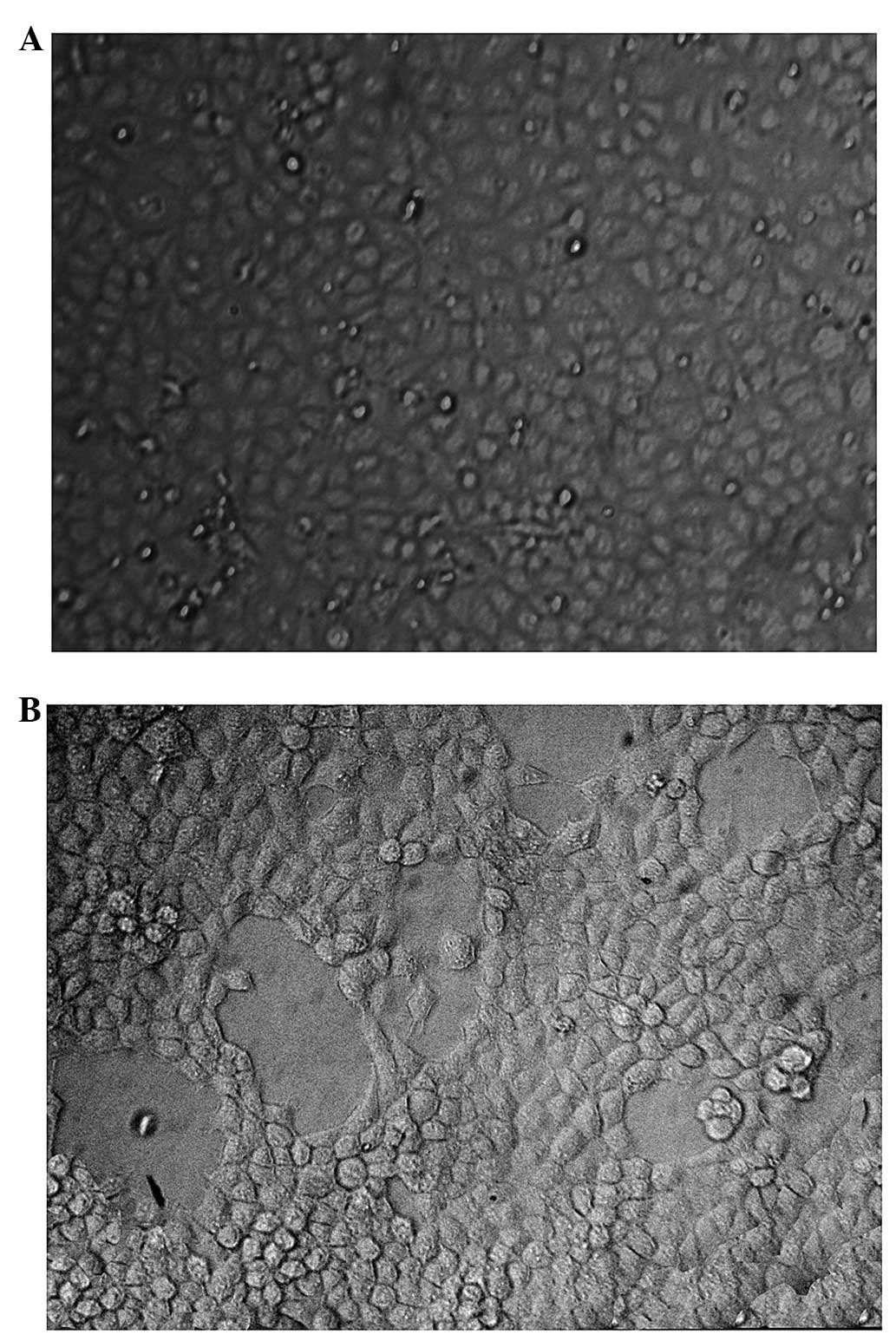
Effect of infection on cell
morphology
MCF-7 cells were treated with the adenoviral vector
at MOI 100 for 24 h. The infection toxicity was then evaluated by
observations of the cell morphology (Fig. 2). No change in cellular morphology
was observed after the 24-h treatment of the cells with the
adenoviral vector (Fig. 2B). The
adherent cells typically show a polygonal, spreading shape, with a
large nucleus. However, we found that cell densities in the
experimental group (transfected cells) were reduced compared to the
control group. We explored whether cell density may be associated
with the cell proliferation rate by cell growth and cell cycle
analyses described below.
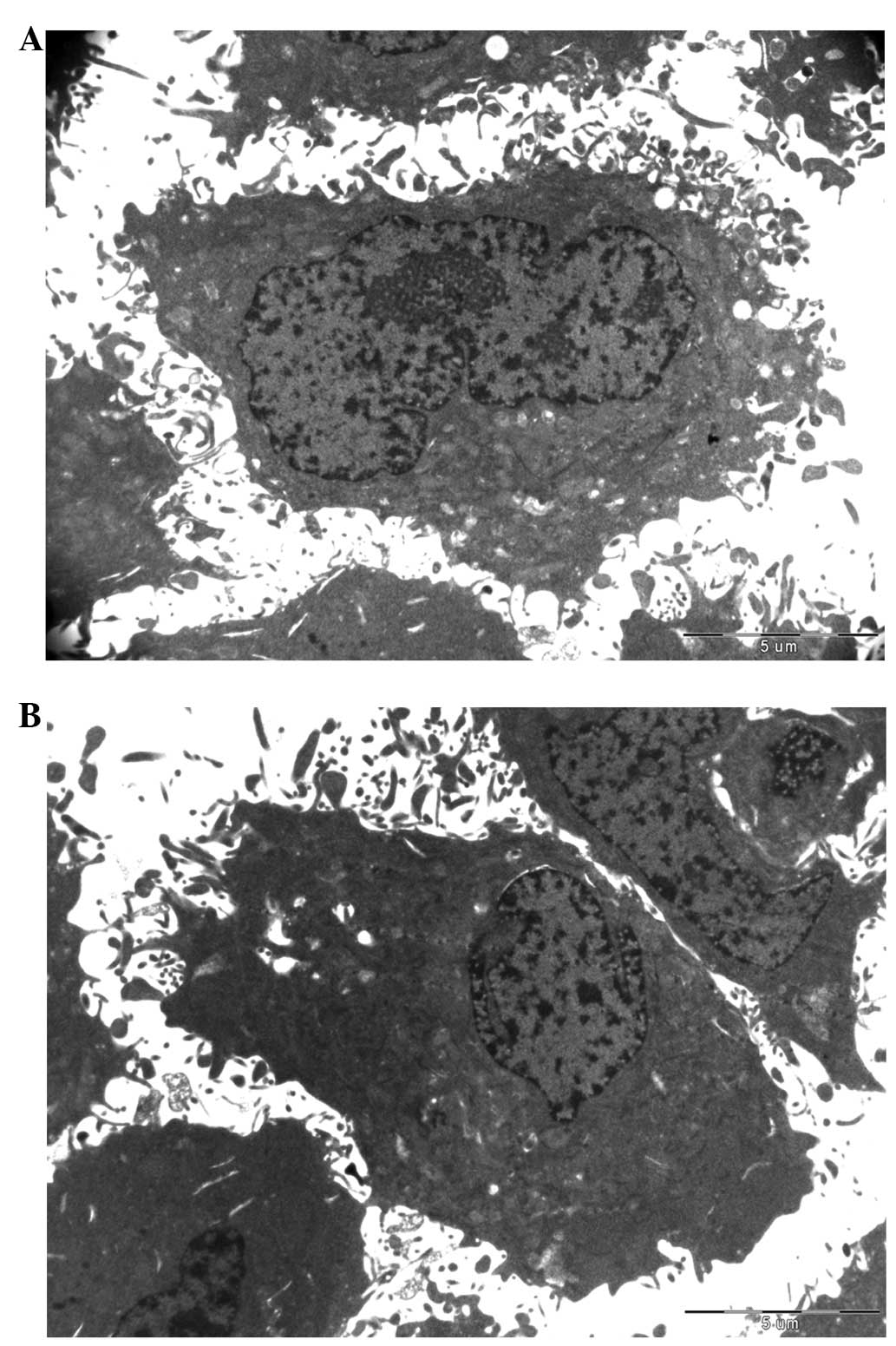
Ultrastructure of MCF-7 cells
As revealed by TEM, the ultrastructure of the
transfected MCF-7 cells (Fig. 3B,
experimental group) was similar to that of the non-transfected
MCF-7 cells (Fig. 3A, control
group). The centrally placed nucleus commonly contained one or two
prominent nucleoli. The nucleus-to-cytoplasm ratio was 1:1.
Expression of CD and TK proteins
The expression of the CD and TK proteins was
analyzed by western blot analysis. The results showed that the CD
and TK proteins are not expressed in the control group, while they
were strongly expressed in the experimental group (Fig. 4).
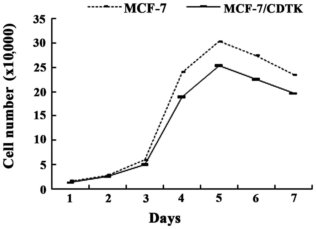
Cell growth curve of MCF-7 cells
MCF-7 cells were observed under a light microscope
following an additional 72-h incubation with the adenoviral vector.
The morphology of MCF-7 cells of the experimental group was similar
to that of the control group. The growth of cells became relatively
slow at one to two days after infection, while growth rates
increased after two days. An increasing number of cells became cell
growth arrested and eventually senescent from the fifth day
onwards. Cell proliferation of the experimental group was reduced
compared to the control group (Fig.
5). The data from cell growth experiments were statistically
analyzed using an independent samples t-test. There was no
significant difference in cell proliferation between the two groups
(P>0.056). This result showed that the adenoviral vector has no
obvious effect on MCF-7 cell growth (Table I and Fig. 5).
 | Table INumber of MCF-7 cells at different
days of infection (mean ×105 ± SD, n=3). |
Table I
Number of MCF-7 cells at different
days of infection (mean ×105 ± SD, n=3).
| Days of
infection |
|---|
|
|
|---|
| Group | 1 | 2 | 3 | 4 | 5 | 6 | 7 |
|---|
| MCF-7 | 1.56±0.14 | 2.80±0.26 | 5.97±0.58 | 23.99±2.92 | 30.32±2.95 | 27.44±2.58 | 23.45±2.09 |
| MCF-7/CDTK | 1.31±0.11 | 2.56±0.19 | 5.01±0.27 | 18.90±2.35 | 25.35±2.14 | 22.51±1.92 | 19.64±1.67 |
| t | 2.463 | 1.265 | 2.574 | 2.349 | 2.363 | 2.659 | 2.469 |
| P | 0.069 | 0.274 | 0.062 | 0.079 | 0.077 | 0.056 | 0.069 |
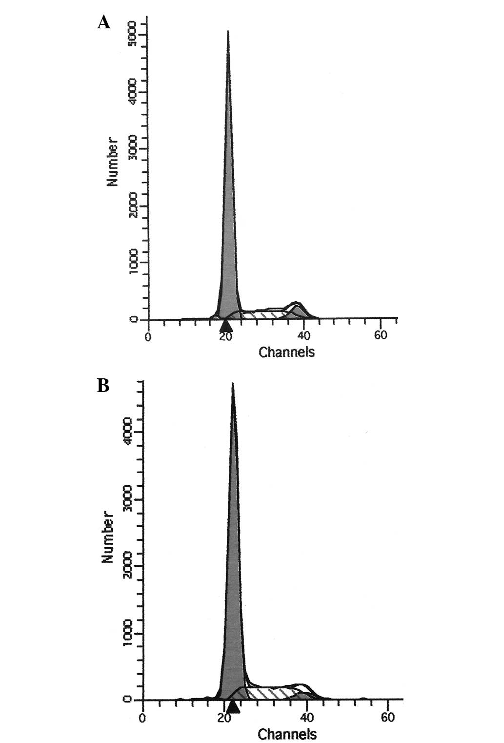
Cell cycle analysis using FCM
The proportion of cells at the different phases of
the cell cycle is shown in Table
II. The data from the experimental and the control group were
statistically analyzed by an independent samples t-test, which
revealed no significant differences (P>0.085) in the number of
cells at the same phase of the cell cycle between the two groups
(Table II). In addition, FCM
analysis revealed that the adenoviral vector has no obvious effect
on the cell cycle distribution of MCF-7 cells (Fig. 6).
| Table IICell cycle changes in MCF-7 cells
following infection with the adenoviral vector (mean% of cells ±
SD, n=3). |
Table II
Cell cycle changes in MCF-7 cells
following infection with the adenoviral vector (mean% of cells ±
SD, n=3).
| Group |
G0-G1 |
G2-M | S |
|---|
| Control | 77.03±3.27 | 7.89±1.43 | 15.01±1.41 |
| Transfected | 73.55±7.34 | 10.77±1.66 | 15.46±1.53 |
| t | 0.749 | 2.279 | 0.375 |
| P | 0.496 | 0.085 | 0.727 |
Discussion
Adenoviral vectors are popular gene delivery vectors
in clinical trials for gene therapy (17–19).
There are several advantages of using adenoviral vectors: first,
the efficiency of transduction is high, as is the level of gene
expression (20,21). In this study, we used an adenoviral
vector containing the GFP reporter gene, which is a commonly
used reporter allowing to unequivocally assess whether a gene is
expressed. GFP expression was evaluated by fluorescence microscopy,
which allowed to assess the efficiency of transduction. This
experiment demonstrated that >95% of the cells were GFP-positive
at a MOI of 100 and 200 pfu/cell. The recombinant adenoviral vector
showed no toxicity to MCF-7 cells at M0I 100 and 200 (Fig. 2B and C). Second, adenoviral vectors
can accommodate relatively large segments of DNA (up to 7.5 kb),
which they can transduce into the target cells. In this study, the
CD/TK fusion gene was 2.4 kb. The CD and TK proteins were
successfully expressed in MCF-7 cells infected with the adenoviral
vector, as shown by western blot analysis (Fig. 4). A major disadvantage of current
adenoviral vectors is their cytotoxicity to target cells. Teramoto
et al (22) showed that
adenoviral vectors directly alter the cell cycle in the infected
airway cells, which may result in slower proliferation: slower
proliferation and cell apoptosis were observed in cells infected by
vector at a MOI of 104. Slower proliferation and cell
apoptosis following Ad vector administration may be due to the high
adenoviral titer. If we select an appropriate adenoviral titer,
cell proliferation may be not affected, as discussed below.
The VEGF protein has been shown to be upregulated in
numerous types of cancer (23,24),
including human breast cancer cells (25,26).
A VEGF promoter-based adenoviral vector strategy has been
successfully adopted to introduce a foreign gene into cancer cells
(27,28). However, this strategy has not been
systematically explored in breast cancer research. Our study will
thus provide valuable information on the use of adenoviral vectors
for breast cancer therapy. Our previous studies successfully
constructed and extensively analyzed the adenoviral vector
Ad-VEGFp-CD/TK (14,29). In the present study, we compared
the expression levels of CD and TK protein (using β-actin as the
loading control) between untransfected and transfected MCF-7 cells
by western blot analysis. This analysis showed that the expression
levels of CD and TK in transfected cells are similar to the
expression level of β-actin. Thus, the VEGF promoter activated CD
and TK protein expression in MCF-7 cells. We conclude that VEGF
promoter-based Ads have good potential to be developed as effective
therapeutic agents for cancer.
This study indicated that infection with the
Ad-VEGFp-CD/TK vector exerts no prominent effect on cell
proliferation in the human breast cancer cell line MCF-7 at a MOI
of 100. Therefore, MOI 100 was selected as the working
concentration. We found that infected MCF-7 cells have a lower
growth rate than the uninfected cells. However, there was no
significant difference (P>0.05) in cell proliferation between
these two groups (Table I). The
result from cell growth analysis was consistent with that of cell
cycle analysis with flow cytometry: no significant difference
(P>0.05) was observed between the two groups with regards to the
number of cells at the same phase of the cell cycle (Table II). It is thus reasonable to
hypothesize that the adenoviral vector does not alter the cell
cycle in the infected MCF-7 cells, which results in the unchanged
proliferation of these cells. In addition, no significant change in
cell morphology or ultrastructure was observed under the light and
the transmission electron microscope. These results indicate that
the vector Ad-VEGFp-CD/TK is non-toxic to MCF-7 cells. Three
additional studies have successfully used this vector (30–32).
Therefore, our study provided the missing information needed for
further use of the vector in the context of suicide gene therapy
for cancer.
In summary, we achieved high-efficiency transduction
of MCF-7 cells by a VEGF promoter-based adenoviral vector, and
stable expression of the CD and TK proteins in vitro. There
were no significant changes in cell morphology, ultrastructure,
proliferation rate and cell-cycle distribution in the infected
MCF-7 cells. We conclude that the VEGF promoter-based suicide gene
system may be an effective strategy for cancer treatment, and that
the Ad-VEGFp-CD/TK vector is non-toxic to MCF-7 cells at the
appropriate titer.
Acknowledgements
This study was supported in part by the Shenzhen
Council for Scientific and Technological Innovation grant
JCYJ20130402151227177, and the Shenzhen Nanshan District Science
and Technology Project grant 2012025.
References
|
1
|
Finzi L, Kraemer A, Capron C, Noullet S,
Goere D, Penna C, Nordlinger B, Legagneux J, Emile JF and Malafosse
R: Improved retroviral suicide gene transfer in colon cancer cell
lines after cell synchronization with methotrexate. J Exp Clin
Cancer Res. 30:922011. View Article : Google Scholar : PubMed/NCBI
|
|
2
|
Jia W, Mei L, Wang Y, Liu L and Che G:
Double suicide genes selectively kill human umbilical vein
endothelial cells. Virol J. 8:742011. View Article : Google Scholar : PubMed/NCBI
|
|
3
|
Liu T, Ye L, He Y, Chen X, Peng J, Zhang
X, Yi H, Peng F and Leng A: Combination gene therapy using
VEGF-shRNA and fusion suicide gene yCDglyTK inhibits gastric
carcinoma growth. Exp Mol Pathol. 91:745–752. 2011. View Article : Google Scholar : PubMed/NCBI
|
|
4
|
Niu HX, Du T, Xu ZF, Zhang XK and Wang RG:
Role of wild type p53 and double suicide genes in interventional
therapy of liver cancer in rabbits. Acta Cir Bras. 27:522–528.
2012. View Article : Google Scholar : PubMed/NCBI
|
|
5
|
Lee SW, Lee YL, Lee YJ, Park SY, Kim IS,
Choi TH, Ha JH, Ahn BC and Lee J: Enhanced antitumor effects by
combination gene therapy using MDR1 gene shRNA and HSV1-tk in a
xenograft mouse model. Cancer Lett. 291:83–89. 2010. View Article : Google Scholar : PubMed/NCBI
|
|
6
|
Kim HA, Nam K, Lee M and Kim SW:
Hypoxia/hepatoma dual specific suicide gene expression plasmid
delivery using bio-reducible polymer for hepatocellular carcinoma
therapy. J Control Release. 171:1–10. 2013. View Article : Google Scholar : PubMed/NCBI
|
|
7
|
Li S, Tokuyama T, Yamamoto J, Koide M,
Yokota N and Namba H: Potent bystander effect in suicide gene
therapy using neural stem cells transduced with herpes simplex
virus thymidine kinase gene. Oncology. 69:503–508. 2005. View Article : Google Scholar : PubMed/NCBI
|
|
8
|
Yi BR, Choi KJ, Kim SU and Choi KC:
Therapeutic potential of stem cells expressing suicide genes that
selectively target human breast cancer cells: Evidence that they
exert tumoricidal effects via tumor tropism (Review). Int J Oncol.
41:798–804. 2012.
|
|
9
|
Jiang YX, Lu Y, Liu TJ, Yang J, Chen Y and
Fang YW: Using HSV-TK/GCV suicide gene therapy to inhibit lens
epithelial cell proliferation for treatment of posterior capsular
opacification. Mol Vis. 17:291–299. 2011.PubMed/NCBI
|
|
10
|
Yu BF, Wu J, Zhang Y, Sung HW, Xie J and
Li RK: Ultrasound-targeted HSVtk and Timp3 gene delivery for
synergistically enhanced antitumor effects in hepatoma. Cancer Gene
Ther. 20:290–297. 2013. View Article : Google Scholar : PubMed/NCBI
|
|
11
|
Kuzmin D, Gogvadze E, Kholodenko R, Grzela
DP, Mityaev M, Vinogradova T, Kopantzev E, Malakhova G, Suntsova M,
Sokov D, Ivics Z and Buzdin A: Novel strong tissue specific
promoter for gene expression in human germ cells. BMC Biotechnol.
10:582010. View Article : Google Scholar : PubMed/NCBI
|
|
12
|
Foekens JA, Peters HA, Grebenchtchikov N,
Look MP, Meijer-van Gelder ME, Geurts-Moespot A, van der Kwast TH,
Sweep CG and Klijn JG: High tumor levels of vascular endothelial
growth factor predict poor response to systemic therapy in advanced
breast cancer. Cancer Res. 61:5407–5414. 2001.PubMed/NCBI
|
|
13
|
Konecny GE, Meng YG, Untch M, Wang HJ,
Bauerfeind I, Epstein M, Stieber P, Vernes JM, Gutierrez J, Hong K,
Beryt M, Hepp H, Slamon DJ and Pegram MD: Association between
HER-2/neu and vascular endothelial growth factor expression
predicts clinical outcome in primary breast cancer patients. Clin
Cancer Res. 10:1706–1716. 2004. View Article : Google Scholar
|
|
14
|
Chen JF, Huang ZH, Huang YY, Song HJ and
Che XY: Construction of recombinant adenoviruses encoding TK
suicide gene driven by VEGF promoter using efficient AdEasier-1
system. Ai Zheng. 23:1093–1097. 2004.(In Chinese).
|
|
15
|
Rabenau HF, Rapp I and Steinmann J: Can
vaccinia virus be replaced by MVA virus for testing virucidal
activity of chemical disinfectants? BMC Infect Dis. 10: View Article : Google Scholar : 2010. View Article : Google Scholar : PubMed/NCBI
|
|
16
|
Zhao W, Song Y, Xu B and Zhan Q:
Overexpression of centrosomal protein Nlp confers breast carcinoma
resistance to paclitaxel. Cancer Biol Ther. 13:156–163. 2012.
View Article : Google Scholar : PubMed/NCBI
|
|
17
|
Puntel M, A K M GM, Farrokhi C, et al:
Safety profile, efficacy, and biodistribution of a bicistronic
high-capacity adenovirus vector encoding a combined
immunostimulation and cytotoxic gene therapy as a prelude to a
phase I clinical trial for glioblastoma. Toxicol Appl Pharmacol.
268:318–330. 2013. View Article : Google Scholar
|
|
18
|
Muhammad AK, Xiong W, Puntel M, Farrokhi
C, Kroeger KM, Salem A, Lacayo L, Pechnick RN, Kelson KR, Palmer D,
Ng P, Liu C, Lowenstein PR and Castro MG: Safety profile of gutless
adenovirus vectors delivered into the normal brain parenchyma:
implications for a glioma phase 1 clinical trial. Hum Gene Ther
Methods. 23:271–284. 2012. View Article : Google Scholar
|
|
19
|
Castro JE, Melo-Cardenas J, Urquiza M,
Barajas-Gamboa JS, Pakbaz RS and Kipps TJ: Gene immunotherapy of
chronic lymphocytic leukemia: a phase I study of intranodally
injected adenovirus expressing a chimeric CD154 molecule. Cancer
Res. 72:2937–2948. 2012. View Article : Google Scholar : PubMed/NCBI
|
|
20
|
Reetz J, Genz B, Meier C, Kowtharapu BS,
Timm F, Vollmar B, Herchenröder O, Abshagen K and Pützer BM:
Development of adenoviral delivery systems to target hepatic
stellate cells in vivo. PLoS One. 8:e670912013. View Article : Google Scholar : PubMed/NCBI
|
|
21
|
Zhang H, Sui A, Wang Z, Liu S and Yao R:
Adenovirus-mediated TRAIL expression and downregulation of Bcl-2
expression suppresses non-small cell lung cancer growth in
vitro and in vivo. Int J Mol Med. 30:358–364.
2012.PubMed/NCBI
|
|
22
|
Teramoto S, Johnson LG, Huang W, Leigh MW
and Boucher RC: Effect of adenoviral vector infection on cell
proliferation in cultured primary human airway epithelial cells.
Hum Gene Ther. 6:1045–1053. 1995. View Article : Google Scholar
|
|
23
|
Lee NO, Park JW, Lee JA, Shim JH, Kong SY,
Kim KT and Lee YS: Dual action of a selective cyclooxygenase-2
inhibitor on vascular endothelial growth factor expression in human
hepatocellular carcinoma cells: novel involvement of discoidin
domain receptor 2. J Cancer Res Clin Oncol. 138:73–84. 2012.
View Article : Google Scholar
|
|
24
|
Eisermann K, Broderick CJ, Bazarov A,
Moazam MM and Fraizer GC: Androgen up-regulates vascular
endothelial growth factor expression in prostate cancer cells via
an Sp1 binding site. Mol Cancer. 12:72013. View Article : Google Scholar : PubMed/NCBI
|
|
25
|
Lee KY, Lee JW, Nam HJ, Shim JH, Song Y
and Kang KW: PI3-kinase/p38 kinase-dependent E2F1 activation is
critical for Pin1 induction in tamoxifen-resistant breast cancer
cells. Mol Cells. 32:107–111. 2011. View Article : Google Scholar : PubMed/NCBI
|
|
26
|
Cao X, Geradts J, Dewhirst MW and Lo HWU:
pregulation of VEGF-A and CD24 gene expression by the tGLI1
transcription factor contributes to the aggressive behavior of
breast cancer cells. Oncogene. 31:104–115. 2012. View Article : Google Scholar
|
|
27
|
Takayama K, Reynolds PN, Adachi Y,
Kaliberova L, Uchino J, Nakanishi Y and Curiel DT: Vascular
endothelial growth factor promoter-based conditionally replicative
adenoviruses for pan-carcinoma application. Cancer Gene Ther.
14:105–116. 2007. View Article : Google Scholar
|
|
28
|
Koshikawa N, Takenaga K, Tagawa M and
Sakiyama S: Therapeutic efficacy of the suicide gene driven by the
promoter of vascular endothelial growth factor gene against hypoxic
tumor cells. Cancer Res. 11:2936–2941. 2000.PubMed/NCBI
|
|
29
|
Kong H, Huang ZH, Li Z, Yu JL, Chen HJ and
Han XJ: Preliminary effect of VEGF promoter-driven recombinant
adenovirus containing double suicide genes on apoptosis of human
gastric carcinoma cells. Nan Fang Yi Ke Da Xue Xue Bao.
27:1152–1155. 2007.(In Chinese).
|
|
30
|
Li XH, Zhou P, Wang LH, Tian SM, Qian Y,
Chen LR and Zhang P: The targeted gene (KDRP-CD/TK) therapy of
breast cancer mediated by SonoVue and ultrasound irradiation in
vitro. Ultrasonics. 52:186–191. 2012. View Article : Google Scholar : PubMed/NCBI
|
|
31
|
Su GQ, Su G and Huang ZH:
Adenovirus-mediated tissue-targeted expression of the CDglyTk gene
for the treatment of breast cancer. Mol Med Rep. 6:321–329.
2012.PubMed/NCBI
|
|
32
|
Kang NH, Hwang KA, Yi BR, Lee HJ, Jeung
EB, Kim SU and Choi KC: Human amniotic fluid-derived stem cells
expressing cytosine deaminase and thymidine kinase inhibits the
growth of breast cancer cells in cellular and xenograft mouse
models. Cancer Gene Ther. 19:412–419. 2012. View Article : Google Scholar
|