Introduction
Bone marrow mesenchymal stem cells (BMSCs) are
multipotent stem cells, which are able to differentiate into a
variety of cell types, including myocardiocytes (1). There is increasing evidence to
suggest that stem cells exist in the adult mammalian heart
(2); however, when a substantial
loss of cardiomyocytes occurs, including in a myocardial
infarction, the endogenous cardiac regenerative mechanisms are
insufficient to replace the lost cardiomyocytes. BMSCs
differentiate into myocardial cells in a suitable in vitro
microenvironment (3). In addition,
BMSCs can differentiate into cardiomyocytes to replace infarcted
myocardium in vivo (1,4),
thereby improving the cardiac function that has been compromised by
repeated ischemic episodes.
The assessment of the efficacy of stem cell
transplantation requires an effective method to track the
migration, proliferation and differentiation of the transplanted
stem cells. The development of molecular imaging techniques has
enabled the visualization of stem cell homing, migration, location
and proliferation (5–8). Magnetic resonance imaging (MRI)
possesses high temporal and spatial resolution, is noninvasive and
is free of ionizing radiation. MRI can be used to observe the
dynamic process of cell migration and may be a desirable approach
for the in vivo tracking of stem cells injected with
contrast agents (9–16).
Superparamagnetic iron oxide (SPIO) nanoparticles,
comprised of single or multiple iron oxide crystals, are
synthesized by the introduction of amine groups onto the surface of
silica-coated composite magnetite nanoparticles (11). They generate superparamagnetism,
thereby resulting in an inhomogeneous local magnetic field, which
shortens the T2 value on MRI (17). By contrast, tissues that do not
contain SPIO may show high signals on T2 and T2* sequences.
Therefore, MRI using SPIO may be effective for tracking
transplanted BMSCs.
SPIO nanoparticles have been used to image the
movement of adipogenic mesenchymal stem cells to the infarcted
myocardium (18,19). In vivo MRI stem cell
tracking has been applied in animal models of cerebral ischemia,
spinal cord injury, myocardial infarction and peripheral nerve
injury (9–16). However, there have been few MRI
studies on repair and functional improvement in animal models of
myocardial infarction (14,16,18).
In the present study, BMSCs were labeled with SPIO
and MRI was used to observe their distribution and migration
following transplantation in a rat model of acute myocardial
infarction.
Materials and methods
Animals
A total of 65 female Sprague-Dawley rats, obtained
from the Laboratory Animal Center of Sun Yat-Sen University,
Guangzhou, China) were used in the present study. The BMSCs were
sourced from five young rats (5–6-weeks old) weighing ~150 g and
the remaining 60 rats (6–8 weeks old; 180–200 g) were used in the
experiments involving myocardial infarction. The rats were housed
at 22°C with a 12 h light/dark cycle. Food and water were available
ad libitum. The study procedure was approved by the
Institutional Review Board of Sun Yat-Sen University and all animal
experiments were performed in accordance with the established
guidelines of the Institutional Animal Care and Use Committee of
Sun Yat-Sen University.
Isolation, culture and purification of
BMSCs
The five young rats were anesthetized with 1.5%
intraperitoneal isoflurane (30 mg/kg), sacrificed by cervical
dislocation and primary BMSCs were collected, as previously
described (1). Briefly, under
aseptic conditions, the epiphyseal regions of the femora and tibia
were removed and marrow plugs were flushed out using Dulbecco’s
modified Eagle’s medium (DMEM; Gibco-BRL, Gaithersburg, MD, USA)
containing 10% (v/v) fetal bovine serum (FBS; Gibco-BRL). A
suspension of single bone marrow cells was obtained by repeated
aspiration. The cells were seeded into 25-ml culture flasks at a
density of 6×10⁴ cells/ml and were cultured in DMEM/F12
(Gibco-BRL) supplemented with 10% (v/v) FBS, 100 U/ml penicillin
(Sigma-Aldrich, St. Louis, MO, USA) and 100 ng/ml streptomycin
(Sigma-Aldrich) in a humidified incubator with 5% CO2 at
37°C. After 2 days, the culture medium and non-adherent cells were
removed. The harvested cells were verified as BMSCs with flow
cytometry using FITC-conjugated rat anti-mouse CD34, CD45, CD29 and
CD90 monoclonal antibodies (Santa Cruz Biotechnology, Inc., Santa
Cruz, CA USA). An isotope antibody was used as the negative
control. The isotype antibody represented the antibody with the
same sources and subgroup with FITC-conjugated rat anti-mouse CD34,
CD45, CD29 and CD90 monoclonal antibodies (Santa Cruz
Biotechnology, Inc.). Cells in passages 3–6 cells were used in the
subsequent experiments.
SPIO labeling of BMSCs
The SPIO
(Fe3+2O3M2+O; Soochow
University, Suzhou, China) used in the present study comprised a
3-aminopropyl triethoxysilane-modified Fe2O3
particle, with a diameter of 10–15 nm. SPIO labeling of the BMSCs
was performed as previously described (20). Briefly, the BMSCs were washed three
times with phosphate-buffered saline (PBS) and then grown in
DMEM/F12 containing 10% FBS, 100 U/ml penicillin and 100 ng/ml
streptomycin. The cells were then labeled with SPIO by culturing
for 24 h at 37°C in an atmosphere of 5% CO2; the iron
concentration of the culture medium was 25 μg/ml. After 24 h, the
cells were washed thoroughly to remove the remaining SPIO. A total
of 5×10⁵ cells were harvested for MRI detection and
unlabeled BMSCs were used as the control. At 24 h after labeling,
the cells were subjected to Prussian blue iron (Beijing Leagene
Biotech, Co., Ltd., Beijing, China) staining using a Mallory’s
method, which was performed according to standard protocols
(21) and images were captured
using an inverted microscope (DMi1; Leica Microsystems GmbH,
Wetzlar, Germany). At least 700 cells were counted. The experiments
were repeated five times independently.
Cell viability assays
Cells (5×10⁵) were incubated with 0.4%
trypan blue dye for 15 min at room temperature prior to microscopic
examination. Images of four randomly selected fields were captured
for each sample. The percentage of viable cells was calculated
using the following formula: Cell survival rate (%) = (total number
of cells - number of dyed cells)/total number of cells × 100.
For the
3-(4,5-dimethylthiazol-2-yl)-2,5-diphenyltetrazolium bromide (MTT)
reduction assay, the cells were plated in a 96-well plate
(1×10⁴ cells/well) and incubated in a humidified
atmosphere with 5% CO2 at 37°C for 1–5 days prior to the
MTT assay (Sigma-Aldrich). Absorbance was measured at 492 nm using
a microtiter plate reader (Wellscan K3; KHB Labsystems, Helsinki,
Finland).
Acute myocardial infarction in rats
The rats were anesthetized using pentobarbital
sodium (30 mg/kg, intraperitoneally), fixed on an operation table
and mechanically ventilated. The ventilator maintained a breathing
ratio of 1:2 at a frequency of 100 times/min and a tidal volume of
14 ml/kg. An electrocardiogram, using the standard II leads from
the RM 6240 BD Multi-channel physiological signal processing system
(Shanghai Huayan Instrument & Equipment Co., Ltd., Shanghai,
China) was used to monitor cardiac function. From the left
parasternal side, a 1.5–2.0-cm longitudinal incision was made
between the third and fourth ribs and the heart was exposed
following thoracotomy. The anterior descending branch of the
coronary artery was ligated 2–3 mm below the left atrial appendage.
The procedure was considered successful when the electrocardiogram
revealed depression of the QRS-wave peak, elevation of the J point
and ST-segment elevation >0.2 mV.
Injection of BMSCs into the infarcted rat
myocardium
The chest was opened under anesthesia (1.5%
intraperitoneal isoflurane; 30 mg/kg) 2 weeks after coronary artery
ligation and the infarcted myocardial region was fully exposed. The
rats were randomly divided into three groups (20/group) and
~5×10⁷ SPIO-labeled BMSCs, an equal quantity of
unlabeled BMSCs or the vehicle (100 μl PBS) was injected into the
myocardial tissues along the margins of the infarcted region, which
was identified by a pale color. The mean weights of the animals in
the three groups were similar.
In vivo MRI tracking of SPIO-labeled
BMSCs
MRI was performed using a Philips Gyroscan Intera
1.5T Superconducting MRI scanner (Philips Medical Systems, Best,
The Netherlands), with a circular surface coil of 5 cm in diameter.
T2 and T2* scans were performed in transverse and oblique sagittal
positions 1 day and 3 weeks after transplantation of the BMSCs. The
scanning parameters for T2 were: repetition time (TR)/echo time
(TE), 2,000/100 msec; slice thickness, 2.0 mm; field of view (FOV),
48×22 mm; matrix size, 256×256; number of signal averages (NSA), 2
and flip angle, 22°. The parameters for T2* were: TR/TE, 253/14
msec; slice thickness, 2.0 mm; interslice gap, 0.2 mm; FOV, 48×23
mm; matrix size, 256×256 and NSA, 3.
Histopathological examination
Following sacrifice, the myocardial tissues were
collected using the MRI images for guidance. The samples were fixed
in 10% formaldehyde for 24 h and then dehydrated in gradient
ethanol (100, 95 amd 75%). The samples were made transparent by
soaking in xylene, then embedded in paraffin and cut into 4-μm
sections. The tissue sections were first stained with hematoxylin
and eosin (H&E) and were then stained using a rat anti-mouse
monoclonal CD90 antibody (Santa Cruz Biotechnology, Inc.). The
tissue sections were examined independently by two experienced
pathologists.
Statistical analysis
Quantitative data is expressed as the mean ±
standard deviation. The differences between the SPIO-label and
unlabeled BMSCs were compared using an unpaired two-sample t-test.
Statistical analyses were performed using SPSS statistical
software, version 15.0 (SPSS, Inc., Chicago, IL, USA). P<0.05
indicated a statistically significant difference.
Results
BMSC isolation
In the present study BMSCs were isolated from the
epiphyseal regions of the femur and tibia of rats and grown ex
vivo. The cells became adherent 6–10 h post-seeding in the
culture bottle, appearing round or polygonal in shape. A few,
short, spindle-like and star-like cells with pseudopodia appeared
2–3 days later. At day 7 post-seeding, radially arranged colonies
were observed; these cells exhibited uneven projections, had a
large nucleus and an apparent nucleolus. At 12–14 days, the cells
were swirl-shaped and between 80 and 90% confluent. The cells at
passages 4 and 5 demonstrated similar spindle and star-like
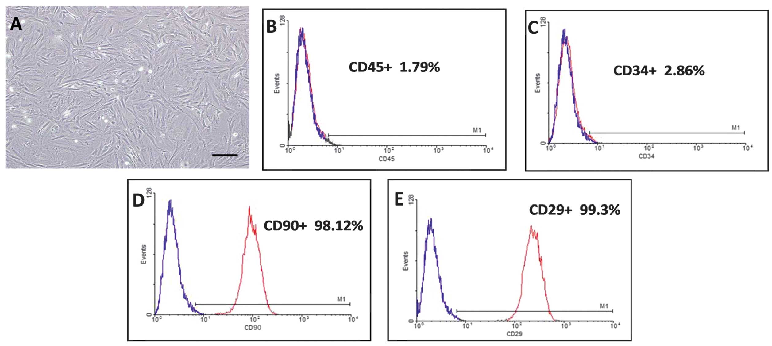
morphological characteristics (Fig.
1A). The hematopoietic stem cell markers CD45 (Fig. 1B) and CD34 (Fig. 1C) were positive in 1.79 and 2.86%
of the cells, respectively. The surface markers CD90 (Fig. 1D) and CD29 (Fig. 1E) were positive in 98.12 and 99.3%
of the cells, respectively.
SPIO labeling of BMSCs
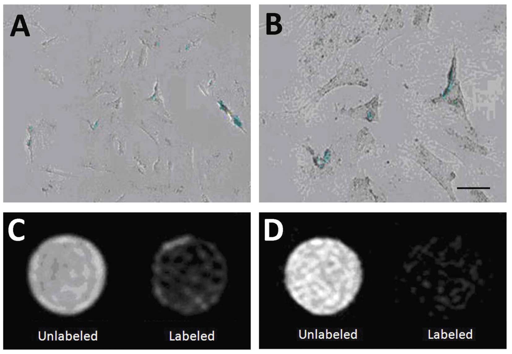
Phase contrast microscopy revealed the presence of
blue iron particles in 99% of the cell cytoplasm in the
SPIO-labeled BMSCs (Fig. 2A and
B). Compared with the unlabeled BMSCs, the SPIO-labeled cells
exhibited a markedly decreased signal on the T2 and T2* sequences
(Fig. 2C and D).
SPIO exerts no immediate toxic effects on
BMSCs
The viability of the SPIO-labeled and unlabeled
BMSCs, examined using the trypan blue exclusion assay, are shown in
Table I. No significant
differences were observed in viability between the SPIO-labeled and
unlabeled cells at any time point. The viabilities of the
SPIO-labeled compared with those of the unlabeled cells were:
98.65±1.63 vs. 98.74±1.42 (P=0.897) after 6 h, 96.35±0.96 vs.
96.42±1.33 (P=0.894) after 12 h and 95.51±1.44 vs. 95.78±1.21
(P=0.655) after 24 h.
 | Table ICell viability of SPIO-labeled and
unlabeled bone marrow mesenchymal stem cells. |
Table I
Cell viability of SPIO-labeled and
unlabeled bone marrow mesenchymal stem cells.
| Time | SPIO-labeled
(n=10) | Unlabeled
(n=10) | P-valuea |
|---|
| 6 h | 98.65±1.63 | 98.74±1.42 | 0.897 |
| 12 h | 96.35±0.96 | 96.42±1.33 | 0.894 |
| 24 h | 95.51±1.44 | 95.78±1.21 | 0.655 |
The results of the MTT assay indicated that SPIO did
not exhibit any antiproliferative effect (data not shown).
In vivo tracking of SPIO-labeled BMSCs by
MRI
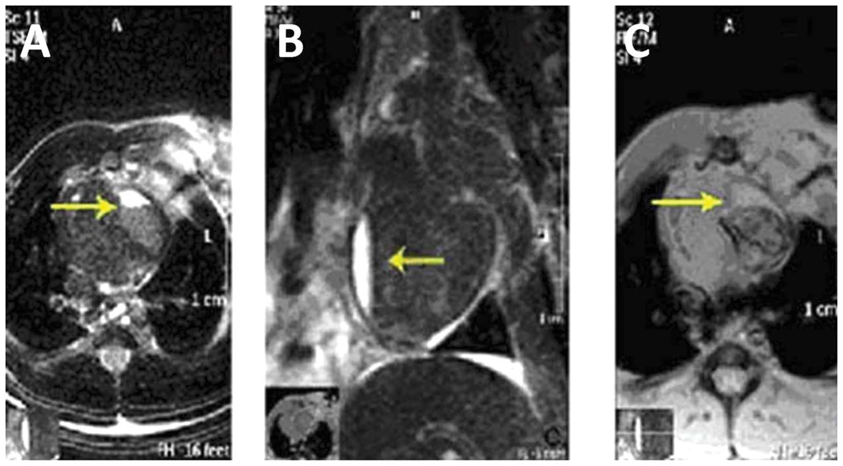
The mean myocardial infarct size of the 60 rats was
1.71±0.13 cm2 and no difference was observed in the size
of the infarct between the three groups. At 2 weeks following
coronary artery ligation, a high-intensity signal was noted in the
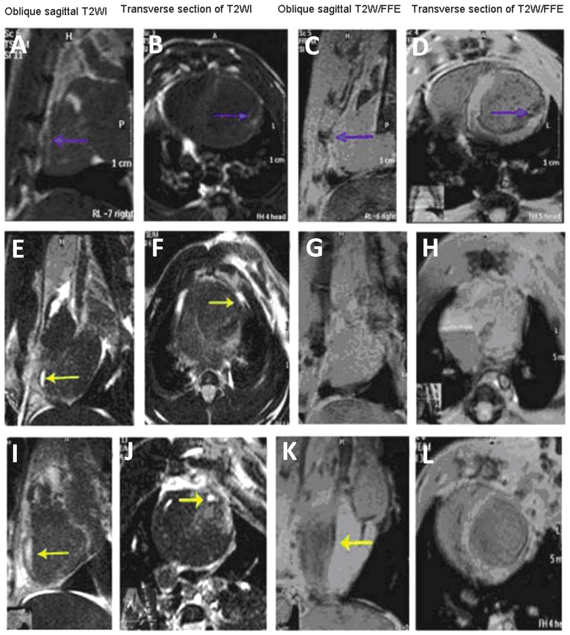
infarcted area on the T2 images (Fig.
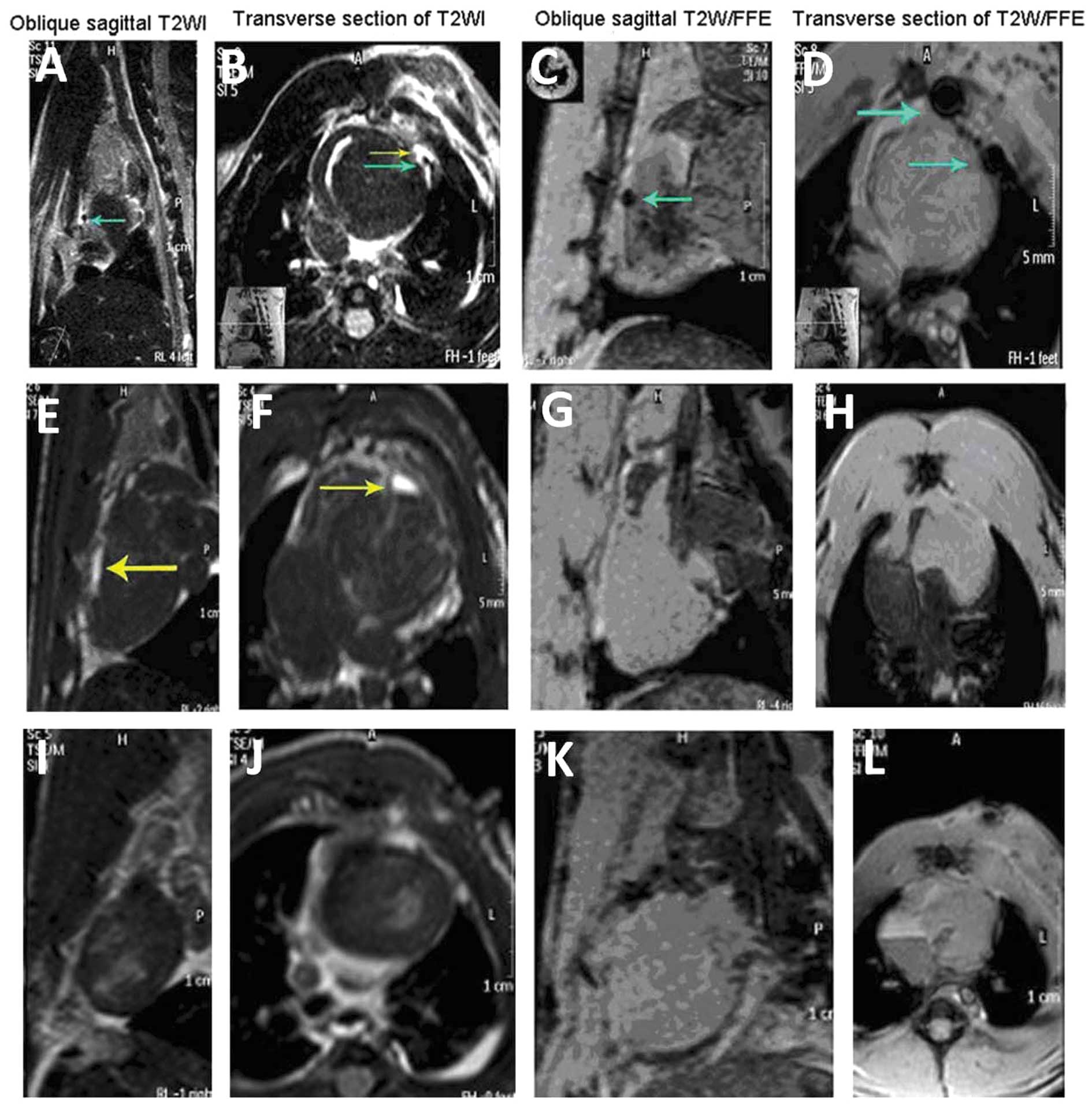
3) and, on the first day following transplantation of the
BMSCs, low-intensity MRI signals were found at the margins of the
infarcted region on the T2 and T2* sequences (Fig. 4). The low-intensity signal
decreased over time and became indistinct from the surrounding
tissue. At 3 weeks, a low-intensity signal began to appear in the
area of infarction; however, subsequently the signal intensity
decreased and became indistinct (Fig.
5).
SPIO-labeled BMSCs in the infarcted
myocardium
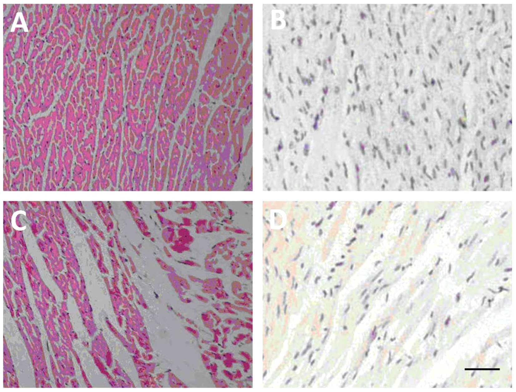
H&E staining revealed the presence of damaged
myocardial cells in the infarcted region 2 weeks after coronary
artery ligation (Fig. 6A and C).
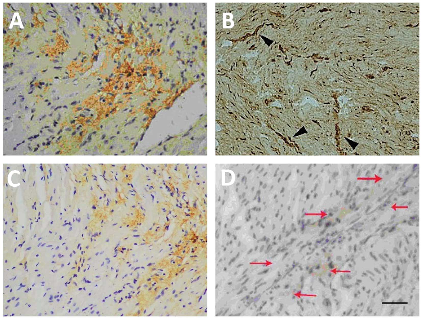
Immunohistochemical staining using the anti-CD90 antibody revealed
low or no expression of CD90 in either the normal or infarcted
myocardial tissues (Fig. 6B and
D). The expression of CD90 was marked in the SPIO-labeled BMSCs
24 h after transplantation, suggesting successful transplantation
of the BMSCs into the infarcted myocardium (Fig. 7A and B). The transplanted BMSCs
remained undifferentiated 3 weeks after transplantation and were
arranged along myocardial fibers in a strip-like manner (Fig. 7C and D).
Discussion
In the present study, BMSCs were labeled with SPIO
and MRI and in vivo tracking of the distribution and
migration of BMSCs was performed 1 day and 3 weeks after BMSC
transplantation. The results revealed that, from day 1
post-transplantation, a low-intensity signal was detected on T2*
sequences. The intensity of the signal decreased with time;
however, the area of the signal increased and gradually expanded
into the infarcted myocardia. This suggests that in vivo
tracking of SPIO-labeled BMSCs by MRI is feasible and effective.
Notably, the signal intensity in the infarcted area further
decreased and became indistinct 3 weeks post-transplantation, which
was likely due to the effects of macrophages.
BMSCs are able to differentiate into myocardial
cells in a suitable microenvironment. Previous studies
investigating the effectiveness of stem cell transplantation in
treating myocardial infarction have provided conflicting results.
Tomita et al (22) first
demonstrated that rat bone marrow stromal cells differentiate into
cardiomyocytes in vitro, induce angiogenesis in ventricular
scar tissue and ultimately improve myocardial function upon
transplantation. In a study of 69 patients with acute myocardial
infarction, intracoronary injection of autologous BMSCs led to
significant improvement in the ejection fraction and the rate of
motion of the infarcted region of the left ventricle (23). Analysis of data from the Autologous
Stem Cell Transplantation in Acute Myocardial Infarction (ASTAMI)
trial demonstrated that, although treatment with stem cells
appeared to be safe in the long-term and was associated with
significantly improved exercise time and heart rate response
(24), further analysis of the
data after 3 years revealed only a small improvement in exercise
time (25). A 3year serial
echocardiographic sub-study of the ASTAMI data indicated that
improvement of the left ventricular ejection fraction and diastolic
function as a result of acute percutaneous coronary intervention
was not affected by the intracoronary injection of stem cells
(26).
The mechanisms by which BMSCs may lead to the repair
infarcted tissue remain to be elucidated. Possible mechanisms
include the improvement of myocardial ischemia by the formation of
new blood vessels and promotion of collateral circulation, an
increase in the number of functional myocardial cells in the
infarcted region, the promotion of host vascular proliferation and
the improvement of cardiac function (27–30).
The present study observed that SPIO-labeled BMSCs remained
undifferentiated at the injection site for 3 weeks following
transplantation.
Stem cells are similar to the surrounding tissue and
magnetic contrast agents are necessary to alter the relaxation of
BMSCs to enable detection by MRI. SPIO nanoparticles produce a
hypointense footprint on MRI and, thus, exhibit a lower signal
intensity on T2 images (12). In
the present study, a significant difference was observed in the T2
images of the myocardium due to SPIO-labeled BMSCs, suggesting that
SPIO labeling provides a measurable effect on MRI (5). In general, contrast agents are not
effectively taken up by non-phagocytic cells; however, contrast
agents are effectively incorporated into macrophages, which are
abundant close to areas of infarction and migrate to the lesion
core (31). Amsalem et al
(18) demonstrated that, 4 weeks
after the transplantation of MSCs, the transplanted cells were not
present in the infarcted myocardium and enhanced MRI signals were
observed that originated from cardiac macrophages that had engulfed
the SPIO nanoparticles. In order to incorporate the contrast agents
into stem cells, positively charged transfection agents, including
polylysine and protamine sulfate, are often used to modify the
surface electrical properties of the cells (32). Labeling stem cells using SPIO
particles does not require a transfection agent and has proven to
be useful for the in vivo detection of low signals in
regions with transplanted cells (14).
The activity and reproductive capability of
magnetically labeled cells determines their clinical application.
The results of the present study indicated that SPIO nanoparticles
at 25 μg/ml effectively labeled cells with no adverse effects on
cell activity, growth or differentiation. Amsalem et al
(18) also found that the SPIO
labeling of stem cells did not affect their protective effect
against progressive left ventricular dilatation and dysfunction is
a rat model of myocardial infarction.
At present, the tracking of stem cells following
transplantation in patients is conducted mainly using in
vitro labeling and the three main imaging techniques used are
labeling with radioisotopes, optical imaging and MRI, each of which
has its own advantages and shortcomings (8). Several studies have examined the use
of MRI/SPIO labeling to trace implanted stem cells (18,33–35).
MRI has high temporal and spatial resolution, is non-invasive and
does not use ionizing radiation. It can image any region of the
body with high resolution and provides a long time-window for
imaging. In addition, MRI can simultaneously obtain physiological,
molecular and anatomical information and enables observation of the
dynamic process of cell migration. However, the technique suffers
from low sensitivity requiring an increased number of cells for
successful tracking (14).
There are a number of limitations of the present
study that must be considered. No Prussian blue staining was used
to colocalize the SPIOs and CD90+ cells. In addition,
CD90 was used as a marker of the grafted cells; however, green
fluorescent protein or other membrane labeling, such as using
dialkylcarbocyanines, is preferable (36). The regions of interest were not
normalized between scans, cardiac function was not assessed and no
quantitative analysis of the signal effects of SPIO in the
infarcted and non-infarcted myocardium was performed. The
limitations of the present study limit the ability to state with
100% certainty that the SPIO was harbored in the stem cells and not
the macrophages; however, the data and temporal findings indicate
that the SPIO was indeed in the stem cells.
Therefore, BMSCs can be readily labeled with SPIO,
without using a transfection agent, using a simple and effective
labeling method. MRI scans can then be used for in vivo
tracking of the distribution of the SPIO-labeled BMSCs.
References
|
1
|
Makino S, Fukuda K, Miyoshi S, et al:
Cardiomyocytes can be generated from marrow stromal cells in vitro.
J Clin Invest. 103:697–705. 1999. View
Article : Google Scholar : PubMed/NCBI
|
|
2
|
Anversa P, Kajstura J, Leri A and Bolli R:
Life and death of cardiac stem cells: a paradigm shift in cardiac
biology. Circulation. 113:1451–1463. 2006. View Article : Google Scholar : PubMed/NCBI
|
|
3
|
Feng X, He X, Li K, et al: The effects of
pulsed electromagnetic fields on the induction of rat bone marrow
mesenchymal stem cells to differentiate into cardiomyocytes-like
cells in vitro. Sheng Wu Yi Xue Gong Cheng Xue Za Zhi. 28:676–682.
2011.(In Chinese).
|
|
4
|
Orlic D, Kajstura J, Chimenti S, et al:
Bone marrow cells regenerate infarcted myocardium. Nature.
410:701–705. 2001. View
Article : Google Scholar : PubMed/NCBI
|
|
5
|
Bulte JW and Kraitchman DL: Iron oxide MR
contrast agents for molecular and cellular imaging. NMR Biomed.
17:484–499. 2004. View
Article : Google Scholar : PubMed/NCBI
|
|
6
|
Ju S, Teng GJ, Lu H, et al: In vivo MR
tracking of mesenchymal stem cells in rat liver after intrasplenic
transplantation. Radiology. 245:206–215. 2007. View Article : Google Scholar : PubMed/NCBI
|
|
7
|
Swijnenburg RJ, van der Bogt KE, Sheikh
AY, Cao F and Wu JC: Clinical hurdles for the transplantation of
cardiomyocytes derived from human embryonic stem cells: role of
molecular imaging. Curr Opin Biotechnol. 18:38–45. 2007. View Article : Google Scholar : PubMed/NCBI
|
|
8
|
Weissleder R: Molecular imaging: exploring
the next frontier. Radiology. 212:609–614. 1999. View Article : Google Scholar : PubMed/NCBI
|
|
9
|
Arbab AS, Bashaw LA, Miller BR, et al:
Characterization of biophysical and metabolic properties of cells
labeled with superparamagnetic iron oxide nanoparticles and
transfection agent for cellular MR imaging. Radiology. 229:838–846.
2003. View Article : Google Scholar : PubMed/NCBI
|
|
10
|
Bos C, Delmas Y, Desmouliere A, et al: In
vivo MR imaging of intravascularly injected magnetically labeled
mesenchymal stem cells in rat kidney and liver. Radiology.
233:781–789. 2004. View Article : Google Scholar : PubMed/NCBI
|
|
11
|
Bruce IJ and Sen T: Surface modification
of magnetic nanoparticles with alkoxysilanes and their application
in magnetic bioseparations. Langmuir. 21:7029–7035. 2005.
View Article : Google Scholar : PubMed/NCBI
|
|
12
|
Geraldes CF and Laurent S: Classification
and basic properties of contrast agents for magnetic resonance
imaging. Contrast Media Mol Imaging. 4:1–23. 2009. View Article : Google Scholar : PubMed/NCBI
|
|
13
|
Henning TD, Saborowski O, Golovko D, et
al: Cell labeling with the positive MR contrast agent Gadofluorine
M. Eur Radiol. 17:1226–1234. 2007. View Article : Google Scholar : PubMed/NCBI
|
|
14
|
Kim D, Hong KS and Song J: The present
status of cell tracking methods in animal models using magnetic
resonance imaging technology. Mol Cells. 23:132–137.
2007.PubMed/NCBI
|
|
15
|
Matuszewski L, Persigehl T, Wall A, et al:
Cell tagging with clinically approved iron oxides: feasibility and
effect of lipofection, particle size, and surface coating on
labeling efficiency. Radiology. 235:155–161. 2005. View Article : Google Scholar : PubMed/NCBI
|
|
16
|
Shen J, Zhong XM, Duan XH, et al: Magnetic
resonance imaging of mesenchymal stem cells labeled with dual (MR
and fluorescence) agents in rat spinal cord injury. Acad Radiol.
16:1142–1154. 2009. View Article : Google Scholar : PubMed/NCBI
|
|
17
|
Hill JM, Dick AJ, Raman VK, et al: Serial
cardiac magnetic resonance imaging of injected mesenchymal stem
cells. Circulation. 108:1009–1014. 2003. View Article : Google Scholar : PubMed/NCBI
|
|
18
|
Amsalem Y, Mardor Y, Feinberg MS, et al:
Iron-oxide labeling and outcome of transplanted mesenchymal stem
cells in the infarcted myocardium. Circulation. 116:I38–I45. 2007.
View Article : Google Scholar : PubMed/NCBI
|
|
19
|
Kraitchman DL, Tatsumi M, Gilson WD, et
al: Dynamic imaging of allogeneic mesenchymal stem cells
trafficking to myocardial infarction. Circulation. 112:1451–1461.
2005. View Article : Google Scholar : PubMed/NCBI
|
|
20
|
Frank JA, Miller BR, Arbab AS, et al:
Clinically applicable labeling of mammalian and stem cells by
combining superparamagnetic iron oxides and transfection agents.
Radiology. 228:480–487. 2003. View Article : Google Scholar : PubMed/NCBI
|
|
21
|
Hansen HA and Weinfeld A: Hemosiderin
estimations and sideroblast counts in the differential diagnosis of
iron deficiency and other anemias. Acta Med Scand. 165:333–356.
1959. View Article : Google Scholar : PubMed/NCBI
|
|
22
|
Tomita S, Li RK, Weisel RD, et al:
Autologous transplantation of bone marrow cells improves damaged
heart function. Circulation. 100:II247–II256. 1999. View Article : Google Scholar : PubMed/NCBI
|
|
23
|
Chen SL, Fang WW, Ye F, et al: Effect on
left ventricular function of intracoronary transplantation of
autologous bone marrow mesenchymal stem cell in patients with acute
myocardial infarction. Am J Cardiol. 94:92–95. 2004. View Article : Google Scholar : PubMed/NCBI
|
|
24
|
Lunde K, Solheim S, Aakhus S, et al:
Exercise capacity and quality of life after intracoronary injection
of autologous mononuclear bone marrow cells in acute myocardial
infarction: results from the Autologous Stem cell Transplantation
in Acute Myocardial Infarction (ASTAMI) randomized controlled
trial. Am Heart J. 154(710): e711–e718. 2007.
|
|
25
|
Beitnes JO, Hopp E, Lunde K, et al:
Long-term results after intracoronary injection of autologous
mononuclear bone marrow cells in acute myocardial infarction: the
ASTAMI randomised, controlled study. Heart. 95:1983–1989. 2009.
View Article : Google Scholar
|
|
26
|
Beitnes JO, Gjesdal O, Lunde K, et al:
Left ventricular systolic and diastolic function improve after
acute myocardial infarction treated with acute percutaneous
coronary intervention, but are not influenced by intracoronary
injection of autologous mononuclear bone marrow cells: a 3 year
serial echocardiographic sub-study of the randomized-controlled
ASTAMI study. Eur J Echocardiogr. 12:98–106. 2011.
|
|
27
|
Lunde K, Solheim S, Aakhus S, et al:
Intracoronary injection of mononuclear bone marrow cells in acute
myocardial infarction. N Engl J Med. 355:1199–1209. 2006.
View Article : Google Scholar : PubMed/NCBI
|
|
28
|
Meyer GP, Wollert KC, Lotz J, et al:
Intracoronary bone marrow cell transfer after myocardial
infarction: eighteen months’ follow-up data from the randomized,
controlled BOOST (Bone marrow transfer to enhance ST-elevation
infarct regeneration) trial. Circulation. 113:1287–1294.
2006.PubMed/NCBI
|
|
29
|
Paxinos G and Katritsis D: Autologous stem
cell transplantation for regeneration of infarcted myocardium:
clinical trials. Hellenic J Cardiol. 49:163–168. 2008.PubMed/NCBI
|
|
30
|
Schächinger V, Erbs S, Elsässer A, et al:
Improved clinical outcome after intracoronary administration of
bone-marrow-derived progenitor cells in acute myocardial
infarction: final 1-year results of the REPAIR-AMI trial. Eur Heart
J. 27:2775–2783. 2006.
|
|
31
|
Terrovitis J, Stuber M, Weiss RG, Leppo M,
Youssef A, Gerstenblith G and Marban E: Iron-labeled stem cells
seen by magnetic resonance imaging: dead or alive? Circulation.
114:2642006.
|
|
32
|
Arbab AS, Yocum GT, Kalish H, et al:
Efficient magnetic cell labeling with protamine sulfate complexed
to ferumoxides for cellular MRI. Blood. 104:1217–1223. 2004.
View Article : Google Scholar : PubMed/NCBI
|
|
33
|
Stuckey DJ, Carr CA, Martin-Rendon E, et
al: Iron particles for noninvasive monitoring of bone marrow
stromal cell engraftment into, and isolation of viable engrafted
donor cells from, the heart. Stem Cells. 24:1968–1975. 2006.
View Article : Google Scholar : PubMed/NCBI
|
|
34
|
Terrovitis J, Stuber M, Youssef A, et al:
Magnetic resonance imaging overestimates ferumoxide-labeled stem
cell survival after transplantation in the heart. Circulation.
117:1555–1562. 2008. View Article : Google Scholar : PubMed/NCBI
|
|
35
|
Zhou R, Idiyatullin D, Moeller S, et al:
SWIFT detection of SPIO-labeled stem cells grafted in the
myocardium. Magn Reson Med. 63:1154–1161. 2010. View Article : Google Scholar : PubMed/NCBI
|
|
36
|
Nagyova M, Slovinska L, Blasko J, et al: A
comparative study of PKH67, DiI, and BrdU labeling techniques for
tracing rat mesenchymal stem cells. In Vitro Cell Dev Biol Anim.
50:656–663. 2014. View Article : Google Scholar : PubMed/NCBI
|