Introduction
Astrocytes are the most predominant and
heterogeneous glial cells in the central nervous system (CNS)
(1,2). According to their morphology and
location, astrocytes are classified into fibrous astrocytes of the
white matter and protoplasmic astrocytes of the grey matter
(3). Traditionally, astrocytes
were viewed as a simple passive cell population able to support
neurons and were deemed to be a homogeneous group of cells.
However, in the late 19th century and the early 20th century
Camillo Golgi and Ramon y Cajal noticed that although astrocytes
share stellate features, their morphology is extremely diverse
(4). Since then, several studies
have demonstrated the morphological diversity of astrocytes in
vitro and in vivo (5–7).
Numerous studies have demonstrated that astrocytes are highly
heterogeneous in terms of their gene expression profiles suggesting
that astrocytes are functionally diverse (8–12).
Previous studies have demonstrated that astrocyte
heterogeneity is not only apparent through cell morphology, but
also gene expression (1,13). Even within a single brain region,
astrocytes can be extremely different (1,8).
Although the heterogeneity of astrocytes was documented over a
century ago, the functional significance of this heterogeneity
remains to be elucidated. This is particularly true with respect to
neural damage and regeneration studies. The failure of neuron
regeneration following injury in the adult mammalian CNS is
considered to be the result of a decreased intrinsic regenerative
capacity in affected neurons (14,15).
The process is affected by numerous factors, including
myelin-associated inhibitory factors (16,17),
glial scars formed by damaged astrocytes and inhibitory molecules
secreted by damaged astrocytes (18,19).
Damaged astrocytes form mechanical and molecular
barriers for axon growth in vivo and in vitro
(20,21). By contrast, primary cortical
astrocytes have been considered a good supportive substrate for
neuron growth (22). This
astrocyte culture also alters the inhibitory effect of
oligodendrocytes (23). These
observations raised the following question: Do these different
populations of astrocyte cells have the same effect on neuron
behavior? It is highly possible that astrocytes are a functionally
heterogeneous population of cells, although experimental evidence
for the functional heterogeneity of astrocytes is scarce (12).
The present study aimed to address this question by
co-culturing purified dorsal root ganglia (DRG) neurons with
primary cortical glial cells. According to their effects on neuron
attachment and neurite growth, two astrocyte subpopulations were
identified. A subpopulation, which supported neuron attachment and
neurite outgrowth, termed typical supportive astrocytes, and a
subpopulation, which inhibited neuron attachment and neurite
outgrowth, termed atypical inhibitory astrocytes. This inhibitory
astrocyte subpopulation may be important in the failure of neuron
regeneration following injury. Further investigation on these
inhibitory astrocytes may deepen our understanding of astrocyte
heterogeneity and their role in the development and maturation of
the nervous system.
Materials and methods
Animals
A total of 24 E14 Sprague Dawley (SD) rats and rat
pups (P2-5) were obtained from the Fourth Military Medical
University animal facility (Xi’an, China). All animal experimental
protocols were approved by the Fourth Military Medical University
Animal Committee and were performed in accordance with the
guidelines of the National Institutes of Health (Bethesda, MD,
USA). All efforts were made to minimize the number of animals used
and their suffering.
Cell culture reagents and antibodies
Fetal bovine serum (FBS) was obtained from HyClone
(Logan, UT, USA). Dulbecco’s modified Eagle’s medium (DMEM), DNase,
trypsin-EDTA, neurobasal medium and B27 serum-free supplement were
obtained from Invitrogen Life Technologies (Carlsbad, CA, USA).
2.5S neural growth factor (NGF), laminin, polylysine and
fluorodeoxyuridine (FudR) were purchased from Sigma-Aldrich (St.
Louis, MO, USA). The following primary antibodies were obtained
from Abcam (Cambridge, MA, USA): Mouse monoclonal
anti-neurofilament [NF; immunoglobulin (Ig)G2a; cat. no. ab128824;
1:1,000], rabbit polyclonal anti-p75 neurotrophin receptor (IgG;
cat. no. ab8874; 1:1,000), mouse monoclonal anti-S100 (IgG2a; cat.
no. ab4066; 1:1,000), rabbit polyclonal anti-NG2 (IgG; cat. no.
ab101807; 1:1,000), mouse monoclonal anti-microglia cell specific
OX42 (IgG2a; cat. no. ab1211; 1:500), rabbit polyclonal
anti-fibronectin (IgG; cat. no. ab23751; 1:1,000) and mouse
monoclonal anti-calcitonin gene related peptide (CGRP; IgG; cat.
no. ab10987; 1:500). Mouse monoclonal anti-oligodendrocyte marker
O4 antibody (IgM; cat. no. NL1326V; 1:50) was purchased from
R&D Systems (Minneapolis, MN, USA). Mouse monoclonal
anti-galactocerebroside antibody (GalC; IgG3; cat. no. MAB342;
1:50) was purchased from EMD Millipore (Boston, MA, USA). Rabbit
polyclonal anti-glial fibril-lary acidic protein antibody (IgG;
GFAP; cat. no. Z0334l 1:1,000) was obtained from Dako (Copenhagen,
Denmark). Alexa Fluor® 594 goat anti-rabbit IgG
polyclonal antibody (cat. no. A-11037, 1:500), Alexa
Fluor® 488 rabbit anti-mouse IgG polyclonal antibody
(cat. no. A-11059; 1:1000), Alexa Fluor® 594 goat
anti-mouse IgG polyclonal antibody (cat. no. A-11032; 1:1000),
Alexa Fluor® 488 goat anti-rabbit IgG polyclonal
antibody (cat. no. A-11008; 1:500) and Alexa Fluor® 594
donkey anti-mouse IgM polyclonal antibody (cat. no. A-21044, 1:50)
were purchased from Life Technologies (Carlsbad, CA, USA).
Primary glial cell culture
Neonatal SD rat pup cortices were isolated and all
meningeal membranes were carefully removed completely under a
stereomicroscope (SZX16-3136; Olympus Corporation, Tokyo, Japan).
Tissue was washed several times in DMEM medium and cut into small
sections (1 mm3) and incubated in 0.125% trypsin-0.05%
EDTA for 15 min at 37°C. DMEM medium containing 10% FBS was added
to terminate digestion. Single cell suspensions were obtained by
triturating through fire polished Pasteur pipettes and washed twice
with 10% FBS DMEM medium containing DNase (10 µg/ml). Cells
were seeded in polylysine coated 6-well plates or 75 cm2
flasks at 1.0×10⁴/cm2. In immunocytochemistry
experiments, cells were seeded at the same density on 12 mm
polylysine coated glass coverslips and cultured in 4-well plates.
Glial cells were maintained in 10% FBS DMEM medium at 37°C with 5%
CO2. The medium was refreshed every 2 days by removing
half and relacing it with fresh medium. After 20 days, cells
reached confluence and were ready for the addition of DRG neurons.
In pilot experiments, another purification step was performed
according to McCarthy and de Vellis (24). The results did not show any
difference, thus in the following experiments, this purification
step was omitted.
DRG neuron culture purification
Pure DRG neuron cultures were generated as described
previously (25). Briefly, SD rats
(E14) were euthanized by CO2 overdose. DRG neurons were
isolated, pooled and digested in 0.125% trypsin-0.05% EDTA at 37°C
for 15 min. Single cell suspensions were obtained by the same
method as glial cells. Following washing twice with DMEM containing
10% FBS and 10 µg/ml DNase, cells were seeded at
0.5–1.5×10⁴/cm2 on laminin-coated coverslips
in normal NBG medium (neurobasal medium with 2% B27 and 50 ng/ml
NGF) and stored at 37°C in a 5% CO2 incubator. After 15
h, stock FudR was added to the NBG medium to a final concentration
of 20 µM and kept for 72 h. Subsequently, FudR medium was
changed to NBG medium and the medium was changed by half every 2
days. After 10 days, the cells were ready for co-culture.
Glial DRG neuron co-culture
When glial cells reached confluence (~20 days after
seeding), DRG neurons (~10 days after seeding) were collected,
dissociated and seeded at 4.0×10³/cm2 on
confluent glial cells in 6-well plates and 4-well coverslips.
Co-cultures were maintained in 10% FBS DMEM containing NGF (50
ng/ml) at 37°C and 5% CO2. Different cell growth pattern
areas were recognized and recorded under a phase contrast
microscope (Ortholux II; Leitz, Wetzlar, Germany).
Neuron attachment and neurite growth
assay
Images of the DRG and glial cell co-culture were
captured 72 h after DRG addition. DRG neuron growth parameters,
including neuronal density, neurite outgrowth (neurite length >5
neuron diameters) and neurites with dystrophic end-bulbs on typical
and atypical areas were calculated, respectively. Nuclei were
counterstained with 4′-6-diamidino-2-phenylindole (DAPI; Life
Technologies).
Immunocytochemistry
Cultures were fixed with 4% paraformaldehyde in 0.01
M PBS for 10 min at room temperature. Following washing three times
with 0.01 M PBS, cultures were blocked by 0.01 M PBS containing 10%
normal goat serum and 0.01% Triton X-100 for 1 h (for
oligodendrocyte staining the Triton X-100 treatment step was
omitted). Coverslips were incubated with primary antibodies at 4°C
overnight. Following rinsing with 0.01 M PBS, cells were incubated
with a mixture of secondary antibodies for 1 h at room
temperature.
Quantitative calculation
Images were acquired using an Olympus IX70
microscope (Olympus, Tokyo, Japan). All the data were collected and
repeated at least four times. Images were assayed using Image J
v3.91 software (National Institutes of Health) and analysis of
variance was used for statistical analysis. In every culture, data
are presented as the mean ± standard error of the mean. Cell
density in the typical and atypical cell growth area were
calculated by counting the number of DAPI+ nuclei in at
least 10 randomly selected visual fields (magnification, ×100).
Oligodendrocyte density on the typical and atypical cell growth
areas were calculated by counting O4+ oligodendrocyte
precursors and GalC+ mature oligodendrocyte cells in at
least 10 randomly selected visual fields (magnification, ×100). The
atypical growth area percentage in the total growth area was
calculated by measuring all atypical growth areas in one culture
and the typical area percentage was calculated using the following
formula: Total growth area - atypical growth area / total growth
area. NF+ DRG neurite number, neurite morphology and
dystrophic endbulbs were examined on typical and atypical astrocyte
patterns in at least 10 randomly selected visual fields
(magnification, ×100). GFAP+ astrocytes were calculated
on typical and atypical astrocyte patterns in a similar manner.
In vivo DRG transplantation and axon
growth
DRG neurons were dissected by microsurgery under
sterile conditions from E14 SD rats. DRG neurons were digested in
trypsin-EDTA (0.125% trypsin with 0.05% EDTA) at 37°C for 20 min
and digestion was terminated by 20% FBS in DMEM. Single cell
suspensions were obtained by aspirating up and down through a fire
polished Pasteur glass pipette. The cell density was adjusted to
1×10³ cells/µl in L15 medium. In total, eight
adult SD rats were used in this experiment. To declare the
injection site coordination on the rat skull surface, Bregma point
was set as 0, anterior Bregma as positive and posterior Bregma as
negative as described previously (26). Cell suspensions (0.5 µl;
5,000 DRG neurons) were injected into either left cortical grey
matter (anterior-posterior direction, posterior Bregma point 0.5
mm; middle-lateral from middle line, lateral 3.0 mm from middle
line; depth from brain surface into cortex 2.0 mm) or the right
corpus callosum (anterior-posterior direction, posterior Bregma
point 0.5 mm; middle-lateral from middle line, lateral 3.0 mm from
middle line; depth from brain surface into cortex 3. 0mm) of adult
SD rats. At 3 weeks after transplantation, animals were perfusion
fixed with 4% paraformaldehyde in 0.01 M PBS and 20 µm
cryostat brain sections were stained using anti-calcitonin gene
related peptide (CGRP) and GFAP antibodies. Neuron survival and
axon outgrowth within the grey matter and white matter were
calculated. In total, eight animals were analyzed in each
group.
Results
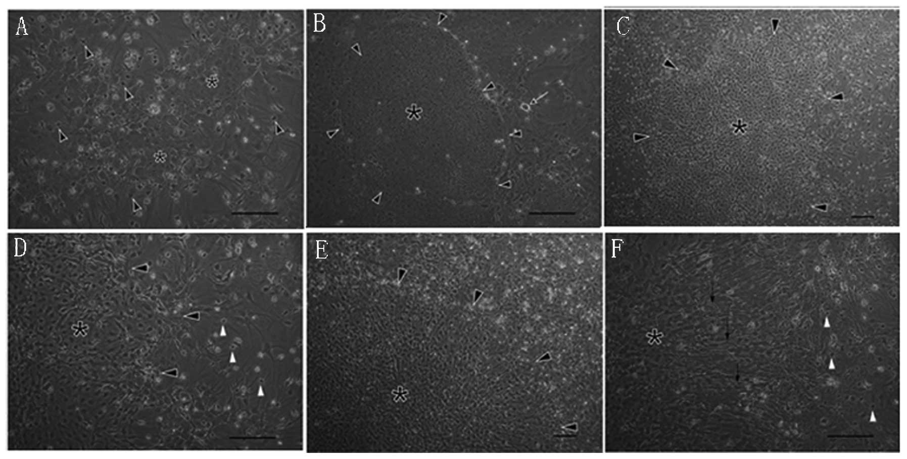
Confluent astrocyte enriched culture
exhibits distinct growth patterns
Following plating at a low density, cortical glial
cells proliferated, expanded and at 20 days, a confluent astrocyte
enriched culture was obtained. Under a phase contrast microscope
(BX45-72P15; Olympus Corporation), typical astrocyte glial cell
morphologies were observed as McCarthy and Vellis reported
(21), including a confluent glial
culture formed by two cellular layers. The top layer was composed
of dark, small and process-bearing cells and the bottom layer was
composed of light, large, fibroblast-like polygonal cells with
prominent nuclei and obscure profiles, and glial cells grew in a
random orientation pattern (Fig.
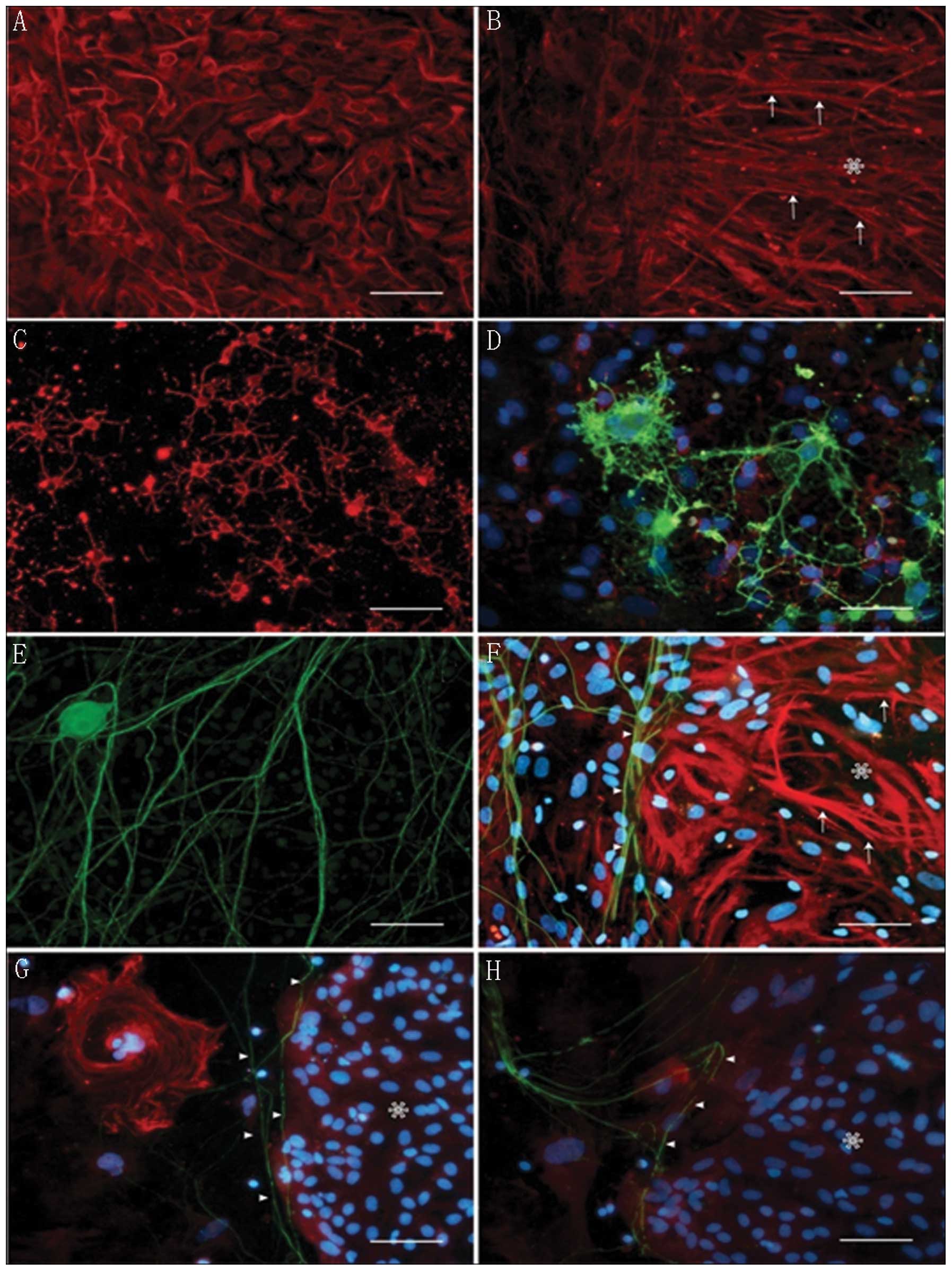
1A). Immunocytochemistry staining demonstrated that the bottom
cells were astrocytes (>95% GFAP+; Fig. 2A and B) and the top layer cells
were composed of immature (>95% O4+; Fig. 2C) and some mature
(CalC+; Fig. 2D)
oligodendrocytes. Typical astrocytes occupied the majority of the
growth area (>70% of the total growth area; Fig. 3C). Besides these typical astrocyte
growth patterns, certain atypical astrocyte growth patterns were
also observed, which occupied a minor growth area (<30% total
growth area; Fig. 3C). These
atypical growth patterns could be easily discriminated from typical
ones by several unique characteristics: These glial cells usually
grew together and formed homogenous cell clusters with few or no
oligodendrocytes on top of them (Figs.
1B–D and 3B) and were usually
small, with a high cell density and grew in random orientation
(Figs. 1B–D and 3A). Certain atypical glial cells
demonstrated a spindle-shaped morphology and polarized orientation
growth patterns (Figs. 1E, F and
2B). Immunocytochemistry
demonstrated that these atypical cells were also GFAP+
(Fig. 2B, F, G and H). The high
density and compact growth pattern of atypical glial cell clusters
formed separated special growth areas with cells of various sizes
(between 10 µm and 10 mm, composed of 100–10,000 cells) and
shapes (from nearly round to irregular). They distributed randomly
within the relatively homologous typical astrocytes giving the
whole glial culture a mosaic appearance.
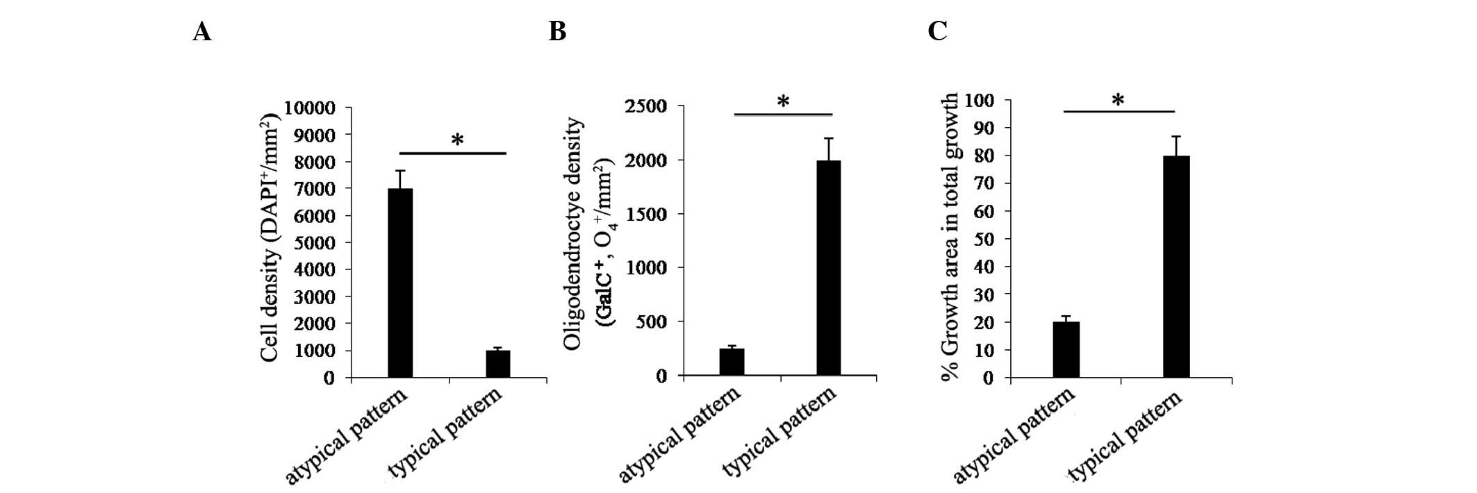
The differences between typical and atypical
astrocyte subpopulations were quantified and shown in Fig. 3A, B and I. The average cell density
of typical astrocytes was significantly lower than atypical
astrocytes (Fig. 3A; P<0.001),
however, the density of oligodendrocytes (O4+ and
GalC+) on top of typical astrocytes was significantly
higher than atypical astrocytes (Fig.
3B; P<0.001). The percentage growth area of typical
astrocytes was significantly higher than atypical astrocytes
(Fig. 3C; P<0.001).
Heterogeneous astrocyte growth patterns
exert different supportive properties on neurite growth and neuron
attachment
The coexistence of different growth patterns of
typical and atypical astrocytes in the same glial culture may, to a
certain degree, reflect the heterogeneity of astrocytes. However,
the functional significance of this heterogeneity remains to be
elucidated. Previous studies have demonstrated that typical
astrocytes prepared using the technique performed by McCarthy and
Vellis formed a good supportive substrate for neuron attachment and
growth (27). The coexistence of
typical and atypical astrocytes provided us with the opportunity to
investigate the effect of astrocyte heterogeneity on neurons. For
this purpose, high purified DRG neurons were added onto confluent
glial cultures and neuronal attachment and growth were evaluated in
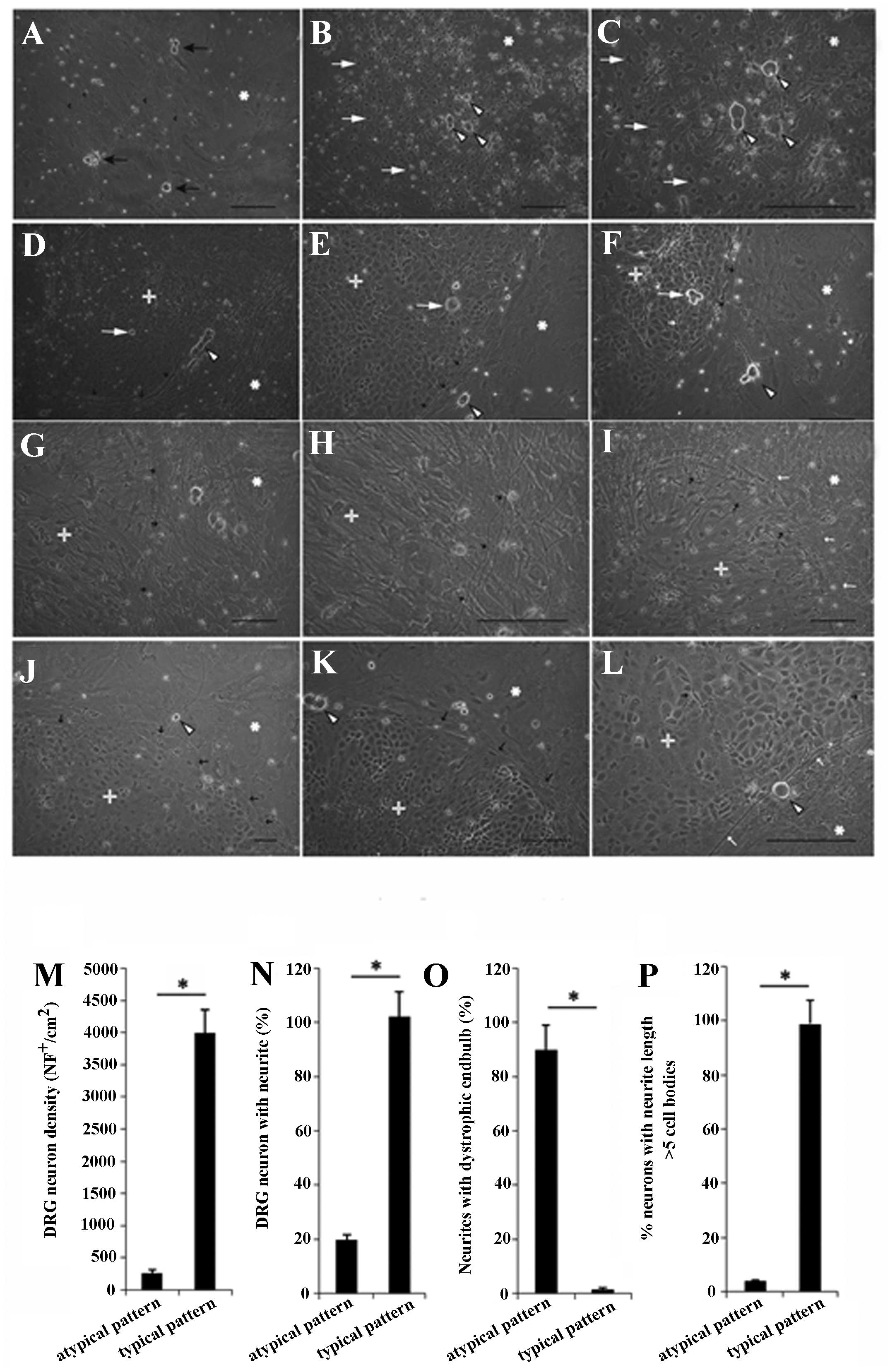
a qualitative and quantitative way. Within 2 h following addition,
DRG neurons settled on top of typical and atypical astrocytes as
single or small clusters (>3) with a similar density and this
loose attachment could be disrupted by partial disturbance of the
culture medium.
For the typical supportive astrocytes, at 6 h after
the addition of the neurons, the neuron attachment was tight and
washing under a medium flow did not allow resuspension of the
neurons. The majority of neurons initiated neurite outgrowth at
this stage and certain neurites extended as long as three cell
bodies. After 24 h, neurite length reached as long as five cell
bodies and at 48 h, the robust outgrowing neurites had formed a
network by connecting with each other and at this stage it was
difficult to discern individual neurites (Fig. 4A). Neurite growth on typical
astrocytes was not inhibited by oligodendrocytes and they grew in a
smooth and flexible way through oligodendrocytes without signs of
extra constraints or abrupt trajectory changes. Even in areas with
a high density of oligodendrocytes, the extending neurite tips
demonstrated no dystrophic end bulbs (Fig. 4B and C).
In contrast to typical astrocytes, atypical
astrocytes demonstrated different effects on DRG neurons. At 6 h
after addition, the majority of settled neuron attachments remained
relatively weak and they detached from the bottom astrocytes
following partial medium disturbance. After 24 h, less than half of
the neurons initiated neurite growth and the length of neurites was
less than three cell bodies. In certain areas, neurons had no
neurite growth after 72 h. Neurons could maintain a normal
morphology without neurite growth even after 7 days and had no
signs of cellular degeneration or death (Fig. 4D and E). Neurites in the atypical
area demonstrated a short, thick, stiff and straight appearance and
were limited within the atypical area, their terminals usually
ending with dystrophic end bulbs (Fig.
4F).
At the boundary of typical and atypical areas, the
dense neurites growing on the typical area usually avoided
penetrating into the neighboring neurite sparse atypical area
(Fig. 4G and H; Fig. 2F). Individual neurites altered
their growth course abruptly on approaching the atypical area and
selectively grew along the boundary on typical astrocytes (Fig. 4J and K; Fig. 2G and H). When short neurites from
typical astrocyte areas grew into the atypical area, their
morphology altered from a flexible curve thread-like morphology to
a stiff straight one (Fig. 4I). In
addition, neurites stopped growing with dystrophic end bulbs
terminals (Fig. 4L).
Quantitative assessment of the different neuron
attachments and neurite growth on typical and atypical areas are
shown in Fig. 4M–P. Neuron
attachment on the typical area was significantly higher than on
atypical astrocytes (Fig. 4M;
P<0.001) 72 h after seeding. Neurite initiation on atypical
astrocytes was markedly delayed (only 20% of neurons initiated
neurite growth) and on typical atrocytes, ~100% showed neurite
growth (Fig. 4N; P<0.001). The
majority of neurites on atypical astrocytes showed dystrophic end
bulbs but few were found on typical astrocytes (Fig. 4O; P<0.001). The percentage of
neurons with neurites longer than five cell bodies on atypical
astrocytes was significantly lower than typical astrocytes
(Fig. 4P; P<0.001).
Immunocytochemistry results demonstrated that typical and atypical
astrocytes were GFAP+ and neurites grew along their
boundary or moved away from crossing into the atypical astrocytes
(Fig. 2F–H).
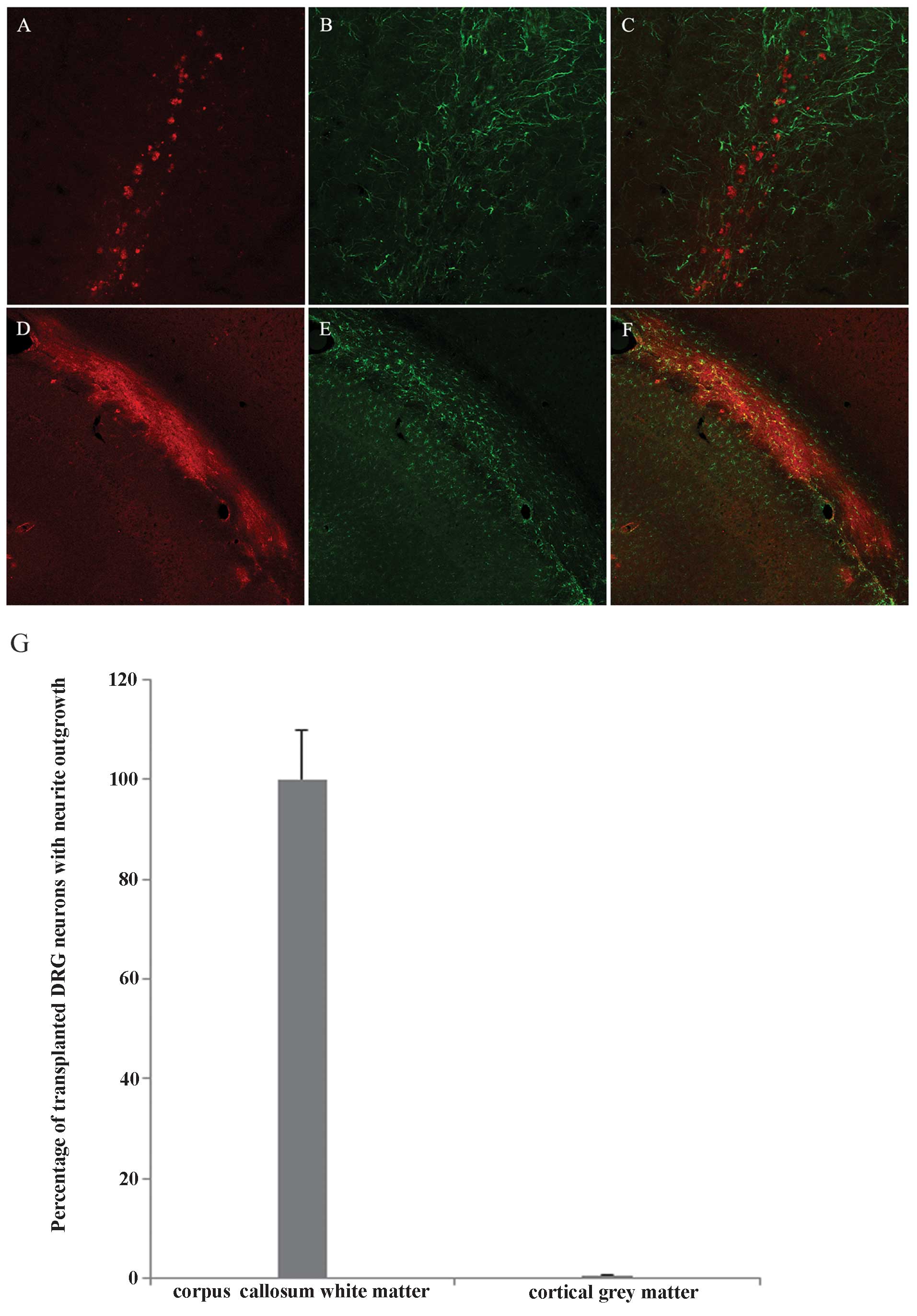
Transplanted DRG neurons show different
neurite growth behavior in cortical grey matter and white
matter
Using the microinjection method, purified DRG neuron
single suspensions were transplanted into cortical grey matter and
the underlying corpus callosum white matter. The growth of neurites
in these two regions was significantly different. DRG neurons in
the grey matter did not show neurite growth, neuron survival was
clearly observed in the injection site and no significant neurite
growth was observed. Grey matter astrocytes around the injection
site demonstrated a mild astrogliosis response to injection
(Fig. 5A–C). By contrast,
transplanted CGRP+ DRG neurons survived and demonstrated
robust axon outgrowth along the corpus callosum. White matter
astrocytes around the injection site demonstrated a mild
astrogliosis response to injection (Fig. 5D–F). All the transplanted DRG
neurons in the corpus callosum demonstrated extensive neurite
outgrowth, but no observable neurite outgrowth was detected in DRG
neurons transplanted into grey matter (Fig. 5G).
Discussion
Astrocytes are the most abundant glial cells in the
CNS. Recent advances in astrocyte research have markedly altered
our understanding of them (28).
In vivo and in vitro evidence indicates that
astrocytes are an extremely heterogeneous population based on their
morphology, antigenicity, receptors, channels, growth factors,
synaptic transmission and responses to brain-blood regulation
(1,28,29).
This heterogeneity reflects the diverse functions of
astrocytes during the development and maturation of the nervous
system. During development, transient glial cell boundaries are
found to restrict and guide axonal pathway finding processes
(30–32). It is well established that damaged
astrocytes inhibit neurite growth in vivo and in
vitro (21,33,34),
however, in normal culture conditions, astrocytes form a good
supportive substrate for neurite growth and alter the inhibitory
effect of oligodendrocytes on neurite regeneration (20,21).
Few studies have noted the effects of astrocyte heterogeneity on
neurite growth (35–37).
These earlier studies prompted us to examine the
effects of astrocyte heterogeneity on neuron growth using purified
DRG neurons with neonatal cortical mixed glial cell cultures. Our
results demonstrated that at least two astrocyte types coexist in
primary cortical astrocyte cultures. The first type were typical
astrocytes, which formed the majority of the culture area with
oligodendrocytes growing on top. These astrocytes supported DRG
neuron attachment and axon growth without inhibition and their
growth pattern was consistent with numerous previous studies
(38,39). The second astrocyte subpopulation
was composed of atypical astrocytes, which formed a small culture
area with few oligodendrocytes growing on top and had a high cell
density and polarized orientation. These atypical astrocytes
exerted strong inhibitory effects on neuron attachment and neurite
outgrowth without affecting neuron survival. When glial cells are
cultured using the method by McCarthy and Vellis, typical
astrocytes dominate the confluent cells and no atypical astrocyte
substructures are formed.
Several modifications of the classical McCarthy and
Vellis method in the present study may contribute to the atypical
astrocyte findings. To reduce earlier cell to cell contact
inhibition, cortical cells were plated at a low density
(1.0×10⁴/cm2 instead of
5.0–10×104/cm2) to prolong glial precursor
cell proliferation and expansion period before they reach
confluence. To maintain their intact state and heterogeneity as
much as possible, purification steps were omitted (this omission
did not change the results), without the purification step,
astrocyte purity was verified by GFAP to be >95% with few
contaminating cells. To minimize the effects from other cell types
during co-culture and monitor neuron behavior at a large scale
(100–1,000 cell cluster), highly purified DRG (99%) neurons were
selected to co-culture with astrocytes due to their
well-established strong intrinsic regeneration capacity, long
neurite growth and the fact that they are easily distinguished
under the microscope (14,40,41).
Atypical astrocytes affected neurons in numerous
ways, including inhibiting neuron attachment, inhibiting neurite
growth initiation, retarding axon growth and provided poor
neurotrophic support, however, they had no effect on cell survival.
Since the atypical inhibitory astrocytes are enclosed by supportive
typical astrocytes, the localized inhibitory effects implied that
the inhibition may be mediated by cellular membrane associated
molecules or short distance effect molecules.
Astrocytes cultured from different brain regions
using the method performed by McCarthy and Vellis have been broadly
used in glial neuron interaction studies (24,42,43).
They form a good substrate for various neurons and support neuron
attachment and neurite growth (23,39,44,45).
In vivo experiments in the present study also confirmed
this. In the present study, the mild astrocyte response at the
injection site indicated that the injury-induced glial response
does not account for the axon outgrowth pattern difference. Fibrous
astrocytes are supportive for axon growth, however, the
protoplasmic astrocytes in grey matter may be inhibitory to axon
growth. Based on several characteristics of the inhibitory
astrocytes, it was hypothesized that they may represent a distinct
astrocyte subtype for the following reasons: i) These cells express
GFAP and usually grow together to form certain substructures with a
similar morphology, which implies they are derived from a common
precursor cell; ii) these cells form inhibitory substructures that
are stable for up to 2 months (the longest time point in the
present study) without alterations (such as changing to supportive
flat cells); iii) neurons growing on them could survive for a long
period of time (2 months) without neurite extension and
degeneration; iv) following a long culture period (2 months), these
cells maintained their inhibitory effects on newly added DRG
neurons (data not shown); v) fibroblast as well as other cell type
(oligodendrocytes and microglia cells) markers were not expressed
on inhibitory astrocytes; vi) these cells demonstrated high
mobility following injury (data not shown).
As supplement to the current opinion about
astrocytes, the results of the present study indicated that
heterogeneous astrocytes are not homologous in their effects on
neuron behavior. A subpopulation of astrocytes exerted inhibitory
effects on neuron attachment and axon growth under normal culture
conditions. Elucidating the nature of these inhibitory astrocytes
may deepen our understanding of the complex glial environment in
normal and pathological conditions and our co-culture model may
also aid in identifying inhibitory astrocytes and the underlying
molecular mechanisms, as well as in developing treatments that may
improve axon regeneration.
References
|
1
|
Barres BA: The mystery and magic of glia:
A perspective on their roles in health and disease. Neuron.
60:430–440. 2008. View Article : Google Scholar : PubMed/NCBI
|
|
2
|
Allen NJ and Barres BA: Neuroscience: Glia
- more than just brain glue. Nature. 457:675–677. 2009. View Article : Google Scholar : PubMed/NCBI
|
|
3
|
Kimelberg HK: Functions of mature
mammalian astrocytes: A current view. Neuroscientist. 16:79–106.
2010. View Article : Google Scholar : PubMed/NCBI
|
|
4
|
Kettenmann H and Ransom BR: The concept of
neuroglia: a historical perpspective. Neuroglia. 2nd edition.
Oxford University Press; New York: pp. 1–9. 2005
|
|
5
|
Raff MC, Abney ER, Cohen J, Lindsay R and
Noble M: Two types of astrocytes in cultures of developing rat
white matter: Differences in morphology, surface gangliosides and
growth characteristics. J Neurosci. 3:1289–1300. 1983.PubMed/NCBI
|
|
6
|
Yong VW, Yong FP, Olivier A, Robitaille Y
and Antel JP: Morphologic heterogeneity of human adult astrocytes
in culture: Correlation with HLA-DR expression. J Neurosci Res.
27:678–688. 1990. View Article : Google Scholar : PubMed/NCBI
|
|
7
|
Bailey MS and Shipley MT: Astrocyte
subtypes in the rat olfactory bulb: morphological heterogeneity and
differential laminar distribution. J Comp Neurol. 328:501–526.
1993. View Article : Google Scholar : PubMed/NCBI
|
|
8
|
Cahoy JD, Emery B, Kaushal A, et al: A
transcriptome database for astrocytes, neurons and
oligodendrocytes: A new resource for understanding brain
development and function. J Neurosci. 28:264–278. 2008. View Article : Google Scholar : PubMed/NCBI
|
|
9
|
Doyle JP, Dougherty JD, Heiman M, et al:
Application of a translational profiling approach for the
comparative analysis of CNS cell types. Cell. 135:749–762. 2008.
View Article : Google Scholar : PubMed/NCBI
|
|
10
|
Ståhlberg A, Andersson D, Aurelius J, et
al: Defining cell populations with single-cell gene expression
profiling: Correlations and identification of astrocyte
subpopulations. Nucleic Acids Res. 39:e242011. View Article : Google Scholar :
|
|
11
|
Yeh TH, Lee da Y, Gianino SM and Gutmann
DH: Microarray analyses reveal regional astrocyte heterogeneity
with implications for neurofibromatosis type 1 (NF1-)regulated
glial proliferation. Glia. 57:1239–1249. 2009. View Article : Google Scholar : PubMed/NCBI
|
|
12
|
Zhang Y and Barres BA: Astrocyte
heterogeneity: An underappreciated topic in neurobiology. Curr Opin
Neurobiol. 20:588–594. 2010. View Article : Google Scholar : PubMed/NCBI
|
|
13
|
Wilkin GP, Marriott DR and Cholewinski AJ:
Astrocyte heterogeneity. Trends Neurosci. 13:43–46. 1990.
View Article : Google Scholar : PubMed/NCBI
|
|
14
|
Neumann S and Woolf CJ: Regeneration of
dorsal column fibers into and beyond the lesion site following
adult spinal cord injury. Neuron. 23:83–91. 1999. View Article : Google Scholar : PubMed/NCBI
|
|
15
|
Goldberg JL, Klassen MP, Hua Y and Barres
BA: Amacrine-signaled loss of intrinsic axon growth ability by
retinal ganglion cells. Science. 296:1860–1864. 2002. View Article : Google Scholar : PubMed/NCBI
|
|
16
|
Yiu G and He Z: Glial inhibition of CNS
axon regeneration. Nat Rev Neurosci. 7:617–627. 2006. View Article : Google Scholar : PubMed/NCBI
|
|
17
|
Chaudhry N and Filbin MT:
Myelin-associated inhibitory signaling and strategies to overcome
inhibition. J Cereb Blood Flow Metab. 27:1096–1107. 2007.
View Article : Google Scholar
|
|
18
|
Busch SA and Silver J: The role of
extracellular matrix in CNS regeneration. Curr Opin Neurobiol.
17:120–127. 2007. View Article : Google Scholar : PubMed/NCBI
|
|
19
|
Verma P, Garcia-Alias G and Fawcett JW:
Spinal cord repair: bridging the divide. Neurorehabil Neural
Repair. 22:429–437. 2008. View Article : Google Scholar : PubMed/NCBI
|
|
20
|
Silver J and Miller JH: Regeneration
beyond the glial scar. Nat Rev Neurosci. 5:146–156. 2004.
View Article : Google Scholar : PubMed/NCBI
|
|
21
|
Wanner IB, Deik A, Torres M, et al: A new
in vitro model of the glial scar inhibits axon growth. Glia.
56:1691–1709. 2008. View Article : Google Scholar : PubMed/NCBI
|
|
22
|
Ard MD, Bunge MB, Wood PM, Schachner M and
Bunge RP: Retinal neurite growth on astrocytes is not modified by
extracellular matrix, anti-L1 antibody, or oligodendrocytes. Glia.
4:70–82. 1991. View Article : Google Scholar : PubMed/NCBI
|
|
23
|
Fawcett JW, Fersht N, Housden L, Schachner
M and Pesheva P: Axonal growth on astrocytes is not inhibited by
oligodendrocytes. J Cell Sci. 103:571–579. 1992.PubMed/NCBI
|
|
24
|
McCarthy KD and de Vellis J: Preparation
of separate astroglial and oligodendroglial cell cultures from rat
cerebral tissue. J Cell Biol. 85:890–902. 1980. View Article : Google Scholar : PubMed/NCBI
|
|
25
|
Liu R, Lin G and Xu H: An efficient method
for dorsal root ganglia neurons purification with a one-time
anti-mitotic reagent treatment. PLoS One. 8:e605582013. View Article : Google Scholar : PubMed/NCBI
|
|
26
|
Paxinos G and Watson C: The Rat Brain in
Stereotaxic Coordinates. 6th. Academic Press; London: 2007
|
|
27
|
Biran R, Noble MD and Tresco PA: Directed
nerve outgrowth is enhanced by engineered glial substrates. Exp
Neurol. 184:141–152. 2003. View Article : Google Scholar : PubMed/NCBI
|
|
28
|
Seifert G, Schilling K and Steinhäuser C:
Astrocyte dysfunction in neurological disorders: a molecular
perspective. Nat Rev Neurosci. 7:194–206. 2006. View Article : Google Scholar : PubMed/NCBI
|
|
29
|
Shen Q, Wang Y, Kokovay E, et al: Adult
SVZ stem cells lie in a vascular niche: a quantitative analysis of
niche cell-cell interactions. Cell Stem Cell. 3:289–300. 2008.
View Article : Google Scholar : PubMed/NCBI
|
|
30
|
Jhaveri S: Midline glia of the tectum: a
barrier for developing retinal axons. Perspect Dev Neurobiol.
1:237–243. 1993.PubMed/NCBI
|
|
31
|
Wu DY, Schneider GE, Silver J, Poston M
and Jhaveri S: A role for tectal midline glia in the unilateral
containment of retinocollicular axons. J Neurosci. 18:8344–8355.
1998.PubMed/NCBI
|
|
32
|
Steindler DA: Glial boundaries in the
developing nervous system. Ann Rev Neurosci. 16:445–470. 1993.
View Article : Google Scholar : PubMed/NCBI
|
|
33
|
McKeon RJ, Schreiber R, Rudge J and Silver
J: Reduction of neurite outgrowth in a model of glial scarring
following CNS injury is correlated with the expression of
inhibitory molecules on reactive astrocytes. J Neurosci.
11:3398–3411. 1991.PubMed/NCBI
|
|
34
|
McKeon RJ, Jurynec MJ and Buck CR: The
chondroitin sulfate proteoglycans neurocan and phosphacan are
expressed by reactive astrocytes in the chronic CNS glial scar. J
Neurosci. 19:10778–10788. 1999.PubMed/NCBI
|
|
35
|
Grierson JP, Petroski RE, Ling DS and
Geller HM: Astrocyte topography and tenascin/cytotactin expression:
correlation with the ability to support neuritic outgrowth. Brain
Res Dev Brain Res. 55:11–19. 1990. View Article : Google Scholar : PubMed/NCBI
|
|
36
|
Fok-Seang J, Smith-Thomas LC, Meiners S,
et al: An analysis of astrocytic cell lines with different
abilities to promote axon growth. Brain Res. 689:207–223. 1995.
View Article : Google Scholar : PubMed/NCBI
|
|
37
|
Meiners S, Powell EM and Geller H: A
distinct subset of tenascin/CS-6-PG-rich astrocytes restricts
neuronal growth in vitro. J Neurosci. 15:8096–8108. 1995.PubMed/NCBI
|
|
38
|
Ard MD and Bunge RP: Heparan sulfate
proteoglycan and laminin immunoreactivity on cultured astrocytes:
relationship to differentiation and neurite growth. J Neurosci.
8:2844–2858. 1988.PubMed/NCBI
|
|
39
|
Powell EM, Meiners S, DiProspero NA and
Geller HM: Mechanisms of astrocyte-directed neurite guidance. Cell
Tissue Res. 290:385–393. 1997. View Article : Google Scholar : PubMed/NCBI
|
|
40
|
Xu XY, Li XT, Peng SW, et al: The
behaviour of neural stem cells on polyhydroxyalkanoate nanofiber
scaffolds. Biomaterials. 31:3967–3975. 2010. View Article : Google Scholar : PubMed/NCBI
|
|
41
|
Plant GW, Currier PF, Cuervo EP, et al:
Purified adult ensheathing glia fail to myelinate axons under
culture conditions that enable Schwann cells to form myelin. J
Neurosci. 22:6083–6091. 2002.PubMed/NCBI
|
|
42
|
Zwain IH and Yen SS: Neurosteroidogenesis
in astrocytes, oligodendrocytes, and neurons of cerebral cortex of
rat brain. Endocrinology. 140:3843–3852. 1999. View Article : Google Scholar : PubMed/NCBI
|
|
43
|
Tien AC, Tsai HH, Molofsky AV, et al:
Regulated temporal-spatial astrocyte precursor cell proliferation
involves BRAF signalling in mammalian spinal cord. Development.
139:2477–2487. 2012. View Article : Google Scholar : PubMed/NCBI
|
|
44
|
Ard MD, Schachner M, Rapp JT and Faissner
A: Growth and degeneration of axons on astrocyte surfaces: effects
on extracellular matrix and on later axonal growth. Glia.
9:248–259. 1993. View Article : Google Scholar : PubMed/NCBI
|
|
45
|
Siddiqui S, Horvat Broecker A and Faissner
A: Comparative screening of glial cell types reveals extracellular
matrix that inhibits retinal axon growth in a chondroitinase
ABC-resistant fashion. Glia. 57:1420–1438. 2009. View Article : Google Scholar : PubMed/NCBI
|