Introduction
The presence of pathogenic bacteria in the root
canal is the major cause of pulpal and periapical pathology
(1–3). Pathogens generate toxins, and induce
progressive and invasive damage to the periradicular tissue.
Enterococcus faecalis, which is a common drug-resistant
pathogen, is able to survive extreme conditions, including
starvation and an alkaline environment, and possesses several
virulence factors that cause persistent pulpal and periapical
infections (4,5). E. faecalis, and other common
pathogens, are able to activate the immune response and destroy
periapical tissue (6).
The balance between osteoblasts and osteoclasts is
responsible for bone resorption and formation (7). Osteoblasts are well known for their
role in bone formation, and are essential in the regeneration and
reconstruction of periapical tissue. Osteoblasts have several key
functions, including bone matrix synthesis, osteoblast regulation
of osteoclastogenesis, endocrine functions and hematopoietic stem
cell regulation (8). In addition,
osteoblasts are associated with various important regulatory
pathways, such as Wnt signaling and runt-related transcription
factor 2 (Runx2) (9,10).
Sodium hypochlorite (NaOCl) and chlorhexidine (CHX)
are common intracanal medicaments, which exert bactericidal effects
on pathogens in the root canal (11,12).
The purpose of root canal therapy is to effectively eradicate
pathogenic bacteria using mechanical instruments and antibacterial
agents. However, little information is currently available
regarding the effects of inactivated pathogens on periapical cells.
Since E. faecalis is frequently isolated in persistent
periapical infection, and due to the important role of osteoblasts
in periapical lesion recovery, the present study aimed to examine
the effects of inactivated E. faecalis on the proliferation
and induction of osteogenic differentiation in the pre-osteoblast
cell line MC3T3-E1. Furthermore, the relative expression levels of
osteogenic genes were detected following exposure to various
concentrations of inactivated E. faecalis.
Materials and methods
Bacteria and cell culture
E. faecalis [American Type Culture Collection
(ATCC)® 29212™; ATCC, Manassas, VA, USA] was streaked on
brain heart infusion agar (BHI; Difco; BD Biosciences, Franklin
Lakes, NJ, USA) and was cultured aerobically at 37°C for 24 h. A
single bacterial colony was inoculated into 5 ml BHI medium and
grown to the exponential phase [bacterial concentration,
~109 colony-forming units (CFU)/ml]. The MC3T3-E1
osteoblast cell line (ATCC, Manassas, VA, USA) was used in the
present study. Osteoblasts were cultured in minimum essential
medium, alpha modified (α-MEM; Invitrogen; Thermo Fisher
Scientific, Inc., Waltham, MA, USA) supplemented with 10% fetal
bovine serum (FBS; Hyclone; GE Healthcare Life Sciences, Logan, UT,
USA), 100 U/ml penicillin and 100 µg/ml streptomycin (Gibco; Thermo
Fisher Scientific, Inc.) at 37°C in a humidified atmosphere
containing 5% CO2.
Inactivation of E. faecalis
The E. faecalis culture (concentration,
109 CFU/ml) was centrifuged at 6,343 × g for 10
min at 4°C, and the supernatant was discarded. The E.
faecalis deposit was then resuspended in 5.25% NaOCl (Shanghai
ChemDo International Trade Co., Ltd., Shanghai, China) or 2% CHX
(Sigma-Aldrich; Merck Millipore, Darmstadt, Germany) for 5 min. The
NaOCl challenge was discontinued by 0.6% sodium thiosulfate, and
the CHX challenge was discontinued by 5% polysorbate 80 and 0.07%
soybean lecithin. To verify the complete inactivation of bacteria,
100 µl inactivated E. faecalis was spread onto BHI agar and
cultured at 37°C for 48 h; no cell growth indicated complete
inactivation. Furthermore, to remove the remnants of NaOCl or CHX
and the neutralizers, the inactivated E. faecalis cultures
were centrifuged 6,343 × g for 5 min at 4°C, and the
supernatants were discarded. Bacterial deposits were resuspended in
PBS after being washed twice in sterile PBS.
Cell proliferation assay
CHX- or NaOCl-inactivated E. faecalis was
gradually diluted ten-fold with α-MEM supplemented with 10% FBS,
100 U/ml penicillin and 100 µg/ml streptomycin. Osteoblasts were
seeded in 96-well tissue culture plates at 2×10⁴
cells/well, and were incubated at 37°C in a humidified atmosphere
containing 5% CO2 for 24 h. Once the cells were attached to the
bottom of the plates, the supernatant was removed and various
concentrations of inactivated E. faecalis (10–104
fold dilution) were added to the wells, in order to evaluate the
effects of inactivated E. faecalis on cell proliferation.
Cells without the addition of E. faecalis were used as the
control group. The medium was replaced every 2 days. Cell
proliferation was assessed using the Cell Counting kit-8 (CCK-8;
Dojindo Laboratories, Kumamoto, Japan) on days 1, 2, 4, 7 and 14
according to the manufacturer's instructions.
Induction of osteogenic
differentiation
For osteogenic induction, osteoblasts were seeded in
6-well plates at a density of 1×10⁵ cells/well, and were
incubated at 37°C in a humidified atmosphere containing 5% CO2.
After cells grew to 50% confluence, CHX- or NaOCl-inactivated E.
faecalis was diluted to 107, 106 and
105 CFU/ml using α-MEM medium (supplemented with 10%
FBS, 100 U/ml penicillin and 100 µg/ml streptomycin), and was added
to the wells. Osteogenic medium (10 mM β-glycerophosphate, 0.2 mM
ascorbic acid and 0.1 µM dexamethasone in α-MEM supplemented with
10% FBS, 100 U/ml penicillin and 100 µg/ml streptomycin) was used
as a positive control, and α-MEM medium was used as a negative
control. The 6-well plates were incubated at 37°C in a humidified
atmosphere containing 5% CO2, and the medium was replaced every 2
days. After 7 days, cells that were adherent to the bottom of the
wells were stained with 0.5% Alizarin Red S solution (pH 4.2;
Sigma-Aldrich; Merck Millipore), and images were captured using a
stereomicroscope (Carl Zeiss, Oberkochen, Germany). Red staining
indicated the presence of calcium deposits. For quantitative
analysis, cells that were adherent to the bottom of the 6-wells for
7 days were washed twice with sterile PBS. The bound dye was then
eluted with 1.5 ml 0.5 M hydrochloric acid-alcohol solution (37%
hydrochloric acid to dehydrated alcohol ratio, 1:24). Osteogenic
induction was quantified by determining the optical density of the
solution at 405 nm. Three independent experiments were conducted,
each of which was performed in triplicate.
Reverse transcription-quantitative
polymerase chain reaction (RT-qPCR)
The relative expression levels of osteogenic genes
under various conditions were analyzed by RT-qPCR. Induction of
osteogenic differentiation by inactivated E. faecalis was
detected as aforementioned. Total RNAs were isolated from
osteoblasts stimulated with 107 and 105
CFU/ml 2% CHX- or 5.25% NaOCl-inactivated E. faecalis for 2,
4, 7 and 14 days using TRIzol Max Bacterial RNA Isolation kit
(Invitrogen; Thermo Fisher Scientific, Inc.). For cDNA synthesis, 2
µg RNA was used as a template and was reverse transcribed using the
PrimeScript First Strand cDNA Synthesis kit (Takara Biotechnology
Co., Ltd., Dalian, China) according to the instructions of the
manufacturer. An additional reaction containing no reverse
transcriptase was used to confirm the absence of genomic DNA. The
primer sequences for the following osteogenic genes: Osteocalcin
(OCN), alkaline phosphatase (ALP), Runx2, osteopontin (OPN) and
osterix (OSX), are presented in Table
I. Total cDNA abundance between the test samples was normalized
using GAPDH as a control. qPCR was conducted using a RealMasterMix
SYBR Green PCR kit [cat. no. FP202; Tiangen Biotech (Beijing) Co.,
Ltd., Beijing, China] in a 7500 Fast Real-Time PCR system (Applied
Biosystems; Thermo Fisher Scientific, Inc.) and was replicated four
times in a 20 µl reaction mixture containing 8 µl 2.5X
RealMasterMix, 1 µl 20X SYBR solution, 0.5 µl primers (10 µM) and 5
ng cDNA. The reaction conditions were as follows: 1 cycle of 95°C
for 60 sec followed by 40 cycles of 95°C for 10 sec, 60°C for 40
sec. Relative gene expressions were calculated using the Cq method
(13).
 | Table I.Primers used to evaluate the
expression of osteogenic genes in MC3T3-E1 cells. |
Table I.
Primers used to evaluate the
expression of osteogenic genes in MC3T3-E1 cells.
| Gene | Primer sequences
(5′-3′) | Amplicon (bp) |
|---|
| OCN | F:
TGACCTCACAGATGCCAAGC; R: GCGCCGGAGTCTGTTCACTA | 94 |
| ALP | F:
CCAGTGAGCAGGACACGATG; R: TGAAGGGAGCCAGTCCAAAG | 108 |
| Runx2 | F:
GAACTACTCCGCCGAGCTCC; R: TGAAACTCTTGCCTCGTCCG | 107 |
| OPN | F:
TGGCAGTGATTTGCTTTTGC; R: GGGTGCAGGCTGTAAAGCTTC | 100 |
| OSX | F:
TCGTCTGACTGCCTGCCTAGT; R: TTGCCTGGACCTGGTGAGAT | 107 |
| GAPDH | F:
CGTGTTCCTACCCCCAATGT; R: TGTCATCATACTTGGCAGGTTTCT | 73 |
Statistical analysis
SPSS 18.0 (SPSS, Inc., Chicago, IL, USA) was used to
perform statistical analyses. One-way analysis of variance and
Tukey honestly significant difference test were used to compare the
proliferation of osteoblasts stimulated with E. faecalis
inactivated by the two drugs. Each experiment was performed in
triplicate. The same statistical tests were used to compare the
degree of mineralization and the relative expression levels of
osteogenic genes in osteoblasts stimulated with the various types
of inactivated E. faecalis. All data are presented as the
mean ± standard deviation. P<0.05 was considered to indicate a
statistically significant difference.
Results
Effects of inactivated E. faecalis on
osteoblast growth
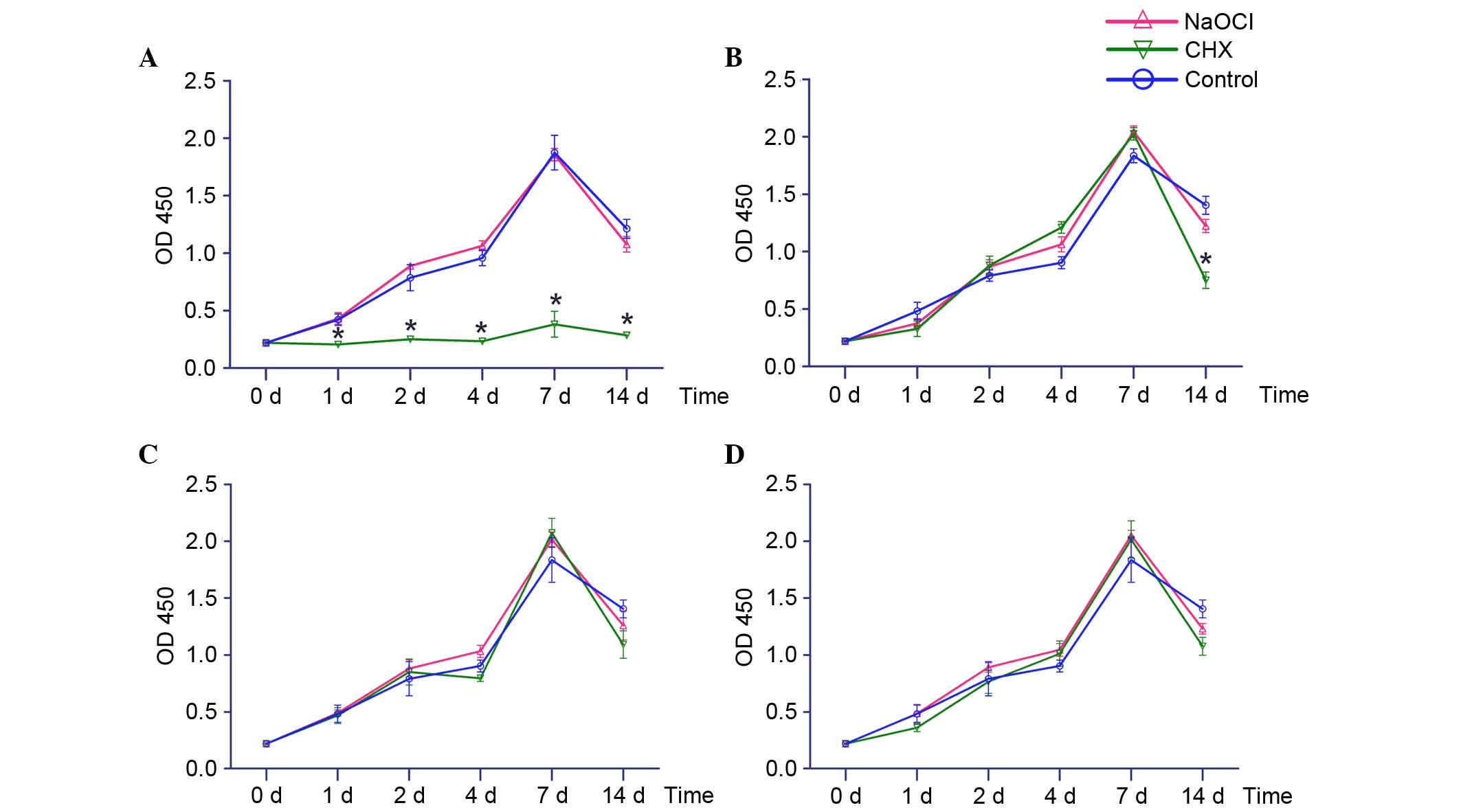
The CCK-8 assay was used to evaluate the effects of
inactivated E. faecalis on osteoblast proliferation
(Fig. 1). At a concentration of
108 CFU/ml, CHX-inactivated E. faecalis
significantly inhibited osteoblast growth at various time points;
however, NaOCl-inactivated E. faecalis did not suppress
osteoblast proliferation, and the osteoblast proliferation curve
was similar to the control group (Fig.
1A). At a concentration of 107 CFU/ml, osteoblasts
exhibited similar growth curves in the CHX-inactivated, NaOCl
-inactivated and control groups between 0 and 7 days; however,
osteoblast proliferation was significantly reduced in the
CHX-inactivated group compared with in the NaOCl-inactivated group
at day 14 (Fig. 1B). When
inactivated E. faecalis was diluted to 106 or
105 CFU/ml, osteoblast proliferation was similar in the
CHX-inactivated, NaOCl-inactivated and control groups (Fig. 1C and D).
Effects of inactivated E. faecalis on
osteogenic induction of osteoblasts
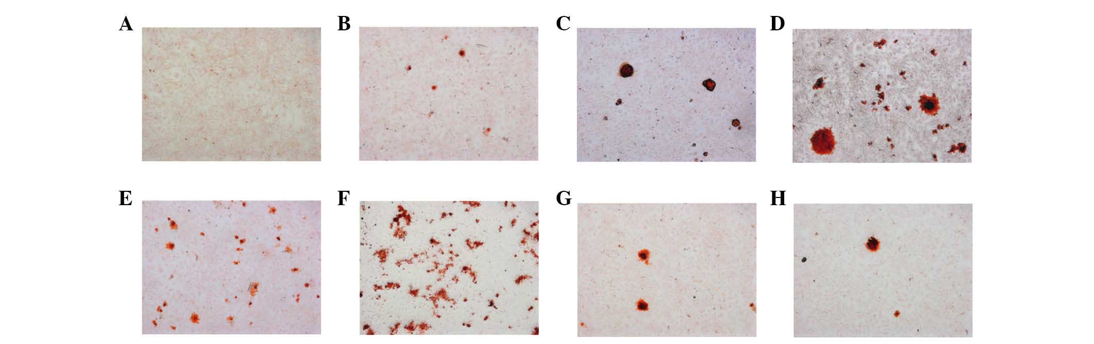
Alizarin red S staining was used to detect
mineralized nodules of osteoblasts stimulated by inactivated E.
faecalis (Fig. 2). Stimulation
of osteoblasts with 107 CFU/ml 2% CHX-inactivated E.
faecalis markedly induced the formation of several mineralized
nodules (Fig. 2F). The number of
mineralized nodules was reduced following stimulation with
106 or 105 CFU/ml E. faecalis;
however, the size of the mineralized nodules was markedly increased
(Fig. 2G and H). Stimulation of
osteoblasts with 107 CFU/ml 5.25% NaOCl-inactivated
E. faecalis induced a slight increase in mineralized nodules
(Fig. 2B), and the size of the
mineralized nodules was markedly increased following stimulation
with 106 CFU/ml E. faecalis, as compared with
107 CFU/ml E. faecalis. Stimulation with
105 CFU/ml E. faecalis resulted in induction of
the largest mineralized nodules (Fig.
2C and D).
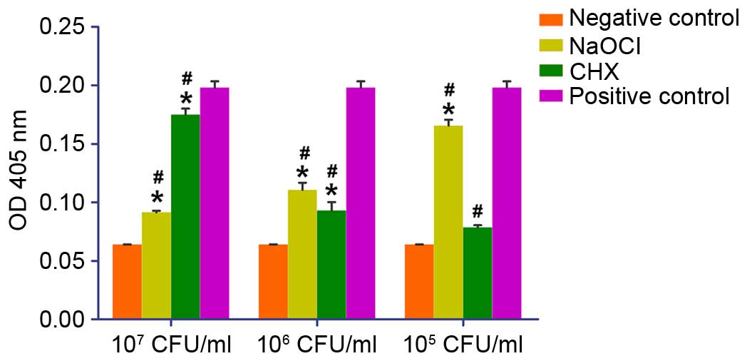
Quantification of the degree of osteoblast
mineralization demonstrated that there was a statistical difference
between cells stimulated with 107 CFU/ml 2% CHX- or
5.25%-inactivated NaOCl E. faecalis (P<0.05); and
mineralization was higher in both groups compared with the negative
control group. Following stimulation with 106 CFU/ml
inactivated E. faecalis, no statistically significant
difference was detected between the NaOCl-inactivated and
CHX-inactivated E. faecalis groups; however, mineralization
was higher in both groups compared with the negative control group,
and was lower compared with the positive control group (P<0.05).
At a concentration of 105 CFU/ml, stimulation with
NaOCl-inactivated E. faecalis resulted in a significantly
higher degree of mineralization compared with in the
CHX-inactivated E. faecalis group, and mineralization was
lower in both groups compared with in the positive control group
(P<0.05; Fig. 3).
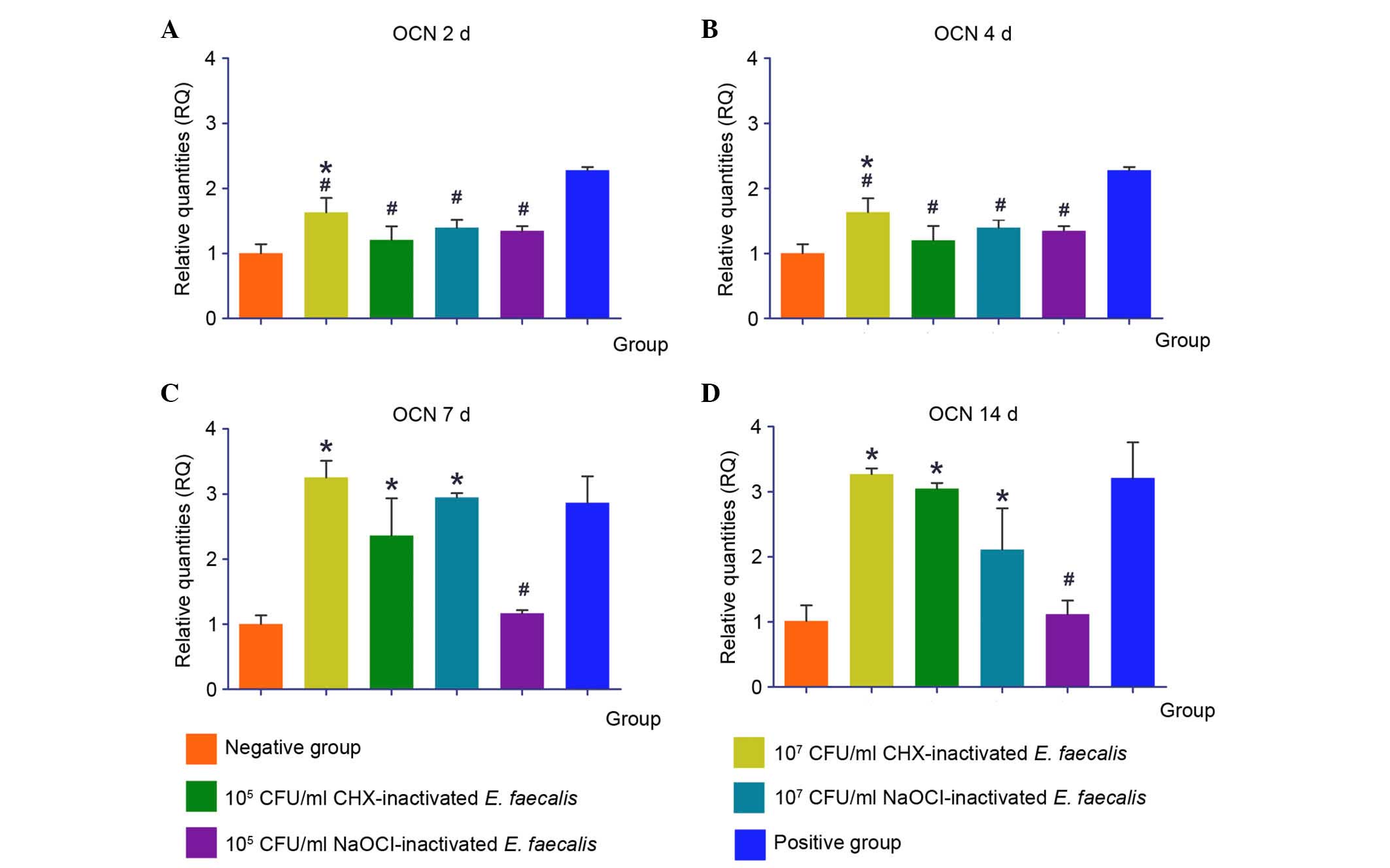
Expression of osteogenic genes in
response to inactivated E. faecalis
Osteoblasts were stimulated with 107
CFU/ml CHX-inactivated E. faecalis (107 CHX-Ef),
105 CFU/ml CHX-inactivated E. faecalis
(105 CHX-Ef), 107 CFU/ml NaOCl-inactivated
E. faecalis (107 NaOCl-Ef) or 105
CFU/ml NaOCl-inactivated E. faecalis (105
NaOCl-Ef). The expression levels of osteogenic genes were then
detected by RT-qPCR. OCN is an important marker of osteogenesis.
Following stimulation with 107 CHX-Ef for 2 or 4 days
the expression levels of OCN were significantly upregulated
compared with the negative control group, and were lower compared
with the positive control group (P<0.05). In addition,
stimulation with 107 CHX-Ef, 105 CHX-Ef or
107 NaOCl-Ef for 7 or 14 days induced the transcription
of OCN; the expression levels of OCN in these groups were
significantly increased compared with in the 105
NaOCl-Ef group (P<0.05; Fig.
4).
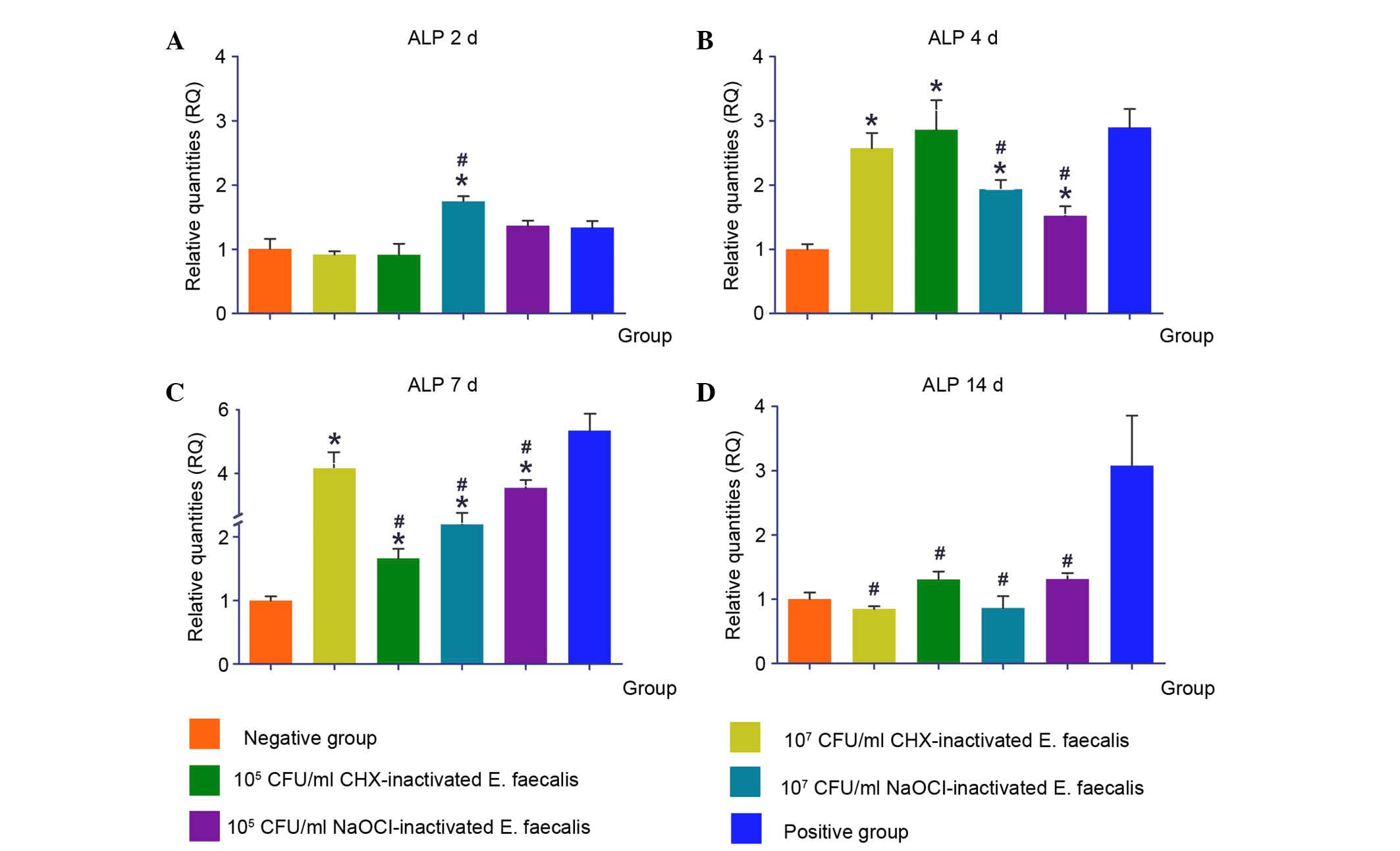
Following 2 days stimulation with
107 NaOCl-Ef the expression levels of ALP were
significantly increased
After 4 and 7 days stimulation with 107
CHX-Ef, 105 CHX-Ef, 107 NaOCl-Ef and
105 NaOCl-Ef the expression levels of ALP were
upregulated. However, after 14 days, the various concentrations of
inactivated E. faecalis did not induce upregulation of ALP,
and the expression levels were similar to those in the negative
control group (Fig. 5).
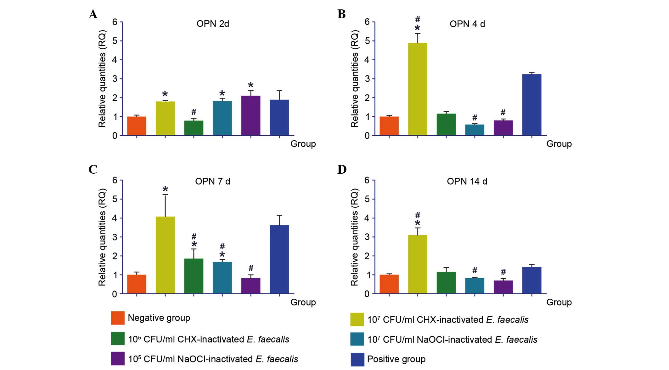
Following 2 days of stimulation with 107
CHX-Ef, 107 NaOCl-Ef and 105 NaOCl-Ef, the
expression levels of OPN were upregulated. Stimulation with
107 CHX-Ef for 4, 7 or 14 days induced the transcription
of OPN to the highest levels, which were higher than in the
positive control group. The expression levels of OPN were not
significantly different between the 105 CHX-Ef,
107 NaOCl-Ef and 105 NaOCl-Ef groups after 4
or 14 days stimulation (Fig.
6).
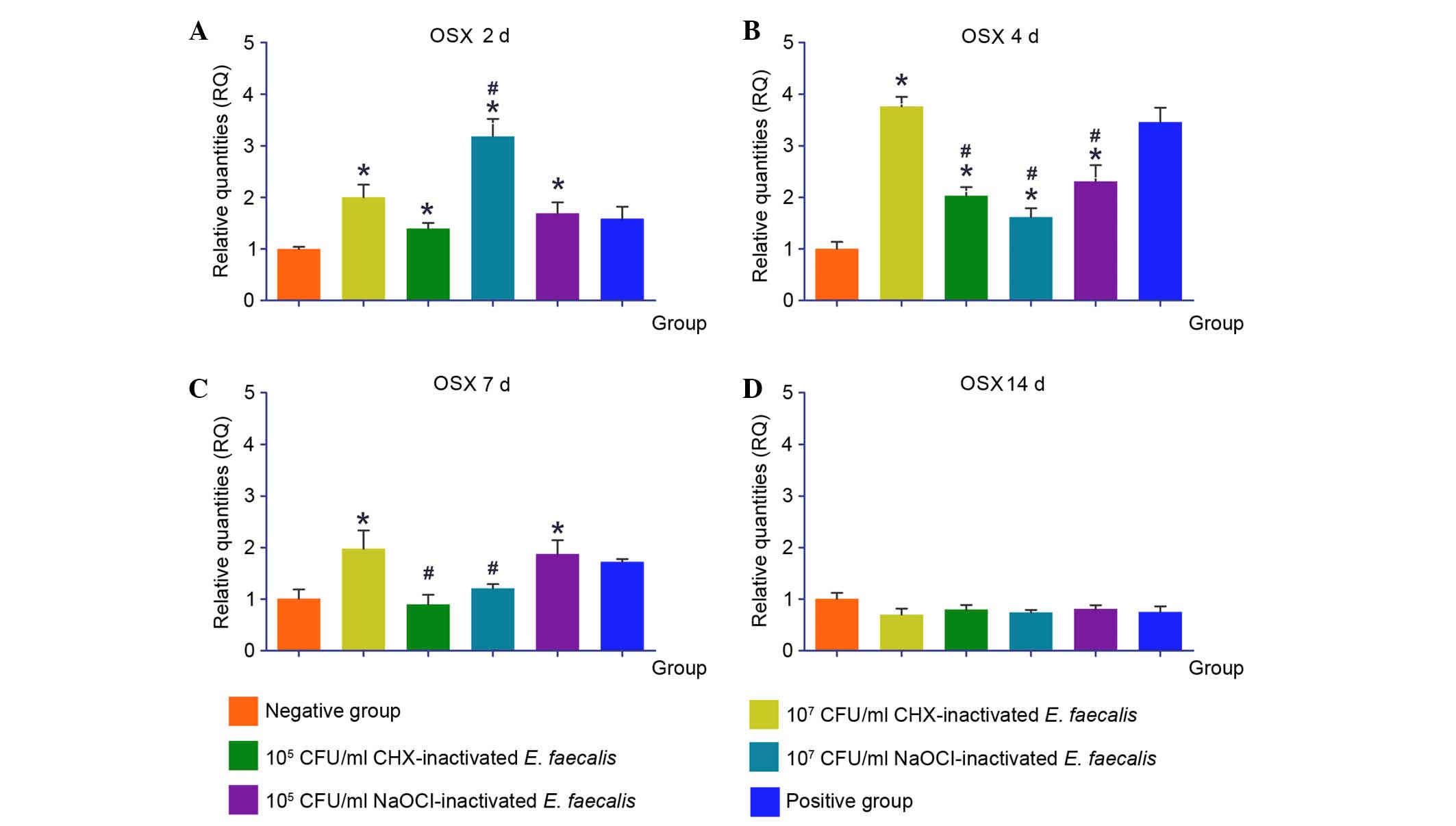
OSX is a vital regulatory factor for osteoblast
differentiation and bone development. The expression levels of OSX
were upregulated in response to stimulation with 107
CHX-Ef, and the highest level was reached after 4 days. Similarly,
following stimulation with 105 CHX-Ef, transcription of
OSX was induced to its highest level after 4 days, and was
decreased to normal levels after 7 and 14 days. After 2 and 4 days
of stimulation, 107 NaOCl-Ef and 105 NaOCl-Ef
upregulated OSX expression. Furthermore, following 14 days of
stimulation, the expression levels of OSX in the experimental
groups were reduced to negative control levels (Fig. 7).
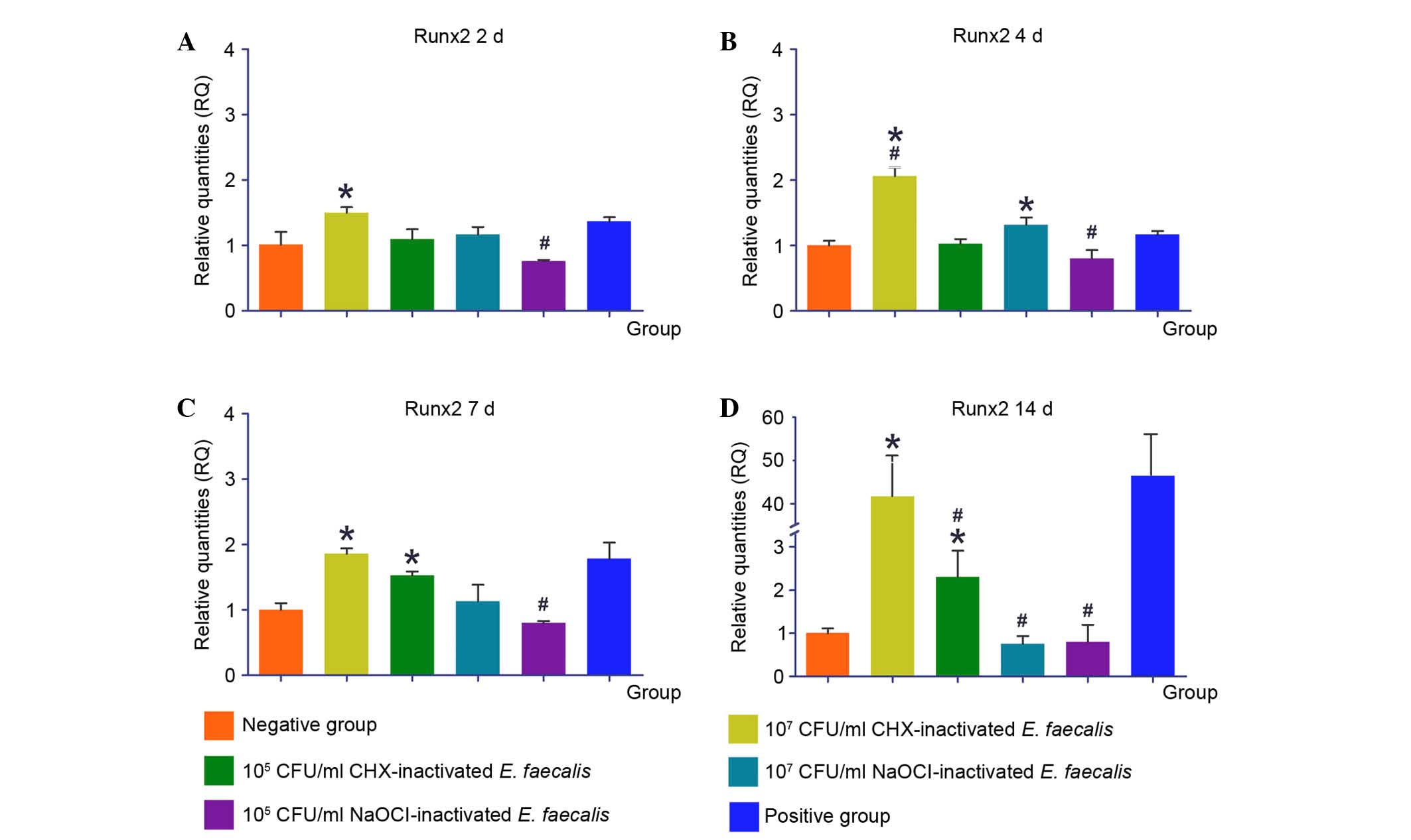
Runx2 is an important transcription factor
associated with bone formation. Transcription of Runx2 was
upregulated in response to stimulation with 107 CHX-Ef,
and a maximum was reached after 14 days. Following stimulation with
105 CHX-Ef, the upregulation of Runx2 was observed after
7 and 14 days. NaOCl-inactivated E. faecalis did not induce
the upregulation of Runx2 compared with CHX-inactivated E.
faecalis during the 14 days of stimulation (Fig. 8).
Discussion
E. faecalis is one of the most commonly
implicated pathogens in persistent and secondary root canal
infection (14,15). The objective of antibacterial
intracanal medicament application is to inactivate pathogens, such
as E. faecalis, in the root canal. It is well known that
living pathogens exert pathogenicity and destroy periapical tissue;
however, when living pathogens in periapical tissue are killed by
intracanal medicaments and are not completely cleared, it remains
unknown as to the effects of the dead bacteria on periapical bone
tissue healing. The present study demonstrated that high
concentrations of E. faecalis inactivated by CHX
significantly inhibited osteoblast proliferation, whereas
NaOCl-inactivated E. faecalis did not affect osteoblast
growth. E. faecalis has several virulence factors, including
lytic enzymes, cytolysin, aggregation substance, pheromones and
lipoteichoic acid (16). CHX at a
concentration of 2% possesses bactericidal activity and induces
precipitation of cytoplasmic contents (17). The cytoplasmic contents of E.
faecalis contain numerous virulence factors, and exert
pathogenicity. Therefore, osteoblast growth was suppressed by the
efflux of pathogenic cytoplasmic components in CHX-inactivated
E. faecalis. NaOCl exerts its bactericidal mechanism via
chlorine. NaOCl releases chlorine, which forms chloramines that
interfere with cell metabolism when exposed to organic tissue.
Chlorine generates irreversible oxidation of sulfhydryl groups of
essential bacterial enzymes, thus inhibiting bacterial enzyme
action and resulting in degradation of E. faecalis virulence
factors (18). Therefore,
osteoblast proliferation was not affected by NaOCl-inactivated
E. faecalis due to the degradation of virulence factors.
The presence of mineralized nodules is a marker of
osteogenic differentiation. Analysis of the degree of osteoblastic
mineralization demonstrated that low concentrations of
NaOCl-inactivated E. faecalis and high concentrations of
CHX-inactivated E. faecalis induced increased levels of
mineralization. High concentrations of CHX can destroy large
amounts of E. faecalis, thus leading to the increased efflux
of cytoplasmic compounds, which may stimulate the generation of
mineralized nodules. This is a self-defense mechanism of
osteoblasts, and mineralized nodules defend osteoblasts against
virulence factors (19). However,
it remains unclear as to how low concentrations of
NaOCl-inactivated E. faecalis increased the number of
mineralized nodules. It may be hypothesized that NaOCl-inactivated
E. faecalis generates a certain substance that inhibits
mineralized nodule formation, and when the substance is at low
concentrations it may not be enough to hinder osteogenesis of
osteoblasts. This hypothesis requires further study.
OCN is a marker of bone formation, which has a
regulatory role in mineralization (20,21).
CHX-inactivated E. faecalis significantly upregulated the
expression levels of OCN after 7 and 14 days, thus indicating that
the virulence factors released by E. faecalis may induce
osteogenesis of osteoblasts. Furthermore, 107 CFU/ml
NaOCl-inactivated E. faecalis also induced OCN expression,
which indicated that the substances released by NaOCl-inactivated
E. faecalis may still induce the formation of OCN, but only
at high concentrations. The expression levels of Runx2 exhibited
similar alterations to OCN gene expression following stimulation
with CHX or NaOCl-inactivated E. faecalis. Runx2 acts
upstream of OCN, and regulates OCN gene transcription. Runx2 serves
a key role in osteoblast differentiation (9). OPN has an important role in
biomineralization, and inhibits the formation and growth of
hydroxyapatite and other biominerals (22). The expression levels of OPN were
not significantly upregulated following stimulation with
inactivated E. faecalis, which were conducive to
biomineralization of osteoblasts. However, 107 CFU/ml
CHX-inactivated E. faecalis was still able to induce
transcription of OPN. The high concentrations of bacterial
virulence factors may induce production of OPN and activate bone
resorption. ALP activity is considered a biochemical marker for
osteoblastic activity (23). The
present study demonstrated that ALP expression was upregulated
after 4 and 7 days stimulation, thus indicating that the
osteoblastic activity of osteoblasts reached the active phase after
4 or 7 days stimulation with CHX- and NaOCl-inactivated E.
faecalis.
OSX is a key transcription factor for osteoblast
differentiation and bone formation, which acts downstream of Runx2
to regulate the expression of several osteogenic factors (24,25).
OSX is a marker of the earlier stages of osteoblast
differentiation. The present study indicated that upregulation of
OSX occurred following 2 and 4 days of exposure to inactivated
E. faecalis, whereas after 14 days, the expression levels of
OSX were not significantly altered. Similar changes in expression
were detected in the positive control group; osteogenic medium did
not induce OSX expression at the late stage. This result indicated
that OSX serves a role in the earlier stages of osteoblast
differentiation.
Metabolites and virulence factors are released after
bacteria are killed by antimicrobial agents. NaOCl exerts
non-specific proteolytic effects, and the substances released by
E. faecalis may be partly degraded (12). The substances released by the
inactivated E. faecalis are very complex and may be
partially degraded by non-specific proteolytic effects of NaOCl
(12). Certain substances may not
be degraded and remain. Therefore, the complex compositions may
induce the different expression of osteogenic genes. CHX does not
exert proteolytic effects, and the pathogenic factors are
predominantly from the substances released by E. faecalis,
and thereby exhibited concentration-dependent regulation of
osteogenic gene transcription.
In conclusion, CHX-inactivated E. faecalis
may inhibit osteoblast proliferation, whereas NaOCl-inactivated
E. faecalis did not affect osteoblast growth. CHX- and
NaOCl-inactivated E. faecalis were associated with different
levels of osteogenic differentiation of osteoblasts, and activated
the expression of osteogenic genes. Additional studies are required
to establish a standard protocol for the use of intracanal
medications in the clinic. The results of the current study
suggested that NaOCl irrigant may be superior to CHX irrigant
regarding its effects on the osteogenic differentiation of
osteoblasts.
Acknowledgements
This study was supported by grants from the
Guangzhou Science Technology Project (201607010271) and the
Guangdong Natural Science Foundation (2014A030313026).
References
|
1
|
Narayanan LL and Vaishnavi C: Endodontic
microbiology. J Conserv Dent. 13:233–239. 2010. View Article : Google Scholar : PubMed/NCBI
|
|
2
|
Peciuliene V, Maneliene R, Balcikonyte E,
Drukteinis S and Rutkunas V: Microorganisms in root canal
infections: A review. Stomatologija. 10:4–9. 2008.PubMed/NCBI
|
|
3
|
Rôças IN and Siqueira JF Jr: Root canal
microbiota of teeth with chronic apical periodontitis. J Clin
Microbiol. 46:3599–3606. 2008. View Article : Google Scholar : PubMed/NCBI
|
|
4
|
Portenier I, Waltimo TMT and Haapasalo M:
Enteroccus faecalis - the root canal survivor and ‘star’ in
posttreatment disease. Endod Top. 6:135–160. 2003. View Article : Google Scholar
|
|
5
|
Stuart CH, Schwartz SA, Beeson TJ and
Owatz CB: Enterococcus faecalis: Its role in root canal treatment
failure and current concepts in retreatment. J Endod. 32:93–98.
2006. View Article : Google Scholar : PubMed/NCBI
|
|
6
|
Siqueira JF Jr and Rôças IN: Bacterial
pathogenesis and mediators in apical periodontitis. Braz Dent J.
18:267–280. 2007. View Article : Google Scholar : PubMed/NCBI
|
|
7
|
Cao X: Targeting osteoclast-osteoblast
communication. Nat Med. 17:1344–1346. 2011. View Article : Google Scholar : PubMed/NCBI
|
|
8
|
Capulli M, Paone R and Rucci N: Osteoblast
and osteocyte: Games without frontiers. Arch Biochem Biophys.
561:3–12. 2014. View Article : Google Scholar : PubMed/NCBI
|
|
9
|
Bruderer M, Richards RG, Alini M and
Stoddart MJ: Role and regulation of RUNX2 in osteogenesis. Eur Cell
Mater. 28:269–286. 2014.PubMed/NCBI
|
|
10
|
Zhang R, Oyajobi BO, Harris SE, Chen D,
Tsao C, Deng HW and Zhao M: Wnt/beta-catenin signaling activates
bone morphogenetic protein 2 expression in osteoblasts. Bone.
52:145–156. 2013. View Article : Google Scholar : PubMed/NCBI
|
|
11
|
Kanisavaran ZM: Chlorhexidine gluconate in
endodontics: An update review. Int Dent J. 58:247–257. 2008.
View Article : Google Scholar : PubMed/NCBI
|
|
12
|
Mohammadi Z: Sodium hypochlorite in
endodontics: An update review. Int Dent J. 58:329–341. 2008.
View Article : Google Scholar : PubMed/NCBI
|
|
13
|
Livak KJ and Schmittgen TD: Analysis of
relative gene expression data using real-time quantitative PCR and
the 2(−Delta Delta C(T)) Method. Methods. 25:402–408. 2001.
View Article : Google Scholar : PubMed/NCBI
|
|
14
|
Naghmouchi K, Le Lay C, Baah J and Drider
D: Antibiotic and antimicrobial peptide combinations: Synergistic
inhibition of Pseudomonas fluorescens and antibiotic-resistant
variants. Res Microbiol. 163:101–108. 2012. View Article : Google Scholar : PubMed/NCBI
|
|
15
|
Collins B, Cotter PD, Hill C and Ross RP:
The impact of nisin on sensitive and resistant mutants of Listeria
monocytogenes in cottage cheese. J Appl Microbiol. 110:1509–1514.
2011. View Article : Google Scholar : PubMed/NCBI
|
|
16
|
Piper C, Draper LA, Cotter PD, Ross RP and
Hill C: A comparison of the activities of lacticin 3147 and nisin
against drug-resistant Staphylococcus aureus and Enterococcus
species. J Antimicrob Chemother. 64:546–551. 2009. View Article : Google Scholar : PubMed/NCBI
|
|
17
|
Ghiselli R, Giacometti A, Cirioni O,
Dell'Acqua G, Mocchegiani F, Orlando F, D'Amato G, Rocchi M,
Scalise G and Saba V: RNAIII-inhibiting peptide and/or nisin
inhibit experimental vascular graft infection with
methicillin-susceptible and methicillin-resistant Staphylococcus
epidermidis. Eur J Vasc Endovasc Surg. 27:603–607. 2004. View Article : Google Scholar : PubMed/NCBI
|
|
18
|
Hartke A, Giard JC, Laplace JM and Auffray
Y: Survival of Enterococcus faecalis in an oligotrophic microcosm:
Changes in morphology, development of general stress resistance,
and analysis of protein synthesis. Appl Environ Microbiol.
64:4238–4245. 1998.PubMed/NCBI
|
|
19
|
Katono T, Kawato T, Tanabe N, Suzuki N,
Iida T, Morozumi A, Ochiai K and Maeno M: Sodium butyrate
stimulates mineralized nodule formation and osteoprotegerin
expression by human osteoblasts. Arch Oral Biol. 53:903–909. 2008.
View Article : Google Scholar : PubMed/NCBI
|
|
20
|
Lombardi G, Perego S, Luzi L and Banfi G:
A four-season molecule: Osteocalcin. Updates in its physiological
roles. Endocrine. 48:394–404. 2015. View Article : Google Scholar : PubMed/NCBI
|
|
21
|
Brennan-Speranza TC and Conigrave AD:
Osteocalcin: An osteoblast-derived polypeptide hormone that
modulates whole body energy metabolism. Calcif Tissue Int. 96:1–10.
2015. View Article : Google Scholar : PubMed/NCBI
|
|
22
|
Gericke A, Qin C, Spevak L, Fujimoto Y,
Butler WT, Sørensen ES and Boskey AL: Importance of phosphorylation
for osteopontin regulation of biomineralization. Calcif Tissue Int.
77:45–54. 2005. View Article : Google Scholar : PubMed/NCBI
|
|
23
|
Farley JR, Hall SL, Tanner MA and Wergedal
JE: Specific activity of skeletal alkaline phosphatase in human
osteoblast-line cells regulated by phosphate, phosphate esters, and
phosphate analogs and release of alkaline phosphatase activity
inversely regulated by calcium. J Bone Miner Res. 9:497–508. 1994.
View Article : Google Scholar : PubMed/NCBI
|
|
24
|
Lee SH, Jeong HM, Han Y, Cheong H, Kang BY
and Lee KY: Prolyl isomerase Pin1 regulates the osteogenic activity
of Osterix. Mol Cell Endocrinol. 400:32–40. 2015. View Article : Google Scholar : PubMed/NCBI
|
|
25
|
Zhang C: Transcriptional regulation of
bone formation by the osteoblast-specific transcription factor Osx.
J Orthop Surg Res. 5:372010. View Article : Google Scholar : PubMed/NCBI
|