Introduction
The rates of overweight and obesity have increased
over recent decades (1–3) with the incidences of pre-diabetes and
diabetes (4,5). Obesity is characterized by
hyperlipidemia, proliferation and hypertrophy in adipocytes with
the deposition of collagen I in adipose tissue (6–9).
Deposition of collagen I in adipose tissue is associated with
insulin sensitivity (10).
Previous reports have demonstrated an inverse correlation between
adipocyte size and collagen I deposition, which is a hallmark of
fibrosis (11–13). These findings suggest a role for
fibrosis in negatively regulating adipocyte hypertrophy.
Over-expression of collagen I adipose tissue in
obesity has been well studied; however, the effect of TLR2 on
collagen I remains unclear. TLR2, along with TLR1 and TLR6,
recognizes a wide variety of pathogen-associated molecules
including lipoproteins, peptidoglycans, lipophilic acids, zymosan,
mannan, and free fatty acid (FFA) (14). Plasma FFA could activate TLR2
(15) and downstream inflammatory
factors, which regulate MMPs and TIMPs (8). MMPs and TIMPs regulate the deposition
of collagen I, which is one of the most important components of the
extracellular matrix (ECM) (16,17).
ECM levels in adipose tissue are also increased in human and murine
obesity (10,13,18,19).
In the pesent study, the role of TLR2 in the deposition of collagen
I in adipose tissue was explored in TLR2 gene knockout mice fed a
high-fat diet.
Materials and methods
Animals and groups
Male C57BL/6J mice [SCXK (Jing) 2009–0015] and
age-matched TLR2 gene knockout mice (022507) were purchased from
HFK (Beijing HFK Bioscience Co., Ltd., Beijing, China) and Jackson
Laboratory (Bar Harbor, ME, USA), respectively. Mice were divided
in 4 groups: Normal control mice (C57BL/6J) fed common chow (NC),
TLR2 knockout mice fed common chow (TK), C57BL/6J mice fed a
high-fat (60%) diet to model obesity (OB), and TLR2 knockout mice
fed a high-fat diet to model obesity (TO). To monitor weight gain,
mice were weighed every 2 weeks, especially the following 3
important time points. Prior to the start of the experiment (4
weeks of age), after the 4th week of the experiment (8 weeks of
age) and at the end of the experiment (20 weeks of age). All animal
experiments were approved by the Ethics Committee for Experimental
Research, Jinzhou Medical University.
Biochemical index measurements
Blood samples were collected from hearts prior to
sacrifice. Fasting plasma glucose (FPG) and levels of triglyceride
(TG), total cholesterol (TC), high-density lipoprotein (HD),
low-density lipoprotein (LDL) and FFA were measured.
Hematoxylin & eosin and Masson
staining
Adipose tissue was collected from fresh sacrificed
mice and fixed with 4% paraformaldehyde for 48 h before paraffin
sections were obtained. H&E and Masson staining were conducted
according to the manufacturer's protocol (Wanleibio Co., Ltd.,
Shenyang, China).
Immunohistochemistry
Section, 5 µm in thickness, were incubated with
rabbit-anti-mouse collagen I (Beijing Biosynthesis Biotechnology
Co., Ltd., Beijing, China) primary antibody overnight at 4°C. Then
slides were washed and incubated with 1:5,000 goat-anti-rabbit
secondary antibody (Absci, Nanjing, China) for 4 h. A DAB kit
(OriGene Technologies, Inc., Beijing, China) was used to bind
secondary antibody, according to the manufacturer's protocol.
Western blotting
Total proteins in adipose tissue were extracted with
RIPA (with 1% PMSF). BCA assay was used to detect protein
concentrations in middle extracting solution. Targeting proteins
were separated by SDS-PAGE, transferred to PVDF membranes and
blocked with skim milk for 2 h at room temperature. Rabbit
anti-mouse primary antibodies to collagen I (Wanleibio Co., Ltd.),
MMP2 (Wanleibio Co., Ltd.), TIMP1 (Wanleibio Co., Ltd.), TLR2
(Beijing Biosynthesis Biotechnology Co., Ltd.), myd88 (Beijing
Biosynthesis Biotechnology Co., Ltd.), TGFβ1 (Wanleibio Co., Ltd.),
p38 MAPK (Bisson, Beijing, China), P-P38MAPK (Wanleibio Co., Ltd.),
and β-actin (Absci) were used to bind targeting proteins. An
ECL-sensitive kit (Wanleibio Co., Ltd.) was used to detect goat
anti-rabbit secondary antibody binding.
RNA extraction and reverse
transcription
Total RNA was extracted from adipose tissue using
TRIzol reagent, then dissolved in RNase free water with RNase
inhibitor. Prior to cDNA synthesis, RNA was monitored by
Nandrop2000, and DEPC water was added to a concentration of 100
µg/µl. Then DNA was removed in a mix (10 µl) with RNA (1 µl), 5X
gDNA eraser buffer (2 µl), gDNA eraser (1 µl), and RNase free dH2O
(6 µl). cDNA was synthesized in a mix (20 µl) with primer script RT
enzyme mix (1 µl), RT primer mix (1 µl), 5X primer script buffer (4
µl), RNase free dH2O (4 µl), and the mix (10 µl) from the previous
step. Reverse transcription was performed in 20-µl reactions at
25°C for 10 min, followed by 37°C for 15 min, and finally
denaturation at 85°C for 5 min. cDNA was stored at −80°C until
further use. All experiments involving the PrimeScript™ RT reagent
kit with gDNA eraser (Takara Bio, Inc., Otsu, Japan) were performed
according to the manufacturer's.
Polymerase chain reaction (PCR)
Expression levels of IL-6 mRNA, TNF-α
mRNA collagen I α1 mRNA, and collagen I α2
mRNA were measured in a 20-µl System with 2XGreenStar Master
mix (10 µl), forward primer (1 µl, 10 pmol/µl), reverse primer (1
µl, 10 pmol/µl), DEPC water (6 µl), and template DNA (2 µl)
obtained by reverse transcription. Primers were designed and
synthesized by Sangon Biotech (Sangon Biotech Co., Ltd, Shanghai,
China) (Table I). All reactions
were performed in a 20-µl volume for 30 sec at 95°C, followed by 45
cycles of 95°C for 5 sec, and 60°C for 34 sec. Relative
quantification of gene expression was performed using the
comparative 2−ΔΔCt method. GAPDH was used as a validated
reference gene. Real-time PCR experiments were performed according
to the protocol provided by the manufacturer of the AccuPower
2xGreenStar qPCR Master Mix kit with gDNA eraser (Bioneer
Corporation, Pangyo R&D Center, Republic of Korea).
 | Table I.Primers of collagen I α1, collagen I
α2, IL6, TNFα and GAPDH. |
Table I.
Primers of collagen I α1, collagen I
α2, IL6, TNFα and GAPDH.
| Genes | Primer sequences
(5′-3′) |
|---|
| Collagen I α1 |
|
|
Forward |
GACGGCTGAGTAGGGAACAC |
|
Reverse |
CTGACTGGAAGAGCGGAGAG |
| Collagen I α2 |
|
|
Forward |
ACCCCTGTGCCCTTTATCAC |
|
Reverse |
GGTTCTGTTGGTCCTGTTGG |
| IL6 |
|
|
Forward |
ATGAAGTTCCTCTCTGCAAGAGACT |
|
Reverse |
CACTAGGTTTGCCGAGTAGATCTC |
| TNFα |
|
|
Forward |
TGTCTCAGCCTCTTCTCATT |
|
Reverse |
AGATGATCTGAGTGTGAGGG |
| GAPDH |
|
|
Forward |
TTGTCAAGCTCATTTCCTGGTATG |
|
Reverse |
GGATAGGGCCTCTCTTGCTCA |
Statistical analysis
Data are presented as means ± standard error
(S.E.M.). Differences between groups were analyzed by one-way ANOVA
with SPSS 20.0 (IBM Corp., Armonk, NY, USA). When appropriate,
differences between groups were evaluated by LSD. Interaction
effects were analyzed by analysis of variance of factorial design.
P<0.05 was considered to indicate a statistically significant
difference.
Results
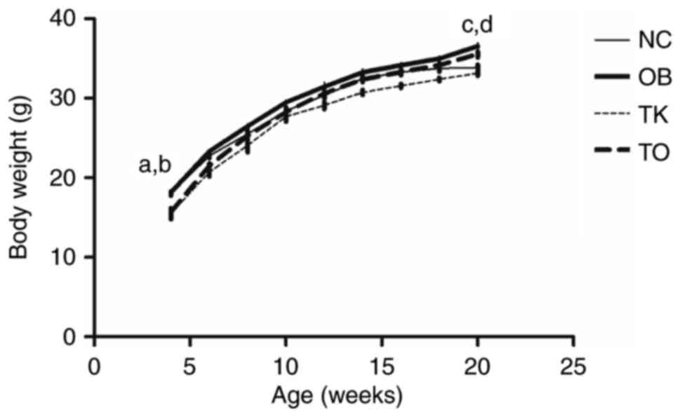
At the start of the experiment, C57BL/6J mice (NC
and OB mice) were heavier than TLR2 gene knockout mice (TK and TO
mice). However, there was no significant difference between the NC
and OB as well as TK and TO mice groups. At 4 weeks, there was no
significant difference among these 4 groups in weight. At 16 weeks,
Weight was increased in the OB group as compared to the NC group,
and decreased in the TO group compared to the OB group (Fig. 1).
Levels of TC, TG, LDL and FFA were higher in the OB
group compared with the NC group. HLD level was lower in the OB
group compared with the NC group. Levels of TC, TG, LDL, HDL and
FFA were lower in the TO group compared with the OB group (Table II).
| Table II.Levels of FPG, TC, TG, HDL, LDL and
FFA in plasma of mice. |
Table II.
Levels of FPG, TC, TG, HDL, LDL and
FFA in plasma of mice.
| Variable | NC | OB | TK | TO |
|---|
| FPG (mmol/l) | 6.78±0.43 |
9.79±0.32a |
6.37±0.46b |
6.88±0.33c,e |
| TC (mmol/l) | 0.26±0.02 |
0.37±0.03a |
0.08±0.01a |
0.12±0.02a,c,e |
| TG (mmol/l) | 2.64±0.06 |
3.03±0.07a |
2.30±0.06b |
2.36±0.06a,c |
| HDL (mmol/l) | 1.14±0.05 |
1.05±0.02a |
0.98±0.03b |
0.94±0.02a,c,f |
| LDL (mmol/l) | 1.46±0.02 |
1.65±0.07a |
1.19±0.03b |
1.2675+0.04a,c,e |
| FFA (mEq/l) | 670.40±22.10 |
1124.89±36.37a |
552.85±23.08b |
563.73+26.37a,c |
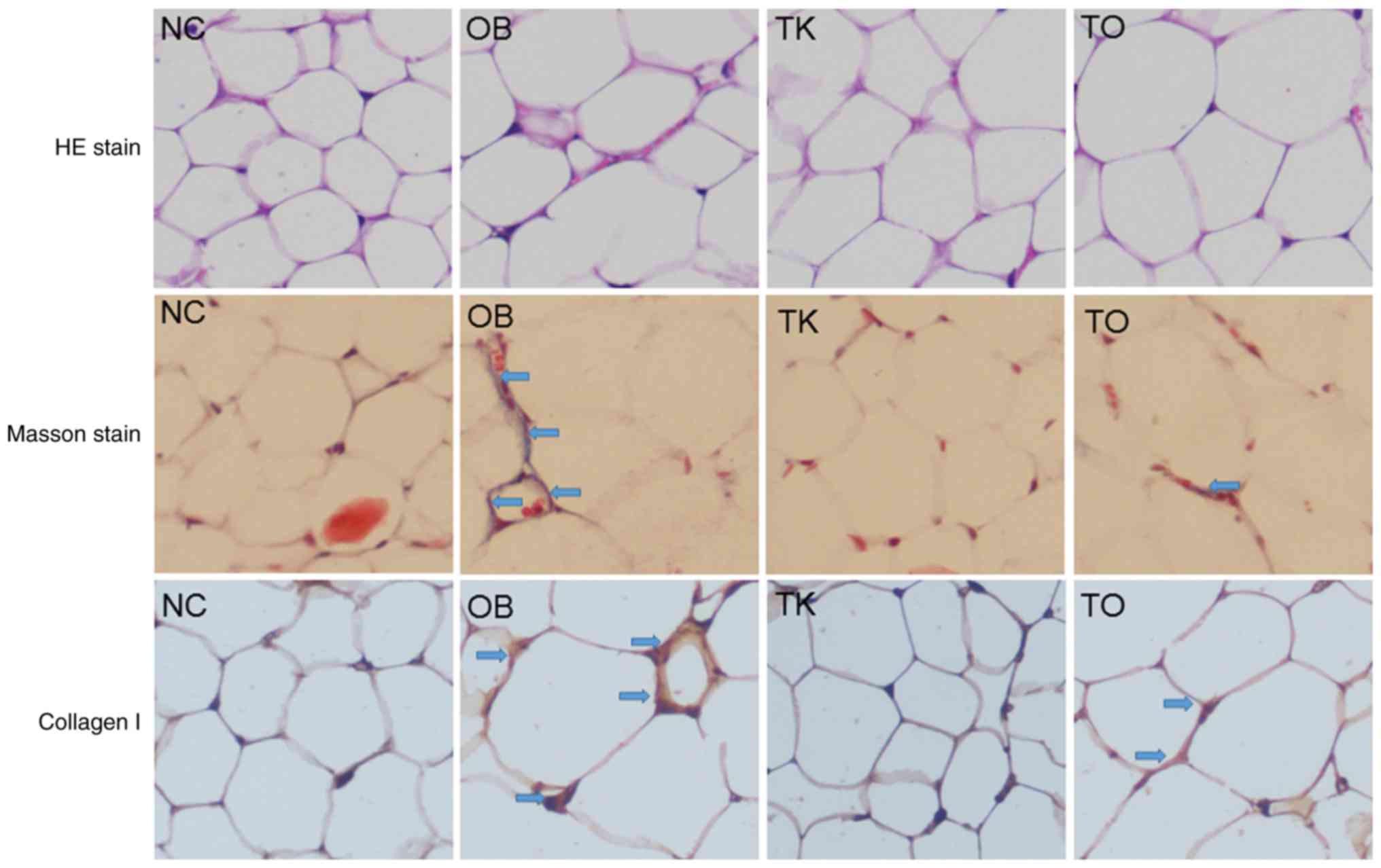
Masson stain revealed more collagen in adipose
tissue in the OB group compared with the NC group. Levels of total
collagen, collagen I, collagen I α1 mRNA, and collagen I α2
mRNA increased in adipose tissue of OB compared with NC mice
but decreased in TO compared with OB mice (Figs. 2 and 3).
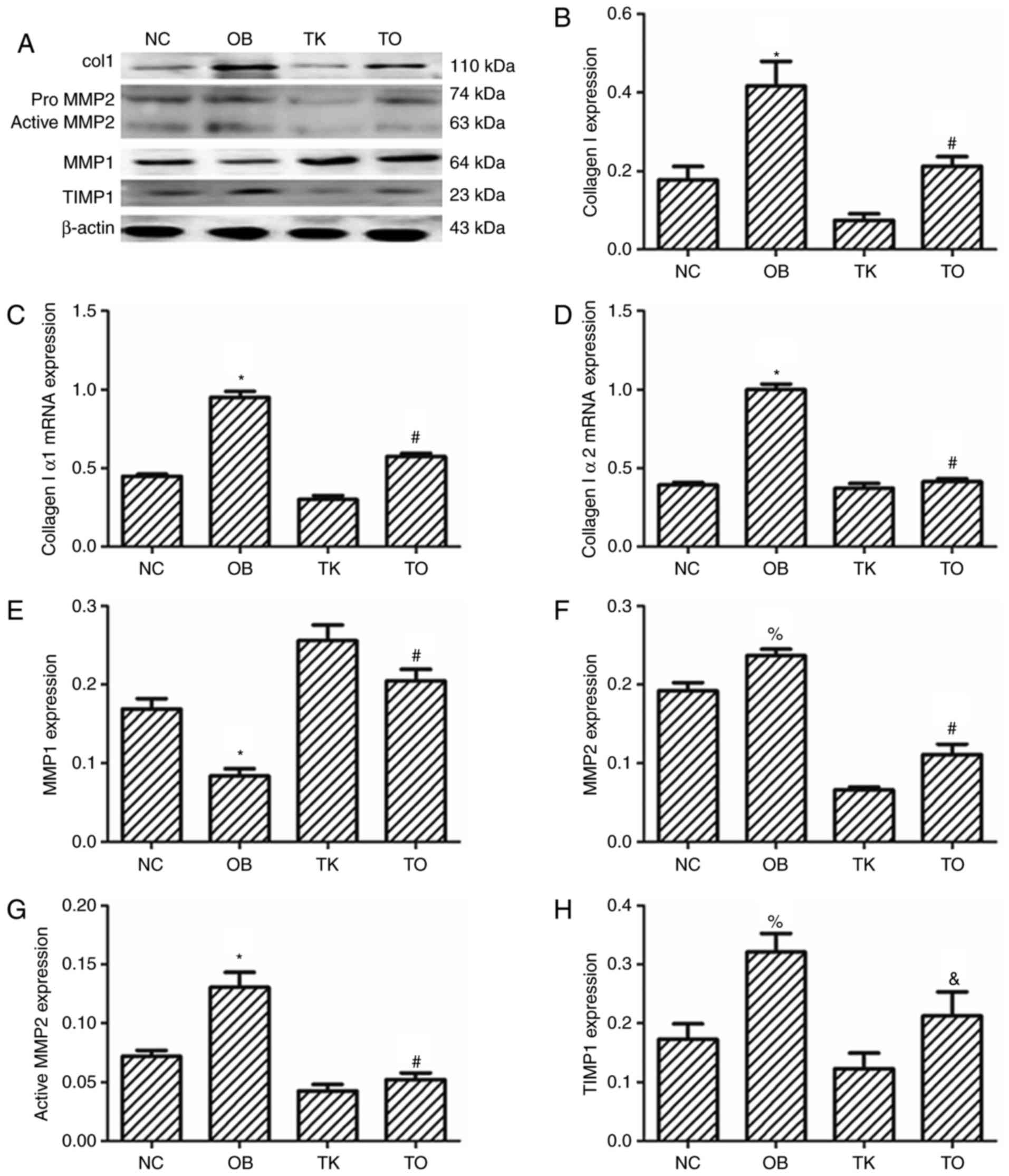 | Figure 3.(A) Western blotting and RT-PCR to
measure levels of (B) collagen I, (C) collagen I α1 mRNA, (D)
collagen I α2 mRNA, (E) MMP1, (F) MMP2, (G) active MMP2, and (H)
TIMP1. Increased collagen I, collagen I α1 mRNA, and collagen I α2
mRNA in the OB group. Decreased collagen I, collagen I α1 mRNA, and
collagen I α2 mRNA in the TO group (*P<0.01 vs. NC;
#P<0.01 vs. OB). Increased levels of MMP2, active
MMP2, and TIMP1 and decreased levels of MMP1 in the OB group.
Decreased levels of MMP2, active MMP2, and TIMP1 and increased
levels of MMP1 were observed in the TO group (*P<0.01,
%P<0.05 vs. NC; #P<0.01, P<0.05 vs.
OB). |
Levels of active MMP2, MMP2, and TIMP1 were higher
in OB compared with NC mice. Expression of MMP1 in adipose tissue
was lower in OB compared with NC mice. Expression of active MMP2,
MMP2, and TIMP1 was lower in TO compared with OB mice. Expression
of MMP1 in adipose tissue was higher in TO compared with OB mice
(Fig. 3).
Levels of TLR2, myd88, p38MAPK, p-p38MAPK and TGF-β1
in adipose tissue were higher in OB compared with NC mice. Levels
of TLR2, myd88, p38MAPK, p-p38MAPK, and TGF-β1 in adipose tissue
were lower in TO compared with OB mice (Fig. 4).
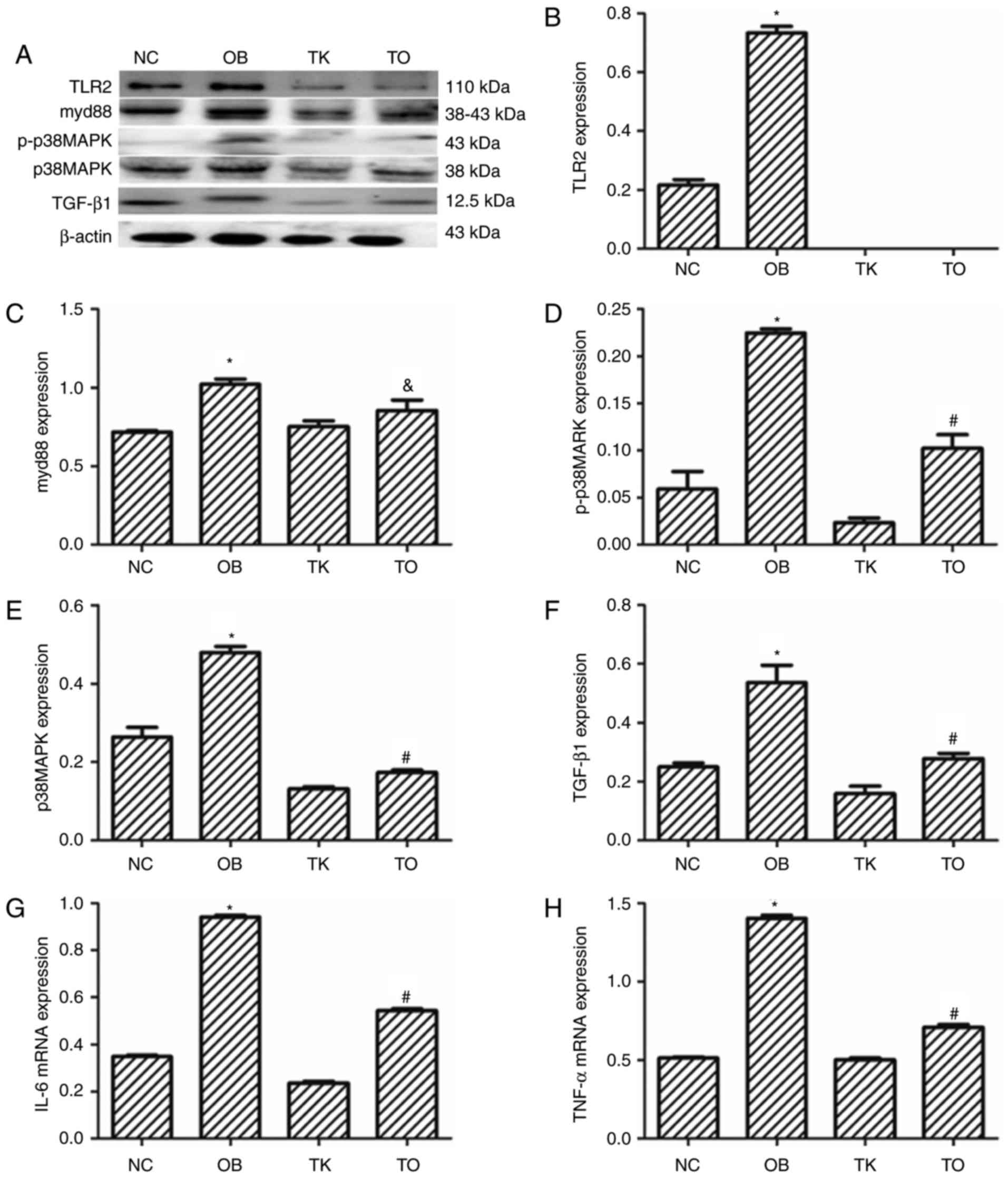 | Figure 4.(A) Western blotting and RT-PCR to
measure levels of (B) TLR2, (C) myd88, (D) p-p38MAPK, (E) p38 MAPK,
(F) TGF-β1, (G) IL6 mRNA and (H) TNF-α mRNA. Increased levels of
TLR2, myd88, p-p38MAPK, p38MAPK, TGFβ1, IL6 mRNA, and TNFα mRNA
levels in the NC group (*P<0.01 vs. NC). myd88, p-p38MAPK,
p38MAPK, TGFβ1, IL6 mRNA, and TNFα mRNA levels in the TO group
(#P<0.01, P<0.05 vs. OB). |
Expression levels of IL-6 mRNA and TNF-α
mRNA were up-regulated in adipose tissue of OB compared with
NC mice, but down-regulated in TO compared with OB mice (Fig. 4).
Discussion
Our findings of increased total collagen and
collagen I in adipose tissue of obese mice support previous reports
(20). Levels of MMP2 and TIMP1,
which regulate collagen I, were also increased in obese mice
(16,17). The increase in levels of MMP2 and
TIMP1 was reported previously (8).
Levels of MMP1, which also regulates collagen deposition, were
observed to decrease in this study. The results presented here
suggest that MMP1 and TIMP1 mediate the deposition of collagen
I.
FFA activates TLR2, which in turn triggers the
overexpression of proinflammatory factors (15). The adipose tissue of obese
individuals is therefore characterized by chronic inflammation,
which may be controlled by TLR2 (21). Such chronic inflammation increases
the expression of MMPs and TIMPs (8). We believe that knocking out TLR2
reduced the expression of MMP1, TIMP1, and other inflammatory
factors, reducing the deposition of collagen I in mice fed a
high-fat diet. In this study, increased levels of myd88, p38 MAPK,
p-p38 MAPK, IL-6, and TNF-α (inflammatory factors downstream of
TLR2) were observed in the adipose tissue of OB compared with NC
mice; levels of these factors were increased in the OB compared
with the TO group. Compared with OB mice, TO mice had lower levels
of collagen I and TIMP1 and higher levels of MMP1. TLR2 knockout
appears to have increased the expression of MMP1 and decreased the
expression of TIMP1. These factors likely reduced the level of
collagen I deposition. These findings support the previous studies
(22,23). Genes knockout resulting in
decreased levels fibrosis factors are reported to have decreased
expression of inflammatory factors.
To further explore deposition of collagen I,
collagen I α1 mRNA and collagen I α2 mRNA expression
were measured in adipose tissue. OB mice showed up-regulation of
collagen I α1 mRNA and collagen I α2 mRNA compared with NC mice.
However, down-regulation of collagen I α1 mRNA and collagen I α2
mRNA were detected in TO mice compared with OB mice (Fig. 3). Statistical analysis revealed an
interaction effect on collagen I α1 and collagen I α2 mRNA between
high fat diet and TLR2 knocking out in adipose tissue of mice.
These findings suggest that the transcription and translation of
collagen I were affected by TLR2 gene knockout and ingestion of a
high-fat diet.
Higher expression of TGFβ1 was observed in OB
compared with NC mice. TGFβ1 expression was lower in TO compared
with OB mice. Elevated levels of cytokines such as TGFβ1 (13,21,24)
trigger SMAD and non-SMAD signaling cascades that contribute to
obesity (25–29) and fibrosis (30). Gene knockouts resulting in
decreased levels of TGFβ1 are reported to have decreased expression
of inflammatory factors (22,23).
TLR2 gene knockout appears to have had similar effects, resulting
in fibrosis and the deposition of collagen I in adipose tissue.
In conclusion, the present results showed that TLR2
gene knockout may reduce collagen I expression in adipose tissue of
mice with obesity. We hypothesize that this effect is mediated by a
balance in pro -vs. anti-inflammatory factors and downstream MMP1,
TIMP1, and TGFβ1.
Acknowledgements
The present study was supported by the Scientific
Foundation for Doctoral Research (20131067) and Natural Scientific
Foundation (201602308) at the Liaoning Science and Technology
Administration Bureau. Professor Zheng and Professor Zhai in the
Department of Brain and Spinal Cord injury in Liaoning province
contributed substantially to this study.
References
|
1
|
Flegal KM, Carroll MD, Ogden CL and Curtin
LR: Prevalence and trends in obesity among US adults, 1999–2008.
JAMA. 303:235–241. 2010. View Article : Google Scholar : PubMed/NCBI
|
|
2
|
Flegal KM, Kruszon-Moran D, Carroll MD,
Fryar CD and Ogden CL: Trends in obesity among adults in the United
States, 2005 to 2014. JAMA. 315:2284–2291. 2016. View Article : Google Scholar : PubMed/NCBI
|
|
3
|
Ogden CL, Carroll MD, Lawman HG, Fryar CD,
Kruszon-Moran D, Kit BK and Flegal KM: Trends in obesity prevalence
among children and adolescents in the United States, 1988–1994
through 2013–2014. JAMA. 315:2292–2299. 2016. View Article : Google Scholar : PubMed/NCBI
|
|
4
|
Chan JC, Zhang Y and Ning G: Diabetes in
China: A societal solution for a personal challenge. Lancet
Diabetes Endocrinol. 2:969–979. 2014. View Article : Google Scholar : PubMed/NCBI
|
|
5
|
Xu Y, Wang L, He J, Bi Y, Li M, Wang T,
Wang L, Jiang Y, Dai M, Lu J, et al: Prevalence and control of
diabetes in Chinese adults. JAMA. 310:948–959. 2013. View Article : Google Scholar : PubMed/NCBI
|
|
6
|
Huber J, Löffler M, Bilban M, Reimers M,
Kadl A, Todoric J, Zeyda M, Geyeregger R, Schreiner M, Weichhart T,
et al: Prevention of high-fat diet-induced adipose tissue
remodeling in obese diabetic mice by n-3 polyunsaturated fatty
acids. Int J Obes (Lond). 31:1004–1013. 2007. View Article : Google Scholar : PubMed/NCBI
|
|
7
|
Khan T, Muise ES, Iyengar P, Wang ZV,
Chandalia M, Abate N, Zhang BB, Bonaldo P, Chua S and Scherer PE:
Metabolic dysregulation and adipose tissue fibrosis: Role of
collagen VI. Mol Cell Biol. 29:1575–1591. 2009. View Article : Google Scholar : PubMed/NCBI
|
|
8
|
Lin Chun TH and Kang L: Adipose
extracellular matrix remodelling in obesity and insulin resistance.
Biochem Pharmacol. 119:8–16. 2016. View Article : Google Scholar : PubMed/NCBI
|
|
9
|
Spencer M, Unal R, Zhu B, Rasouli N,
McGehee RE Jr, Peterson CA and Kern PA: Adipose tissue
extracellular matrix and vascular abnormalities in obesity and
insulin resistance. J Clin Endocrinol Metab. 96:E1990–E1998. 2011.
View Article : Google Scholar : PubMed/NCBI
|
|
10
|
Vila IK, Badin PM, Marques MA, Monbrun L,
Lefort C, Mir L, Louche K, Bourlier V, Roussel B, Gui P, et al:
Immune cell Toll-like receptor 4 mediates the development of
obesity- and endotoxemia-associated adipose tissue fibrosis. Cell
Rep. 7:1116–1129. 2014. View Article : Google Scholar : PubMed/NCBI
|
|
11
|
Dankel SN, Svärd J, Matthä S, Claussnitzer
M, Klöting N, Glunk V, Fandalyuk Z, Grytten E, Solsvik MH, Nielsen
HJ, et al: COL6A3 expression in adipocytes associates with insulin
resistance and depends on PPARγ and adipocyte size. Obesity (Silver
Spring). 22:1807–1813. 2014. View Article : Google Scholar : PubMed/NCBI
|
|
12
|
Divoux A, Tordjman J, Lacasa D, Veyrie N,
Hugol D, Aissat A, Basdevant A, Guerre-Millo M, Poitou C, Zucker
JD, et al: Fibrosis in human adipose tissue: Composition,
distribution, and link with lipid metabolism and fat mass loss.
Diabetes. 59:2817–2825. 2010. View Article : Google Scholar : PubMed/NCBI
|
|
13
|
Spencer M, Yao-Borengasser A, Unal R,
Rasouli N, Gurley CM, Zhu B, Peterson CA and Kern PA: Adipose
tissue macrophages in insulin-resistant subjects are associated
with collagen VI and fibrosis and demonstrate alternative
activation. Am J Physiol Endocrinol Metab. 299:E1016–E1027. 2010.
View Article : Google Scholar : PubMed/NCBI
|
|
14
|
Barton GM and Medzhitov R: Toll-like
receptor signaling pathways. Science. 300:1524–1525. 2003.
View Article : Google Scholar : PubMed/NCBI
|
|
15
|
Kim S, Jin Y, Choi Y and Park T:
Resveratrol exerts anti-obesity effects via mechanisms involving
down-regulation of adipogenic and inflammatory processes in mice.
Biochem Pharmacol. 81:1343–1351. 2011. View Article : Google Scholar : PubMed/NCBI
|
|
16
|
Hopps E and Caimi G: Matrix
metalloproteinases in metabolic syndrome. Eur J Intern Med.
23:99–104. 2012. View Article : Google Scholar : PubMed/NCBI
|
|
17
|
Hopps E and Caimi G: Matrix
metalloproteases as a pharmacological target in cardiovascular
diseases. Eur Rev Med Pharmacol Sci. 19:2583–2589. 2015.PubMed/NCBI
|
|
18
|
Bourlier V, Sengenès C, Zakaroff-Girard A,
Decaunes P, Wdziekonski B, Galitzky J, Villageois P, Esteve D,
Chiotasso P, Dani C and Bouloumié A: TGFbeta family members are key
mediators in the induction of myofibroblast phenotype of human
adipose tissue progenitor cells by macrophages. PLoS One.
7:e312742012. View Article : Google Scholar : PubMed/NCBI
|
|
19
|
Henegar C, Tordjman J, Achard V, Lacasa D,
Cremer I, Guerre-Millo M, Poitou C, Basdevant A, Stich V, Viguerie
N, et al: Adipose tissue transcriptomic signature highlights the
pathological relevance of extracellular matrix in human obesity.
Genome Biol. 9:R142008. View Article : Google Scholar : PubMed/NCBI
|
|
20
|
Luo T, Nocon A, Fry J, Sherban A, Rui X,
Jiang B, Xu XJ, Han J, Yan Y, Yang Q, et al: AMPK activation by
metformin suppresses abnormal extracellular matrix remodeling in
adipose tissue and ameliorates insulin resistance in obesity.
Diabetes. 65:2295–2310. 2016. View Article : Google Scholar : PubMed/NCBI
|
|
21
|
Samad F, Yamamoto K, Pandey M and
Loskutoff DJ: Elevated expression of transforming growth
factor-beta in adipose tissue from obese mice. Mol Med. 3:37–48.
1997.PubMed/NCBI
|
|
22
|
Adapala VJ, Ward M and Ajuwon KM: Adipose
tissue biglycan as a potential anti-inflammatory target of sodium
salicylate in mice fed a high fat diet. J Inflamm (Lond). 9:152012.
View Article : Google Scholar : PubMed/NCBI
|
|
23
|
Nokhbehsaim M, Keser S, Nogueira AV, Jäger
A, Jepsen S, Cirelli JA, Bourauel C, Eick S and Deschner J: Leptin
effects on the regenerative capacity of human periodontal cells.
Int J Endocrinol. 2014:1803042014. View Article : Google Scholar : PubMed/NCBI
|
|
24
|
Alessi MC, Bastelica D, Morange P, Berthet
B, Leduc I, Verdier M, Geel O and Juhan-Vague I: Plasminogen
activator inhibitor 1, transforming growth factor-beta1 and BMI are
closely associated in human adipose tissue during morbid obesity.
Diabetes. 49:1374–1380. 2000. View Article : Google Scholar : PubMed/NCBI
|
|
25
|
Johnson DL, Carnes D, Steffensen B and
Cochran DL: Cellular effects of enamel matrix derivative are
associated with different molecular weight fractions following
separation by size-exclusion chromatography. J Periodontol.
80:648–656. 2009. View Article : Google Scholar : PubMed/NCBI
|
|
26
|
Okubo K, Kobayashi M, Takiguchi T, Takada
T, Ohazama A, Okamatsu Y and Hasegawa K: Participation of
endogenous IGF-I and TGF-beta 1 with enamel matrix
derivative-stimulated cell growth in human periodontal ligament
cells. J Periodontal Res. 38:1–9. 2003. View Article : Google Scholar : PubMed/NCBI
|
|
27
|
Prime SS, Pring M, Davies M and Paterson
IC: TGF-beta signal transduction in oro-facial health and
non-malignant disease (part I). Crit Rev Oral Biol Med. 15:324–336.
2004. View Article : Google Scholar : PubMed/NCBI
|
|
28
|
Sieber C, Kopf J, Hiepen C and Knaus P:
Recent advances in BMP receptor signaling. Cytokine Growth Factor
Rev. 20:343–355. 2009. View Article : Google Scholar : PubMed/NCBI
|
|
29
|
Suzuki S, Nagano T, Yamakoshi Y, Gomi K,
Arai T, Fukae M, Katagiri T and Oida S: Enamel matrix derivative
gel stimulates signal transduction of BMP and TGF-{beta}. J Dent
Res. 84:510–514. 2005. View Article : Google Scholar : PubMed/NCBI
|
|
30
|
Wynn TA: Cellular and molecular mechanisms
of fibrosis. J Pathol. 214:199–210. 2008. View Article : Google Scholar : PubMed/NCBI
|