Introduction
Advanced tumors are occasionally non-responsive to
mono-therapies. By contrast, combination chemotherapy may stimulate
the regression of specific types of tumors even at progressive
stages, and this particularly has served an important role in
cancer treatment for a number of years (1,2).
Furthermore, it has been suggested that effective and favorable
drug combinations should be explored to improve combination
chemotherapeutics, which may markedly improve the current outcomes
of cancer treatment.
Tumor necrosis factor-related apoptosis-inducing
ligand (TRAIL) is a well-known transmembrane cytokine, and a member
of the tumor necrosis factor family that mediates apoptosis in a
variety of tumor cell types (3,4).
TRAIL has been proposed as one of the most effective anti-cancer
therapeutic agents, owing to its efficiency to selectively induce
cell death in a variety of tumor cells whilst exhibiting minimal
toxicity in the majority of normal cells (5,6). The
binding of TRAIL to death receptors may also engage the extrinsic
apoptotic pathway and trigger apoptotic signaling (7). TRAIL induces apoptosis by binding to
death receptors and activating the Fas-associated death domain
protein, ultimately leading to the activation of the effector
caspase cascade, involving caspase-8, −9, −10, and −3 (8,9). A
previous study has demonstrated that a number of cancer cells,
together with lung A549 cells, acquire resistance to the apoptotic
effects of TRAIL (10).
Celastrol is a triterpenoid, initially isolated from
the Chinese herb Tripterygium wilfordii, or ‘Thunder God
Vine’, that has been extensively applied in the treatment of
autoimmune and neurodegenerative diseases (11–14).
Previous studies have revealed that celastrol possesses multiple
biological and pharmacological functions, in vitro and in
vivo, including anti-microbial, anti-inflammatory and
anti-cancer activities against prostate, breast and pancreatic
cancer cells (15,16). It was also identified to have
multiple mechanisms or signaling pathways that contribute to its
anti-cancer activity, based on its efficiency to selectively
initiate apoptosis in numerous tumor cell types (15,17).
Autophagy is an intracellular catabolic mechanism,
whose primary functions are to sustain cellular homeostasis by
recycling long-lived unrequired proteins, remove exhausted,
redundant and defective cellular elements, and promote cell
survival during brief periods of nutrient-starvation and other
stressors (18,19). During autophagy, double membrane
vesicles develop to form autophagic vacuoles named autophagosomes.
These fuse with lysosomes to create auto-lysosomes, wherein
sequestered segments are degraded by lysosomal enzymes (20,21).
The autophagosome formation is mediated by the ubiquitin-like
protein ATG12-autophagy protein (Atg)5-Atg16 complex and
microtubule-associated proteins 1A/1B light chain 3B (LC3)-II, an
LC3-I-phospholipid conjugate widely applied as an autophagy marker
(22,23). Sequestosome 1 (p62) is a
ubiquitin-binding protein and a well-known autophagy marker. It
organizes into autophagosomes by directly interacting with LC3, and
is completely degraded by autophagy. Inhibition of autophagy
results in low levels of p62 accumulation (19). It was previously revealed that the
inhibition of autophagy flux sensitizes cancer cells to
conventional radio and chemotherapy treatment (24–26),
confirming that autophagy flux inhibition may be an appropriate and
effective technique for cancer treatment. The antimalarial drug
chloroquine (CQ), which functions as an autophagy inhibitor,
prevents lysosome acidification and lysosomal fusion with
autophagosomes, and also inhibits the degradation of metabolic
stress, thereby inducing apoptosis (27–30).
The mitochondria serve a pivotal role in the
intrinsic pathway of cell death by transmitting apoptotic signals
to the cytosol, leading to the activation of the caspase cascade.
Changes in mitochondria appear following the activation of cell
death signals, including the formation of reactive oxygen species
(ROS), loss of mitochondrial membrane potential (ΔΨm), opening of
the permeability alteration pore, and release of apoptotic bodies
into the cytosol (31–36). ROS-inducing agents have been
applied to kill tumor cells in cancer therapy (37,38).
The present study aimed to explore the use of
celastrol as a sensitizing agent to TRAIL-initiated apoptosis in
A549 cells. It was revealed that a combined regimen of celastrol
and TRAIL resulted in an improved outcome compared with treatment
with either celastrol or TRAIL alone.
Materials and methods
Cell culture
Human lung A549, HCC-15 and Calu-3 cell lines
(American Type Culture Collection, Manassas, VA, USA) were
maintained in RPMI-1640 medium (Gibco; Thermo Fisher Scientific,
Inc., Waltham, MA, USA) containing with 10% fetal bovine serum
(FBS; Sigma-Aldrich; Merck KGaA, Darmstadt, Germany). During
experimentation, cells were switched to RPMI containing 1% FBS.
Reagents
Recombinant celastrol and chloroquine (20 µM) were
obtained from Sigma-Aldrich; Merck KGaA (Darmstadt, Germany). TRAIL
(200 ng/ml) was purchased from Abfrontier; Young in Frontier, Co.,
Ltd. (Seoul, South Korea).
Cell viability assay
A549, HCC-15 and Calu-3 cells were plated in 12-well
plates and pre-exposed to celastrol (1, 2 and 4 µM) for 12 h in
37°C incubator, and were then additionally treated with TRAIL
protein (200 ng/ml) for an additional 2 h. Cell morphology was
assessed under a light microscope (Nikon Corporation, Tokyo, Japan,
magnification, ×100), and cell viability was evaluated by a crystal
violet staining method. Cells were stained with a staining solution
(0.5% crystal violet in 30% ethanol and 3% formaldehyde) for 10 min
at room temperature, washed four times with PBS, and were dried.
Cells were then lysed with 1% SDS solution. The absorbance value
was then measured at wavelength of 550 mm using a plate reader.
Cell viability was expressed as relative dye intensity compared
with that of the control.
Trypan blue exclusion assay
Cell viability was evaluated by trypan blue
exclusion assay (Sigma-Aldrich; Merck KGaA), using a hemocytometer.
Following each treatment, the cells at 1.0×10⁴
cells/well in 12-well plates, were trypsinized and re-suspended in
PBS. Trypan blue dye solution (0.4%) was added to the cell
suspension for 5 min at room temperature. Unstained cells were
viable and stained cells were dead. The total cell number and
trypan blue-positive cells were counted using a light microscope in
a blinded manner. The percentage of surviving cells was calculated
using the formula: Number of stained cells/number of total cells
×100. Each treatment was performed in triplicate.
ROS determination
A549 cells (1.0×10⁴ cells/well) in
12-well plates, were pre-incubated with N-acetyl cysteine
(NAC) for 1 h and then incubated with TRAIL alone or combined with
CQ (10 mM). The formation of ROS was ascertained through
application of the cell permeable fluorescent marker
dihydroethidium (DHE). Briefly, cells were treated with 5 µM DHE
for 30 min at 37°C in the dark. The fluorescence was then measured
using a fluorescence plate reader at excitation and emission
wavelengths of 518 and 605 nm, respectively.
ΔΨm analysis
The changes in ΔΨm were assessed using a cationic
fluorescent marker. A549 cells were maintained on cover slips in a
24-well plate, incubated with 10 ml JC-1 at 37°C for 30 min and
then washed with PBS. The cells were then mounted with
DakoCytomation fluorescent mounting medium (Dako; Agilent
Technologies, Inc., Santa Clara, CA, USA) and visualized under a
fluorescence microscope (magnification, ×400).
Immunofluorescent staining
A549 cells (5.0×10³ cells/well) were
cultured on poly-L-lysine coated coverslips. Following
differentiation and specific treatment, the cells were fixed with
4% paraformaldehyde at room temperature 15 min and permeabilized
with 0.1% Triton X-100. The cells were then incubated for 60 min at
room temperature with blocking solution (5% FBS in Tris-buffered
saline) followed by overnight incubation at 4°C with anti-p62
(1:250; cat. no. PA5-20839; Invitrogen; Thermo Fisher Scientific,
Inc.) and cleaved caspase-3 [(1:250; cat. no. 9665; Cell Signaling
Technology, Inc., Danvers, MA, USA) antibodies. Subsequent to
washing with PBS, the cells were incubated with secondary antibody
(Alexa Fluor® 488-conjugated donkey polyclonal
anti-rabbit; 1:500; cat. no. A-21206; Thermo Fisher Scientific,
Inc.; and Texas Red-X-conjugated goat polyclonal anti-mouse; 1:500;
cat. no. T-6390; Thermo Fisher Scientific, Inc.) for 2 h in the
dark. Finally, immunostaining was visualized under a fluorescence
microscope (magnification, ×400).
Western blot analysis
Western blot analysis was performed as described
previously (39). Briefly,
immunoprecipitation assay buffer (Qiagen, Inc, Valencia, CA, USA)
was used to extract total protein in A549 cells. The supernatant
was collected by centrifugation (13,282 × g; 4°C; 10 min). The
protein concentration was determined using the Pierce Bicinchoninic
Protein Assay kit (Thermo Fisher Scientific, Inc.). Proteins (30
µg) were separated on 10% SDS-PAGE gels and blotted onto
polyvinylidene fluoride membranes. Membranes were blocked with 5%
non-fat dried milk at 25°C for 1 h, followed by incubation with
primary antibodies overnight at 4°C. Antibodies against β-actin
were purchased from Sigma-Aldrich; Merck KGaA, and those against
cleaved caspase-8 were obtained from BD Pharmingen (BD Biosciences,
Franklin Lakes, NJ, USA). Cleaved caspase-3 and LC3 antibodies were
purchased from Cell Signaling Technology, Inc., and p62 antibodies
were purchased from EMD Millipore (Billerica, MA, USA). Membranes
were incubated with horseradish peroxidase conjugated secondary
antibody (cat. no. 4410; 1:2,000; Cell Signaling Technology, Inc.)
at 25°C for 1 h. The immune-reactive protein bands were visualized
using an enhanced chemiluminescence detection system (GE Healthcare
Life Sciences, Chalfont, UK). Densitometry of the signal bands was
conducted using the Bio-1D densitometer (Vilber Lourmat,
Eberhardzell, Germany). Images were examined using a Fusion-FX7
imaging system (Vilber Lourmat).
Statistical analysis
Statistical analysis was performed using GraphPad
Prim (version 5.03; GraphPad Software, Inc., La Jolla, CA, USA).
All experiments were performed in triplicate, and the data are
expressed as the mean ± standard error. Significant differences
between control and treated samples were analyzed using one-way
analysis of variance followed by Duncan's post hoc test.
Results
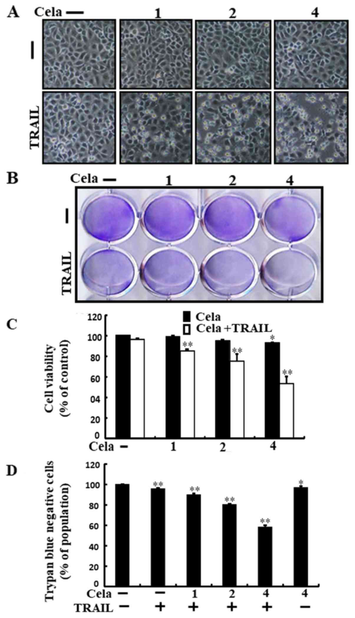
Celastrol sensitizes TRAIL-initiated
apoptosis in A549 cells
Alterations in cell morphologies were examined under
a light microscope. Treatment with either celastrol or TRAIL alone
did not or only marginally induced cell death (Fig. 1), and no morphological changes were
observed. However, a combined regimen of TRAIL and different
concentrations of celastrol markedly increased the number of
apoptotic cell deaths compared with celastrol or TRAIL alone
(Fig. 1A-D). These data indicate
that celastrol promoted TRAIL-initiated apoptosis in A549
cells.
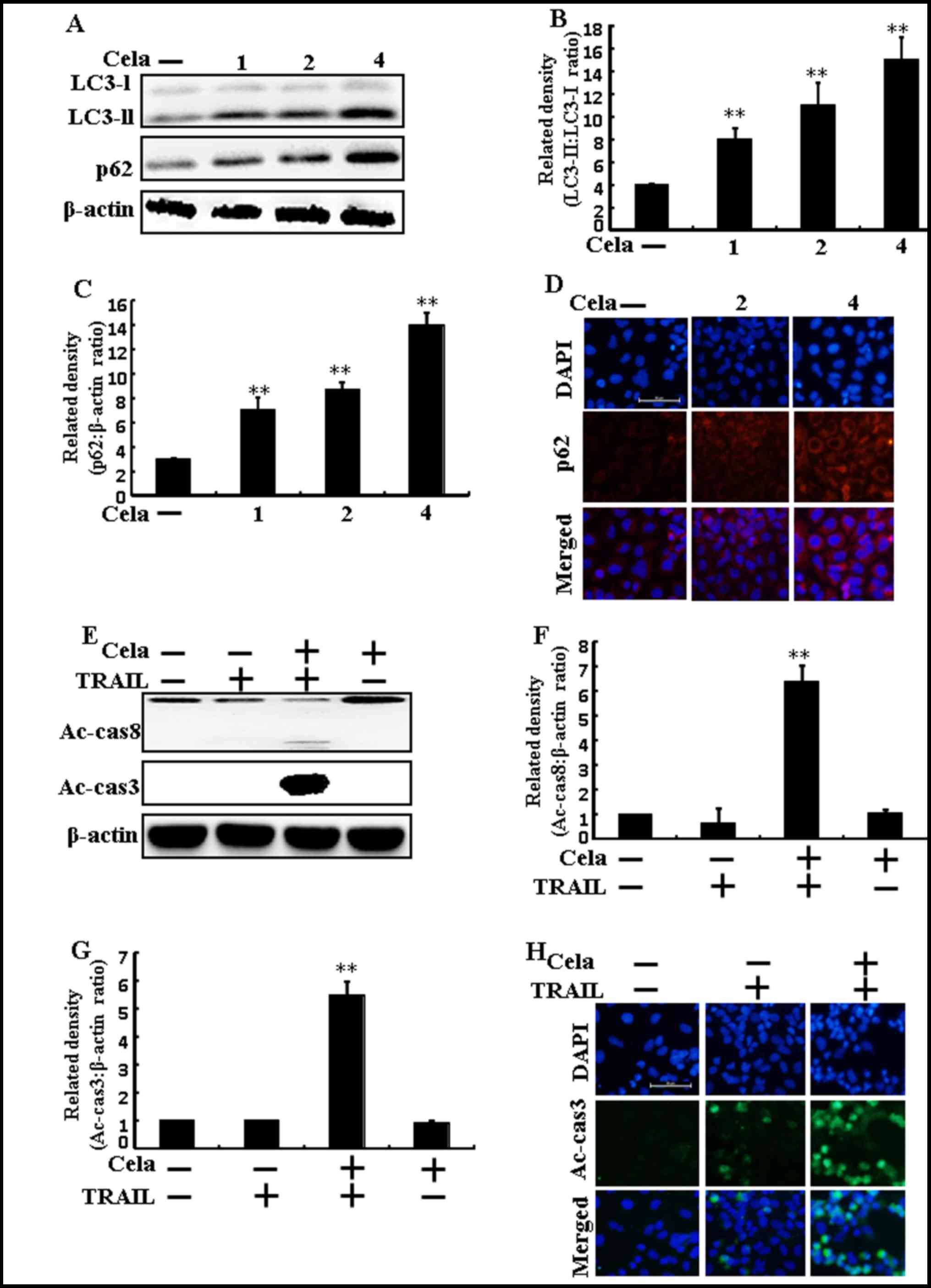
Autophagy flux is inhibited by
celastrol in A549 cells
LC3-II and p62 expression increased following
celastrol treatment (Fig. 2A-C).
Immunofluorescence staining also demonstrated that varying doses of
celastrol increased p62 protein levels (Fig. 2D). The combined regimen of TRAIL
and celastrol increased active (Ac)-caspase 3 and Ac-caspase 8
levels (Fig. 2E-G).
Immunofluorescent staining results also suggested that Ac-caspase 3
levels of combined celastrol and TRAIL treatment were increased
than only TRAIL treatment (Fig.
2H), indicating that autophagy flux was inhibited by
celastrol.
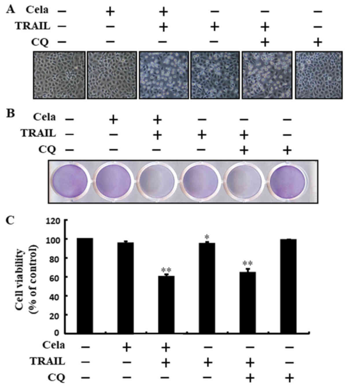
Celastrol increases TRAIL-mediated
cell death by attenuating autophagy flux
The cell morphology results indicated that increased
apoptosis was achieved by co-treatment of TRAIL and celastrol or CQ
(Fig. 3A). The combined regimen of
CQ and TRAIL markedly increased cell death and attenuated cell
viability (Fig. 3B and C). These
results suggested that celastrol sensitized TRAIL-initiated cell
death by attenuating autophagy flux.
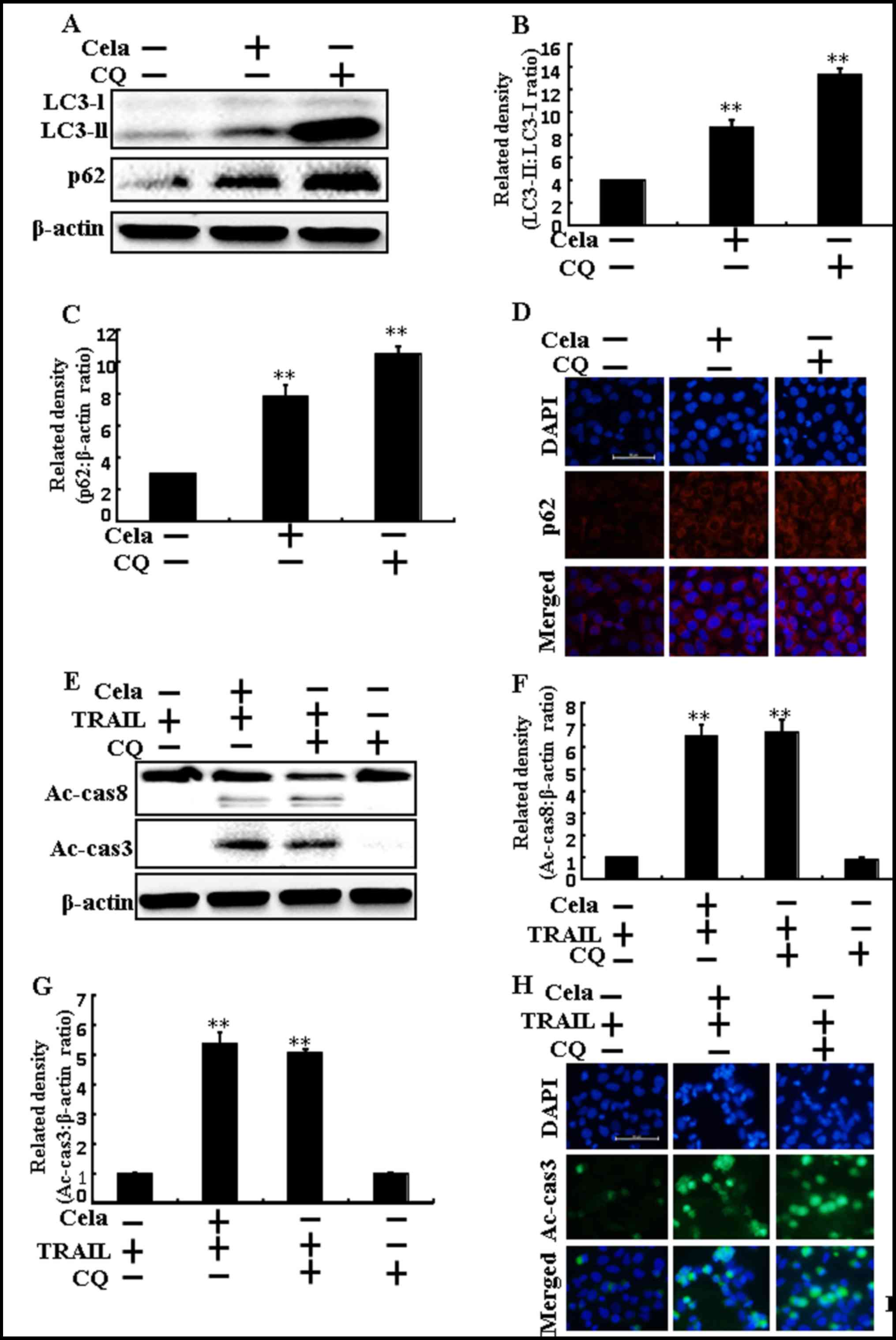
Celastrol-mediated autophagy flux
inhibition promotes TRAIL-initiated apoptosis
LC3-II and p62 expression were markedly increased in
A549 cells treated with celastrol or CQ alone, confirming that
celastrol inhibits autophagy flux (Fig. 4A-C). Immunofluorescence staining
results also demonstrated that p62 protein levels were increased
(Fig. 4D). The combined regimen of
TRAIL and CQ increased Ac-caspase 3 and Ac-caspase 8 protein levels
(Fig. 4E-G). The immunofluorescent
staining results also demonstrated that Ac-caspase 3 expression was
increased (Fig. 4H). These results
also indicated that the promotion of the TRAIL-initiated apoptotic
mechanism by celastrol was due to the attenuation of autophagy
flux.
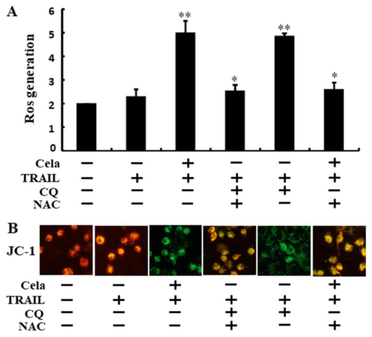
Attenuation of autophagy stimulates
ROS generation and changes in ΔΨm
As demonstrated in Fig.
5A, ROS levels were increased in cells treated with celastrol
combined with TRAIL or CQ. These increases were effectively
attenuated when the cells were pre-incubated with N-acetyl
cysteine (NAC) for 1 h. Green fluorescence observed in the cells
treated with celastrol combined with TRAIL or CQ suggested
decreased ΔΨm values compared with that from the TRAIL alone
treatment, but treatment with NAC restored these ΔΨm values
(Fig. 5B). These data suggest that
celastrol-mediated autophagy flux attenuation increased
TRAIL-initiated apoptosis via increases in ROS generation and
decreasing ΔΨm.
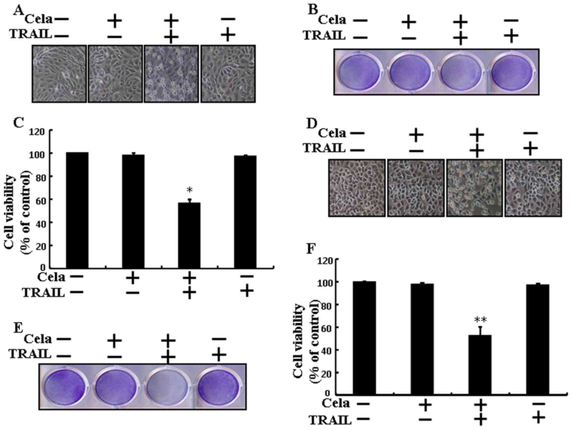
Celastrol sensitizes TRAIL-initiated
apoptosis in different lung cells
Celastrol or TRAIL treatment alone did not, or only
marginally affected, the cell viability of Calu-3 and HCC-15 cells
(Fig. 6). Nevertheless, the
combined TRAIL and celastrol regimen significantly decreased the
cell viability of Calu-3 and HCC-15 cells (Fig. 6A-F). These results suggested that
celastrol significantly increased TRAIL-initiated apoptosis in
Calu-3 and HCC-15 cells.
Discussion
The aim of the present study was to determine the
function of celastrol with or without TRAIL, in lung cancer cells.
The results revealed that celastrol-mediated autophagy flux
inhibition sensitized TRAIL-mediated apoptosis via regulation of
ROS and ΔΨm.
TRAIL is considered one of the most promising
anti-cancer agents identified in the previous 25 years, owing to
the specificity of its action, initiating apoptosis in specific
cell types (5,40–44)
and prompting the destruction of cancer cells without causing
toxicity to normal cells. Previous studies have confirmed that the
repeated administration of TRAIL proteins adequately attenuated
tumor growth without causing serious side effects (6,45).
In this manner, considerable attention has been paid to TRAIL as a
anti-cancer therapy for human cancer. The antitumor activities of
celastrol are not only limited to in vitro systems, as this
triterpenoid was also identified to inhibit the uncontrolled growth
and metastasis of melanoma in xenograft mouse models (46). Previous studies have demonstrated
that multiple mechanisms or signaling pathways lead to
celastrol-induced cancer cell death (15,17,47).
Autophagy flux is a complete system of autophagy, starting with the
amalgamation of the autophagosome with the lysosome, resulting in
the degradation and recycling of their cargo (18,48).
ROS serves an important role in response to cancer therapeutics,
considering that ROS formation is associated with cancer initiation
and progression.
A previous study have attempted to examine TRAIL
sensitizers that are capable of overcoming TRAIL resistance in
cancer cells (49). The present
study revealed that a combined regimen of TRAIL and different doses
of celastrol significantly increased the level of apoptotic cell
death compared with treatment with celastrol or TRAIL alone. A
previous study has suggested that celastrol treatment attenuated
cell proliferation and the induction of apoptosis and autophagy in
gastric cancer cells (50).
However, the results from the present study indicated that LC3-II
and p62 expression increased following celastrol treatment, where
co-treatment of celastrol and TRAIL increased Ac-cas3 and Ac-cas8
levels. It has bene demonstrated previously that treatment of
celastrol in MCF-7 cells induces apoptosis through mitochondrial
caspase-dependent and independent signaling pathways (51). Furthermore, Kannaiyan et al
(52) identified that celastrol
treatment inhibits proliferation and stimulates apoptosis in
RPMI-8226 cells, alongside the activation of c-Jun N-terminal
kinase and attenuation of the phosphoinositide 3-kinase/protein
kinase B signaling pathways. The present study also demonstrated
that the combined regimen of TRAIL and celastrol or chloroquine
significantly attenuated cell viability and increased cell death.
Previous studies have suggested that the inhibition of autophagy
stimulates dysfunctional mitochondria and ROS generation in
prostate cancer cells (53,54).
The data from the present study suggest that celastrol-mediated
autophagy flux inhibition increased TRAIL-induced apoptosis via ROS
generation and decreases in ΔΨm. In conclusion, the present study
demonstrated that celastrol improved TRAIL-initiated apoptosis via
inhibiting autophagy flux. In addition, the use of TRAIL in
combination with celastrol may provide an adequate therapeutic
strategy for safe treatment of TRAIL-resistant lung cancer,
suggesting that celastrol-mediated autophagy flux inhibition
sensitized TRAIL-initiated apoptosis via the regulation of ROS and
the ΔΨm.
Acknowledgements
Not applicable.
Funding
The present study was supported by a grant from the
National Research Foundation of Korea, funded by the Korean
Government (Grant no. 2016R1A2B2009293).
Availability of data and materials
The analyzed data sets generated during the study
are available from the corresponding author on reasonable
request.
Authors' contributions
UMN and SYP designed the study. UMN and HHY
performed the experiments. UMN and SYP analyzed the data and wrote
the manuscript. UMN, HHY and SYP revised and approved the final
version of the manuscript.
Ethics approval and consent to
participate
Not applicable.
Patient consent of publication
Not applicable.
Competing interests
The authors declare that they have no competing
interests.
References
|
1
|
Garcia G and Odaimi M: Systemic
combination chemotherapy in elderly pancreatic cancer: A review. J
Gastrointest Cancer. 48:121–128. 2017. View Article : Google Scholar : PubMed/NCBI
|
|
2
|
Sun W, Sanderson PE and Zheng W: Drug
combination therapy increases successful drug repositioning. Drug
Discov Today. 21:1189–1195. 2016. View Article : Google Scholar : PubMed/NCBI
|
|
3
|
Aggarwal BB: Signalling pathways of the
TNF superfamily: A double-edged sword. Nat Rev Immunol. 3:745–756.
2003. View
Article : Google Scholar : PubMed/NCBI
|
|
4
|
Pitti RM, Marsters SA, Ruppert S, Donahue
CJ, Moore A and Ashkenazi A: Induction of apoptosis by Apo-2
ligand, a new member of the tumor necrosis factor cytokine family.
J Biol Chem. 271:12687–12690. 1996. View Article : Google Scholar : PubMed/NCBI
|
|
5
|
Ashkenazi A, Pai RC, Fong S, Leung S,
Lawrence DA, Marsters SA, Blackie C, Chang L, McMurtrey AE, Hebert
A, et al: Safety and antitumor activity of recombinant soluble Apo2
ligand. J Clin Invest. 104:155–162. 1999. View Article : Google Scholar : PubMed/NCBI
|
|
6
|
Bellail AC, Qi L, Mulligan P, Chhabra V
and Hao C: TRAIL agonists on clinical trials for cancer therapy:
The promises and the challenges. Rev Recent Clin Trials. 4:34–41.
2009. View Article : Google Scholar : PubMed/NCBI
|
|
7
|
Wang S and El-Deiry WS: TRAIL and
apoptosis induction by TNF-family death receptors. Oncogene.
22:8628–8633. 2003. View Article : Google Scholar : PubMed/NCBI
|
|
8
|
Gonzalvez F and Ashkenazi A: New insights
into apoptosis signaling by Apo2L/TRAIL. Oncogene. 29:4752–4765.
2010. View Article : Google Scholar : PubMed/NCBI
|
|
9
|
Johnstone RW, Frew AJ and Smyth MJ: The
TRAIL apoptotic pathway in cancer onset, progression and therapy.
Nat Rev Cancer. 8:782–798. 2008. View
Article : Google Scholar : PubMed/NCBI
|
|
10
|
Jin CY, Park C, Hwang HJ, Kim GY, Choi BT,
Kim WJ and Choi YH: Naringenin up-regulates the expression of death
receptor 5 and enhances TRAIL-induced apoptosis in human lung
cancer A549 cells. Mol Nutr Food Res. 55:300–309. 2011. View Article : Google Scholar : PubMed/NCBI
|
|
11
|
Li H, Zhang YY, Huang XY, Sun YN, Jia YF
and Li D: Beneficial effect of tripterine on systemic lupus
erythematosus induced by active chromatin in BALB/c mice. Eur J
Pharmacol. 512:231–237. 2005. View Article : Google Scholar : PubMed/NCBI
|
|
12
|
Xu X, Wu Z, Xu C, Ren Y and Ge Y:
Observation on serum anti-double stranded DNA antibodies of
tripterine in systemic lupus erythematosus of (NZBxW)F1 mice. Ann
Rheum Dis. 62:377–378. 2003. View Article : Google Scholar : PubMed/NCBI
|
|
13
|
Pinna GF, Fiorucci M, Reimund JM, Taquet
N, Arondel Y and Muller CD: Celastrol inhibits pro-inflammatory
cytokine secretion in Crohn's disease biopsies. Biochem Biophys Res
Commun. 322:778–786. 2004. View Article : Google Scholar : PubMed/NCBI
|
|
14
|
Cleren C, Calingasan NY, Chen J and Beal
MF: Celastrol protects against MPTP- and 3-nitropropionic
acid-induced neurotoxicity. J Neurochem. 94:995–1004. 2005.
View Article : Google Scholar : PubMed/NCBI
|
|
15
|
Yang H, Chen D, Cui QC, Yuan X and Dou QP:
Celastrol, a triterpene extracted from the Chinese ‘Thunder of God
Vine,’ is a potent proteasome inhibitor and suppresses human
prostate cancer growth in nude mice. Cancer Res. 66:4758–4765.
2006. View Article : Google Scholar : PubMed/NCBI
|
|
16
|
Zhang T, Hamza A, Cao X, Wang B, Yu S,
Zhan CG and Sun D: A novel Hsp90 inhibitor to disrupt Hsp90/Cdc37
complex against pancreatic cancer cells. Mol Cancer Ther.
7:162–170. 2008. View Article : Google Scholar : PubMed/NCBI
|
|
17
|
Nagase M, Oto J, Sugiyama S, Yube K,
Takaishi Y and Sakato N: Apoptosis induction in HL-60 cells and
inhibition of topoisomerase II by triterpene celastrol. Biosci
Biotechnol Biochem. 67:1883–1887. 2003. View Article : Google Scholar : PubMed/NCBI
|
|
18
|
Mizushima N: Autophagy: Process and
function. Genes Dev. 21:2861–2873. 2007. View Article : Google Scholar : PubMed/NCBI
|
|
19
|
Klionsky DJ, Abdalla FC, Abeliovich H,
Abraham RT, Acevedo-Arozena A, Adeli K, Agholme L, Agnello M,
Agostinis P, Aguirre-Ghiso JA, et al: Guidelines for the use and
interpretation of assays for monitoring autophagy. Autophagy.
8:445–544. 2012. View Article : Google Scholar : PubMed/NCBI
|
|
20
|
Todde V, Veenhuis M and van der Klei IJ:
Autophagy: Principles and significance in health and disease.
Biochim Biophys Acta. 1792:3–13. 2009. View Article : Google Scholar : PubMed/NCBI
|
|
21
|
Mizushima N and Komatsu M: Autophagy:
Renovation of cells and tissues. Cell. 147:728–741. 2011.
View Article : Google Scholar : PubMed/NCBI
|
|
22
|
Kabeya Y, Mizushima N, Ueno T, Yamamoto A,
Kirisako T, Noda T, Kominami E, Ohsumi Y and Yoshimori T: LC3, a
mammalian homologue of yeast Apg8p, is localized in autophagosome
membranes after processing. EMBO J. 19:5720–5728. 2000. View Article : Google Scholar : PubMed/NCBI
|
|
23
|
Tanida I, Minematsu-Ikeguchi N, Ueno T and
Kominami E: Lysosomal turnover, but not a cellular level, of
endogenous LC3 is a marker for autophagy. Autophagy. 1:84–91. 2005.
View Article : Google Scholar : PubMed/NCBI
|
|
24
|
Apel A, Herr I, Schwarz H, Rodemann HP and
Mayer A: Blocked autophagy sensitizes resistant carcinoma cells to
radiation therapy. Cancer Res. 68:1485–1494. 2008. View Article : Google Scholar : PubMed/NCBI
|
|
25
|
Carew JS, Espitia CM, Esquivel JA II,
Mahalingam D, Kelly KR, Reddy G, Giles FJ and Nawrocki ST:
Lucanthone is a novel inhibitor of autophagy that induces cathepsin
D-mediated apoptosis. J Biol Chem. 286:6602–6613. 2011. View Article : Google Scholar : PubMed/NCBI
|
|
26
|
Boya P, Gonzalez-Polo RA, Casares N,
Perfettini JL, Dessen P, Larochette N, Métivier D, Meley D,
Souquere S, Yoshimori T, et al: Inhibition of macroautophagy
triggers apoptosis. Mol Cell Biol. 25:1025–1040. 2005. View Article : Google Scholar : PubMed/NCBI
|
|
27
|
Poole B and Ohkuma S: Effect of weak bases
on the intralysosomal pH in mouse peritoneal macrophages. J Cell
Biol. 90:665–669. 1981. View Article : Google Scholar : PubMed/NCBI
|
|
28
|
Fan C, Wang W, Zhao B, Zhang S and Miao J:
Chloroquine inhibits cell growth and induces cell death in A549
lung cancer cells. Bioorg Med Chem. 14:3218–3222. 2006. View Article : Google Scholar : PubMed/NCBI
|
|
29
|
Jiang PD, Zhao YL, Deng XQ, Mao YQ, Shi W,
Tang QQ, Li ZG, Zheng YZ, Yang SY and Wei YQ: Antitumor and
antimetastatic activities of chloroquine diphosphate in a murine
model of breast cancer. Biomed Pharmacother. 64:609–614. 2010.
View Article : Google Scholar : PubMed/NCBI
|
|
30
|
Yoon YH, Cho KS, Hwang JJ, Lee SJ, Choi JA
and Koh JY: Induction of lysosomal dilatation, arrested autophagy,
and cell death by chloroquine in cultured ARPE-19 cells. Invest
Ophthalmol Vis Sci. 51:6030–6037. 2010. View Article : Google Scholar : PubMed/NCBI
|
|
31
|
Kroemer G and Reed JC: Mitochondrial
control of cell death. Nat Med. 6:513–519. 2000. View Article : Google Scholar : PubMed/NCBI
|
|
32
|
Lim ML, Minamikawa T and Nagley P: The
protonophore CCCP induces mitochondrial permeability transition
without cytochrome c release in human osteosarcoma cells. FEBS
Lett. 503:69–74. 2001. View Article : Google Scholar : PubMed/NCBI
|
|
33
|
Linsinger G, Wilhelm S, Wagner H and
Häcker G: Uncouplers of oxidative phosphorylation can enhance a Fas
death signal. Mol Cell Biol. 19:3299–3311. 1999. View Article : Google Scholar : PubMed/NCBI
|
|
34
|
Mlejnek P: Caspase-3 activity and carbonyl
cyanide m-chlorophenylhydrazone-induced apoptosis in HL-60. Altern
Lab Anim. 29:243–249. 2001.PubMed/NCBI
|
|
35
|
Nagata S: Apoptosis by death factor. Cell.
88:355–365. 1997. View Article : Google Scholar : PubMed/NCBI
|
|
36
|
Newmeyer DD and Ferguson-Miller S:
Mitochondria: Releasing power for life and unleashing the
machineries of death. Cell. 112:481–490. 2003. View Article : Google Scholar : PubMed/NCBI
|
|
37
|
Zou ZZ, Nie PP, Li YW, Hou BX, Rui-Li, Shi
XP, Ma ZK, Han BW and Luo XY: Synergistic induction of apoptosis by
salinomycin and gefitinib through lysosomal and mitochondrial
dependent pathway overcomes gefitinib resistance in colorectal
cancer. Oncotarget. 8:22414–22432. 2017.PubMed/NCBI
|
|
38
|
Mi YJ, Geng GJ, Zou ZZ, Gao J, Luo XY, Liu
Y, Li N, Li CL, Chen YQ, Yu XY and Jiang J: Dihydroartemisinin
inhibits glucose uptake and cooperates with glycolysis inhibitor to
induce apoptosis in non-small cell lung carcinoma cells. PLoS One.
10:e01204262015. View Article : Google Scholar : PubMed/NCBI
|
|
39
|
Nazim UM, Moon JH, Lee YJ, Seol JW and
Park SY: PPARγ activation by troglitazone enhances human lung
cancer cells to TRAIL-induced apoptosis via autophagy flux.
Oncotarget. 8:26819–26831. 2017. View Article : Google Scholar : PubMed/NCBI
|
|
40
|
Kischkel FC, Lawrence DA, Chuntharapai A,
Schow P, Kim KJ and Ashkenazi A: Apo2L/TRAIL-dependent recruitment
of endogenous FADD and caspase-8 to death receptors 4 and 5.
Immunity. 12:611–620. 2000. View Article : Google Scholar : PubMed/NCBI
|
|
41
|
Van Geelen CM, de Vries EG and de Jong S:
Lessons from TRAIL-resistance mechanisms in colorectal cancer
cells: Paving the road to patient-tailored therapy. Drug Resist
Updat. 7:345–358. 2004. View Article : Google Scholar : PubMed/NCBI
|
|
42
|
Srivastava RK: TRAIL/Apo-2L: Mechanisms
and clinical applications in cancer. Neoplasia. 3:535–546. 2001.
View Article : Google Scholar : PubMed/NCBI
|
|
43
|
Shankar S and Srivastava RK: Enhancement
of therapeutic potential of TRAIL by cancer chemotherapy and
irradiation: Mechanisms and clinical implications. Drug Resist
Updat. 7:139–156. 2004. View Article : Google Scholar : PubMed/NCBI
|
|
44
|
LeBlanc H, Lawrence D, Varfolomeev E,
Totpal K, Morlan J, Schow P, Fong S, Schwall R, Sinicropi D and
Ashkenazi A: Tumor-cell resistance to death receptor-induced
apoptosis through mutational inactivation of the proapoptotic Bcl-2
homolog Bax. Nat Med. 8:274–281. 2002. View Article : Google Scholar : PubMed/NCBI
|
|
45
|
Walczak H, Miller RE, Ariail K, Gliniak B,
Griffith TS, Kubin M, Chin W, Jones J, Woodward A, Le T, et al:
Tumoricidal activity of tumor necrosis factor-related
apoptosis-inducing ligand in vivo. Nat Med. 5:157–163. 1999.
View Article : Google Scholar : PubMed/NCBI
|
|
46
|
Abbas S, Bhoumik A, Dahl R, Vasile S,
Krajewski S, Cosford ND and Ronai ZA: Preclinical studies of
celastrol and acetyl isogambogic acid in melanoma. Clin Cancer Res.
13:6769–6778. 2007. View Article : Google Scholar : PubMed/NCBI
|
|
47
|
Sethi G, Ahn KS, Pandey MK and Aggarwal
BB: Celastrol, a novel triterpene, potentiates TNF-induced
apoptosis and suppresses invasion of tumor cells by inhibiting
NF-kappaB-regulated gene products and TAK1-mediated NF-kappaB
activation. Blood. 109:2727–2735. 2007.PubMed/NCBI
|
|
48
|
Klionsky DJ, Abeliovich H, Agostinis P,
Agrawal DK, Aliev G, Askew DS, Baba M, Baehrecke EH, Bahr BA,
Ballabio A, et al: Guidelines for the use and interpretation of
assays for monitoring autophagy in higher eukaryotes. Autophagy.
4:151–175. 2008. View Article : Google Scholar : PubMed/NCBI
|
|
49
|
Dimberg LY, Anderson CK, Camidge R,
Behbakht K, Thorburn A and Ford HL: On the TRAIL to successful
cancer therapy? Predicting and counteracting resistance against
TRAIL-based therapeutics. Oncogene. 32:1341–1350. 2013. View Article : Google Scholar : PubMed/NCBI
|
|
50
|
Lee HW, Jang KS, Choi HJ, Jo A, Cheong JH
and Chun KH: Celastrol inhibits gastric cancer growth by induction
of apoptosis and autophagy. BMB Rep. 47:697–702. 2014. View Article : Google Scholar : PubMed/NCBI
|
|
51
|
Yang HS, Kim JY, Lee JH, Lee BW, Park KH,
Shim KH, Lee MK and Seo KI: Celastrol isolated from Tripterygium
regelii induces apoptosis through both caspase-dependent and
-independent pathways in human breast cancer cells. Food Chem
Toxicol. 49:527–532. 2011. View Article : Google Scholar : PubMed/NCBI
|
|
52
|
Kannaiyan R, Manu KA, Chen L, Li F,
Rajendran P, Subramaniam A, Lam P, Kumar AP and Sethi G: Celastrol
inhibits tumor cell proliferation and promotes apoptosis through
the activation of c-Jun N-terminal kinase and suppression of PI3
K/Akt signaling pathways. Apoptosis. 16:1028–1041. 2011. View Article : Google Scholar : PubMed/NCBI
|
|
53
|
Saleem A, Dvorzhinski D, Santanam U,
Mathew R, Bray K, Stein M, White E and DiPaola RS: Effect of dual
inhibition of apoptosis and autophagy in prostate cancer. Prostate.
72:1374–1381. 2012. View Article : Google Scholar : PubMed/NCBI
|
|
54
|
Wang J, Tan X, Yang Q, Zeng X, Zhou Y, Luo
W, Lin X, Song L, Cai J, Wang T and Wu X: Inhibition of autophagy
promotes apoptosis and enhances anticancer efficacy of adriamycin
via augmented ROS generation in prostate cancer cells. Int J Bioch
Cell Biol. 77:80–90. 2016. View Article : Google Scholar
|