Introduction
Colorectal cancer is the third most common type of
tumor worldwide, and is considered to be the second most common
cause of cancer-associated mortality in China (1,2). A
number of risk factors have been identified to be involved in the
development of colon cancer, including environmental, lifestyle and
genetic factors (3). Previous
studies have demonstrated that the incidence of colon cancer has
increased due to worsening of lifestyle and environmental factors
(4,5). The progression of colon cancer
consists of sequential processes in which adenomas may develop into
carcinomas, characterized by significant morbidity and mortality
(6). Although chemotherapy has
been widely used in clinical therapy, drug resistance and rapid
tumor growth are the principal obstacles in the treatment of colon
cancer. Therefore, novel approaches are required to improve the
effectiveness of chemotherapy, inhibiting the initiation and
progression of colon cancer. Previous studies demonstrated that
microRNAs (miRNAs) can inhibit tumor growth, improving the
effectiveness of chemotherapy drugs (7–9).
miRNAs are a class of short noncoding RNAs that
serve as important regulators of gene expression (10). The miRBase database contains
>1,500 human miRNAs that have a role in regulating gene
expression at the post-transcriptional level (11), affecting numerous cellular
processes during embryonic development and disease (12–14).
A number of previous studies demonstrated that miRNAs are
associated with diagnosis, progression and prognosis of colon
cancer (15,16). miRNA (miR)-143 and miR-145 were
identified to be downregulated in precancerous and neoplastic
colorectal tissue in 2003 (17).
Furthermore, certain miRNAs, including miR-192 and miR-215, have
been demonstrated to be associated with carcinogenesis by
regulating cell proliferation, invasion, capillary tube formation,
cell cycle and angiogenesis (18,19).
Previous studies have demonstrated that miR-101 may
be a promising tumor inhibitor and miR-101 expression levels were
identified to be decreased in several types of cancer, including
embryonal rhabdomyosarcoma, endometrial cancer and hepatocellular
carcinoma (20–22). Furthermore, miR-101 may serve as a
tumor suppressor by inhibiting the expression level of multiple
oncogenes (23–25). However, to the best of our
knowledge, the role of miR-101 in the progression of colon cancer
is not understood and the potential targets of miR-101 in colon
cancer have not yet been identified.
The present study aimed to investigate the
expression levels of miR-101 in various colorectal cancer cell
lines and colon cancer tissues. Furthermore, the role of miR-101
during colon cancer progression was examined using in vitro
and in vivo models. Additionally, the direct targets of
miR-101 were investigated.
Materials and methods
Patient information and sample
collection
Between February 2016 and May 2018, 20 patients with
colon cancer who underwent surgical resection at The Yantai Yeda
Hospital (Yantai, China) were selected. Patients with a confirmed
cancer diagnosis followed by postoperative pathological examination
were enrolled in the study. Patients exhibiting additional types of
tumors and patients who underwent preoperative radiotherapy or
chemotherapy were excluded from the study. A total of 12 males and
8 females were included in the present study, with an average age
of 45.1±6.9 years. In total, 4 patients exhibited T1 primary tumor
stage, 7 patients presented T2 and nine patients T3. According to
the tumor, node and metastasis staging system, 4 patients exhibited
colon cancer at stage I, 4 at stage II, 7 at stage III and 5 at
stage IV (26). A total of 12
patients exhibited low and middle degrees of differentiation
(27) and 8 patients presented
high differentiation. Lymph node metastasis was observed in 13
patients. The present study was approved by The Ethics Committee of
Yantai Yeda Hospital and informed consent was obtained from all
patients. The tumor tissues and adjacent tissues were collected and
stored at −80°C. Healthy tissues, as confirmed by histopathological
assays, at 2 cm away from the tumor tissue were considered as
adjacent normal tissue controls.
Cell culture
Colorectal cancer cell lines (HCT116, SW480 and
HT29) and a normal human intestinal epithelial cell line (FHC) were
purchased from The Shanghai Institute of Biochemistry and Cell
Biology (Chinese Academy of Science, Shanghai, China; http://www.sibcb.ac.cn/). The cells were maintained in
Dulbecco's modified Eagle's medium supplemented with 10% fetal
bovine serum (both from Gibco; Thermo Fisher Scientific, Inc.,
Waltham, MA, USA) in a humidified water-jacketed incubator with 5%
CO2 at 37°C. The cells were subcultured at 90%
confluence.
Reverse transcription-quantitative
polymerase chain reaction analysis (RT-qPCR)
Total RNA was isolated from colorectal carcinoma
tissues, adjacent normal tissues and cell lines, and the expression
levels of miR-101 and CREB1 were examined. The experiments were
conducted as previously described (28). Total RNA was extracted using TRIzol
reagent (Thermo Fisher Scientific, Inc.) in accordance with the
manufacturer's protocol. DNA was synthesized using the TransScript
miRNA RT Enzyme Mix (TransGen Biotech Co., Ltd., Beijing, China),
according to the manufacturer's protocol, as follows: RT at 50°C
for 60 min and inactivation of reverse transcriptase at 70°C for 15
min. The primer sequences used were: miR-101 forward,
5′-GCGGCGTACAGTACTGTGATAA-3′, reverse, 5′-GTGCAGGGTCCGAGGT-3′;
CREB1 forward, 5′-AACAATGGTACGGATGGGGT-3′, reverse,
5′-GCCATAACAACTCCAGGGGC-3′; GAPDH forward,
5′-AGAAGGCTGGGGCTCArTTG-3′, reverse, 5′-AGGGGCCATCCACAGTCTTC-3′.
PCR amplification was conducted using SYBR Premix Ex Taq™ II
(Takara Biotechnology Co., Ltd., Dalian, China) with a 20-µl
reaction system under the conditions of 95°C for 30 sec, 95°C for
30 sec and 60°C for 30 sec for 40 cycles, following the
manufacturer's instructions. RT-qPCR analysis was conducted using
the LightCycler® 480 Instrument (Roche Applied Science,
Penzberg, Germany) GAPDH small nuclear RNA was used as internal
reference gene. The 2−ΔΔCq method (29) was used to quantify expression.
Cell viability and wound healing
assay
HT29 cells transfected with negative control mimics
(miR-NC; Thermo Fisher Scientific, Inc.) or miR-101 mimics (Thermo
Fisher Scientific, Inc.) were seeded into 96-well plates, using
Lipofectamine® 2000 reagent (Invitrogen; Thermo Fisher
Scientific, Inc.). The following sequences were used: miR-101
mimics 5′-UACAGUACUGUGAUAACUGAA-3′. The negative control for the
agomir was sense 5′-UUCUCCGAACGUGUCACGUTT-3′. The final
concentration of miRNA mimics was 50 nM. Following a 48-h
incubation, viability was evaluated using a Cell Counting Kit-8
(CCK-8; Dojindo Molecular Technologies, Inc., Kumamoto, Japan)
according to the manufacturer's protocol. Cell proliferation was
analyzed by measuring the absorbance at 450 nm using a microplate
reader. HT29 cells were seeded at a density of 5×10⁵
into 6-well plates and cultured under standard conditions. When the
confluence reached 100%, a scratch was made using a 200 µl pipette
tip. Cell migration was determined by counting the cells migrating
into the scraped area. The process of wound closure was monitored
and images were captured at 0 and 12 h under an inverted microscope
at ×400 magnification and counted.
Bioinformatics analysis
The targets and binding sites of miR-101 were
predicted using a number of online tools and databases, including
EIMMO (version2; http://www.mirz.unibas.ch/EIMMo2/) (30), miRBase (version21; http://www.mirbase.org/) (31) and TargetScan (version 7.2;
http://www.targetscan.org/) (32). CREB1 was predicted to be a target
of miR-101 in silico and was validated in vitro using
a dual luciferase assay.
Dual luciferase assays
HT29 cells were plated in a 24-well plate and
cotransfected with 200 ng pMIR-CREB1-wild-type (WT) or
pMIR-CREB1-mutant (Mut)plasmid (Promega Corporation, Madison, WI,
USA), together with miR-101 or NC mimics using
Lipofectamine® 2000 reagent (Invitrogen; Thermo Fisher
Scientific, Inc.) together with miR-101 mimics or miR-NC at a
concentration of 100 nM. A total of 10 ng of the pRL-TK vector
containing the Renilla luciferase gene (Promega Corporation)
was transfected using Lipofectamine® 2000 (Invitrogen;
Thermo Fisher Scientific, Inc.). Reporter activity was determined
at 48 h after transfection. The luciferase activity was analyzed
using a Dual-Luciferase Reporter Assay system (Promega Corporation)
according to the manufacturer's protocols.
Western blot analysis
Cellular proteins were extracted by RIPA buffer
(ab156034; Abcam, Cambridge, UK). Total protein concentration was
quantified using BCA Protein Assay kit (Pierce; Thermo Fisher
Scientific, Inc). The proteins (50 µg/well) were transferred onto a
polyvinylidene difluoride membrane following electrophoresis with
12% SDS-PAGE. The membrane was incubated for 1 h at 37°C in 5%
non-fat dry milk powder. A rabbit polyclonal antibody against CREB1
(ab31387, 1:1,000, Abcam), and a mouse monoclonal antibody for
GAPDH (ab8245, 1:5,000, Abcam) were used as the primary antibodies.
The membranes were subsequently probed with a goat anti-rabbit
horseradish peroxidase-conjugated secondary antibody (ab150077,
1:20,000, Abcam) at room temperature for 2 h. Signals were detected
following incubation with a SuperSignal West Pico chemiluminescent
substrate (Pierce; Thermo Fisher Scientific, Inc.), and imaged
using a ChemiDoc XRS system (Bio-Rad Laboratories, Inc., Hercules,
CA, USA), and quantified using Quantity One software (version
4.6.8; Bio-Rad Laboratories, Inc.).
Mouse xenograft model
HT29 cells were transfected with 150 nM miR-101
mimics or miR-NC using Amaxa-Nucleofector II (Amaxa Biosystems,
Inc, Gaithers- burg, MD). Following transfection, cells were
incubated in fresh medium for 24 h at 37°C. Subsequently, the cells
were collected and washed with ice-cold PBS three times and
subsequently suspended in PBS at a concentration of
5×10⁶ cells/ml. Nude mice (n=12, male; age, 06–08 weeks;
weight, 18–22 g) were purchased from the Shanghai Laboratory Animal
Center Laboratory Animal Co., Ltd. (Shanghai, China). These mice
were housed five per cage under the following conditions: Constant
temperature, 25°C; humidity, 40–75%; 12 h light/dark cycle and free
access to food and water. These mice were divided into two groups;
one group (n=6) was inoculated with cells transfected with miR-101
mimics, and the other group (n=6) with cells transfected with
miR-NC. Mice were xenografted with transfected tumor cells by
subcutaneous injection of 5×10⁵ cells in 100 µl. Tumor
growth was measured every 3 days from the 7th day. Tumor volume (V)
was monitored by measuring the length (L) and width (W) with
calipers and calculated with the following formula: V=(L ×
W2) ×0.5. The tumors were isolated from the mice with a
surgical scissor, weighed on the 22nd day, and subsequently
preserved in 4% paraformaldehyde at 4°C for subsequent
immunohistochemical analysis. All experiments involving animals
were approved by The Ethics Committee of Yantai Yeda Hospital.
Immunohistochemical analysis
Tumor tissues were fixed overnight in 4%
paraformaldehyde at room temperature, dehydrated, permeabilized and
embedded in paraffin using a Leica embedding machine (Leica
Microsystems, Inc., Buffalo Grove, IL, USA). Using an RM2016
slicing machine (Leica, Wetzlar, Germany), the paraffin blocks were
sliced into 5-µm serial paraffin sections, which were placed in an
oven overnight. Hydrated tissue sections were treated with 3%
hydrogen peroxide solution to block endogenous peroxidase; the
sections were subsequently acid-fixed using a pre-configured
citrate buffer and the sections were placed in liquid using a
microwave heating method. Following repair, blocking was performed
with 5% normal goat serum albumin (Thermo Fisher Scientific, Inc.)
at room temperature for 20 min. Sections were incubated overnight
at 4°C with primary antibodies (rabbit polyclonal antibodies
against CREB1, ab31387, 1:100; Abcam) and proliferating cell
nuclear antigen (PCNA, ab18197, 1:50, Abcam). Then incubated with
the secondary goat anti-rabbit IgG (horseradish peroxidase)
antibody (ab150077, 1:200; Abcam) at 37°C for 30 min. Following
3,3′-diaminobenzidine staining at room temperature for 10 min,
hematoxylin staining was performed at room temperature for 2 min,
followed by dehydration and neutral resin mounting. Images were
observed with a routine microscope at ×400 magnification and
counted. The intensity of immunohistochemical staining was analyzed
and quantified in three randomly selected fields per section using
Image-Pro Plus software (version 6.0; Media Cybernetics, Inc.,
Rockville, MD, USA).
Statistical analysis
All quantitative data for statistical analyses were
from at least three independent experiments. Data are presented as
the mean ± standard deviation. Comparisons between two groups were
performed using Student's t-test, and paired Student's t-test was
used to analyze paired data. Comparisons among three or more groups
were performed using analysis of variance followed by Bonferroni
post hoc test. P<0.05 was considered to indicate a statistically
significant difference.
Results
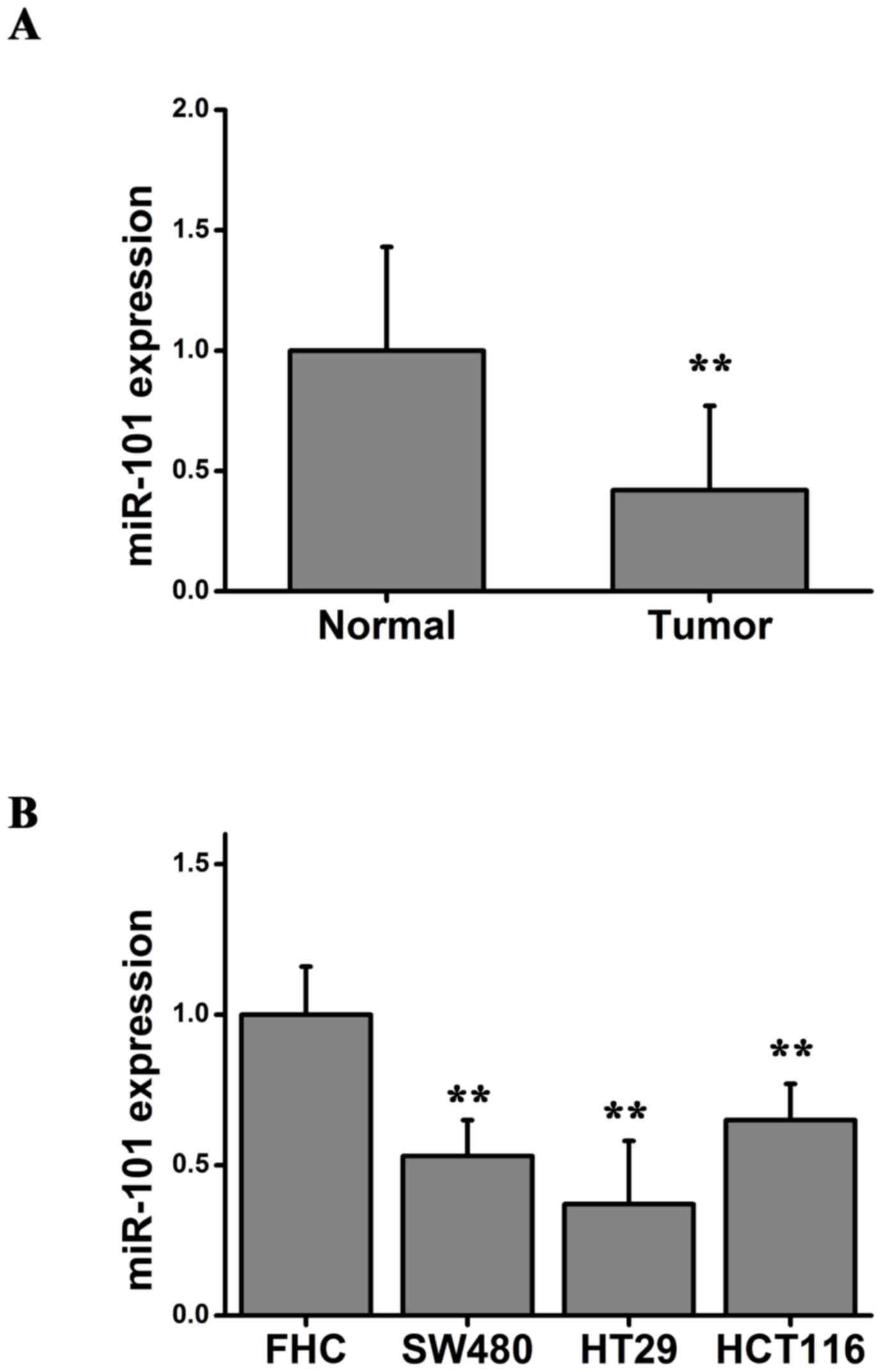
miR-101 is significantly downregulated
in cancer cell lines and colon cancer tissues
The expression of miR-101 was determined in colon
cancer tissues from 20 patients. The expression level of miR-101 in
colorectal carcinoma tissues and the adjacent normal tissues was
measured using RT-qPCR. Significant downregulation of the
expression level of miR-101 was identified in all 20 colorectal
carcinoma tissues compared with adjacent normal tissue (Fig. 1A). Furthermore, the expression
level of miR-101 was significantly upregulated in the normal human
intestinal epithelial cell line (FHC) compared with colorectal
cancer cell lines (SW480, HT29 and HCT116; Fig. 1B). Collectively, the RT-qPCR
results suggested that miR-101 may be involved in the development
and progression of colon cancer.
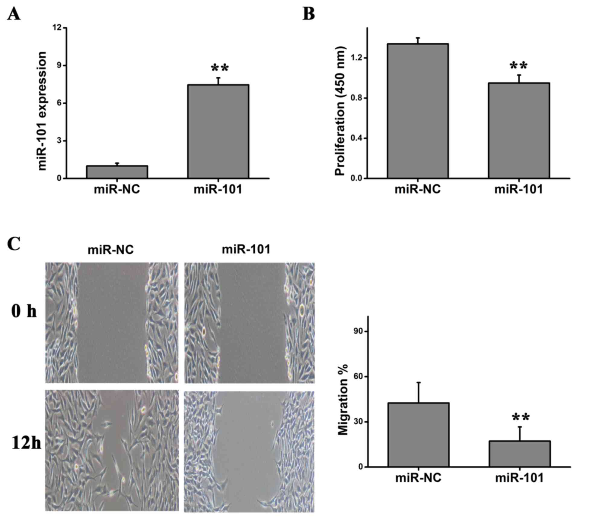
miR-101 inhibits tumor cell
proliferation and migration
Due to the downregulation of the expression level of
miR-101 in colorectal cancer tissues and cell lines, it was
hypothesized that miR-101 may inhibit tumor cell proliferation and
migration. In order to investigate the role of miR-101 on the
proliferation of HT29 cells, HT29 cells were transfected with
miR-101 mimics or miR-NC. RT-qPCR was performed to examine the
expression level of miR-101, and a significant upregulation of
miR-101 was detected following miR-101 overexpression (Fig. 2A). CCK-8 cell viability assay
suggested that overexpression of miR-101 significantly inhibited
the proliferation of HT29 cells (Fig.
2B). Using a wound healing assay, it was identified that
overexpression of miR-101 significantly repressed the migration of
HT29 cells (Fig. 2C).
Collectively, the present results suggested that overexpression of
miR-101 levels may inhibit the malignant features of colorectal
cancer, including cell proliferation and migration.
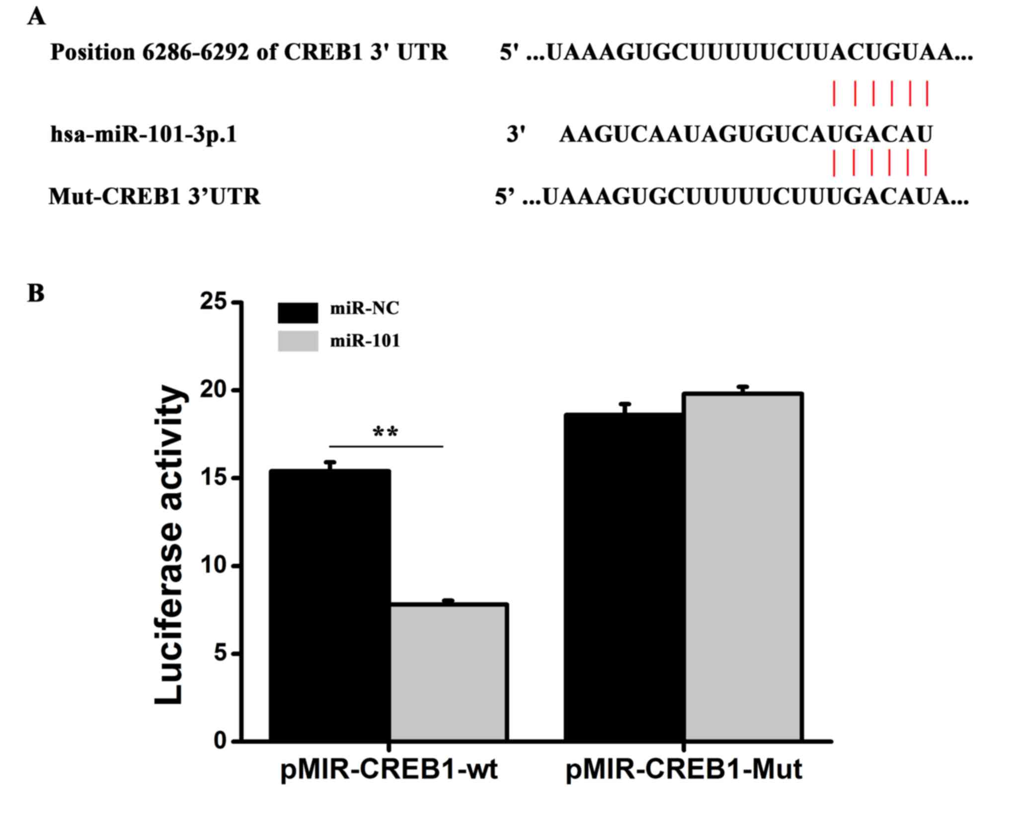
miR-101 regulates CREB1 expression by
targeting the 3′-untranslated region (UTR) of CREB1
Potential targets of miR-101 were predicted using
miRBase (http://www.mirbase.org/), and miR-101
binding sites were identified in the 3′-UTR of CREB1, one of the
putative targets (Fig. 3A). To
investigate whether miR-101 interacts with the CREB1 transcript
in vitro, the miR-101 binding sites in the 3′-UTR of CREB1
were mutated, and a dual luciferase assay was conducted (Fig. 3A). The pMIR-CREB1-WT plasmid,
containing the 3′-UTR of CREB1, or the pMIR-CREB1-Mut reporter,
containing a mutated form of the 3′-UTR, was cotransfected into
HT29 cells together with miR-101 mimics or miR-NC mimics and
luciferase assays were performed. Transfection of miR-101 mimics
significantly decreased the luciferase activity of pMIR-CREB1-WT
reporter plasmid compared with the miR-NC mimics. However, miR-101
overexpression did not affect the luciferase activity of the
pMIR-CREB1-Mut reporter plasmid compared with the miR-NC mimics
(Fig. 3B). Collectively, the
present results suggested that miR-101 may be able to bind directly
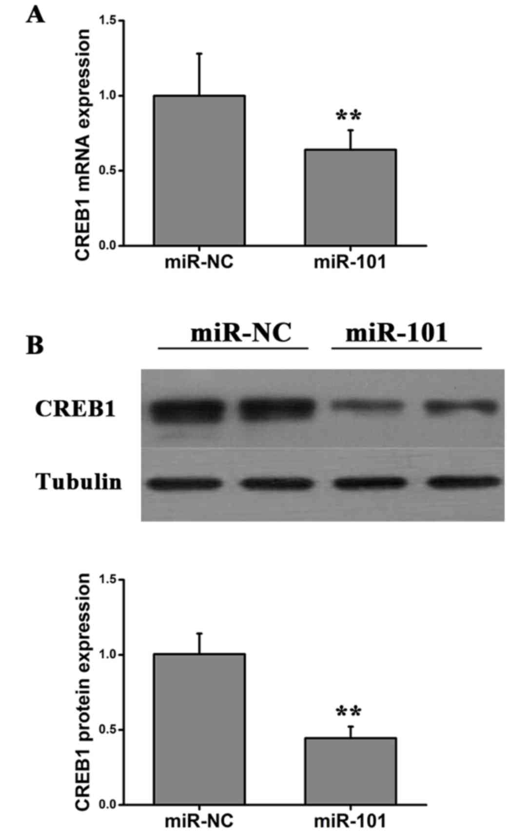
to the 3′-UTR of CREB1 to regulate gene expression. To investigate
whether miR-101 regulated the expression of CREB1, HT29 were
transfected with miR-101 and the protein and mRNA expression levels
of CREB1 were examined using western blot and RT-qPCR analysis,
respectively. miR-101 overexpression significantly decreased the
mRNA and protein expression levels of CREB1 (Fig. 4). The present results suggested
that miR-101 was able to decrease the expression level of CREB1 in
colon cancer cells by directly binding the 3′-UTR of CREB1.
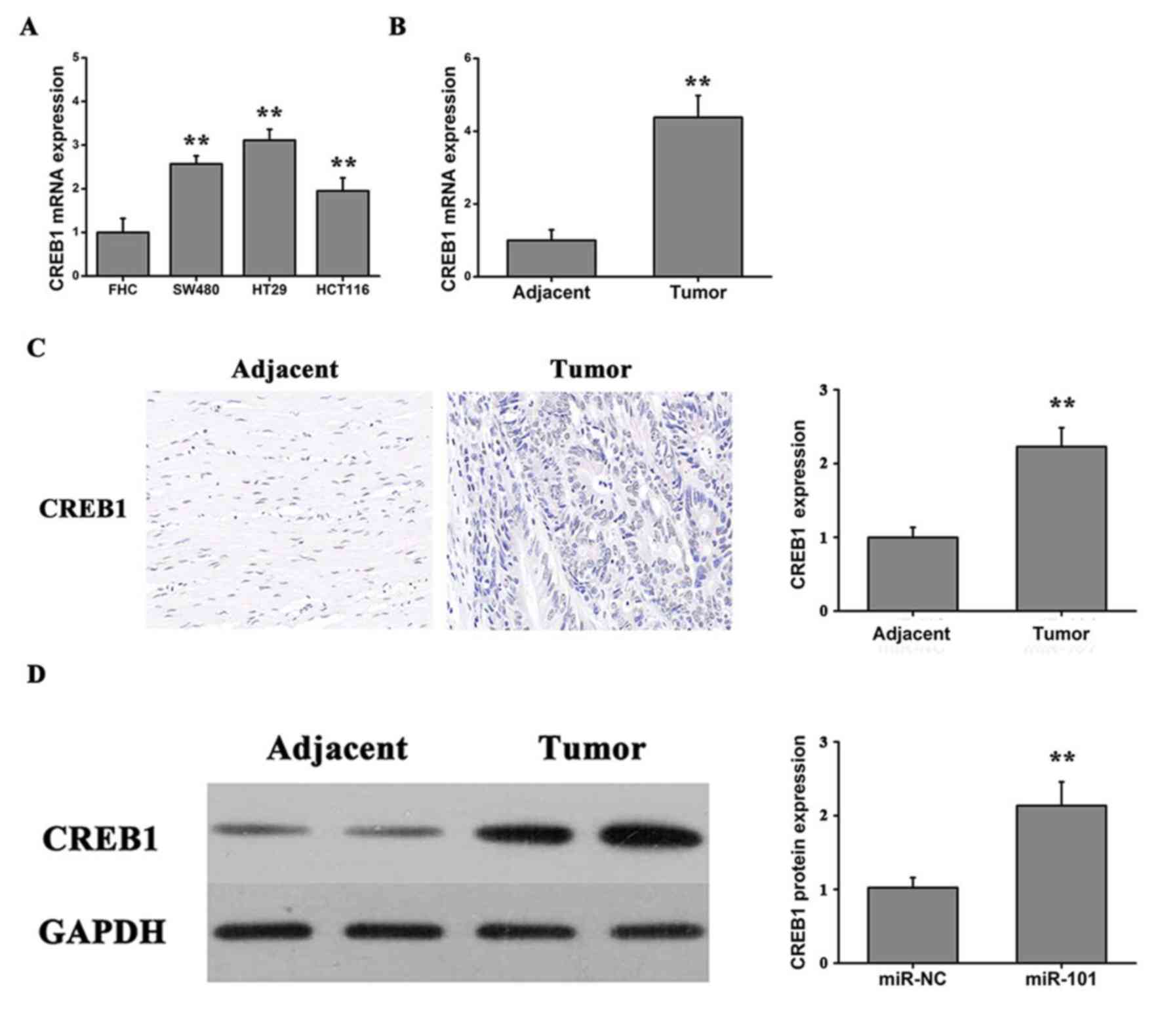
Expression of CREB1 in cell lines and
colon cancer tissues
To investigate the role of CREB1 in colon cancer
progression, the expression level of CREB1 was assessed using
RT-qPCR in three human colorectal cancer cell lines (SW480, HT29
and HTC116) and the FHC cell line. Furthermore, the expression
level of CREB1 was investigated in colorectal carcinoma tissues and
adjacent normal tissues. The expression level of CREB1 was
significantly increased in the three tumor cell lines compared with
the FHC cell line (Fig. 5A).
Furthermore, the expression levels of CREB1 were significantly
upregulated in the tumor tissues compared with the adjacent normal
tissues (Fig. 5B). Notably, the
immunochemistry results suggested that the protein expression of
CREB1 was significantly upregulated in the tumor tissues compared
with the normal tissues (Fig. 5C).
Additionally, it was identified, by western blotting, that the
protein expression level of CREB1 in colon cancer tumor tissues was
significantly increased compared with adjacent normal tissues
(Fig. 5D). Collectively, the
present results suggested that miR-101 may serve an important role
in colon cancer by inhibiting CREB1 expression.
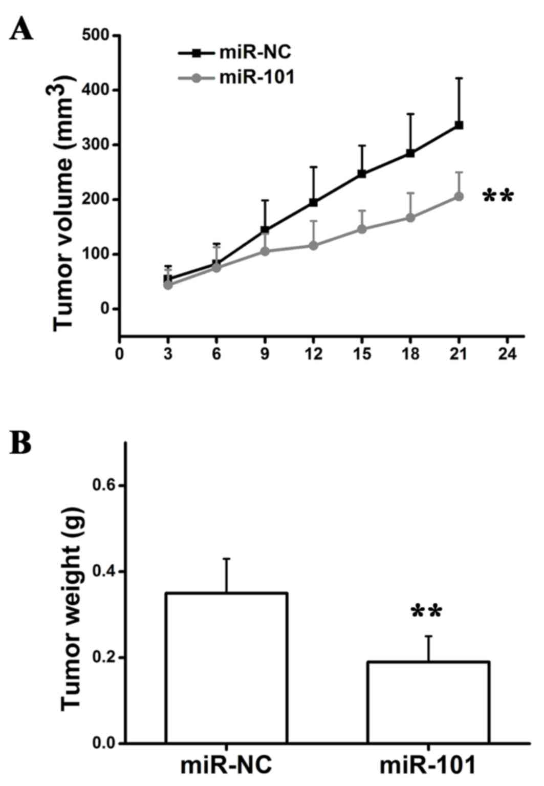
miR-101 inhibits tumor growth in
mouse
HT29 cells were injected into mice to generate a
murine xenograft model of colon carcinoma. Transfected cells were
injected subcutaneously into the back of nude mice. The tumor
growth rate was measured every 3 days and at the end of the
experiment the tumors were surgically removed, weighed and imaged
(Fig. 6). The present results
suggested that overexpression of miR-101 suppressed tumor growth
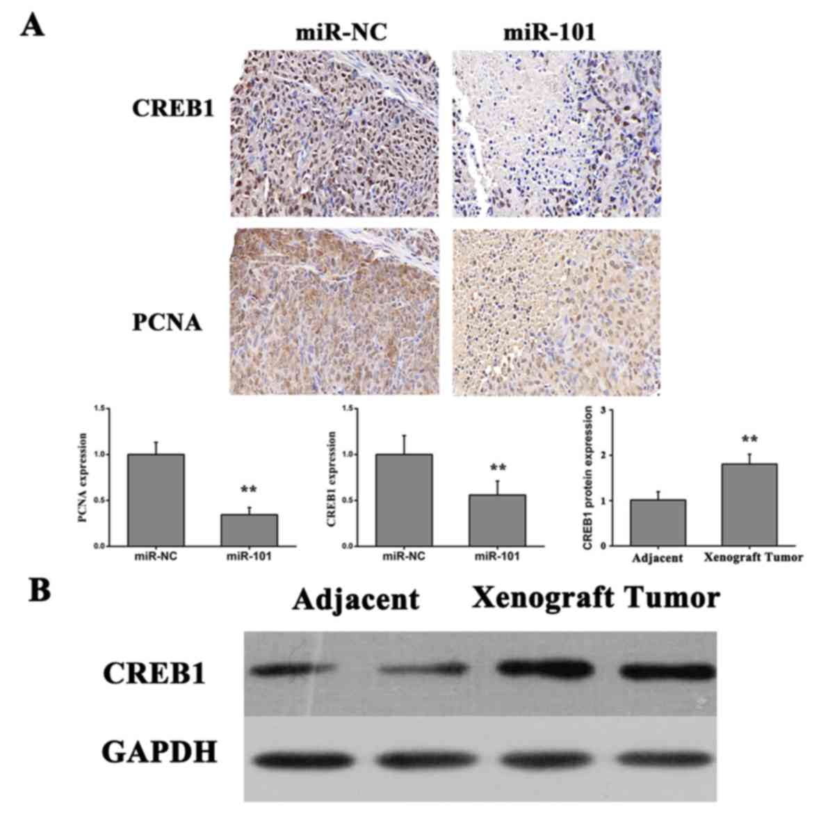
in vivo. Subsequently, immunohistochemical analysis was
performed using anti-PCNA and anti-CREB1 antibodies to detect
proliferating cells and CREB1 protein expression levels,
respectively. The number of cells positive for PCNA and CREB1
staining was significantly decreased in xenograft tumor tissues
produced from HT29 cells transfected with miR-101 compared with
miR-NC xenografts (Fig. 7A).
Additionally, the protein expression level of CREB1 was
significantly increased compared with adjacent normal tissue in
nude mice xenografted with colon cancer cells, as detected by
western blotting (Fig. 7B),
suggesting that CREB1 may be involved the development and
progression of colon cancer. The present results suggested that
overexpression of miR-101 led to downregulation of the expression
level of CREB1 and significant inhibition of tumor growth.
Discussion
miRNAs are important gene regulators and are
involved in numerous cellular processes, including proliferation,
migration, apoptosis, differentiation and carcinogenesis (33–36).
Furthermore, miRNAs have been identified to be associated with the
initiation and progression of malignant tumors (37). Although previous studies
investigated the role of miR-101 in colon cancer (38), the present results suggested a
number of novel findings. Overexpression of miR-101 served
anti-proliferative and anti-migratory roles in HT29 cells. In
addition, in vivo results suggested that miR-101
downregulated CREB1 in a tumor xenograft murine model. Furthermore,
CREB1 was identified as a potential target of miR-101 and
downregulation of CREB1 may be one of the underlying mechanisms
that mediate the effect of miR-101 in colon cancer suppression.
Therefore, the tumor suppressive effects of miR-101 in colon cancer
may involve the direct targeting of CREB1.
Multiple miRNAs have been observed to be
dysregulated in different types of human colorectal cancer
(39–43). Among them, miR-143 and miR-145 were
the first miRNAs observed to be downregulated in colorectal
neoplasms (17). Schepeler et
al (44) demonstrated that the
proliferation of three colorectal cancer cell lines (LS174T, DLD1
and HCT116) was significantly inhibited following transfection with
miR-145, suggesting that miR-145 may have tumor suppressive roles.
Additionally, miR-143 may serve an important role in colorectal
cancer cell proliferation by targeting the KRAS proto-oncogene
GTPase, which is involved in various signaling pathways that
regulate multiple cellular biological processes (45,46).
By contrast, a group of oncogenic miRNAs, including miR-17, miR-18a
and miR-19a, may be involved in the development of adenoma into
carcinoma (47,48).
Numerous studies have observed that miR-101
(sequence TACAGTACTGTGATAACTGAA) may function as a tumor inhibitor
(49). Vella et al
(20) demonstrated that miR-101
may repress the migration of embryonal rhabdomyosarcoma cells by
suppressing enhancer of zeste homolog 2. Furthermore,
overexpression of miR-101 inhibited the proliferation and migration
of endometrial cancer cells and induced cell apoptosis. However,
the mechanism underlying the association between miR-101 and colon
cancer progression remains unclear. In the present study, it was
hypothesized that downregulation of miR-101 may lead to colon
cancer progression, and the occurrence of a malignant phenotype.
The present study observed that miR-101 was significantly
downregulated in the human cancer tissues compared with the
adjacent normal colon tissues. In addition, the expression level of
miR-101 was significantly downregulated in three colorectal cancer
cell lines, SW480, HT29 and HCT116, compared with a normal human
intestinal epithelial cell line (FHC). The present results
suggested that miR-101 may contribute to the initiation and
progression of colon cancer.
Therefore, downregulation of miR-101 may represent a
feature of metastatic colon cancer cells. miR-101 expression was
significantly downregulated in invasive tumor cells, and the
expression of miR-101 has been identified to be downregulated in
undifferentiated colon cancer cells, which exhibit an aggressive
phenotype. The in vitro experiments in the present study
supported the hypothesis that overexpression of miR-101 may
regulate the malignant features of colon cancer cells. HT29 cells
overexpressing miR-101 exhibited a significant decrease in cell
proliferation and migration. The present finding supports the
observations in a previous study by Strillacci et al
(50).
Accumulating evidence demonstrated that numerous
biological events, including inflammation (51), epithelial-mesenchymal transition
(52) and hypoxia response
(53), are involved in the
establishment of a malignant phenotype in colon cancer cells. The
present study investigated the underlying mechanisms of miR-101 in
the development and progression of colon cancer. Notably, miR-101
significantly inhibited the expression level of CREB1 by directly
binding to the 3′-UTR of CREB1. The association between miR-101 and
CREB1 was supported by experiments performed in colon cancer cells
transfected with miR-101. Overexpression and activation of miR-101
led to a significant decrease of the mRNA and protein expression
levels of CREB1. Furthermore, CREB1 expression levels were
significantly increased in various colon cancer cell lines and
colon cancer tissues, and the immunohistochemical results confirmed
that the protein expression level of CREB1 in tumor tissues was
significantly upregulated compared with adjacent normal tissues.
Furthermore, in vivo experiments suggested that CREB1 may be
a key target of miR-101, as overexpression of miR-101 inhibited
tumor growth and downregulated the expression level of CREB1. The
miR-101/CREB1 pathway may represent a potential novel mechanism of
colon cancer progression and it may facilitate the prevention and
the treatment of colon cancer.
Collectively, the present study suggested that
miR-101 may serve a role as a tumor inhibitor. Overexpression of
miR-101 suppressed cell proliferation and migration in vitro
and inhibited tumor growth in xenograft mouse models in
vivo, with CREB1 being a potential functional target of
miR-101. The present results suggested an association between the
miR-101/CREB1 pathway and tumor progression, and miR-101 may be an
important factor involved in this signaling pathway. Collectively,
this study suggested that miR-101 could be a therapeutic target in
colon cancer, and that investigating the miR-101/CREB1 pathway may
facilitate the development of a novel therapeutic strategy to treat
colon cancer.
Acknowledgements
Not applicable.
Funding
No funding was received.
Availability of data and materials
The datasets used and/or analyzed during the current
study are available from the corresponding author on reasonable
request.
Authors' contributions
QY and WY designed the study, performed experiments,
analyzed the data and wrote the manuscript. WY and XH performed the
experiments. QY and XH analyzed the data and drafted the
manuscript.
Ethics approval and consent to
participate
The present study was approved by The Ethics
Committee of Yantai Yeda Hospital and informed consent was obtained
from all patients.
Patient consent for publication
Not applicable.
Competing interests
The authors have no competing interests.
References
|
1
|
Gellad ZF and Provenzale D: Colorectal
cancer: National and international perspective on the burden of
disease and public health impact. Gastroenterology. 138:2177–2190.
2010. View Article : Google Scholar : PubMed/NCBI
|
|
2
|
Siegel R, Naishadham D and Jemal A: Cancer
statistics, 2013. CA Cancer J Clin. 63:11–30. 2013. View Article : Google Scholar : PubMed/NCBI
|
|
3
|
Jeon J, Du M, Schoen RE, Hoffmeister M,
Newcomb PA, Berndt SI, Caan B, Campbell PT, Chan AT, Chang-Claude
J, et al: Determining risk of colorectal cancer and starting age of
screening based on lifestyle, environmental, and genetic factors.
Gastroenterology. 154:2152–2164.e19. 2018. View Article : Google Scholar : PubMed/NCBI
|
|
4
|
Torre LA, Bray F, Siegel RL, Ferlay J,
Lortet-Tieulent J and Jemal A: Global cancer statistics, 2012. CA
Cancer J Clin. 65:87–108. 2015. View Article : Google Scholar : PubMed/NCBI
|
|
5
|
Sjo OH, Berg M, Merok MA, Kolberg M,
Svindland A, Lothe RA and Nesbakken A: Peritoneal carcinomatosis of
colon cancer origin: Highest incidence in women and in patients
with right-sided tumors. J Surg Oncol. 104:792–797. 2011.
View Article : Google Scholar : PubMed/NCBI
|
|
6
|
Vermeer NC, Snijders HS, Holman FA,
Liefers GJ, Bastiaannet E, van de Velde CJ and Peeters KC:
Colorectal cancer screening: Systematic review of screen-related
morbidity and mortality. Cancer Treat Rev. 54:87–98. 2017.
View Article : Google Scholar : PubMed/NCBI
|
|
7
|
Mori F, Ferraiuolo M, Santoro R, Sacconi
A, Goeman F, Pallocca M, Pulito C, Korita E, Fanciulli M, Muti P,
et al: Multitargeting activity of miR-24 inhibits long-term
melatonin anticancer effects. Oncotarget. 7:20532–20548. 2016.
View Article : Google Scholar : PubMed/NCBI
|
|
8
|
Yogin P, Nirav S, Lee JS, Markoutsa E, Jie
C, Liu S, Botbyl R, Reisman D, Xu P and Chen H: A novel
double-negative feedback loop between miR-489 and the
HER2-SHP2-MAPK signaling axis regulates breast cancer cell
proliferation and tumor growth. Oncotarget. 7:18295–18308.
2016.PubMed/NCBI
|
|
9
|
Konishi H, Fujiya M, Ueno N, Moriichi K,
Sasajima J, Ikuta K, Tanabe H, Tanaka H and Kohgo Y: microRNA-26a
and −584 inhibit the colorectal cancer progression through
inhibition of the binding of hnRNP A1-CDK6 mRNA. Biochem Biophys
Res Commun. 467:847–852. 2015. View Article : Google Scholar : PubMed/NCBI
|
|
10
|
Tili E, Michaille JJ and Calin GA:
Expression and function of micro-RNAs in immune cells during normal
or disease state. Int J Med Sci. 5:73–79. 2008. View Article : Google Scholar : PubMed/NCBI
|
|
11
|
He L and Hannon GJ: MicroRNAs: Small RNAs
with a big role in gene regulation. Nat Rev Genet. 5:522–531. 2004.
View Article : Google Scholar : PubMed/NCBI
|
|
12
|
Kantharidis P, Wang B, Carew RM and Lan
HY: Diabetes complications: The microRNA perspective. Diabetes.
60:1832–1837. 2011. View Article : Google Scholar : PubMed/NCBI
|
|
13
|
Trionfini P and Benigni A: MicroRNAs as
master regulators of glomerular function in health and disease. J
Am Soc Nephrol. 28:1686–1696. 2017. View Article : Google Scholar : PubMed/NCBI
|
|
14
|
Kato M and Natarajan R: MicroRNAs in
diabetic nephropathy: Functions, biomarkers, and therapeutic
targets. Ann N Y Acad Sci. 1353:72–88. 2015. View Article : Google Scholar : PubMed/NCBI
|
|
15
|
Srivastava K and Srivastava A:
Comprehensive review of genetic association studies and
meta-analyses on miRNA polymorphisms and cancer risk. PLoS One.
7:e509662012. View Article : Google Scholar : PubMed/NCBI
|
|
16
|
Tokarz P and Blasiak J: The role of
microRNA in metastatic colorectal cancer and its significance in
cancer prognosis and treatment. Acta Biochim Pol. 59:467–474. 2012.
View Article : Google Scholar : PubMed/NCBI
|
|
17
|
Michael MZ, O' Connor SM, van Holst
Pellekaan NG, Young GP and James RJ: Reduced accumulation of
specific microRNAs in colorectal neoplasia. Mol Cancer Res.
1:882–891. 2003.PubMed/NCBI
|
|
18
|
Song B, Wang Y, Kudo K, Gavin EJ, Xi Y and
Ju J: miR-192 Regulates dihydrofolate reductase and cellular
proliferation through the p53-microRNA circuit. Clin Cancer Res.
14:8080–8086. 2008. View Article : Google Scholar : PubMed/NCBI
|
|
19
|
Braun CJ, Zhang X, Savelyeva I, Wolff S,
Moll UM, Schepeler T, Ørntoft TF, Andersen CL and Dobbelstein M:
p53-Responsive micrornas 192 and 215 are capable of inducing cell
cycle arrest. Cancer Res. 68:10094–10104. 2008. View Article : Google Scholar : PubMed/NCBI
|
|
20
|
Vella S, Pomella S, Leoncini PP, Colletti
M, Conti B, Marquez VE, Strillacci A, Roma J, Gallego S, Milano GM,
et al: MicroRNA-101 is repressed by EZH2 and its restoration
inhibits tumorigenic features in embryonal rhabdomyosarcoma. Clin
Epigenetics. 7:822015. View Article : Google Scholar : PubMed/NCBI
|
|
21
|
Konno Y, Dong P, Xiong Y, Suzuki F, Lu J,
Cai M, Watari H, Mitamura T, Hosaka M, Hanley SJ, et al:
MicroRNA-101 targets EZH2, MCL-1 and FOS to suppress proliferation,
invasion and stem cell-like phenotype of aggressive endometrial
cancer cells. Oncotarget. 5:6049–6062. 2014. View Article : Google Scholar : PubMed/NCBI
|
|
22
|
Xu L, Beckebaum S, Iacob S, Wu G, Kaiser
GM, Radtke A, Liu C, Kabar I, Schmidt HH, Zhang X, et al:
MicroRNA-101 inhibits human hepatocellular carcinoma progression
through EZH2 downregulation and increased cytostatic drug
sensitivity. J Hepatol. 60:590–598. 2014. View Article : Google Scholar : PubMed/NCBI
|
|
23
|
Su H, Yang JR, Xu T, Huang J, Xu L, Yuan Y
and Zhuang SM: MicroRNA-101, down-regulated in hepatocellular
carcinoma, promotes apoptosis and suppresses tumorigenicity. Cancer
Res. 69:1135–1142. 2009. View Article : Google Scholar : PubMed/NCBI
|
|
24
|
Yan D, Ng WL, Zhang X, Wang P, Zhang Z, Mo
YY, Mao H, Hao C, Olson JJ, Curran WJ and Wang Y: Targeting
DNA-PKcs and ATM with miR-101 sensitizes tumors to radiation. PLoS
One. 5:e113972010. View Article : Google Scholar : PubMed/NCBI
|
|
25
|
Buechner J, Tømte E, Haug BH, Henriksen
JR, Løkke C, Flægstad T and Einvik C: Tumour-suppressor microRNAs
let-7 and mir-101 target the proto-oncogene MYCN and inhibit cell
proliferation in MYCN-amplified neuroblastoma. Br J Cancer.
105:296–303. 2011. View Article : Google Scholar : PubMed/NCBI
|
|
26
|
Tuttle RM, Haugen B and Perrier ND:
Updated American Joint Committee on cancer/tumor-node-metastasis
staging system for differentiated and anaplastic thyroid cancer
(Eighth Edition): What changed and why? Thyroid. 27:751–756. 2017.
View Article : Google Scholar : PubMed/NCBI
|
|
27
|
Chuang-Bo Y, Tai-Ping H, Hai-Feng D,
Yong-Jun J, Xi-Rong Z, Guang-Ming M, Chenglong R, Jun W and Yong Y:
Quantitative assessment of the degree of differentiation in colon
cancer with dual-energy spectral CT. Abdom Radiol (NY).
42:2591–2596. 2017. View Article : Google Scholar : PubMed/NCBI
|
|
28
|
Liu G, Friggeri A, Yang Y, Park YJ,
Tsuruta Y and Abraham E: miR-147, a microRNA that is induced upon
Toll-like receptor stimulation, regulates murine macrophage
inflammatory responses. Proc Natl Acad Sci USA. 106:15819–15824.
2009. View Article : Google Scholar : PubMed/NCBI
|
|
29
|
Livak KJ and Schmittgen TD: Analysis of
relative gene expression data using real-time quantitative PCR and
the 2(-Delta Delta C(T)) method. Methods. 25:402–408. 2001.
View Article : Google Scholar : PubMed/NCBI
|
|
30
|
Gaidatzis D, van Nimwegen E, Hausser J and
Zavolan M: Inference of miRNA targets using evolutionary
conservation and pathway analysis. BMC Bioinformatics. 8:692007.
View Article : Google Scholar : PubMed/NCBI
|
|
31
|
Van Peer G, Lefever S, Anckaert J, Beckers
A, Rihani A, Van Goethem A, Volders PJ, Zeka F, Ongenaert M,
Mestdagh P and Vandesompele J: miRBase tracker: Keeping track of
microRNA annotation changes. Database (Oxford). 2014(pii):
bau0802014.PubMed/NCBI
|
|
32
|
Agarwal V, Bell GW, Nam JW and Bartel DP:
Predicting effective microRNA target sites in mammalian mRNAs.
eLife. 4:e050052015. View Article : Google Scholar :
|
|
33
|
Chen CZ, Li L, Lodish HF and Bartel DP:
MicroRNAs modulate hematopoietic lineage differentiation. Science.
303:83–86. 2004. View Article : Google Scholar : PubMed/NCBI
|
|
34
|
Cheng AM, Byrom MW, Shelton J and Ford LP:
Antisense inhibition of human miRNAs and indications for an
involvement of miRNA in cell growth and apoptosis. Nucleic Acids
Res. 33:1290–1297. 2005. View Article : Google Scholar : PubMed/NCBI
|
|
35
|
Croce CM and Calin GA: miRNAs, cancer, and
stem cell division. Cell. 122:6–7. 2005. View Article : Google Scholar : PubMed/NCBI
|
|
36
|
Karp X and Ambros V: Developmental
biology. Encountering microRNAs in cell fate signaling. Science.
310:1288–1289. 2005. View Article : Google Scholar : PubMed/NCBI
|
|
37
|
Mitomo S, Maesawa C, Ogasawara S, Iwaya T,
Shibazaki M, Yashima-Abo A, Kotani K, Oikawa H, Sakurai E, Izutsu
N, et al: Downregulation of miR-138 is associated with
overexpression of human telomerase reverse transcriptase protein in
human anaplastic thyroid carcinoma cell lines. Cancer Sci.
99:280–286. 2008. View Article : Google Scholar : PubMed/NCBI
|
|
38
|
Chen LG, Xia YJ and Cui Y: Upregulation of
miR-101 enhances the cytotoxic effect of anticancer drugs through
inhibition of colon cancer cell proliferation. Oncol Rep.
38:100–108. 2017. View Article : Google Scholar : PubMed/NCBI
|
|
39
|
Gramantieri L, Ferracin M, Fornari F,
Veronese A, Sabbioni S, Liu CG, Calin GA, Giovannini C, Ferrazzi E,
Grazi GL, et al: Cyclin G1 is a target of miR-122a, a microRNA
frequently down-regulated in human hepatocellular carcinoma. Cancer
Res. 67:6092–6099. 2007. View Article : Google Scholar : PubMed/NCBI
|
|
40
|
Chan JA, Krichevsky AM and Kosik KS:
MicroRNA-21 is an antiapoptotic factor in human glioblastoma cells.
Cancer Res. 65:6029–6033. 2005. View Article : Google Scholar : PubMed/NCBI
|
|
41
|
Yan LX, Huang XF, Shao Q, Huang MY, Deng
L, Wu QL, Zeng YX and Shao JY: MicroRNA miR-21 overexpression in
human breast cancer is associated with advanced clinical stage,
lymph node metastasis and patient poor prognosis. RNA.
14:2348–2360. 2008. View Article : Google Scholar : PubMed/NCBI
|
|
42
|
Iorio MV, Ferracin M, Liu CG, Veronese A,
Spizzo R, Sabbioni S, Magri E, Pedriali M, Fabbri M, Campiglio M,
et al: MicroRNA gene expression deregulation in human breast
cancer. Cancer Res. 65:7065–7070. 2005. View Article : Google Scholar : PubMed/NCBI
|
|
43
|
Nicoloso MS and Calin GA: MicroRNA
involvement in brain tumors: From bench to bedside. Brain Pathol.
18:122–129. 2008. View Article : Google Scholar : PubMed/NCBI
|
|
44
|
Schepeler T, Reinert JT, Ostenfeld MS,
Christensen LL, Silahtaroglu AN, Dyrskjøt L, Wiuf C, Sørensen FJ,
Kruhøffer M, Laurberg S, et al: Diagnostic and prognostic microRNAs
in stage II colon cancer. Cancer Res. 68:6416–6424. 2008.
View Article : Google Scholar : PubMed/NCBI
|
|
45
|
Omerovic J, Laude AJ and Prior IA: Ras
proteins: Paradigms for compartmentalised and isoform-specific
signalling. Cell Mol Life Sci. 64:2575–2589. 2007. View Article : Google Scholar : PubMed/NCBI
|
|
46
|
Bonfrate L, Altomare DF, Di Lena M,
Travaglio E, Rotelli MT, De Luca A and Portincasa P: MicroRNA in
colorectal cancer: New perspectives for diagnosis, prognosis and
treatment. J Gastrointestin Liver Dis. 22:311–320. 2013.PubMed/NCBI
|
|
47
|
Frampton AE, Krell J, Gall TM, Castellano
L, Stebbing J and Jiao LR: miR-15b and miR-17 are tumor-derived
plasma microRNAs dysregulated in colorectal neoplasia. Ann Surg.
262:e61–e62. 2015. View Article : Google Scholar : PubMed/NCBI
|
|
48
|
Diosdado B, van de Wiel MA, Terhaar Sive
Droste JS, Mongera S, Postma C, Meijerink WJ, Carvalho B and Meijer
GA: MiR-17-92 cluster is associated with 13q gain and c-myc
expression during colorectal adenoma to adenocarcinoma progression.
Br J Cancer. 101:707–714. 2009. View Article : Google Scholar : PubMed/NCBI
|
|
49
|
Chandramouli A, Onyeagucha BC,
Mercado-Pimentel ME, Stankova L, Shahin NA, LaFleur BJ, Heimark RL,
Bhattacharyya AK and Nelson MA: MicroRNA-101 (miR-101)
post-transcriptionally regulates the expression of EP4 receptor in
colon cancers. Cancer Biol Ther. 13:175–183. 2012. View Article : Google Scholar : PubMed/NCBI
|
|
50
|
Strillacci A, Valerii MC, Sansone P,
Caggiano C, Sgromo A, Vittori L, Fiorentino M, Poggioli G, Rizzello
F, Campieri M and Spisni E: Loss of miR-101 expression promotes
Wnt/β-catenin signalling pathway activation and malignancy in colon
cancer cells. J Pathol. 229:379–389. 2013. View Article : Google Scholar : PubMed/NCBI
|
|
51
|
Tsujii M and DuBois RN: Alterations in
cellular adhesion and apoptosis in epithelial cells overexpressing
prostaglandin endoperoxide synthase 2. Cell. 83:493–501. 1995.
View Article : Google Scholar : PubMed/NCBI
|
|
52
|
Kalluri R and Weinberg RA: The basics of
epithelial-mesenchymal transition. J Clin Invest. 119:1420–1428.
2009. View Article : Google Scholar : PubMed/NCBI
|
|
53
|
Demir R, Naschberger L, Demir I, Melling
N, Dimmler A, Papadopoulus T, Sturzl M, Klein P and Hohenberger W:
Hypoxia generates a more invasive phenotype of tumour cells: An in
vivo experimental setup based on the chorioallantoic membrane.
Pathol Oncol Res. 15:417–422. 2009. View Article : Google Scholar : PubMed/NCBI
|