Introduction
Intestinal ischemia reperfusion (I/R) injury is one
of the commonest tissue and organ injuries that occur during
surgeries, such as abdominal aortic aneurysm surgery,
cardiopulmonary bypass surgery and intestinal transplantation
surgery (1). Intestinal I/R injury
can cause pathophysiological changes of the intestinal mucosa
(2) and not only causes the
destruction of intestinal tissue, but also results in secondary
damage to distant organs and tissues, thus inducing multi-organ
dysfunction syndrome and systemic inflammatory response syndrome
(3). As a result of these changes,
the small intestine is often called ‘the origin of multiple organ
failure after trauma’ (4,5). Previous studies have revealed the
pathological mechanisms of intestinal I/R injury, which include
microvascular damage to ischemic organs, the release of
inflammatory cytokines, ATP depletion, oxygen free radical injury,
leukocyte adhesion, calcium overload and apoptosis (2,6). I/R
injury evokes reactive oxygen species (ROS) production and results
in DNA damage, cell apoptosis and tubule destruction (7). In previous studies, cell and animal
experiments have investigated the pathogenesis of intestinal I/R
injuries (8,9). However, due to their complicated
mechanisms, the underlying pathological factors involved in
intestinal I/R injuries remain to be elucidated. Therefore,
additional studies on the mechanism of intestinal I/R injuries can
provide a theoretical basis for their clinical treatment.
Remote ischemic pre-conditioning (RIPOC) is a new
type of organ protection method that allows the short-term ischemia
and reperfusion of one organ to protect another distal organ
(10,11). The first study of RIPOC found that
preconditioning of the anterior descending artery of a dog heart
with 4 cycles of 5 min ischemia followed by 5 min of reperfusion
helped to protect against distant myocardial I/R injury (12). A subsequent study demonstrated that
I/R preconditioning of tissues and organs in the farther regions
could also help to protect organs (13). Those studies notably enhanced the
operability of RIPOC and the possibility of its clinical use.
However, due to the unpredictability of clinical ischemic events,
such as anesthesia methods, anesthetic drugs, complications and
individual differences, the current clinical applications of RIPOC
remain limited (14,15). RIPOC can be implemented following
the occurrence of ischemic events with good controllability and its
use and applications are being extensively studied (16,17).
However, the mechanism by which RIPOC protects against intestinal
I/R injury has not yet been fully elucidated.
High-mobility group box 1 (HMGB1) is a highly
conserved DNA-binding protein that is passively released by dead
cells or actively secreted into the extracellular environment by
inflammatory cells under conditions of cellular stress to regulate
innate and adaptive immunity (18).
The known HMGB1 receptors include receptor for advanced glycation
end products (RAGE), Toll-like receptor (TLR) 2 and TLR4 (19). Studies have verified that RAGE is
the earliest identified HMGB1 receptor that belongs to the
immunoglobulin superfamily (20,21).
The binding of RAGE to its ligands can result in the activation of
multiple protein kinases including MAPKs, RaC/cell division control
protein 42 homolog and Janus kinase/STATs and thereby activate the
NF-κB signaling pathway (22). In
addition, the biological function of HMGB1 also depends on the
involvement of RAGE (20). However,
it is unclear whether the RAGE/HMGB pathway is involved in the
protective effect of RIPOC against intestinal I/R injury.
The present study established a mouse model of
intestinal I/R injury and observed the influence of RIPOC on
histopathological features, antioxidant capacity and inflammation
in the injured mice. In addition, the regulatory effects of RIPOC
on the RAGE/HMGB pathway in the intestinal I/R injury mouse models
was also confirmed. The results of the present study suggested
RIPOC as an approach for protecting tissues against I/R injury by
attenuating oxidative stress and preventing inflammation.
Materials and methods
Animals
A total of 40 male specific pathogen-free (SPF) C57
BL/6 mice (8 weeks old; weight range, 20–30 g) were acclimated for
a week prior to being used for experiments. The mice were housed in
a room with a 12-h light/dark cycle, 40–60% humidity and a
controlled temperature of 18–23°C; food and water were available
ad libitum. All animal experiments were performed in
compliance with ethical standards of Shandong Provincial Hospital
Affiliated to Shandong University. The present study was approved
by the Ethics committee of Shandong Provincial Hospital affiliated
to Shandong University (approval no. 2019-330).
Establishment of an intestinal I/R
injury model
Mice were anesthetized by intraperitoneal injection
of 60 mg/kg pentobarbital sodium before surgery to avoid pain.
Next, the superior mesenteric artery (SMA) was exposed and the root
of the superior mesenteric artery was blocked using a non-traumatic
vascular clip, resulting in complete intestinal ischemia for 1 h,
followed by reperfusion of the clip for 1 h. Heart rate, blood
pressure and body temperature were monitored during experiments to
evaluated the stress response. Mice in the sham group were also
anesthetized, however, the SMA was isolated but not blocked. All
the mice were euthanized with pentobarbital sodium (i.p., 60 mg/kg)
and then decapitated when they reached a humane end of life (gentle
heartbeat, even breathing, stable body temperature). Mice which did
not breath for over 3 min and without heartbeat were identified as
having succumbed. Samples of colorectal tissue were collected and
fixed in 4% paraformaldehyde for further examination by hematoxylin
and eosin (H&E) staining and immunohistochemistry. Other tissue
samples were quick-frozen in liquid nitrogen and stored at −80°C.
Serum samples were collected and stored at −80°C. No animal
succumbed during the experiment before sacrifice. The whole
experiments lasted for ≤1.5 h.
Animal groups
The mice were randomly assigned to four separate
groups: i) the Sham group (only laparotomy was performed and the
SMA was separated without clamping); ii) the I/R group (treated as
aforementioned; iii) I/R+RIPOC group (following ischemia for 45
min, three cycles of 30 sec artery perfusion/30 sec artery blocking
were performed and the remaining steps were the same as for the I/R
group) and 10 mice per group; and iv) the I/R+RIPOC+CC-90003 group.
CC9003 was purchased from MedChemExpress (cat. no. HY-112570).
H&E staining
The separated intestinal tubes were fixed with 4%
paraformaldehyde (EMD Millipore) for 12 h at 4°C, dehydrated by
gradient alcohol (70% alcohol for 1 h, 85% alcohol for 1 h, 95%
alcohol for 1 h, 100% alcohol for 30 min, 100% alcohol for 1 h,
100% alcohol for 30 min). Then the samples were embedded in
paraffin and cut into 4-µm sections. After being roasted at 38°C
for 6 h, the sections were subjected to a series of operations
including dewaxing with xylene, hydration with a gradient alcohol
series, hematoxylin staining at room temperature for 8 min,
differentiation in 1% alcohol and hydrochloric acid for 3 sec,
bluing in Scott blue buffer for 10 min, and eosin for 10 sec. After
rehydrating and clearing with a gradient alcohol series, the
sections were observed under a light microscope (CKX41, Olympus
Corporation) at ×200 and ×400 magnification with 5 fields. The
histological assessment was performed according to Chiu's score
(23). Briefly, it was as follows:
0, normal; 1, development of Gruenhagen's space, along with
capillary congestion; 2, increase in epithelial space with moderate
lifting of epithelial layer; 3, markedly lifted epithelial; 4,
denuded villi with lamina propria and dilated capillaries; 5,
digestion and disintegration of lamina propria and hemorrhage and
ulceration.
Immunohistochemistry (IHC) assays
As described in previous studies (24,25),
the 4-µm sections were dewaxed in xylene and then rehydrated with a
gradient alcohol series (100-70%); following which, they were
subjected to antigen retrieval with 0.01 M citrate buffer (cat. no.
AR0024; Wuhan Boster Biological Technology, Ltd.) and incubation
with 3% H2O2 at room temperature for 10 min. Next, the sections
were blocked with 10% bovine serum albumin (BSA; Sigma-Aldrich;
Merck KGaA; cat. no. A2153), incubated with primary antibodies
against HMGB1 (1:100; cat. no. ab77302; Abcam) and RAGE (1:100;
cat. no. ab3611; Abcam) overnight at 4°C, and then incubated with
an HRP-labeled secondary antibody (1:50, Abcam; cat. no. ab205719)
at room temperature for 2 h. Subsequently, the sections were
hatched by exposure to 50 µl of peroxidase-labeled polymer (cat.
no. K4003; Dako; Agilent Technologies, Inc.) and 100 µl of
substrate-chromogen (cat. no. K3464; Dako; Agilent Technologies,
Inc.) for 2 min. The results were examined under a light microscope
(Olympus Corporation) at ×200 magnification with 6 fields.
Reverse transcription-quantitative
polymerase chain reaction (RT-qPCR)
Total tissue RNAs were extracted from the separated
tissues, which were then homogenized on ice using
TRIzol® reagent (Thermo Fisher Scientific, Inc.). The
RNA concentration was detected with a GeneQuant 1300
spectrophotometer (Cytiva) according to the manufacturer's
protocols. A 1-µl sample of total RNA was reverse-transcribed into
cDNA in a 20 µl system using an All-in-One First-Strand cDNA
Synthesis kit (GeneCopoeia, Inc.) based on the manufacturer's
protocols. Then RT-qPCR was performed using a SYBR-Green qPCR kit
(cat. no. F-416L; Finnzymes; Thermo Fisher Scientific, Inc.) on a
CFX96 Real-time PCR Detection System C1000 (Bio-Rad Laboratories,
Inc.). The thermocycling conditions were as follows: 95°C for 2
min; followed by 40 cycles of 95°C for 10 sec, 60°C for 34 sec and
72°C for 33 sec. The primers used for RT-qPCR were synthesized by
Takara Biotechnology Co., Ltd. and the gene expression was
quantitated by the 2−ΔΔCq method (26) from 3 repeated experiments. The
primer sequences were as following: GAPDH forward,
5′-CCTCGTCTCATAGACAAGATGGT-3′ and reverse,
5′-GGGTAGAGTCATACTGGAACATG-3′; HMGB1 forward,
5′-TGTTCTGAGTACCGCCCAAA-3′ and reverse, 5′-CTTGGCGGCCTTCTTTTCAT-3′;
and RAGE forward, 5′-TCACAGAAACCGGTGATGAAG-3′ and reverse,
5′-CTCGAGTCTGGGTTGTCGTT-3′.
Western blot analysis
The intestinal tube tissues in each group were
homogenized on ice and their total proteins were extracted using
RIPA lysate buffer (cat. no. AR0105; Wuhan Boster Biological
Technology, Ltd.). The total protein concentration in each sample
was determined using a BCA Protein Assay kit (cat. no. 233225;
Thermo Fisher Scientific, Inc.). A 20-µg aliquot of total protein
from each sample was separated by 10% SDS-PAGE (cat. no. NP0322BOX;
Thermo Fisher Scientific, Inc.) and the protein bands were
transferred onto PVDF membranes (PerkinElmer, Inc.), which were
subsequently blocked with 5% powdered skimmed milk at room
temperature for 2 h. Following blocking, the membranes were
incubated with primary antibodies at 4°C overnight, and then
incubated with HRP-conjugated secondary antibodies (1:2,000; cat.
nos. ab205719 or ab6721; Abcam) at room temperature for 1 h. Next,
ECL reagent (Pierce; Thermo Fisher Scientific, Inc.) was used to
visualize the immunostained proteins and the relative amounts of
protein were determined using a ChemiDoc Imaging System (Bio-Rad
Laboratories, Inc.). The primary antibodies used were: HMGB1
(1:1,000; cat. no. ab77302), RAGE (1:1,000; cat. no. ab3611),
p-ERK1/2 (1:1,000; cat. no. ab214362), ERK1/2 (1:1,000; cat. no.
ab17942), p-p65 (1:1,000; cat. no. ab86299), p65 (1:1,000; cat. no.
ab32536), p-IKBa (1:1,000; cat. no. ab24783), IKBa (1:1,000; cat.
no. ab7217), NLRP3 (1:1,000; cat. no. ab214185) and GAPDH (1:2,000;
cat. no. ab9485; all from Abcam). GAPDH was used as an internal
reference. The gray value in each group was determined using ImageJ
software (version 1.51d; National institutes of Health).
Flow cytometric analysis for ROS
The level of ROS was analyzed using blue fluorescent
dye dihydroethidium (DHE, Invitrogen; Thermo Fisher Scientific,
Inc.) as described in a previous study (23). The prepared samples were incubated
with 2.5 mmol/l DHE solution for 25 min at 37°C. Following washing
with PBS three times, the fluorescence intensity in each group was
determined by flow cytometry (Beckman CytoFLEX; Beckman Coulter,
Inc.).
ELISA assays
A total of 5 ml blood was collected from the vein
and heparin using anticoagulation. Following centrifugation (3,000
× g for 10 min at 4°C), the blood serum was collected and stored at
−20°C until use. The concentrations of IL-18 (cat. no.
E-EL-M0730c), IL-33 (cat. no. E-EL-M2642c), malondialdehyde (MDA;
cat. no. E-EL-0060c) and thioredoxin (Trx; cat. no. E-EL-M1134c;
all from Elabscience, Inc.), superoxide dismutase (SOD; cat. no.
CK-E20348; Yuanye Bio-Technology, Co. Ltd.), glutathione peroxidase
(GPx; cat. no. 703102, Cayman Chemical Company) and 8-OHdG (cat.
no. E-EL-0028c; Elabscience, Inc.) in blood serum were determined
using the corresponding ELISA kits according to instructions
provided by the manufacturer. The experimental data were obtained
by measuring the absorbance at 450 nm with a microplate reader
(Thermo Fisher Scientific, Inc.).
Statistical analysis
All data were analyzed using SPSS version 19.0 (IBM
Corp.) and results are presented as the mean value ± standard
deviation of data obtained from at least 3 independent experiments.
One-way ANOVA with post hoc Tukey's test was used to analyze the
experimental data. P<0.05 was considered to indicate a
statistically significant difference.
Results
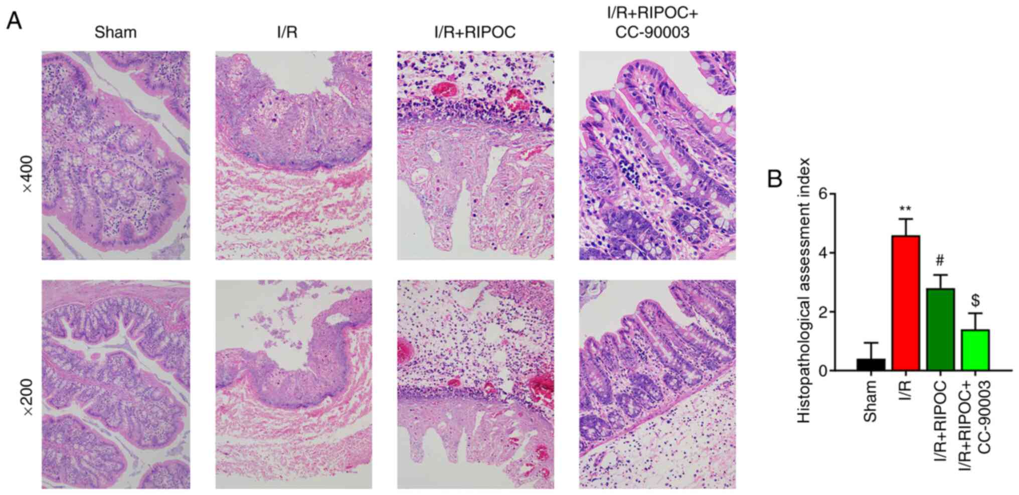
RIPOC ameliorates the
histopathological features of intestinal mucosa in mice with
intestinal I/R injury via the ERK pathway
To investigate the effects of I/R, mice were used in
the present study, as described by Gubernatorova et al
(27). H&E staining was
performed to determine whether RIPOC could protect the intestinal
tissue of mice following intestinal I/R injury. As demonstrated in
Fig. 1, the intestinal mucosa in
the sham group had a normal appearance, whereas in the I/R group,
the small intestine contained villi that exhibited obvious edema.
Furthermore, the mucosa and villi in the I/R group were
disorganized; most of the villous mucosa were deciduous and the
glands were damaged. Following treatment with RIPOC, these
histopathological features in the I/R group demonstrated
improvement. Furthermore, it was found that CC-90003 treatment
could further ameliorate the histopathological features (Fig. 1B).
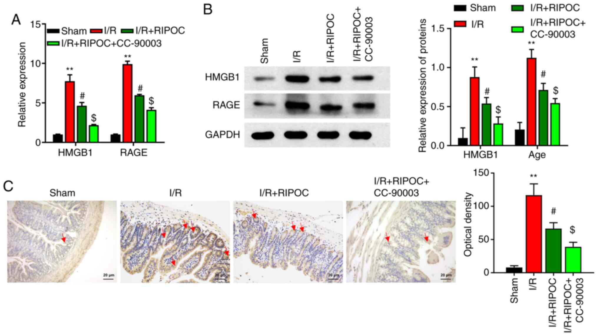
RIPOC downregulates HMGB1 and RAGE
expression in the intestinal I/R injury model mice via the ERK
pathway
To further explore the possible molecular mechanism
by which RIPOC protects against intestinal lesions induced by
intestinal I/R injury, the changes in expression of I/R
injury-related proteins were analyzed. As demonstrated in Fig. 2A, intestinal I/R injury
significantly increased the levels of HMGB1 and RAGE expression,
whereas RIPOC reduced these increases. It was also determined that
CC-90003 could reduce the levels of HMGB1 and RAGE mediated by
RIPOC in the intestinal I/R injury mouse models even further
(P<0.05, P<0.01; Fig. 2A). In
addition, western blot and IHC studies were performed to verify the
influence of RIPOC or CC-90003 on HMGB1 and RAGE expression in the
intestinal I/R injury mouse models and the results were similar to
those obtained from the RT-qPCR assays (Fig. 2B and C).
RIPOC decreases ROS, MDA and Trx
levels and increases SOD and GSH-Px levels in an intestinal I/R
injury mouse model via the ERK pathway
To confirm whether RIPOC exerted an antioxidant
affect in the intestinal I/R injury mouse model, multiple oxidative
stress indices were examined. A flow cytometric analysis revealed
that the levels of ROS in the intestinal I/R injury mouse models
were significantly higher compared with the sham mice and those
increased levels could be reduced by RIPOC; in addition, the
inhibitory effect of RIPOC on ROS production could be further
accentuated by CC-90003 (P<0.05, P<0.01; Fig. 3A). In addition, MDA and Trx levels
were higher and the SOD and GSH-Px levels were lower in the I/R
model group compared with the sham group; and these changes could
be attenuated by RIPOC. Concurrently, it was also revealed that
CC-90003 could further accentuate the decreases in MDA and Trx
levels as well as the increases in SOD and GSH-Px levels mediated
by RIPOC in the intestinal I/R injury mouse model (P<0.05,
P<0.01; Fig. 3B-E). 8-OHdG, a
marker of oxidative stress, was enhanced in the intestinal I/R
injury mouse model (P<0.01), while it was significantly
decreased by RIPOC treatment compared with I/R mouse model
(P<0.05) and further blocked by ERK inhibitor, CC-90003
(P<0.05; Fig. 3F).
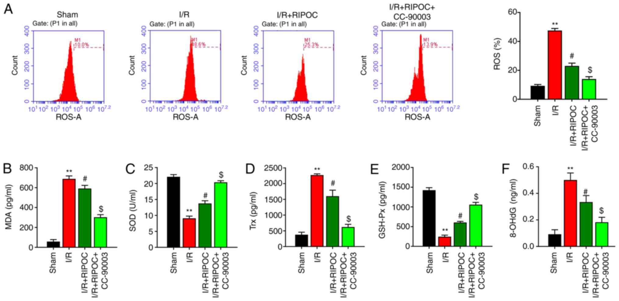 | Figure 3.RIPOC decreases ROS, MDA and Trx
levels and increases SOD and GSH-Px levels in intestinal I/R injury
mouse models via the ERK pathway. (A) Following treatment of the
I/R injury mouse models with RIPOC and/or CC-90003, the levels of
ROS were identified using a flow cytometer equipped with a
dihydroethidium fluorescent probe. The concentrations of (B) MDA,
(C) SOD, (D) Trx, (E) GSH-Px and (F) 8-OHdG in each group were
examined using the corresponding commercial ELISA kits. **P<0.01
vs. the sham group; #P<0.05 vs. the I/R group;
$P<0.05 vs. the I/R+RIPOC group. RIPOC, remote
ischemic post-conditioning; ROS, reactive oxygen species; MDA,
malondialdehyde; Trx, thioredoxin; SOD, superoxide dismutase;
GSH-Px, glutathione peroxidase; I/R, ischemia reperfusion. |
RIPOC reduces IL-1β, IL-18 and IL-33
levels in mice with intestinal I/R injury via the ERK pathway
The present study next investigated the possible
mechanism by which RIPOC affects the levels of interleukin-1 family
cytokines (IL-1β, IL-18 and IL-33) in mice following intestinal I/R
injury. As demonstrated in Fig.
4A-C, the levels of IL-1β, IL-18 and IL-33 in the I/R injury
mice were significantly increased and RIPOC treatment efficiently
reversed those increases. The data also demonstrated that CC-90003
could further enhance the reversal effect of RIPOC on the
upregulation of IL-1β, IL-18 and IL-33 in mice following intestinal
I/R injury.
 | Figure 4.RIPOC reduces the levels of IL-1β,
IL-18, IL-33 and NLRP3 in mice with intestinal I/R injury via the
ERK pathway. Intestinal I/R injury mouse models were treated with
RIPOC or/and CC-90003, respectively. The levels of (A) IL-1β, (B)
IL-18 and (C) IL-33 were analyzed by ELISA. (D) Western blot assays
were performed to evaluate p-ERK1/2, ERK1/2, p-p65, p65, p-IKBα,
IKBα and NLRP3 expression. The relative levels of p-ERK1/2 and
p-IKBα were defined as the ratio of phosphorylated protein to total
protein. **P<0.01 vs. the sham group; #P<0.05 vs.
the I/R group; $P<0.05 vs. the I/R+RIPOC group.
RIPOC, remote ischemic post-conditioning; NLRP3, NLR family pyrin
domain containing 3; I/R, ischemia reperfusion; p-,
phosphorylated. |
RIPOC attenuates p-ERK1/2, p-p65 and
NLRP3 expression and enhances p-IKBα expression in mice following
intestinal I/R injury via the ERK pathway
To further determine the mechanism by which RIPOC
protects against intestinal I/R injury, the intestinal I/R injury
mouse model were treated with RIPOC and/or CC-90003. A western
blotting analysis demonstrated that compared with the sham group,
the levels of p-ERK1/2, p-p65, p-IKBα and NLRP3 were significantly
increased in intestinal I/R injury group, while RIPOC could
significantly attenuate the increases in p-ERK1/2, p-p65, p-IKBα
and NLRP3 expressions in intestinal I/R injury mice. In addition,
CC-90003 could further inhibit the increases in p-ERK1/2, p-p65,
p-IKBα and NLRP3 expressions mediated by RIPOC following intestinal
I/R injury (Fig. 4D).
Discussion
The small intestine is crucial for maintaining
normal physiological activities of the human body and is
responsible for the uptake, digestion and absorption of nutrients
(28). The small intestine is also
the largest reservoir of bacteria and endotoxins and a vital
neuroendocrine and immune organ (29). In pathological conditions, the small
intestine performs functions that influence all systems of the body
(29). Previous studies have
demonstrated that intestinal I/R injury is a universal
pathophysiological process that occurs in patients suffering from
shock, severe trauma and undergoing resuscitation, all of which
cause a series of pathophysiological changes to occur in intestinal
tissues (30,31). Intestinal I/R injury can also cause
secondary damage to distant organs, in addition to intestinal
tissue damage (32). However, the
relevant mechanisms and treatment strategies of Intestinal I/R
injury are still not entirely clear. The present study established
a mouse model of intestinal I/R injury and verified that intestinal
I/R injury could seriously damage the structure of intestinal
mucosa.
RIPOC, as a new non-pharmaceutical intervention, can
alleviate long-term I/R-injured target organs via transient
ischemic preconditioning of limbs and other distant organs
(33). Studies have demonstrated
that RIPOC has a significant protective effect when used in
treatment of ischemic stroke (34,35),
myocardial ischemia reperfusion injury (36,37),
or acute ST-elevation myocardial infarction (38). For example, RIPOC improves the
transplantation of mesenchymal stem cells in reperfused myocardium
(17). Furthermore, limb RIPOC was
demonstrated to relieve cerebral I/R injury via AMPK-dependent
autophagy (16), attenuate I/R
injury in rat skin flaps by reducing oxidative stress (39) and help to protect against cerebral
I/R injury in a rat model (40).
The present study verified that RIPOC could improve the
histopathological features of intestinal mucosa in mice following
intestinal I/R injury.
Previous studies have verified that the intestinal
mucosal barrier damage caused by I/R injury is mainly attributable
to low perfusion pressure, oxygen-free radical injuries and the
action of cytokines (41,42). While free radicals are constantly
produced, there are also certain enzymes that decompose them in an
organism (43). The main endogenous
antioxidant enzymes include SOD, CAT and GSH-Px (44). Cytokines including TNF-α, IL-6,
IL-1β, IL-18 and IL-33 serve important roles in I/R injuries and
greatly contribute to the occurrence of diseases caused by I/R
injuries (45,46). I/R injury in the liver significantly
promotes ROS expression and the activation of NLRP3 inflammasomes.
In contrast, a ROS antagonist such as NAC alleviates hepatic injury
by suppressing the activation of NLRP3 (47). The present study detected relevant
indicators and found that intestinal I/R injury could markedly
reduce the antioxidant capacity of intestinal tissue and promote an
inflammatory response, while RIPOC could enhance the antioxidant
capacity of intestinal tissue and reduce an inflammatory
response.
ERK, as one of the most characteristic members of
the MAPK family, regulates a range of cellular properties and
activities, including cell metabolism, cell viability,
inflammation, cell necrosis and apoptosis (48). The present study demonstrated that
the protective effect of RIPOC against intestinal I/R injury was
regulated by the ERK pathway. Furthermore, the levels of HMGB1 and
RAGE expression could be markedly downregulated by RIPOC in the
intestinal I/R injury mouse models. The main HMGB1 receptors in
cells are RAGE and TLRs, such as TLR2 and TLR4 (49). The activation of those receptors can
subsequently activate various signaling pathways, such as the MAPK
pathway and NF-κB signaling pathway and thus upregulate various
inflammatory factors and promote an inflammatory cascade reaction
(50). The present study further
demonstrated that RIPOC could reduce the levels of p-ERK1/2, p-p65,
p-IKBα and NLRP3 in mice with intestinal I/R injury by affecting
the ERK pathway. Other studies have suggested that the HMGB1/NF-κB
pathway participates in cerebral and myocardial I/R injuries
(51–53). A previous study demonstrated that
hepatic ischemia/reperfusion injury is mediated by the ERK/NF-κB
pathway (54). Furthermore, Yang
et al (51) suggested that
quercetin attenuates hepatic I/R injury by mediating the ERK/NF-κB
pathway (55). The present study
demonstrated for the first time, to the best of the authors'
knowledge, that HMGB1 and RAGE expression can be attenuated by an
ERK inhibitor and thus NF-κB and NLRP3 expression was also
suppressed. Therefore, it was hypothesized that RIPOC may protect
against I/R injury by mediating the ERK/HMGB1/RAGE/NF-κB
pathway.
In fact, RIPOC is a harmless approach providing a
practical and potential tool to attenuate I/R injury (56). Although it has demonstrated a
beneficial outcome to patients, it remains to be further explored.
Therefore, further investigation is required to explore the effect
of RIPOC at various doses on normal animals.
The conclusion of the present study is that RIPOC
exerted strong antioxidant and anti-inflammatory effects in mice
following intestinal I/R injury and it produced those effects via
an ERK-mediated HMGB1/RAGE/NF-κB pathway. The present study
suggested RIPOC as a potential method for treating intestinal I/R
injury induced by serious infection and traumatic shock. However,
the specific mechanism of the therapeutical effect has not been
fully investigated and further studies are required.
Acknowledgements
Not applicable.
Funding
No funding was received.
Availability of data and materials
The datasets used and/or analyzed during the current
study are available from the corresponding author upon reasonable
request.
Authors' contributions
LM and JW designed the experiments. LM, JW and JL
performed the experiments. LM, JW, NZ and MC performed the
experiments and collected experimental data. LM, JW, JL and XZ
analyzed the data. XZ verified the accuracy of the data analysis.
XZ and JL provided the resource supports. LM and JW drafted the
manuscript. All authors read and approved the final manuscript.
Ethics approval and consent to
participate
All animal experiments were performed in compliance
with ethical standards of Shandong Provincial Hospital Affiliated
to Shandong University. The present study was approved by the
Ethics Committee of Shandong Provincial Hospital Affiliated to
Shandong University (approval no. 2019-330).
Patient consent for publication
Not applicable.
Competing interests
The authors declare that they have no competing
interests.
References
|
1
|
Gonzalez LM, Moeser AJ and Blikslager AT:
Animal models of ischemia-reperfusion-induced intestinal injury:
Progress and promise for translational research. Am J Physiol
Gastrointest Liver Physiol. 308:G63–G75. 2015. View Article : Google Scholar : PubMed/NCBI
|
|
2
|
Hu Q, Ren H, Ren J, Liu Q, Wu J, Wu X, Li
G, Wang G, Gu G, Guo K, et al: Released mitochondrial DNA following
intestinal ischemia reperfusion induces the inflammatory response
and gut barrier dysfunction. Sci Rep. 8:73502018. View Article : Google Scholar : PubMed/NCBI
|
|
3
|
Wang M, Verhaegh R, Tsagakis K, Brencher
L, Zwanziger D, Jakob HG, Groot H and Dohle DS: Impact of acute
intestinal ischemia and reperfusion injury on hemodynamics and
remote organs in a rat model. Thorac Cardiovasc Surg. 66:99–108.
2018. View Article : Google Scholar : PubMed/NCBI
|
|
4
|
Ferrada P, Wolfe L, Duchesne J, Fraga GP,
Benjamin E, Alvarez A, Campbell A, Wybourn C, Garcia A, Morales C,
et al: Management of duodenal trauma: A retrospective review from
the Panamerican Trauma Society. J Trauma Acute Care Surg.
86:392–396. 2019. View Article : Google Scholar : PubMed/NCBI
|
|
5
|
Li J, Kong P, Chen C, Tang J, Jin X, Yan J
and Wang Y: Targeting IL-17A improves the dysmotility of the small
intestine and alleviates the injury of the interstitial cells of
cajal during sepsis. Oxid Med Cell Longev. 2019:14757292019.
View Article : Google Scholar : PubMed/NCBI
|
|
6
|
Mester A, Magyar Z, Sogor V, Tanczos B,
Stark Y, Cherniavsky K, Bidiga L, Peto K and Nemeth N: Intestinal
ischemia-reperfusion leads to early systemic micro-rheological and
multiorgan microcirculatory alterations in the rat. Clin Hemorheol
Microcirc. 68:35–44. 2018. View Article : Google Scholar : PubMed/NCBI
|
|
7
|
Ameli M, Hashemi MS, Moghimian M and
Shokoohi M: Protective effect of tadalafil and verapamil on
testicular function and oxidative stress after torsion/detorsion in
adult male rat. Andrologia. 50:e130682018. View Article : Google Scholar : PubMed/NCBI
|
|
8
|
Jia Z, Lian W, Shi H, Cao C, Han S, Wang
K, Li M and Zhang X: Ischemic post-conditioning protects against
intestinal ischemia/reperfusion injury via the HIF-1α/miR-21 axis.
Sci Rep. 7:161902017. View Article : Google Scholar : PubMed/NCBI
|
|
9
|
Sheu EG, Wakatsuki K, Oakes S, Carroll MC
and Moore FD Jr: Prevention of intestinal ischemia-reperfusion
injury in humanized mice. Surgery. 160:436–442. 2016. View Article : Google Scholar : PubMed/NCBI
|
|
10
|
Giannopoulos G, Vrachatis DA, Panagopoulou
V, Vavuranakis M, Cleman MW and Deftereos S: Remote ischemic
conditioning and renal protection. J Cardiovasc Pharmacol Ther.
22:321–329. 2017. View Article : Google Scholar : PubMed/NCBI
|
|
11
|
Heusch G: Remote ischemic conditioning in
cardiovascular surgery. J Cardiovasc Pharmacol Ther. 22:297–301.
2017. View Article : Google Scholar : PubMed/NCBI
|
|
12
|
Przyklenk K, Bauer B, Ovize M, Kloner RA
and Whittaker P: Regional ischemic ‘preconditioning’ protects
remote virgin myocardium from subsequent sustained coronary
occlusion. Circulation. 87:893–899. 1993. View Article : Google Scholar : PubMed/NCBI
|
|
13
|
Kharbanda RK, Mortensen UM, White PA,
Kristiansen SB, Schmidt MR, Hoschtitzky JA, Vogel M, Sorensen K,
Redington AN and MacAllister R: Transient limb ischemia induces
remote ischemic preconditioning in vivo. Circulation.
106:2881–2883. 2002. View Article : Google Scholar : PubMed/NCBI
|
|
14
|
Anttila V, Haapanen H, Yannopoulos F,
Herajärvi J, Anttila T and Juvonen T: Review of remote ischemic
preconditioning: From laboratory studies to clinical trials. Scand
Cardiovasc J. 50:355–361. 2016. View Article : Google Scholar : PubMed/NCBI
|
|
15
|
Gill R, Kuriakose R, Gertz ZM, Salloum FN,
Xi L and Kukreja RC: Remote ischemic preconditioning for myocardial
protection: Update on mechanisms and clinical relevance. Mol Cell
Biochem. 402:41–49. 2015. View Article : Google Scholar : PubMed/NCBI
|
|
16
|
Guo H, Zhao L, Wang B, Li X, Bai H, Liu H,
Yue L, Guo W, Bian Z, Gao L, et al: Remote limb ischemic
post-conditioning protects against cerebral ischemia-reperfusion
injury by activating AMPK-dependent autophagy. Brain Res Bull.
139:105–113. 2018. View Article : Google Scholar : PubMed/NCBI
|
|
17
|
Jiang Q, Yu T, Huang K, Lu J, Zhang H and
Hu S: Remote ischemic post-conditioning ameliorates the mesenchymal
stem cells engraftment in reperfused myocardium. PLoS One.
11:e01460742016. View Article : Google Scholar : PubMed/NCBI
|
|
18
|
Wang S and Zhang Y: HMGB1 in inflammation
and cancer. J Hematol Oncol. 13:1162020. View Article : Google Scholar : PubMed/NCBI
|
|
19
|
Yang WS, Kim JJ, Lee MJ, Lee EK and Park
SK: Ectodomain shedding of RAGE and TLR4 as a negative feedback
regulation in high-mobility group box 1-activated aortic
endothelial cells. Cell Physiol Biochem. 51:1632–1644. 2018.
View Article : Google Scholar : PubMed/NCBI
|
|
20
|
Bangert A, Andrassy M, Müller AM,
Bockstahler M, Fischer A, Volz CH, Leib C, Göser S, Korkmaz-Icöz S,
Zittrich S, et al: Critical role of RAGE and HMGB1 in inflammatory
heart disease. Proc Natl Acad Sci USA. 113:E155–E164. 2016.
View Article : Google Scholar : PubMed/NCBI
|
|
21
|
Mou K, Liu W, Han D and Li P: HMGB1/RAGE
axis promotes autophagy and protects keratinocytes from ultraviolet
radiation-induced cell death. J Dermatol Sci. 85:162–169. 2017.
View Article : Google Scholar : PubMed/NCBI
|
|
22
|
Qiu Y, Chen Y, Zeng T, Guo W, Zhou W and
Yang X: High-mobility group box-B1 (HMGB1) mediates the
hypoxia-induced mesenchymal transition of osteoblast cells via
activating ERK/JNK signaling. Cell Biol Int. 40:1152–1161. 2016.
View Article : Google Scholar : PubMed/NCBI
|
|
23
|
Chiu CJ, McArdle AH, Brown R, Scott HJ and
Gurd FN: Intestinal mucosal lesion in low-flow states. I. A
morphological, hemodynamic, and metabolic reappraisal. Arch Surg.
101:478–483. 1970. View Article : Google Scholar : PubMed/NCBI
|
|
24
|
Qu H, Yin H, Yan S, Tao M, Xie Y and Chen
W: Inhibitor of growth 4 suppresses colorectal cancer growth and
invasion by inducing G1 arrest, inhibiting tumor angiogenesis and
reversing epithelial-mesenchymal transition. Oncol Rep.
35:2927–2935. 2016. View Article : Google Scholar : PubMed/NCBI
|
|
25
|
Ma X, Du T, Zhu D, Chen X, Lai Y, Wu W,
Wang Q, Lin C, Li Z, Liu L, et al: High levels of glioma tumor
suppressor candidate region gene 1 predicts a poor prognosis for
prostate cancer. Oncol Lett. 16:6749–6755. 2018.PubMed/NCBI
|
|
26
|
Livak KJ and Schmittgen TD: Analysis of
relative gene expression data using real-time quantitative PCR and
the 2(-Delta Delta C(T)) method. Methods. 25:402–408. 2001.
View Article : Google Scholar : PubMed/NCBI
|
|
27
|
Gubernatorova EO, Perez-Chanona E,
Koroleva EP, Jobin C and Tumanov AV: Murine model of intestinal
ischemia-reperfusion injury. J Vis Exp. 111:538812016.PubMed/NCBI
|
|
28
|
Zhang Y, Zulfiqar M, Bluth MH, Bhalla A
and Beydoun R: Molecular diagnostics in the neoplasms of small
intestine and appendix: 2018 update. Clin Lab Med. 38:343–355.
2018. View Article : Google Scholar : PubMed/NCBI
|
|
29
|
Volk N and Lacy B: Anatomy and physiology
of the small bowel. Gastrointest Endosc Clin N Am. 27:1–13. 2017.
View Article : Google Scholar : PubMed/NCBI
|
|
30
|
Zhang X, Moriwaki T, Kawabata T, Goto S,
Liu KX, Guo CY and Li TS: Nicaraven attenuates post-operative
systemic inflammatory responses-induced tumor metastasis. Ann Surg
Oncol. 27:1068–1074. 2020. View Article : Google Scholar : PubMed/NCBI
|
|
31
|
Li X, Ling Y, Cao Z, Shen J, Chen S, Liu
W, Yuan B and Wen S: Targeting intestinal epithelial
cell–programmed necrosis alleviates tissue injury after intestinal
ischemia/reperfusion in rats. J Surg Res. 225:108–117. 2018.
View Article : Google Scholar : PubMed/NCBI
|
|
32
|
Hummitzsch L, Zitta K, Berndt R, Wong YL,
Rusch R, Hess K, Wedel T, Gruenewald M, Cremer J, Steinfath M, et
al: Remote ischemic preconditioning attenuates intestinal mucosal
damage: Insight from a rat model of ischemia-reperfusion injury. J
Transl Med. 17:1362019. View Article : Google Scholar : PubMed/NCBI
|
|
33
|
Peng B, Guo QL, He ZJ, Ye Z, Yuan YJ, Wang
N and Zhou J: Remote ischemic post-conditioning protects the brain
from global cerebral ischemia/reperfusion injury by up-regulating
endothelial nitric oxide synthase through the PI3K/Akt pathway.
Brain Res. 1445:92–102. 2012. View Article : Google Scholar : PubMed/NCBI
|
|
34
|
Zhao JJ, Xiao H, Zhao WB, Zhang XP, Xiang
Y, Ye ZJ, Mo MM, Peng XT and Wei L: Remote ischemic
post-conditioning for ischemic stroke: A systematic review and
meta-analysis of randomized controlled trials. Chin Med J (Engl).
131:956–965. 2018. View Article : Google Scholar : PubMed/NCBI
|
|
35
|
Liang D, He XB, Wang Z, Li C, Gao BY, Wu
JF and Bai YL: Remote limb ischemic post-conditioning promotes
motor function recovery in a rat model of ischemic stroke via the
up-regulation of endogenous tissue kallikrein. CNS Neurosci Ther.
24:519–527. 2018. View Article : Google Scholar : PubMed/NCBI
|
|
36
|
Wang X, Wang J, Tu T, Iyan Z, Mungun D,
Yang Z and Guo Y: Remote ischemic post-conditioning protects
against myocardial ischemia-reperfusion injury by inhibition of the
RAGE-HMGB1 pathway. BioMed Res Int. 2018:45656302018.PubMed/NCBI
|
|
37
|
Zhang ZX, Li H, He JS, Chu HJ, Zhang XT
and Yin L: Remote ischemic post-conditioning alleviates myocardial
ischemia/reperfusion injury by up-regulating ALDH2. Eur Rev Med
Pharmacol Sci. 22:6475–6484. 2018.PubMed/NCBI
|
|
38
|
Ghaffari S, Pourafkari L, Manzouri S and
Nader ND: Effect of remote ischemic post-conditioning during
thrombolysis in STEMI. Herz. 43:161–168. 2018. View Article : Google Scholar : PubMed/NCBI
|
|
39
|
Zhang Y, Xu H, Wang T, He J, Wei J, Wang T
and Dong J: Remote limb ischemic post-conditioning attenuates
ischemia-reperfusion injury in rat skin flapby limiting oxidative
stress. Acta Cir Bras. 31:15–21. 2016. View Article : Google Scholar : PubMed/NCBI
|
|
40
|
Ramagiri S and Taliyan R: Protective
effect of remote limb post-conditioning via upregulation of heme
oxygenase-1/BDNF pathway in rat model of cerebral ischemic
reperfusion injury. Brain Res. 1669:44–54. 2017. View Article : Google Scholar : PubMed/NCBI
|
|
41
|
Ren G, Yuan X, Zhao X, Hao Q, Cao J, Wang
Y, Gao Q, Dou J and Zeng Q: Characterization and evolution of
intestine injury at the anhepatic phase in portal hypertensive
rats. Exp Ther Med. 16:4765–4771. 2018.PubMed/NCBI
|
|
42
|
Jiang Q: Research progress on intestinal
barrier dysfunction and treatment of severe acute pancreatitis. Adv
Emerg Med. 6:1–6. 2017.
|
|
43
|
Mason RP: Imaging free radicals in
organelles, cells, tissue, and in vivo with immuno-spin trapping.
Redox Biol. 8:422–429. 2016. View Article : Google Scholar : PubMed/NCBI
|
|
44
|
Goc Z, Szaroma W, Kapusta E and Dziubek K:
Protective effects of melatonin on the activity of SOD, CAT, GSH-Px
and GSH content in organs of mice after administration of SNP. Chin
J Physiol. 60:1–10. 2017. View Article : Google Scholar : PubMed/NCBI
|
|
45
|
Huo Y, Liu S-X, Li L, Feng J, Li H-Y and
Song G-Y: Circulating vaspin and IL-6 concentrations in second
trimester pregnancy with gestational diabetes. Clin Exp Obstet
Gynecol. 46:211–215. 2019.
|
|
46
|
Opal SM and DePalo VA: Anti-inflammatory
cytokines. Chest. 117:1162–1172. 2000. View Article : Google Scholar : PubMed/NCBI
|
|
47
|
Shi C, Wang Q, Rao Z, Shi Y, Wei S, Wang
H, Lu X, Wang P, Lu L, Zhou H, et al: Diabetes induces hepatocyte
pyroptosis by promoting oxidative stress-mediated NLRP3
inflammasome activation during liver ischaemia and reperfusion
injury. Ann Transl Med. 8:7392020. View Article : Google Scholar : PubMed/NCBI
|
|
48
|
Sun Y, Liu WZ, Liu T, Feng X, Yang N and
Zhou HF: Signaling pathway of MAPK/ERK in cell proliferation,
differentiation, migration, senescence and apoptosis. J Recept
Signal Transduct Res. 35:600–604. 2015. View Article : Google Scholar : PubMed/NCBI
|
|
49
|
Tirone M, Tran NL, Ceriotti C, Gorzanelli
A, Canepari M, Bottinelli R, Raucci A, Di Maggio S, Santiago C,
Mellado M, et al: High-mobility group box 1 orchestrates tissue
regeneration via CXCR4. J Exp Med. 215:303–318. 2018. View Article : Google Scholar : PubMed/NCBI
|
|
50
|
Bianchi ME, Crippa MP, Manfredi AA,
Mezzapelle R, Rovere Querini P and Venereau E: High-mobility group
box 1 protein orchestrates responses to tissue damage via
inflammation, innate and adaptive immunity, and tissue repair.
Immunol Rev. 280:74–82. 2017. View Article : Google Scholar : PubMed/NCBI
|
|
51
|
Yang M, Ruidi A, Minghang L, Tian X, Lu X
and Dong Z: β-caryophyllene mitigates cerebral ischemia reperfusion
injury in mice by inhibiting HMGB1/TLR4/NF-κB pathway. Chin J
Immunol. 33:1009–1013. 2017.
|
|
52
|
Dong LY, Chen F, Xu M, Yao LP, Zhang YJ
and Zhuang Y: Quercetin attenuates myocardial ischemia-reperfusion
injury via downregulation of the HMGB1-TLR4-NF-κB signaling
pathway. Am J Transl Res. 10:1273–1283. 2018.PubMed/NCBI
|
|
53
|
Xie W, Zhu T, Dong X, Nan F, Meng X, Zhou
P, Sun G and Sun X: HMGB1-triggered inflammation inhibition of
notoginseng leaf triterpenes against cerebral ischemia and
reperfusion injury via MAPK and NF-κB signaling pathways.
Biomolecules. 9:5122019. View Article : Google Scholar : PubMed/NCBI
|
|
54
|
Yu Q, Wu L, Liu T, Li S, Feng J, Mao Y,
Fan X, Guo C and Wu J: Protective effects of
levo-tetrahydropalmatine on hepatic ischemia/reperfusion injury are
mediated by inhibition of the ERK/NF-κB pathway. Int
Immunopharmacol. 70:435–445. 2019. View Article : Google Scholar : PubMed/NCBI
|
|
55
|
Wu L, Zhang Q, Dai W, Li S, Feng J, Li J,
Liu T, Xu S, Wang W, Lu X, et al: Quercetin pretreatment attenuates
hepatic ischemia reperfusion-induced apoptosis and autophagy by
inhibiting ERK/NF-κB Pathway. Gastroenterol Res Pract.
9724217:20172017.PubMed/NCBI
|
|
56
|
Nielsen MB, Krogstrup NV, Oltean M,
Nieuwenhuijs-Moeke GJ, Dor FJMF, Birn H and Jespersen B: Remote
ischaemic conditioning and early changes in plasma creatinine as
markers of one year kidney graft function-A follow-up of the
CONTEXT study. PLoS One. 14:e02268822019. View Article : Google Scholar : PubMed/NCBI
|