Introduction
Melanogenesis is a crucial physiological process
that occurs in melanocytes by which the melanosomes that synthesize
and store melanin pigment are loaded with melanin and are
translocated into the epidermal keratinocytes (1). Melanin biosynthesis is a tightly
regulated process, with different pathways controlled by several
enzymes and regulators. Tyrosinase is the main enzyme, initiating
and regulating melanogenesis. However, tyrosinase-related protein
(TRP)-1 and −2 are eumelanogenic enzymes contributing to the
completion of the process and acting as modifiers for pathway
velocity. TRP-1 and TRP-2 stabilize tyrosinase activity and TRP-1
possibly maintains melanosome structural integrity. Moreover,
several regulators are involved in melanin biosynthesis, such as
microphthalmia-associated transcription factor (MITF), which is
considered the main transcriptional regulator of melanogenesis,
functioning as the ‘central switchboard’ for the routing of various
signals involved in the expression of melanogenesis-related genes
(2,3).
Melanin pigment plays a crucial role in the
protection of epidermal cell DNA from solar ultraviolet radiation
damage, and in determining skin, hair and eye color (4,5).
Furthermore, it can modulate skin immune responses and serves as a
scavenger of reactive oxygen species (ROS), cellular toxins and
miscellaneous chemical compounds, preventing further skin damage
(6,7). The excessive reduction in melanin
production (hypopigmentation) is associated with abnormal
melanocyte development and dysfunction (8,9),
which subsequently reduces protection from harmful UV radiations
present in sunlight. On the other hand, the aberrant excessive
production and the accumulation of melanin (hyperpigmentation) can
lead to the development of skin disorders, such as melasma,
post-inflammatory hyperpigmentation, solar lentigo, ephelides and
café-au-lait macules (10).
Moreover, the overproduction of melanin is recognized not only as a
pathological concern, but also as a cosmetic issue. In this regard,
individuals from a number of countries in Asia, Africa, South
America and the Middle East have decided to reduce skin
pigmentation to obtain a lighter skin tone, as fair skin is
considered synonymous with youth, health, wealth and beauty in
different cultures (11,12). However, hyperpigmentation may be
congenital as a result of skin issues/systemic disease or it may be
caused by environmental factors (13).
The active agents used to suppress melanin
production and lighten the skin for therapeutic or cosmetic
purposes are either natural or synthetic, and may function at
various levels during melanogenesis. However, several of these
agents have undesired adverse effects, such as irritation, rashes,
inflamed skin, itchiness, toxicity and pain (14–17), and some of these agents exhibit
relatively poor skin permeability (17,18). Therefore, there is a need for new
safe and effective skin depigmenting agents to overcome these
issues. The use of natural products, including essential oils as
functional ingredients in cosmetics and depigmenting agents, has
received increasing attention due to the growing interest of
consumers in ingredients from natural sources. Moreover, several of
these products have multiple pharmacological activities, including
anti-melanogenic activity (19,20).
The aim of the present study was to identify
naturally-sourced agents for skin whitening purposes and for use in
the cosmetic industry with beneficial proliferative properties. For
this purpose, the anti-melanogenic activity of essential oils
extracted from 10 medicinal plants was evaluated using the B16F10
melanoma cell line by measuring the melanin content. The effects of
essential oils with potent anti-melanogenic activity on cell
proliferation, protection against
H2O2-induced cell death, and the expression
of certain melanogenesis-related proteins, namely MITF, tyrosinase,
TRP-1 and TRP-2, were also evaluated.
Materials and methods
Extraction of essential oils
The hydrodistillation method was used to extract the
essential oils from 10 medicinal plants using different plant parts
(National Institute of Forest Science; Republic of Korea) (Table I). In brief, 1 kg of the plant
part was mixed with 10 liters of distilled water and heated at
102°C using a heating mantle (cat. no. MS-DM608; Misung Scientific
Co., Ltd.). The volatile steam was then condensed using a
Dean-Stark trap (National Institute of Forest Science; Republic of
Korea), and the acquired whole essential oil was dehydrated using
anhydrous sodium sulfite and stored at 4°C until use.
 | Table I.List of the scientific names, common
names, and parts used of the investigated medicinal plants. |
Table I.
List of the scientific names, common
names, and parts used of the investigated medicinal plants.
| No. | Scientific
name | Common name | Parts used |
|---|
| 1 | Citrus
unshiu | Satsuma orange | Peels |
| 2 | Citrus
natsudaidai Hayata | Natsumikan | Peels |
| 3 | Citrus pseudo
gulgul | Hill lemon | Peels |
| 4 | Juniperus
chinensis L | Chinese
juniper | Leaves |
| 5 | Juniperus
chinensis var. sargentii | Sargent
juniper | Leaves |
| 6 | Zanthoxylum
piperitum | Japanese
pepper | Fruits |
| 7 | Zanthoxylum
schinifolium | Peppertree | Fruits |
|
| (Siebold &
Zucc) |
|
|
| 8 | Artemisia
capillaris | Yin Chen Hao | Grass clumps |
| 9 | Aster
glehnii F. Schmidt |
Ezo-goma-naa | Grass clumps |
| 10 | Cinnamomum
cassia | Chinese
cinnamon | Leaves |
Cells and cell culture
B16F10 mouse melanoma cells (Korean Cell Line Bank)
were cultured in Dulbecco's modified Eagle's medium (DMEM; Welgene
Inc.) with or without phenol red supplemented with 10% fetal bovine
serum (FBS; Sigma-Aldrich; Merck KGaA) and 1%
streptomycin/penicillin (Welgene Inc.) under standard culture
conditions for 24 h for recovery. The cultured cells were then
treated with the assigned concentrations of the tested essential
oils for a further 24 or 72 h (Table
II) for further analysis. For cell viability assay and western
blot analysis, the B16F10 cells were cultured in DMEM in 24-well
plates at a density of 55×104 and 3×10⁵
cells/well, respectively, and treated with the essential oils for
24 h. For the 5-bromo-2-deoxyuridine (BrdU) and melanin
quantification assays, the B16F10 cells were cultured in DMEM
without phenol red in 6-well plates at a density of
1×10⁵ cells/ml and treated with the essential oils for
24 or 72 h, respectively.
| Table II.The concentrations of the tested
essential oils used in the different assays in the present
study. |
Table II.
The concentrations of the tested
essential oils used in the different assays in the present
study.
|
|
| Tested
concentrations (ppm) |
|---|
|
|
|
|
|---|
| No. | Essential oil
source | Cell viability
assay | Melanin
quantification assays | BrdU assay | Western blot
analysis |
|---|
| 1 | Citrus
unshiu | 0.31, 1.25, 5, 20
and 80 | 0.31 and 1.25 | 0.31 and 1.25 | 1.25a |
| 2 | Citrus
natsudaidai Hayata | 0.08, 0.31, 1.25, 5
and 20 | 0.08, 0.31, 1.25
and 5 | - | - |
| 3 | Citrus pseudo
gulgul | 0.08, 0.31, 1.25, 5
and 20 | 0.08, 0.31 and
1.25 | - | - |
| 4 | Juniperus
chinensis L | 0.08, 0.31, 1.25, 5
and 20 | 0.08, 0.31 and
1.25 | 0.08, 0.31 and
1.25 | 1.25a |
| 5 | Juniperus
chinensis var. sargentii | 0.31, 1.25, 5, 20
and 80 | 0.31 | - | - |
| 6 | Zanthoxylum
piperitum | 0.08, 0.31, 1.25, 5
and 20 | 0.08, 0.31, 1.25
and 5 | 0.08, 0.31, 1.25
and 5 | 5a |
| 7 | Zanthoxylum
schinifolium (Siebold & Zucc) | 0.31, 1.25, 5, 20
and 80 | 0.31, 1.25 and
5 | - | - |
| 8 | Artemisia
capillaris | 0.08, 0.31, 1.25, 5
and 20 | 0.08, 0.31, 1.25
and 5 | 0.08, 0.31, 1.25
and 5 | 5a |
| 9 | Aster
glehnii F. Schmidt | 0.31, 1.25, 5, 20
and 80 | 0.31 and 1.25 | - | - |
| 10 | Cinnamomum
cassia | 0.08, 0.31, 1.25, 5
and 20 | 0.08, 0.31, 1.25
and 5 | - | - |
Cell viability assay and determination
of half maximal inhibitory concentration (IC50)
values
MTT assay was used to construct a cell viability
curve and to determine the IC50 values. The cultured
cells were incubated with various concentrations of the essential
oils for 24 h. After treatment, the medium containing essential
oils was replaced with a solution of 5 mg/ml MTT (Sigma-Aldrich;
Merck KGaA) and incubated at 37°C for 2 h. The optical density (OD)
was measured at 570 nm using a microplate reader (BioTek Inc.).
Cell viability was calculated using the following formula: OD
sample/OD control ×100 for each concentration. The cell survival
curve and the IC50 value for each treatment were
calculated from these values using the SigmaPlot software program
(V. 10.0; Systat Software, Inc.).
Measurement of melanin content
For the melanin quantification assay, the cells were
pre-incubated with 200 nM α-melanocyte-stimulating hormone (α-MSH;
Sigma-Aldrich; Merck KGaA) for 1 h at 37°C before adding the
essential oils to promote melanin production. The assigned tested
essential oils at non-toxic concentrations (Table II) or anti-melanogenic agent
arbutin (250 µM) were added to the culture medium and incubated for
a further 72 h at 37°C. α-MSH was used without essential oils as a
positive control. DMSO alone was used as a standard control.
Following treatment, the extracellular melanin content in 200 µl of
culture media was measured using a microplate reader using a
microplate reader (Agilent Technologies, Inc.) at 405 nm. To
measure the intracellular melanin content, the cells were washed
twice with phosphate-buffered saline (PBS) and collected using
trypsinization. Centrifugation at 20,000 × g for 15 min at 4°C was
performed, and the melanin pellets were dissolved in 1 N NaOH
containing 10% DMSO for 1 h at 60°C. The mixed homogenate (100 µl)
was placed in a 96-well microplate, and the OD values were measured
using a microplate reader (Agilent Technologies, Inc.) at 405 nm.
The extracellular and intracellular melanin contents per well were
calculated and expressed as a percentage of the control.
Assessment of inhibitory effects of
essential oil extracts on tyrosinase activity
Mushroom tyrosinase activity assay was performed
according to the manufacturer's recommendations. In a 96-well
plates, 20 µl mushroom tyrosinase (2,000 U/ml, Sigma-Aldrich; Merck
KGaA), 30 µl essential oil extracts or arbutin (Sigma-Aldrich;
Merck KGaA) as a positive control, 210 µl phosphate buffer (0.1 M;
pH 6.8) and 40 µl tyrosine (1.5 mM, Sigma-Aldrich; Merck KGaA) were
mixed and incubated at 37°C for 20 min. The OD value was then
measured using a microplate reader (Agilent Technologies, Inc.) at
490 nm. The tyrosinase activity in the samples were expressed using
the following formula: OD sample/OD control ×100.
Measurement of cell proliferation
BrdU assay was carried out using a cell
proliferation ELISA BrdU kit (Roche Diagnostics) according to the
manufacturer's recommendations. In brief, following the treatment
period with the assigned concentrations of the essential oils, 100
µl BrdU solution (100 µM) were added to each well in 1 ml medium,
and the plates were then incubated at 37°C for 4 h. Subsequently,
the cells were fixed using 1 ml FixDenat (Roche Diagnostics) in
each well for 30 min and incubated with the kit-supplied anti-BrdU
antibody (1:100; Roche Diagnostics) for 90 min at room temperature.
After washing, the cells were incubated with 500 µl substrate for
20 min at room temperature, and 125 µl H2SO4
(1 M) were added. The plates were analyzed at 450 nm using a
spectrometer (Agilent Technologies, Inc.).
Assessment of the protective effects
of the essential oils against H2O2-induced
cell death
B16F10 cells were cultured in DMEM containing the
assigned concentrations of the tested essential oils (Table II) for 24 h, as described above.
H2O2 (Sigma-Aldrich; Merck KGaA) was then
added at a final concentration of 400 µM for 4 h. Cell viability
was assessed using MTT assay as aforementioned.
Western blot analysis
Protein samples from the B16F10 cells treated with
the assigned concentrations of the essential oils for 24 h were
extracted using Pro-Prep solution (iNtRON Biotechnology Inc.)
according to the manufacturer's protocol. The concentration of
protein was determined by performing a bicinchoninic acid assay.
Subsequently, 5 µg protein were loaded and separated using sodium
dodecyl sulfate-polyacrylamide gel electrophoresis on 8–10% gels
and transferred onto nitrocellulose membranes (Daeillab Lab Service
Co., Ltd.) using the wet transfer system. The membranes were
blocked for 2 h with 5% skimmed milk (BD Biosciences) in PBS with
0.05% Tween-20 (PBST) at room temperature. Subsequently, the
membranes were incubated with antibodies against MITF (1:300; cat.
no. sc-56725; Santa Cruz Biotechnology, Inc.), tyrosinase (1:300;
cat. no. sc-20035; Santa Cruz Biotechnology, Inc.), TRP-1 (1:300;
cat. no. sc-25543; Santa Cruz Biotechnology, Inc.), TRP-2 (1:300;
cat. no. sc-25544; Santa Cruz Biotechnology, Inc.) and β-actin
(1:3,000; cat. no. #4967; Cell Signaling Technology, Inc.), which
served as an internal control overnight at 4°C, followed by
incubation with horseradish peroxidase-conjugated secondary
antibodies (1:5,000; cat. nos. ADI-SAB-100 and ADI-SAB-300; Enzo
Life Science Inc.) in 5% skimmed milk in PBST for 1 h at room
temperature. Luminol reagent (Bio-Rad Laboratories, Inc.) was used
to visualize antibody binding. The blots were scanned using Gel Doc
1000, version 1.5 (Bio-Rad Laboratories, Inc.), and band
intensities were normalized to β-actin levels.
Statistical analyses
Data are presented as the mean ± standard deviation
(SD). Data were analyzed using one-way analysis of variance (ANOVA)
with SPSS 10.10 standard version (IBM Corp.). Means obtained from
three independent experiments were evaluated using one-way ANOVA
and Tukey's post hoc t-test for multiple comparisons. A value of
P<0.05 was considered to indicate a statistically significant
difference.
Results
Effects of the tested essential oils
on cell viability
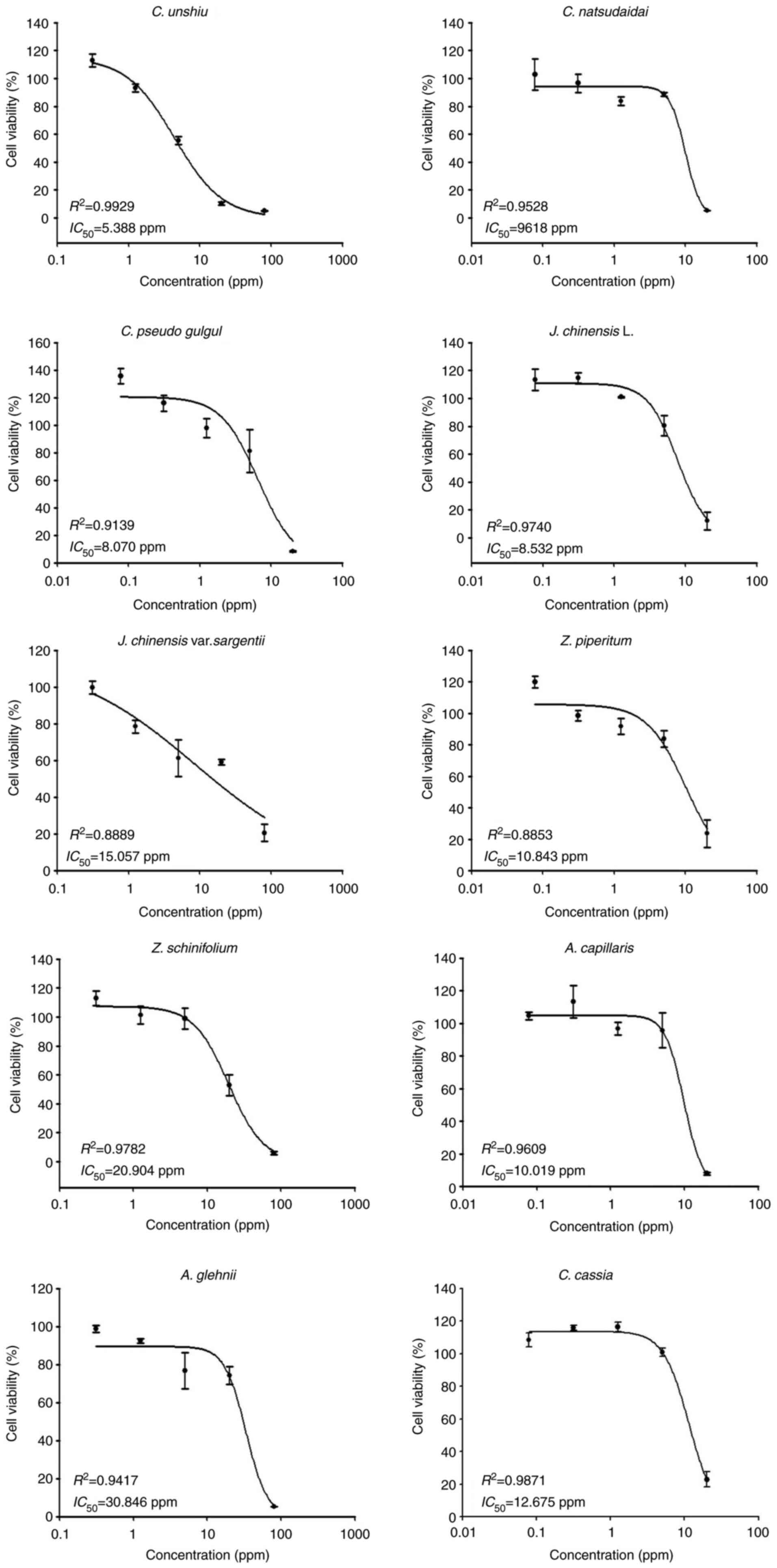
Cell viability assays were conducted using a wide
range of concentrations (0.31–80 ppm) for the essential oils
extracted from Citrus unshiu (C. unshiu),
Juniperus chinensis var. sargentii (J.
chinensis var. sargentii), Zanthoxylum
schinifolium (Siebold & Zucc) (Z. schinifolium) and
Aster glehnii F. Schmidt (A. glehnii), and from 0.08
to 20 ppm for the essential oils extracted from Citrus
natsudaidai Hayata (C. natsudaidai), Citrus pseudo
gulgul (C. pseudo gulgul), Juniperus chinensis (J.
chinensis L.), Zanthoxylum piperitum (Z.
piperitum), Artemisia capillaris (A. capillaris)
and Cinnamomum cassia (C. cassia) to screen their
toxic effects on B16F10 cells. The tested essential oils exhibited
variable toxicity levels in the B16F10 cells, as revealed by the
cell viability curves and IC50 values (Fig. 1). The essential oils extracted
from C. unshiu exhibited the highest toxicity level
(IC50, 5.388 ppm), whereas the essential oil extracted
from A. glehnii exhibited the lowest toxicity level
(IC50, 30.846 ppm) (Fig.
1).
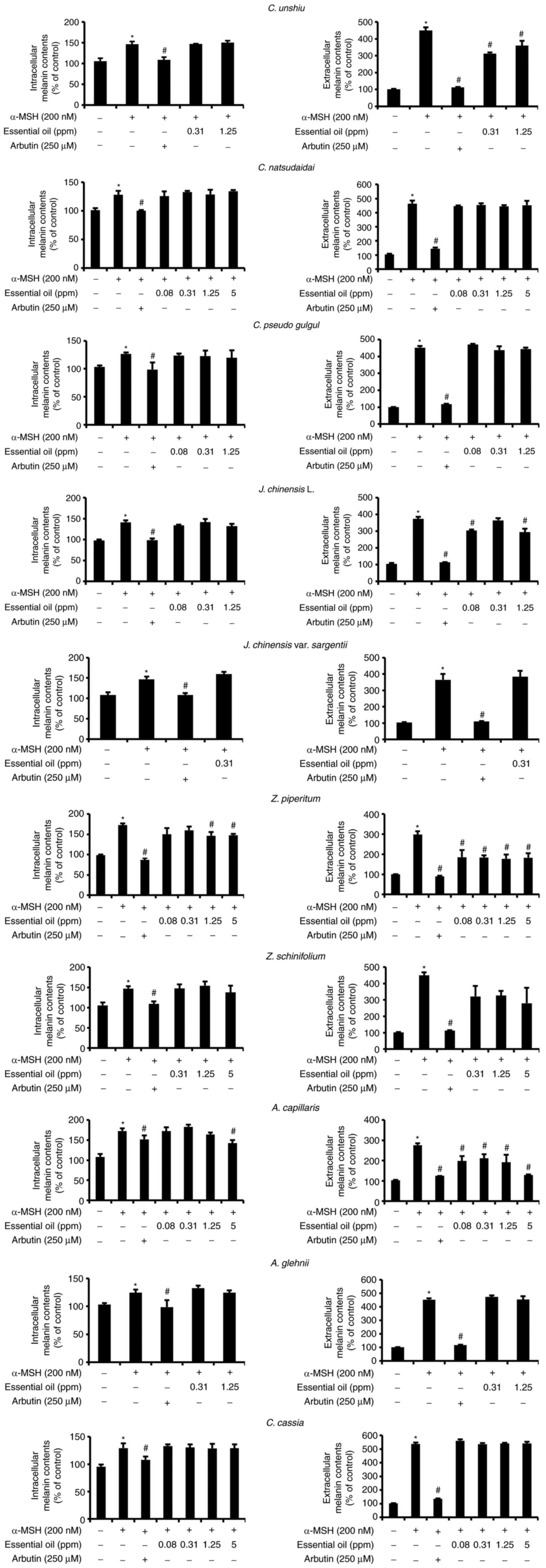
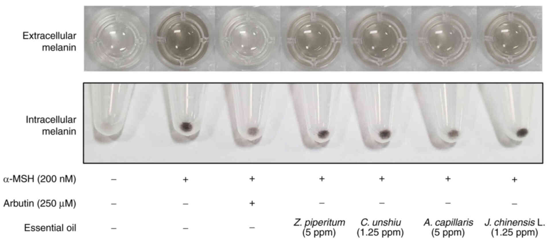
Effect of essential oils on melanin
content
A set of non-toxic concentrations of the tested
essential oils was used to investigate their anti-melanogenic
activity in B16F10 cells. The extracellular and intracellular
melanin contents were quantified after culturing the cells with the
assigned treatments for 72 h (Fig.
2). The extracellular and intracellular melanin contents were
significantly higher (P<0.05) in the positive control group
(α-MSH) than in the other treatment groups. However, arbutin
treatment significantly decreased the extracellular and
intracellular melanin content compared to that in the positive
control group (P<0.05). Of note, the essential oils extracted
from C. unshiu, J. chinensis L., Z. piperitum and
A. capillaris significantly decreased the extracellular
melanin contents at all concentrations tested compared to the
positive control group (P<0.05), excluding the concentration of
0.31 ppm for J. chinensis L. The highest concentrations of
these essential oils decreased the extracellular melanin content by
20, 38.8, 21 and 53.5% compared with the positive control group,
respectively. However, only the elevated concentrations of Z.
piperitum and A. capillaris significantly decreased the
intracellular melanin content (P<0.05) compared to that in the
positive control group by 14.4 and 17.5%, respectively (Figs. 2 and 3). Of note, none of these essential oils
significantly altered the tyrosinase activity (Fig. S1).
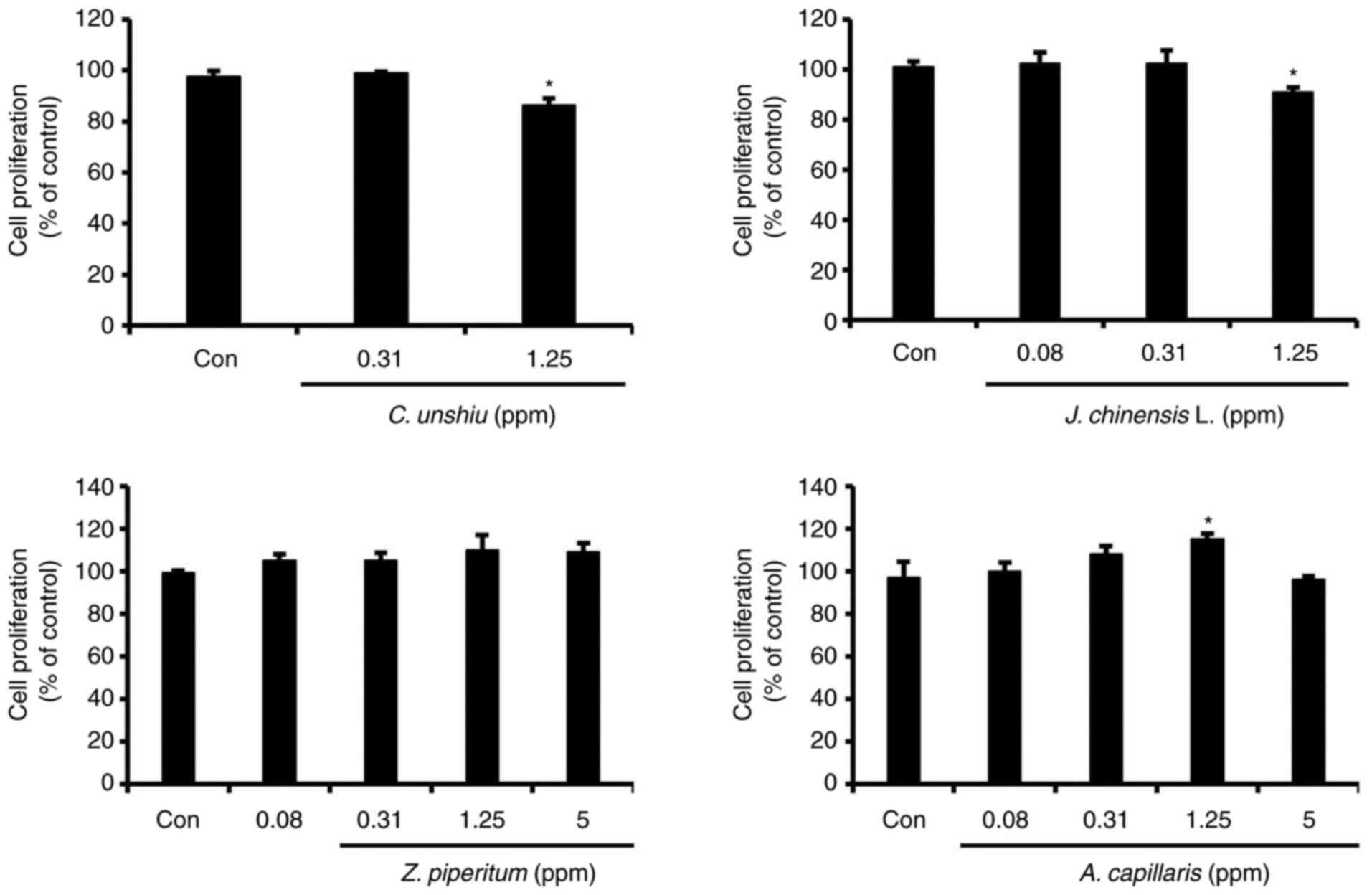
Effect of essential oils on B16F10
cell proliferation
Among the 10 essential oils examined in the present
study, only four essential oils with anti-melanogenic activity
(C. unshiu, J. chinensis L., Z. piperitum, and A.
capillaris) were selected to investigate their effects on cell
proliferation using BrdU assay. In general, the essential oils
extracted from Z. piperitum and A. capillaris
enhanced cell proliferation, although only A. capillaris
extract at a concentration of 1.25 ppm significantly (P<0.05)
increased cell proliferation by 18.7% compared to that in the
control group. However, the highest concentrations tested for the
C. unshiu and J. chinensis L. extracts significantly
decreased cell proliferation by 11.5 and 10.1%, respectively
(P<0.05; Fig. 4).
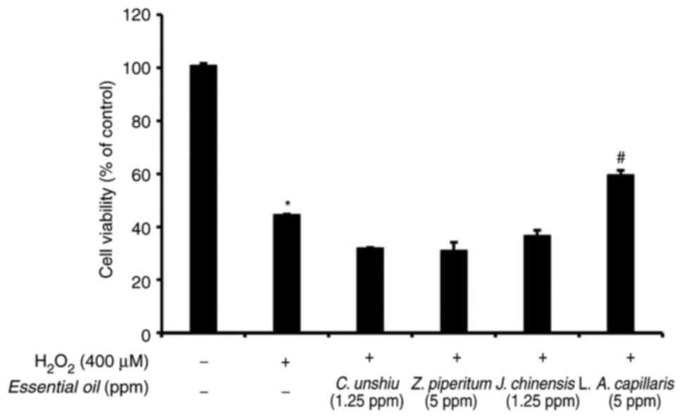
Assessment of the protective effects
of the essential oils against H2O2-induced
cell death
The protective effects of the essential oils
extracted from C. unshiu, J. chinensis L., Z.
piperitum, and A. capillaris against
H2O2-induced cell death were assessed using
MTT assay. The most effective concentration of the essential oils
for an anti-melanogenic effect was used for MTT assay. As shown in
Fig. 5, only the essential oil
extracted from A. capillaris attenuated the effects of
H2O2 on cell death induction and
significantly increased cell viability in the presence of
H2O2 in the culture media compared to the
other essential oils (P<0.05).
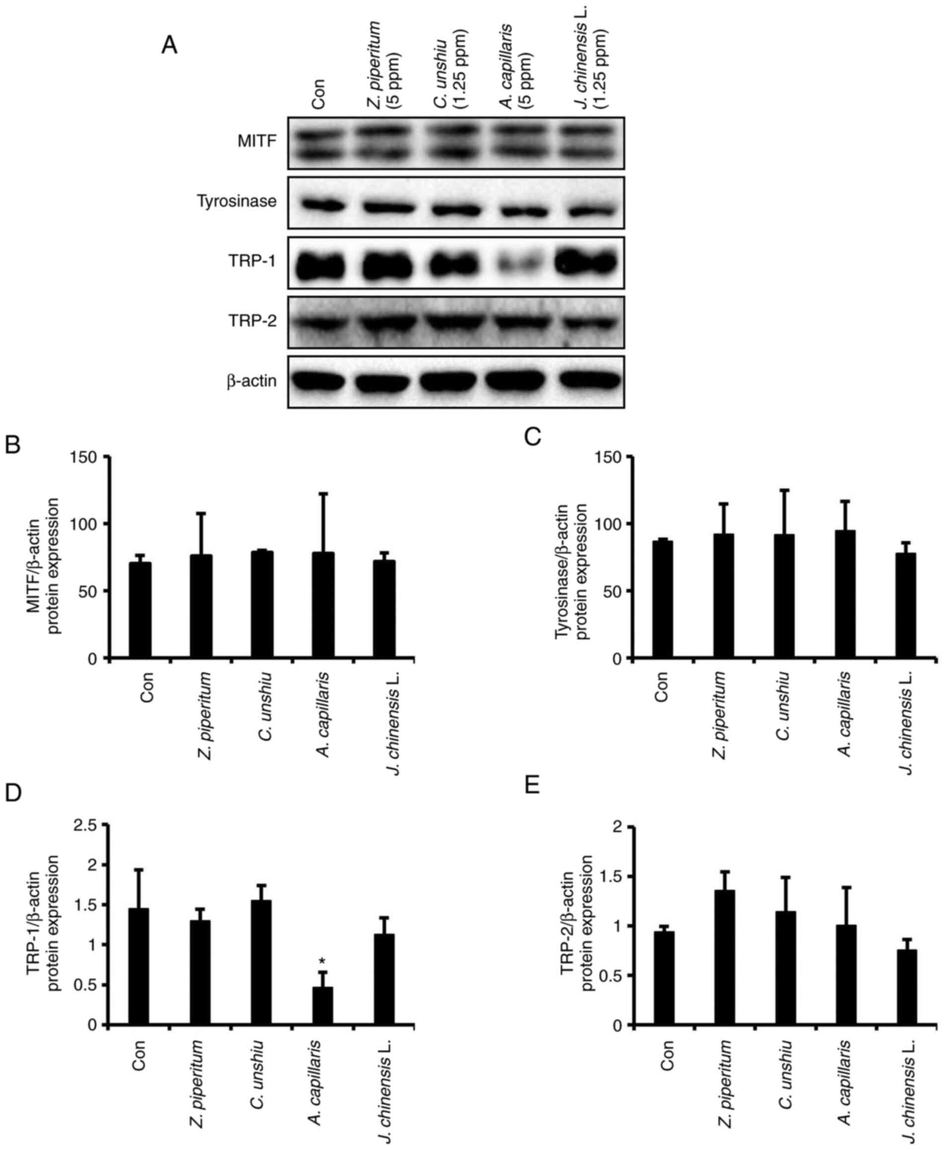
Effects of the essential oils on the
translational levels of melanogenesis-related genes
The effects of the four essential oils extracted
from C. unshiu, J. chinensis L., Z. piperitum, and
A. capillaris on the MITF, tyrosinase, TRP-1 and TRP-2
translational levels in B16F10 cells were examined using western
blot analysis. The most effective concentration of the essential
oils for an anti-melanogenic effect was used in western blot
analysis. The essential oil extracted from A. capillaris at
a concentration of 5 ppm significantly decreased the TRP-1 protein
level by ~68% compared to that in the control group (P<0.05).
However, the other essential oils did not induce any significant
changes in the protein levels of tyrosinase, TRP-1 and TRP-2
(Figs. 6 and S2). Among the proteins related to
melanogenesis, only MITF exhibited double bands. The upper band has
been assigned as a shift of the lower band due to phosphorylation
(21). However, none of the
essential oils altered the expression levels of MITF (Figs. 6 and S2).
Discussion
Essential oils extracted from 10 medicinal plants
were assessed in the present study to determine their
anti-melanogenic activities using B16F10 cell line model. These
plants represent various groups of medicinal plants that are widely
distributed in a number of Asian countries and have a long history
of use in folk medicine to treat various diseases. Moreover, over
the past few decades, increased attention has been directed towards
the use of functional components from these plants in biomedical
applications to treat various diseases, such as cancer (22–25), allergies (26), dermatopathology (27,28) and other diseases (29–32). However, little is known about
their effects and functions as natural anti-melanogenic agents.
Melanogenesis is a complex and multistep process
that results in melanin formation. Therefore, the measurement of
the melanin content is a direct strategy which can be used to
assess the melanogenic activity. It has been reported that in an
in vitro culture system, melanin is synthesized
intracellularly and is then transported into the surrounding
culture medium (33,34). In the present study, the
extracellular and intracellular melanin contents were determined
simultaneously to measure the total amount of melanin synthesized
under various treatment conditions in B16F10 cells. The
anti-melanogenic effects of the essential oils were further
examined on the human melanoma cell line, A375SM; however, when
treated with α-MSH as a positive control, the cells did not produce
a sufficient amount of melanin to evaluate the anti-melanogenic
effects (data not shown). Among the essential oil extracts
examined, only four essential oils from C. unshiu, J.
chinensis L., Z. piperitum and A. capillaris
successfully decreased melanogenesis compared to the other
extracts, which reflects their potency as anti-melanogenic agents.
In agreement with these findings, previous studies have reported
the inhibitory effects of some fractions extracted from these four
plants on melanin formation using various cell models (31,35–37). Notably, the non-toxic
concentrations of all essential oils used in the present study,
which inhibited melanin formation, were very low compared to the
fraction concentrations used in a previous study, which reached up
to 100 µg/ml (36). Although the
whole extracts of the essential oils exerted beneficial effects on
melanogenesis, active substances were not evaluated in the present
study. Therefore, further studies on the active compounds for each
essential oil are warranted. However, using the whole essential
oils has an advantage over purified components as they have
multi-pharmacological activities, and the different components may
exert a synergistic or potentiating effects and be important for
the bioactivity of the essential oils (38,39). Moreover, unlike previous studies
(35–37), the present study used the
hydrodistillation method for essential oil extraction, avoiding the
hazards of organic solvents and emulsion formation using other
methods (40). To v further
alidate efficiency of the extracts, their effects on cell
proliferation were assessed using BrdU assay, which established the
positive effects of two extracts, Z. piperium and A.
capillaris, at specific concentrations on cell proliferation.
Additionally, as the skin, more than other tissues, is exposed to
numerous external stresses generating several types of ROS, such as
H2O2 that is also produced as a response to a
multitude of very complex cellular events causing various
deleterious effects and apoptosis in keratinocytes (41,42), the protective effects of A.
capillaris against H2O2-induced cell
death were also examined. Of note, A. capillaris was also
able to significantly reduce H2O2-induced
cell death, suggesting that it is an anti-melanogenic agent with
proliferative and antioxidant properties. Hong et al
(43) reported that ethyl acetate
fraction from A. capillaris exerted significant ROS
scavenging and protective effects against oxidative DNA.
The anti-melanogenic effects of the extracted
essential oils on the B16F10 cell line were also investigated at
the molecular level by assessing the MITF, tyrosinase, TRP-1, and
TRP-2 protein expression levels using western blot analysis.
Although the four essential oils tested successfully decreased the
synthesized melanin content, they did not affect the expression
levels of MITF, tyrosinase and TRP-2 proteins, or even tyrosinase
activity, and only the essential oil extracted from A.
capillaris significantly decreased the expression level of
TRP-1 compared to that in the control group. Although tyrosinase is
the key enzyme in melanogenesis, TRP-1 is considered an
eumelanogenic enzymes with a vital role in the completion of
melanogenesis. TRP-1 is a protein producing eumelanin in the last
stage of the melanogenesis. Eumelanin is the most common type of
melanin comprising cross-linked 5,6-dihydroxyindole (DHI) and
5,6-dihydroxyindole-2-carboxylic acid (DHICA). TRP-1 induces the
oxidative conversion of DHICA to indole-5,6-quinone-2-carboxylic
acid, which is a structural unit of eumelanin (2,3).
Therefore, the overexpression of TRP-1 causes skin color to darken
(44). Eberle et al
(45) examined the expression
levels of tyrosinase family genes in melanoma and normal melanocyte
human cell lines and found that the expression levels of tyrosinase
and TRP-2 are regulated differently than TRP-1. Accordingly, in the
present study, A. capillaris at a concentration of the 5 ppm
did not affect the MITF, tyrosinase and TRP-2 expression levels or
tyrosinase activity, whereas it significantly decreased TRP-1
expression compared to that in the control group. It appears that
A. capillaris inhibits melanogenesis via a
tyrosinase-independent pathway. Therefore, any materials or
components that suppress TRP-1 expression may affect melanogenesis
by reducing the oxidation of DHICA to a carboxylated
indole-quinone. The findings of the present study suggest that the
low expression of TRP-1 can reduce melanin synthesis. The present
study initially examined various concentrations of A.
capillaris (0.08, 0.13, 1.25 and 5 ppm) on cell viability and
melanin synthesis. While the concentrations <5 ppm (0.08, 0.13
and 1.25 ppm) did not affect cytotoxicity, these concentrations did
not reduce intracellular melanin contents. The concentration of 5
ppm exerted the optimal effect on melanin synthesis, and
significantly decreased the extracellular and intracellular melanin
contents by 53.5 and 17.5%, respectively compared to the control
group. Therefore, this concentration was used to examine the effect
of A. capillaris against the protein expression levels of
TRP-1. Although the C. unshiu, J. chinensis L. and Z.
piperitum extracts decreased the melanin content, none of them
altered the translational level of the proteins involved in melanin
synthesis. These extracts appear to exert their effects on
melanogenesis via mechanisms different from those of A.
capillaris. As melanogenesis is a tightly regulated process
that includes various enzymes and other factors controlling
different pathways, materials that exert an inhibitory effect on
any of these factors are anticipated to inhibit melanogenesis.
In conclusion, the essential oils extracted from
C. unshiu, J. chinensis L., Z. piperitum and A.
capillaris using the hydrodistillation method inhibited melanin
synthesis. A. capillaris extract was the most potent
inhibitor of melanin synthesis, with good potential to enhance cell
viability and anti-H2O2 activity. A.
capillaris extract inhibited melanin synthesis by
downregulating the TRP-1 expression level. The present study did
not perform animal experiments to reveal the effects of the
essential oils. Further animal studies are thus required to address
the systemic effects of the essential oils. However, in general,
animal experiments to evaluate the functional effects of certain
materials on the skin are prohibited for animal protection. Even
though the present study did not determine the effects of the
extracts in in vivo conditions, these four essential oil
extracts, particularly A. capillaris, may be considered as
natural anti-melanogenic agents with beneficial proliferative
properties for the treatment of skin pigmentary disorders and for
skin whitening in the cosmetic industry. However, future studies
using in vivo models are required for further validation and
to investigate the effects of these extracts at the molecular level
through various mechanisms and pathways.
Supplementary Material
Supporting Data
Acknowledgements
Not applicable.
Funding
The present study was performed with the support of the National
Institute of Forest Science (Project no. FP0702-2016-03-2020) and
was partially supported by the BK21 FOUR Program (grant no.
F20YY8109033) through the National Research Foundation of Korea
(NRF), funded by the Ministry of Education, Korea.
Availability of data and materials
The datasets used and/or analyzed during the current
study are available from the corresponding author on reasonable
request.
Authors' contributions
MJK, EAM and BSA designed the experiments and wrote
the manuscript. MJK, DSK, MJP and BJA performed the experiments and
analyzed the data. EAM and BSA confirmed the authenticity of all
the raw data. EAM, EBJ and BSA analyzed the data and revised the
manuscript. All authors have read and approved the final
manuscript.
Ethics approval and consent to
participate
Not applicable.
Patient consent for publication
Not applicable.
Competing interests
The authors declare that they have no competing
interests.
References
|
1
|
Tayarani-Najaran Z, Akaberi M, Vatani M
and Emami SA: Evaluation of antioxidant and anti-melanogenic
activities of different extracts from aerial parts of Nepeta
binaludensis Jamzad in murine melanoma B16F10 cells. Iran J Basic
Med Sci. 19:662–669. 2016.PubMed/NCBI
|
|
2
|
Slominski A, Tobin DJ, Shibahara S and
Wortsman J: Melanin pigmentation in mammalian skin and its hormonal
regulation. Physiol Rev. 84:1155–1228. 2004. View Article : Google Scholar : PubMed/NCBI
|
|
3
|
Slominski A, Zmijewski MA and Pawelek J:
L-tyrosine and L-dihydroxyphenylalanine as hormone-like regulators
of melanocyte functions. Pigment Cell Melanoma Res. 25:14–27. 2012.
View Article : Google Scholar : PubMed/NCBI
|
|
4
|
Wang L, Oh JY, Kim YS, Lee HG, Lee JS and
Jeon YJ: Anti-photoaging and anti-melanogenesis effects of fucoidan
isolated from Hizikia fusiforme and its underlying mechanisms. Mar
Drugs. 18:4272020. View Article : Google Scholar : PubMed/NCBI
|
|
5
|
Rok J, Otręba M, Buszman E and Wrześniok
D: Melanin-from melanocyte to keratinocyte, that is how melanin is
transported within the skin. Ann Acad Med Siles. 66:60–66.
2012.
|
|
6
|
Slominski A, Wortsman J, Luger T, Paus R
and Solomon S: Corticotropin releasing hormone and
proopiomelanocortin involvement in the cutaneous response to
stress. Physiol Rev. 80:979–1020. 2000. View Article : Google Scholar : PubMed/NCBI
|
|
7
|
Slominski AT, Zmijewski MA, Skobowiat C,
Zbytek B, Slominski RM and Steketee JD: Sensing the environment:
Regulation of local and global homeostasis by the skin's
neuroendocrine system. Adv Anat Embryol Cell Biol. 212:vvii, 1,
115. 2012.PubMed/NCBI
|
|
8
|
Otręba M, Buszman E, Miliński M and
Wrześniok D: Hypomelanoses transmitted from generation to
generation. Postepy Hig Med Dosw (Online). 68:1081–1090. 2014.(In
Polish). View Article : Google Scholar : PubMed/NCBI
|
|
9
|
Otręba M, Miliński M, Buszman E, Wrześniok
D and Beberok A: Hereditary hypomelanocytoses: the role of PAX3,
SOX10, MITF, SNAI2, KIT, EDN3 and EDNRB genes. Postepy Hig Med Dosw
(Online). 67:1109–1118. 2013.(In Polish). View Article : Google Scholar : PubMed/NCBI
|
|
10
|
Goswami P and Sharma HK: Skin
hyperpigmentation disorders and use of herbal extracts: A review.
Curr Trends Pharm Res. 7:81–104. 2020.
|
|
11
|
Burger P, Landreau A, Azoulay S, Michel T
and Fernandez X: Skin whitening cosmetics: Feedback and challenges
in the development of natural skin lighteners. Cosmetics. 3:362016.
View Article : Google Scholar
|
|
12
|
Pollock S, Taylor S, Oyerinde O,
Nurmohamed S, Dlova N, Sarkar R, Galadari H, Manela-Azulay M, Chung
HS, Handog E and Kourosh AS: The dark side of skin lightening: An
international collaboration and review of a public health issue
affecting dermatology. Int J Womens Dermatol. 7:158–164. 2020.
View Article : Google Scholar : PubMed/NCBI
|
|
13
|
Cestari TF, Dantas LP and Boza JC:
Acquired hyperpigmentations. An Bras Dermatol. 89:11–25. 2014.
View Article : Google Scholar : PubMed/NCBI
|
|
14
|
Blaut M, Braune A, Wunderlich S, Sauer P,
Schneider H and Glatt H: Mutagenicity of arbutin in mammalian cells
after activation by human intestinal bacteria. Food Chem Toxicol.
44:1940–1947. 2006. View Article : Google Scholar : PubMed/NCBI
|
|
15
|
Hwang KS, Yang JY, Lee JY, Lee YR, Kim SS,
Kim GR, Chae JS, Ahn JH, Shin DS, Choi TY and Bae MA: A novel
anti-melanogenic agent, KDZ-001, inhibits tyrosinase enzymatic
activity. J Dermatol Sci. 89:165–171. 2018. View Article : Google Scholar : PubMed/NCBI
|
|
16
|
Saeedi M, Eslamifarb M and Khezri K: Kojic
acid applications in cosmetic and pharmaceutical preparations.
Biomed Pharmacother. 110:582–593. 2019. View Article : Google Scholar : PubMed/NCBI
|
|
17
|
Lee R, Ko HJ, Kim K, Sohn Y, Min SY, Kim
JA, Na D and Yeon JH: Anti-melanogenic effects of extracellular
vesicles derived from plant leaves and stems in mouse melanoma
cells and human healthy skin. J Extracell Vesicles. 9:17034802019.
View Article : Google Scholar : PubMed/NCBI
|
|
18
|
Fong P and Tong HH: In silico prediction
of the cosmetic whitening effects of naturally occurring lead
compounds. Nat Prod Commun. 7:1287–1294. 2012.PubMed/NCBI
|
|
19
|
Huang HC, Wang HF, Yih KH, Chang LZ and
Chang TM: The dual antimelanogenic and antioxidant activities of
the essential oil extracted from the leaves of acorus
macrospadiceus (Yamamoto) F. N. Wei et Y.K. Li. Evid Based
Complement Alternat Med. 2012:7812802012. View Article : Google Scholar : PubMed/NCBI
|
|
20
|
Huang HC, Ho YC, Lim JM, Chang TY, Ho CL
and Chang TM: Investigation of the anti-melanogenic and antioxidant
characteristics of eucalyptus camaldulensis flower essential oil
and determination of its chemical composition. Int J Mol Sci.
16:10470–10490. 2015. View Article : Google Scholar : PubMed/NCBI
|
|
21
|
Wu M, Hemesath TJ, Takemoto CM, Horstmann
MA, Wells AG, Price ER, Fisher DZ and Fisher DE: c-Kit triggers
dual phosphorylations, which couple activation and degradation of
the essential melanocyte factor Mi. Genes Dev. 14:301–12. 2000.
View Article : Google Scholar : PubMed/NCBI
|
|
22
|
Herdwiani W, Soemardji AA and Elfahmi Tan
MI: Review of cinnamon as a potent anticancer drug. Asian J Pharma
Clin Res. 9:8–13. 2016.
|
|
23
|
Kuo ZK, Lin MW, Lu IH, Yao HJ, Wu HC, Wang
CC, Lin SH, Wu SY, Tong TS, Cheng YC, et al: Antiangiogenic and
antihepatocellular carcinoma activities of the Juniperus
chinensis extract. BMC Complement Altern Med. 16:2772016.
View Article : Google Scholar : PubMed/NCBI
|
|
24
|
Chung KS, Cheon SY, Roh SS, Lee M and An
HJ: Chemopreventive effect of Aster glehni on
inflammation-induced colorectal carcinogenesis in mice. Nutrients.
10:2022018. View Article : Google Scholar : PubMed/NCBI
|
|
25
|
Kim J, Jung KH, Yan HH, Cheon MJ, Kang S,
Jin X, Park S, Oh MS and Hong SS: Artemisia capillaris
leaves inhibit cell proliferation and induce apoptosis in
hepatocellular carcinoma. BMC Complement Altern Med. 18:1472018.
View Article : Google Scholar : PubMed/NCBI
|
|
26
|
Kono R, Nomura S, Okuno Y, Kagiya T,
Nakamura M, Utsunomiya H and Ueno M: Two Japanese pepper
(Zanthoxylum piperitum) fruit-derived compounds attenuate
IgE-mediated allergic response in vitro and in vivo via inhibition
of mast cell degranulation. Eur J Pharmacol. 885:1734352020.
View Article : Google Scholar : PubMed/NCBI
|
|
27
|
Kim SS, Baik JS, Oh TH, Yoon WJ, Lee NH
and Hyun CG: Biological activities of Korean Citrus obovoides and
Citrus natsudaidai essential oils against acne-inducing
bacteria. Biosci Biotechnol Biochem. 72:2507–2513. 2008. View Article : Google Scholar : PubMed/NCBI
|
|
28
|
Kang GJ, Han SC, Yi EJ, Kang HK and Yoo
ES: The inhibitory effect of premature Citrus unshiu extract
on atopic dermatitis in vitro and in vivo. Toxicol Res. 27:173–180.
2011. View Article : Google Scholar : PubMed/NCBI
|
|
29
|
Choi SY, Ko HC, Ko SY, Hwang JH, Park JG,
Kang SH, Han SH, Yun SH and Kim SJ: Correlation between flavonoid
content and the NO production inhibitory activity of peel extracts
from various citrus fruits. Biol Pharm Bull. 30:772–778. 2007.
View Article : Google Scholar : PubMed/NCBI
|
|
30
|
Jin KS, Lee JY, Hyun SK, Kim BW and Kwon
HJ: Juniperus chinensis and the functional compounds, cedrol
and widdrol, ameliorate α-melanocyte stimulating hormone-induced
melanin formation in B16F10 Cells. Food Sci Biotechnol. 24:611–618.
2015. View Article : Google Scholar
|
|
31
|
Jin S, Yun HJ, Jeong HY, Oh YN, Park HJ,
Yun SG, Kim BW and Kwon HJ: Widdrol, a sesquiterpene isolated from
Juniperus chinensis, inhibits angiogenesis by targeting
vascular endothelial growth factor receptor 2 signaling. Oncol Rep.
34:1178–1184. 2015. View Article : Google Scholar : PubMed/NCBI
|
|
32
|
Lee SW, Lim JM, Mohan H, Seralathan KK,
Park YJ, Lee JH and Oh BT: Enhanced bioactivity of Zanthoxylum
schinifolium fermented extract: Anti-inflammatory,
anti-bacterial, and anti-melanogenic activity. J Biosci Bioeng.
129:638–645. 2020. View Article : Google Scholar : PubMed/NCBI
|
|
33
|
Laskin JD, Piccinini L, Engelhardt DL and
Weinstein IB: Control of melanin synthesis and secretion by B16/C3
melanoma cells. J Cell Physiol. 113:481–486. 1982. View Article : Google Scholar : PubMed/NCBI
|
|
34
|
Bhatnagar V, Srirangam A and Abburi R: In
vitro modulation of proliferation and melanization of melanoma
cells by citrate. Mol Cell Biochem. 187:57–65. 1998. View Article : Google Scholar : PubMed/NCBI
|
|
35
|
Jeong CH and Shim KH: Tyrosinase inhibitor
isolated from the leaves of Zanthoxylum piperitum. Biosci
Biotechnol Biochem. 68:1984–1987. 2004. View Article : Google Scholar : PubMed/NCBI
|
|
36
|
Saba E, Oh MJ, Lee YY, Kwak D, Kim S and
Rhee MH: Artemisia capillaris thunb. Inhibits melanin
synthesis activity via ERK-dependent MITF pathway in B16/F10
melanoma cells. Korean J Vet Res. 58:1–7. 2018. View Article : Google Scholar
|
|
37
|
Kim JK, Park NH and Hwang JS: Skin
lightening effect of the dietary intake of citrus peel extract
against UV-induced pigmentation. Nat Prod Commun.
14:1934578X198599792019.
|
|
38
|
Burt S: Essential oils: Their
antibacterial properties and potential applications in foods-a
review. Int J food Microbiol. 94:223–253. 2004. View Article : Google Scholar : PubMed/NCBI
|
|
39
|
Popa M, Măruțescu L, Oprea E, Bleotu C,
Kamerzan C, Chifiriuc MC and Grădișteanu Pircalabioru G: In vitro
evaluation of the antimicrobial and immunomodulatory activity of
culinary herb essential oils as potential perioceutics. Antibiotics
(Basel). 9:4282020. View Article : Google Scholar : PubMed/NCBI
|
|
40
|
Dangkulwanich M and Charaslertrangsi T:
Hydrodistillation and antimicrobial properties of lemongrass oil
(Cymbopogon citratus, Stapf): An undergraduate laboratory
exercise bridging chemistry and microbiology. J Food Sci Educ.
19:41–48. 2020. View Article : Google Scholar
|
|
41
|
Baldea I, Mocan T and Cosgarea R: The role
of ultraviolet radiation and tyrosine stimulated melanogenesis in
the induction of oxidative stress alterations in fair skin
melanocytes. Exp Oncol. 31:200–208. 2009.PubMed/NCBI
|
|
42
|
Kim ES, Park SJ, Goh MJ, Na YJ, Jo DS, Jo
YK, Shin JH, Choi ES, Lee HK, Kim JY, et al: Mitochondrial dynamics
regulate melanogenesis through proteasomal degradation of MITF via
ROS-ERK activation. Pigment Cell Melanoma Res. 27:1051–1062. 2014.
View Article : Google Scholar : PubMed/NCBI
|
|
43
|
Hong JH, Lee JW, Park JH and Lee IS:
Antioxidative and cytoprotective effects of Artemisia
capillaris fractions. Biofactors. 31:43–53. 2007. View Article : Google Scholar : PubMed/NCBI
|
|
44
|
Kim ZH, Hwang JW, Lee JH, Kim H, Lim DS,
Kang S, Lee HS and Choi YS: Whitening effect of storage protein 2
from silkworm hemolymph. Adv Biosci Biotechnol. 5:758–767. 2014.
View Article : Google Scholar
|
|
45
|
Eberle J, Garbe C, Wang N and Orfanos CE:
Incomplete expression of the tyrosinase gene family (tyrosinase,
TRP-1, and TRP-2) in human malignant melanoma cells in vitro.
Pigment Cell Res. 8:307–313. 1995. View Article : Google Scholar : PubMed/NCBI
|