Introduction
The estimated data for 2022 indicated that 12.5% of
all new cancer cases and 18.7% of total cancer deaths worldwide
were associated with lung cancer (1). Notably, research has indicated that
the incidence of lung cancer in most countries will continue to
rise until 2035, highlighting it as a significant global public
health concern (2). Moreover, lung
adenocarcinoma (LUAD) has been estimated to account for ~40% of all
lung cancer cases (3). Therefore,
it is crucial to identify new biomarkers that can accurately
diagnose LUAD.
With technological advancements and the broad use of
high-throughput RNA sequencing technology, the role of transfer RNA
(tRNA)-derived small RNAs (tsRNAs), as a new type of non-coding
small RNA, has been determined in the medical field (4,5). The
length of tsRNAs is generally 18–40 nucleotides, which mainly
includes tRNA-derived RNA fragments (tRfs) and tRNA halves.
Depending on the cleavage site of the parent tRNA, tRfs can be
categorized into four types: tRF-1, tRF-3, tRF-5 and i-tRF, while
tRNA-derived stress-induced RNAs (tiRNAs) can be classified into 5′
and 3′ tiRNAs (6,7). Furthermore, it has been indicated
that tiRNAs are crucially involved in gene expression modulation,
protein translation inhibition and epigenetic regulation, all of
which contribute to viral replication and intercellular
communication (8). Moreover,
tsRNAs, as untranslated RNA molecules, serve various roles in
different types of cancer. For example, tsRNA-42 has been reported
to be inactivated in chronic lymphocytic leukemia primarily through
promoter methylation (9).
Additionally, epigallocatechin gallate can promote ferroptosis in
non-small cell lung cancer by downregulating tsRNA-13502 (10). Furthermore, the downregulation of
tRFdb-3013a/b has been shown to be associated with a poor prognosis
for survival in patients with colon and rectal adenocarcinoma
(11). In addition, tsRNAs have
been reported to be associated with cancer development, and may
have oncogenic or oncostatic functions (12).
Several studies on tsRNAs have indicated that they
can serve as novel biomarkers for the diagnosis of various diseases
(13). For example,
tRF-Pro-AGG-004 and tRF-Leu-CAG-002 in the serum have been
identified as novel biomarkers for diagnosing pancreatic cancer
(14). Furthermore, tRNA-26576 has
been shown to enhance the proliferation of breast cancer cells,
indicating its potential as a diagnostic marker and therapeutic
target for breast cancer (15).
However, there is limited research on the role of small molecule
RNAs in LUAD. At present, the studies on small molecule RNAs and
screening mechanisms have mainly focused on microRNAs (miRNAs)
(16,17). tsRNAs are highly stable and
abundant in various body fluids, and are extensively involved in
pathological processes (18),
sometimes more so than miRNAs; therefore, the identification of
abnormally expressed tsRNAs in LUAD may improve clinical diagnosis
and prognosis.
The present study aimed to investigate LUAD-related
tsRNAs using the OncotRF database. Reverse
transcription-quantitative PCR (RT-qPCR) analysis of the
differential expression of tsRNAs in serum and tissue samples from
patients with LUAD and normal subjects was conducted to identify
hsa_tsr011468. Moreover, receiver operating characteristic (ROC)
curves and survival analyses were performed to evaluate the
diagnostic and prognostic value of hsa_tsr011468 in LUAD. The
relative expression levels of hsa_tsr011468 in the preoperative and
postoperative serum samples from patients with LUAD were also
compared to assess its dynamic monitoring ability. Additionally,
functional enrichment analyses of Gene Ontology (GO) and Kyoto
Encyclopedia of Genes and Genomes (KEGG) predicted target genes and
signaling pathways, providing new research directions for exploring
effective biomarkers and specific mechanisms of action in LUAD.
Materials and methods
Collection of serum and tissue
specimens
Serum samples were collected from a total of 84
patients with LUAD (mean patient age: 64±9 years, male-to-female
ratio: 45%) and 88 healthy individuals (mean patient age: 59±9
years, male-to-female ratio: 63%) between April 2020 and April 2022
from Nantong First People's Hospital (Nantong, China). A total of
70 of the 84 patients diagnosed with LUAD underwent surgery, and
postoperative serum samples were also collected for analysis.
Furthermore, 20 serum samples were collected from patients with
squamous cell carcinoma (SCC) (mean patient age: 66±8 years,
male-to-female ratio: 54%) and 20 serum samples were collected from
patients with large cell carcinoma (LCC) (mean patient age: 62±9
years, male-to-female ratio: 67%) at The Affiliated Hospital of
Nantong University (Nantong, China) between September 2023 and May
2024. A total of 42 LUAD tissue samples (mean patient age: 66±9
years, male-to-female ratio: 57%) and their corresponding paired
paracancerous tissue samples (2–3 cm away from the tumor) were
collected between April 2020 and April 2022 from the Affiliated
Hospital of Nantong University. None of the patients from whom
tissues were collected underwent prior radiotherapy or
chemotherapy. The present study was approved by the First People's
Hospital of Nantong City (approval no. 2023KT197) and the
Affiliated Hospital of Nantong University (approval no. 2019-L071),
and was performed in accordance with The Declaration of
Helsinki.
Cell culture
The human LUAD cell lines (A549, NCI-H1299 and PC9)
and the human normal lung epithelial cell line BEAS-2B used in the
present study were obtained from The Cell Bank of Type Culture
Collection of The Chinese Academy of Sciences. The cells were
cultured in RPMI 1640 medium (Corning, Inc.) supplemented with 10%
fetal bovine serum (Gibco; Thermo Fisher Scientific, Inc.) and 1%
penicillin-streptomycin. Cells were passaged by trypsin digestion
when they entered the logarithmic growth phase and reached a
maximum of 70–80% confluence. Passage was performed 1–2 times per
experiment. The medium was changed when the cells reached 80%
density, approximately once every 2 days. The cells were observed
microscopically to be of good condition with a translucent
appearance and intact cell membranes. The cells were cultured in an
incubator at 37°C and 5% CO2 until they reached the
logarithmic growth phase for subsequent experiments.
LUAD tissue, cell and serum RNA
extraction
Total RNA was extracted from cells (A549, NCI-H1299
and PC9), LUAD serum and milled tissue using TRIzol®
reagent (Invitrogen; Thermo Fisher Scientific, Inc.). The
concentration and purity of acquired RNA were assessed using a UV
spectrophotometer. The RNA was then subjected to RT or was stored
at −80°C.
Database screening
The OncotRF database (http://bioinformatics.zju.edu.cn/OncotRF/) was
utilized to screen meaningful tsRNAs in LUAD. Basic information on
tsRNAs was obtained and defined using the tsRBase
database(https://ngdc.cncb.ac.cn/databasecommons/database/id/7266#:~:text=Taken%20together,%20tsRBase%20is%20the%20most%20comprehensive%20and%20systematic%20tsRNA),
the tsRFun database (https://rna.sysu.edu.cn/tsRFun/), and the UCSC
database (https://genome.ucsc.edu/). TargetScan
(https://www.targetscan.org/vert_80/),
miRanda (http://mirtoolsgallery.tech/mirtoolsgallery/node/1055),
RNAhybrid (https://bibiserv.cebitec.uni-bielefeld.de/rnahybrid/submission.html/)
and tRFTar (http://www.rnanut.net/tRFTar/) databases were used to
predict target gene of selected tsRNA. KOBAS software (http://bioinfo.org/kobas) was used to perform GO and
KEGG pathway analyses.
Evaluation of hsa_tsr011468 assay
The linearity of hsa_tsr011468 was evaluated using
continuous 10-fold dilutions of hsa_tsr011468 cDNA extracted from
A549 cells. Mixed serum samples, prepared by combining multiple
normal human sera, were maintained at room temperature for 0, 4, 8,
12 and 24 h, and were subjected to repeated freeze-thaw cycles for
1, 3, 5, 7 and 9 times. Subsequently, the expression levels of
these samples were measured using RT-qPCR to analyze their
stability. The specificity of the hsa_tsr011468 mRNA assay was
evaluated by unimodal dissolution curve. RNA integrity was assessed
by electrophoresis on 1% agarose gels containing ethidium bromide.
The PCR product and the loading buffer were mixed in proportion and
loaded into the agarose gel for electrophoresis. After 43 min of
separation at 110 V, the results were observed in comparison to DNA
markers.
RT-qPCR
RNA concentration and purity were evaluated using UV
spectrophotometry, while RNA integrity was determined through
agarose gel electrophoresis (2% concentration). RNA was then used
to synthesize cDNA using the Revert Aid First Strand cDNA Synthesis
Kit (Thermo Fisher Scientific, Inc.) according to the
manufacturer's instructions. The amplification conditions were as
follows: Incubation at 42°C for 60 min and 70°C for 5 min, followed
by maintenance at 4°C. The cDNA was then utilized as a template for
qPCR amplification to detect the expression of target genes. The
reaction mix comprised SYBR Green I Mix (ABclonal Biotech Co.,
Ltd.), forward primer, reverse primer, cDNA and DEPC water in a
ratio of 10:1:1:3:5. The qPCR reaction was performed using the
QuantStudioQ5 system (Thermo Fisher Scientific, Inc.), and the
cycle conditions were as follows: 95°C for 10 min, followed by 40
cycles of 95°C for 15 sec and 60°C for 30 sec. The primers used for
qPCR analysis were: 5′-tRNA-Ala-AGC-11-1_L25, forward
5′-GGGGGAATTAGCTCAAATGG′, reverse 5′-AGTGCAGGGTCCGAGGTATT-3′, based
on its sequence 5′-GGGGAATTAGCTCAAATGGTAGAGC-3′;
5′-M-tRNA-Gln-CTG-5-1_L24, forward 5′-CGGGTTCCATGGTGTAATGG-3′,
reverse 5′-AGTGCAGGGTCCGAGGTATT-3′, based on its sequence
5′-GGTTCCATGGTGTAATGGTTAGCA-3′; 3′-mito-tRNA-Arg-TCG_L25, forward
5′-CGCGCGTTATGATAATCATATTTAC-3′, reverse
5′-AGTGCAGGGTCCGAGGTATT-3′, based on its sequence
5′-TTATGATAATCATATTTACCAACCA-3′; 3′-M-tRNA-Glu-TTC-4-2_L28, forward
5′-GTTCGATTCCCGGTCAGG-3′, reverse 5′-AGTGCAGGGTCCGAGGTATT-3′, based
on its sequence 5′-CCGGGTTCGATTCCCGGTCAGGGAACCA-3′;
3′-M-tRNA-iMet-CAT-1-8_L24, forward 5′-CGGATCGAAACCATCCTCTG-3′,
reverse 5′-AGTGCAGGGTCCGAGGTATT-3′, based on its sequence
5′-GATCGAAACCATCCTCTGCTACCA-3′; hsa_tsr011468, forward
5′-CCTCGTGGCGCAATGG-3′ and reverse 5′-AGTGCAGGGTCCGAGGTATT-3′,
based on its sequence 5′-GACCTCGTGGCGCAATGGTAGCGC-3′ (all from
GeneAdv Co., Ltd.). The sequences of the above molecules were
obtained from the tsRBase. U6 was employed as a standard control
and its primer sequence was: Forward 5′-CGCTTCGGCAGCCACATATAC-3′
and reverse 5′-TTCACGAATTTGCGTGTCATC-3′. 18S rRNA was also employed
as a standard control and its primer sequence was: Forward
5′-CGGCTACCACATCCAAGGAA-3′ and reverse 5′-GCTGGAATTACCGCGGCT-3′
(Guangzhou RiboBio Co., Ltd.). The expression levels of the product
were determined using the 2−ΔΔCq method (19).
Nuclear and cytoplasmic RNA extraction
assay
Trypsin digestion of adherent cells (A549,
NCI-H1299, PC9 and BEAS-2B) was performed and the cell precipitate
was collected. Nuclear and cytoplasmic RNA were extracted using a
Nucleus and Cytoplasmic Protein Extraction Kit (Beyotime Institute
of Biotechnology), where the protease inhibitor was substituted
with an RNase inhibitor. The extracted RNA was reverse transcribed
and stored at −80°C for qPCR analysis.
Statistical analysis
All data were statistically analyzed using GraphPad
8.0 (Dotmatics). The data are presented as the mean ± SD. Unpaired
Student's t-tests were performed to compare the results between
patients with LUAD and healthy controls, as well as between lung
cancer cells and normal lung epithelial cells. Paired Student's
t-tests were performed to compare pre-operative and post-operative
serum samples, as well as tumor and paracancerous tissue samples
from patients with LUAD. A one-way ANOVA followed by Bonferroni
post hoc test was used to compare the differences in the levels of
hsa_tsr011468 in mixed sera at different times and the number of
freeze-thaw cycles. Associations between hsa_tsr011468 expression
and clinicopathological features were analyzed using the
χ2 test or Fisher's exact test. The log-rank test was
employed to compare Kaplan-Meier survival curves. The diagnostic
and combined diagnostic values of hsa_tsr011468 and other serum
diagnostic markers for lung cancer were evaluated via receiver ROC
analysis, with the area under the curve (AUC) values assessed. To
determine the use of hsa_tsr011468, Cyfra21-1, CEA and SCC antigen
to distinguish patients with LUAD from healthy controls,
sensitivity, specificity, overall accuracy, positive predictive
value and negative predictive value were calculated as follows:
Sensitivity=number of true positives/total number of patients with
LUAD in the population; Specificity=number of true negatives/total
number of healthy controls in the population; Overall
accuracy=(true positive + true negative)/total number of samples;
Positive predictive value=true positive/(true positive + false
positive); Negative predictive value=true negative/(true negative +
false negative). P<0.05 was considered to indicate a
statistically significant difference.
Results
Database screening and validation of
hsa_tsr011468 by qPCR
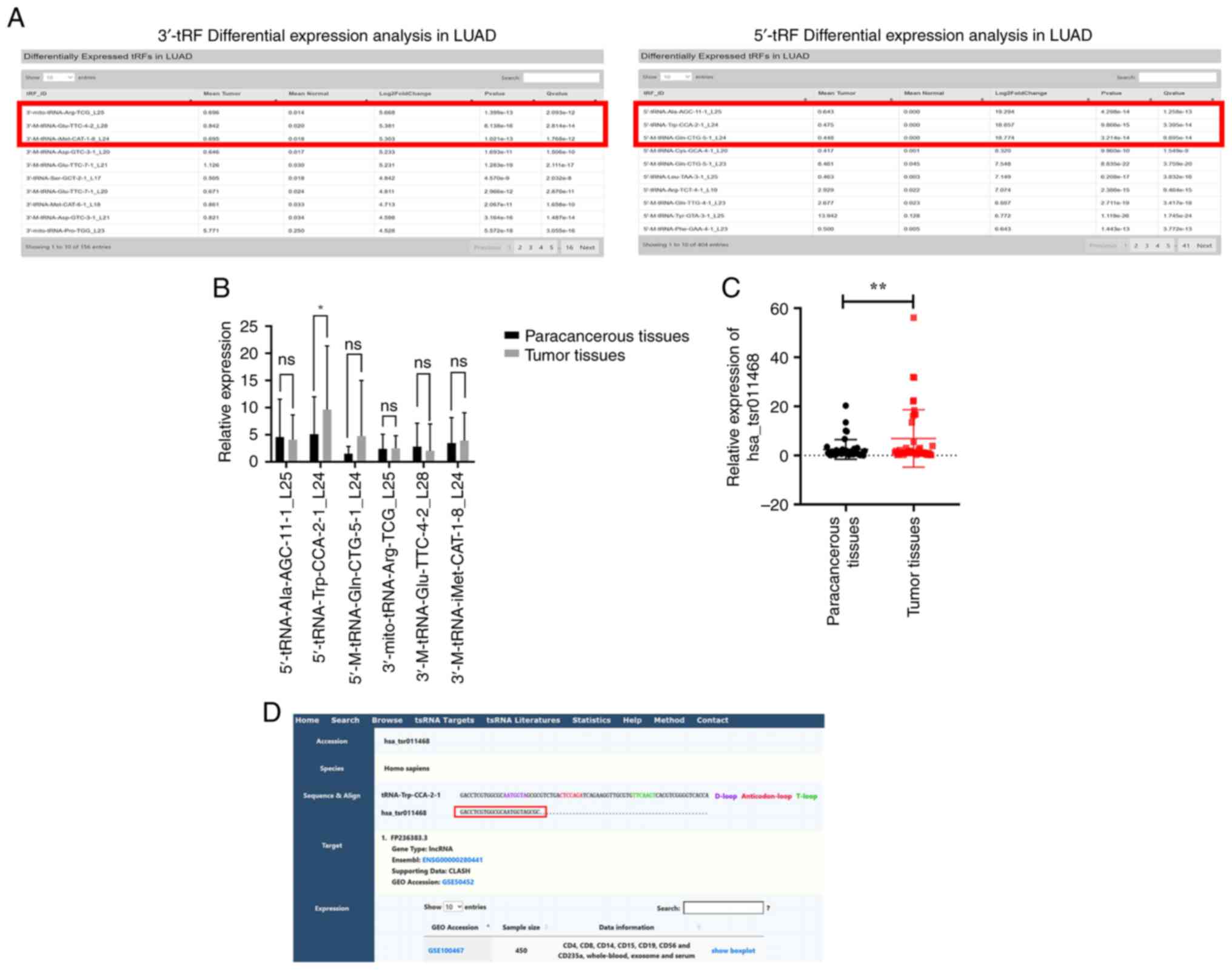
The OncotRF database was searched to investigate the
significance of tsRNAs in LUAD. The search results were ranked
based on the criterion of log2 fold-change and the top three ranked
3′tRF and 5′tRF were selected for validation (Fig. 1A). To further screen for tsRNAs
significantly associated with LUAD, the expression of the top three
ranked 3′tRF and 5′tRF were analyzed in 12 pairs of LUAD tissues.
The results showed that 5′-tRNA-Trp-CCA-2-1_L24 was differentially
expressed in LUAD tissues compared with in paracancerous tissues
(Fig. 1B). This finding was later
confirmed by the addition of 30 pairs of tissues to assess their
relative expression levels; the results indicated that the
expression of 5′-tRNA-Trp-CCA-2-1_L24 was higher in LUAD tissues
than that in paracancerous tissues (Fig. 1C). Furthermore, to simplify the
nomenclature of 5′-tRNA-Trp-CCA-2-1_L24, the tsRBase database was
searched using molecular data, which revealed that
5′-tRNA-Trp-CCA-2-1_L24 could be named hsa_tsr011468 (Fig. 1D).
Methodological evaluation of
hsa_tsr011468
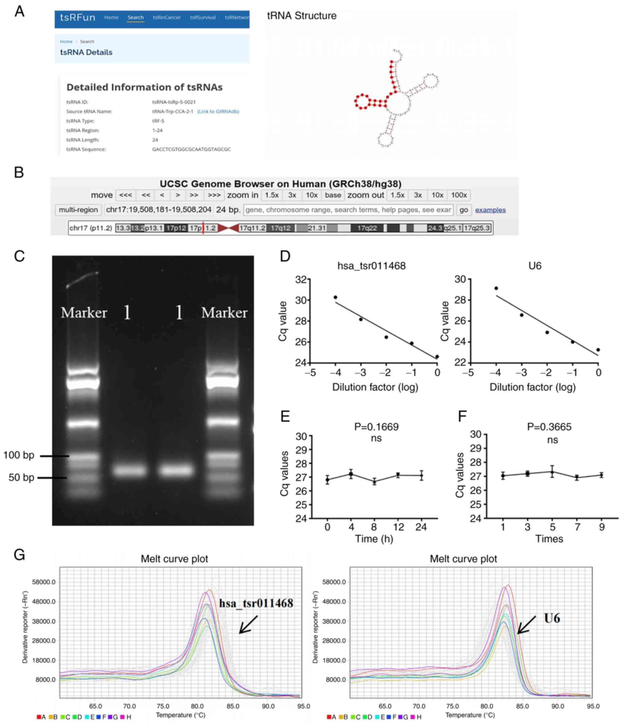
Detailed information was obtained and the molecular
structure of hsa_tsr011468 was determined using the tsRFun database
(Fig. 2A). Furthermore, the UCSC
database was utilized to identify the chromosomal location of
hsa_tsr011468, which was chr 17:19,508,181-19,508,204 (Fig. 2B). The length of the hsa_tsr011468
PCR product was confirmed to be ~75 bp through agarose gel
electrophoresis (Fig. 2C).
To ensure the reliability of the experimental
results, a methodological evaluation of hsa_tsr011468 was
conducted. The regression equation for hsa_tsr011468 was Y=−1.357 ×
X + 24.37 (R2=0.960) and for U6 was Y=−1.429 × X + 22.72
(R2=0.935), indicating good linearity for hsa_tsr011468
(Fig. 2D). Subsequently, the
relative expression levels of hsa_tsr011468 were measured in the
mixed sera after being incubated for varying durations or being
subjected to different numbers of freeze-thaw cycles. The RT-qPCR
results indicated that there were no significant changes in the
expression levels of hsa_tsr011468, demonstrating that
hsa_tsr011468 exhibits good stability (Fig. 2E and F). The specificity of the PCR
amplification was validated by a smooth single-peak melting curve
(Fig. 2G). These data indicated
that all experiments on hsa_tsr011468 had good reliability.
Clinical advantages of hsa_tsr011468
as a serum marker for LUAD
A total of 20 serum samples were collected from
patients with LCC, SCC, and LUAD (mean patient age: 63±8 years,
male-to-female ratio: 33%), and 20 samples were collected from
normal controls (mean patient age: 58±8 years, male-to-female
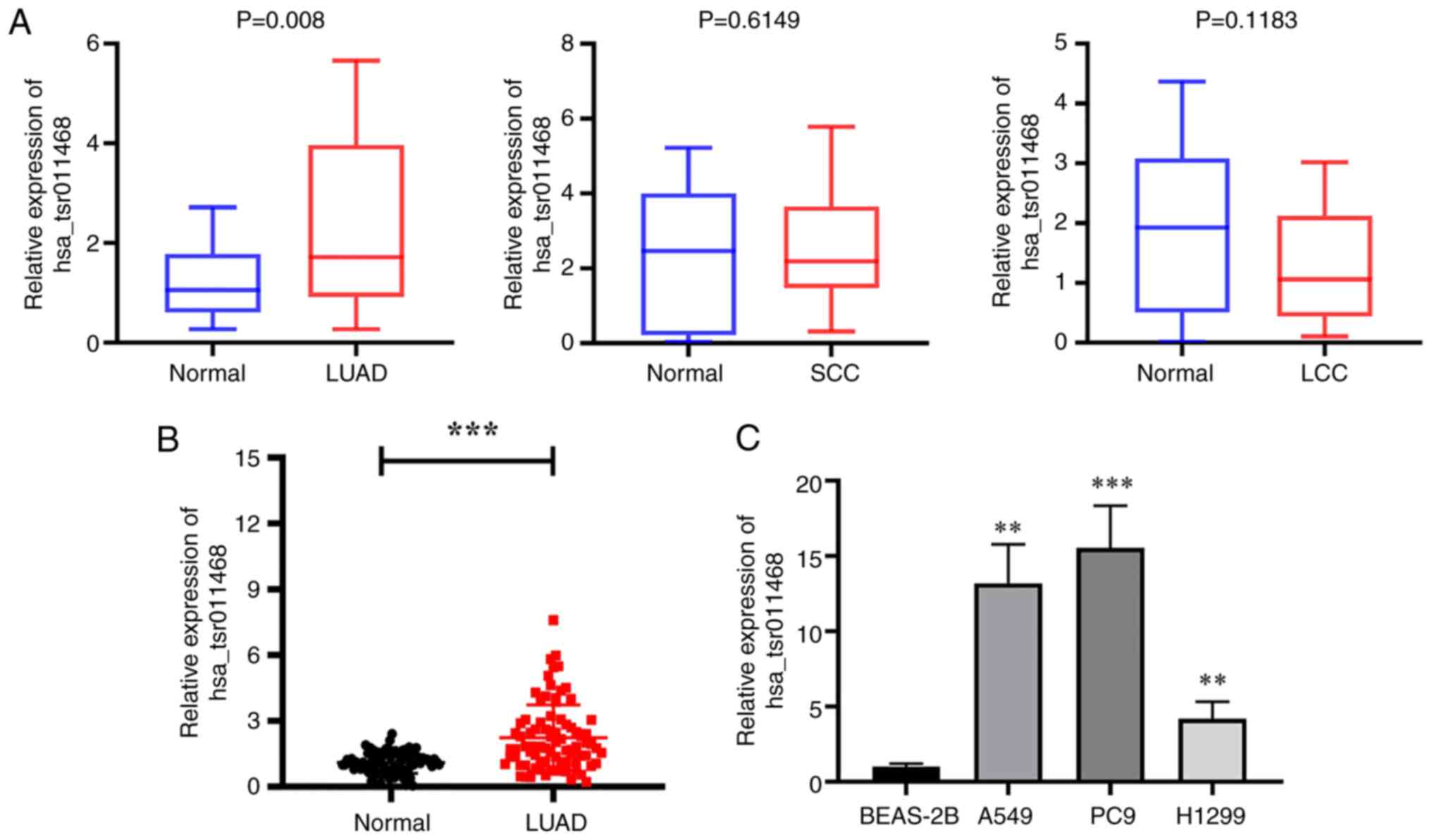
ratio: 54%). The expression levels of hsa_tsr011468 in these
non-small cell lung cancer were detected by RT-qPCR. It was
revealed that the expression of hsa_tsr011468 was not statistically
different in the serum of patients with SCC and LCC compared with
that from healthy controls; however, the expression of
hsa_tsr011468 was significantly higher in serum samples from
patients with LUAD compared with those from normal subjects
(Fig. 3A). These findings
suggested that hsa_tsr011468 can be used as a specific diagnostic
indicator of LUAD to distinguish it from other types of non-small
cell lung cancer.
To validate that hsa_tsr011468 can serve as a serum
diagnostic marker for LUAD, we continued to collect and expand the
sample size, ultimately totaling 84 serum samples from patients
with LUAD and 88 control samples from healthy subjects for qPCR
analysis. The results indicated that the relative expression levels
of hsa_tsr011468 were higher in samples from patients with LUAD
compared with those from normal controls (Fig. 3B). Furthermore, the expression
levels of hsa_tsr011468 were elevated in LUAD cell lines (Fig. 3C). Subsequently, the 84 patients
with LUAD were categorized into high-expression (n=42) and
low-expression (n=42) groups based on the median expression of
hsa_tsr011468, in order to investigate the association between
hsa_tsr011468 and clinicopathological features.
Tumor-node-metastasis classification was performed according to the
eighth edition of tumor-node-metastasis staging developed by the
International Association for the Study of Lung Cancer (20). The χ2 test and Fisher's
exact test revealed that hsa_tsr011468 was associated with tumor
size (Table I). Overall, these
data indicated that hsa_tsr011468 could serve as a specific
biomarker for LUAD.
 | Table I.Relationship of hsa_tsr011468
expression with clinicopathological features in patients with lung
adenocarcinoma. |
Table I.
Relationship of hsa_tsr011468
expression with clinicopathological features in patients with lung
adenocarcinoma.
|
|
| hsa_tsr011468
expression |
|---|
|
|
|
|
|---|
| Characteristic | Cases (n=84) | High (n=42) | Low (n=42) | P-value |
|---|
| Age, years |
|
|
| 0.498 |
|
≤60 | 31 | 14 | 17 |
|
|
>60 | 53 | 28 | 25 |
|
| Sex |
|
|
| 0.059 |
|
Male | 26 | 17 | 9 |
|
|
Female | 58 | 25 | 33 |
|
| Tumor size, cm |
|
|
| 0.023a |
|
<5 | 54 | 22 | 32 |
|
| ≥5 | 30 | 20 | 10 |
|
| Tumor
differentiation |
|
|
| 0.078 |
| Well +
Moderate | 48 | 20 | 28 |
|
|
Poor | 36 | 22 | 14 |
|
| Lymphatic
metastasis |
|
|
| 0.503 |
| No | 51 | 27 | 24 |
|
|
Yes | 33 | 15 | 18 |
|
| TNM stage |
|
|
| 0.818 |
|
I–II | 55 | 27 | 28 |
|
|
III–IV | 29 | 15 | 14 |
|
| Smoking status |
|
|
| 0.287 |
|
Smoker | 18 | 11 | 7 |
|
|
Never-smoker | 66 | 31 | 35 |
|
| Serum ProGRP,
pg/ml |
|
|
| 0.801 |
|
≤50 | 63 | 31 | 32 |
|
|
>50 | 21 | 11 | 10 |
|
| Serum Cyfra21-1,
ng/ml |
|
|
| 0.763 |
|
≤3.3 | 71 | 36 | 35 |
|
|
>3.3 | 13 | 6 | 7 |
|
| Serum NSE,
ng/ml |
|
|
| 0.533 |
|
≤16 | 72 | 35 | 37 |
|
|
>16 | 12 | 7 | 5 |
|
| Serum CEA,
ng/ml |
|
|
| 0.067 |
| ≤5 | 71 | 32 | 39 |
|
|
>5 | 13 | 10 | 3 |
|
| Serum SCC antigen,
mg/l |
|
|
| 0.313 |
|
<1.5 | 74 | 35 | 39 |
|
|
≥1.5 | 10 | 7 | 3 |
|
Diagnostic value of hsa_tsr011468 in
LUAD
As aforementioned, hsa_tsr011468 is significantly
associated with LUAD and is a highly expressed molecule in this
type of cancer; therefore, it may be considered a potential LUAD
biomarker. However, its diagnostic value in LUAD requires further
investigation. The results of ROC analysis showed that the AUC
values of the common lung cancer diagnostic markers cytokeratin 19
fragment, carcinoembryonic antigen (CEA) and SCC antigen were
0.581, 0.686 and 0.641, respectively, whereas the AUC value of
hsa_tsr011468 was 0.763, indicating a better diagnostic value
(Fig. 4A). When combined with
other common lung cancer diagnostic indicators, the AUC value for
hsa_tsr011468 was notably increased (Fig. 4B and C). Furthermore, the AUC value
increased to 0.840 after hsa_tsr011468 was combined with all common
lung cancer indicators (Fig. 4D).
In addition, the sensitivity of the diagnosis increased from 57.14
to 67.86% when combined with all other common indicators of lung
cancer (Table II).
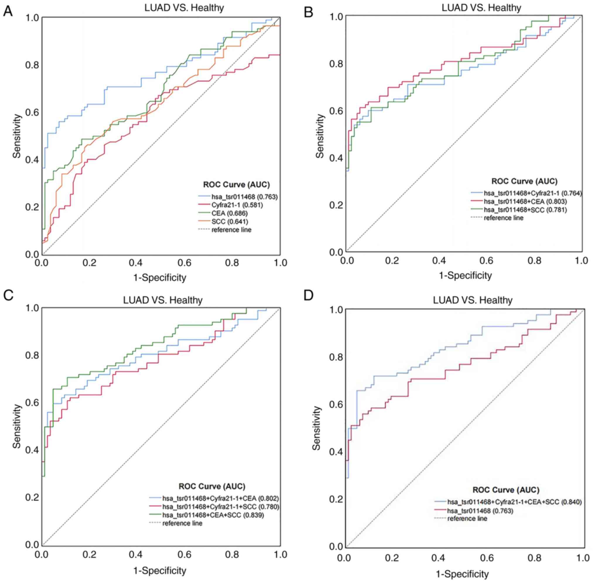 | Figure 4.Evaluation of the diagnostic use of
hsa_tsr011468 in LUAD. (A) ROC analysis of the independent use of
plasma hsa_tsr011468, Cyfra21-1, CEA and SCC antigen in
distinguishing patients with LUAD from healthy controls. (B) ROC
curve analysis of the combined diagnostic value of hsa_tsr011468 +
Cyfra21-1, CEA or SCC. (C) ROC curve analysis of the combined
diagnostic value of hsa_tsr011468 + Cyfra21-1 + CEA or SCC, and of
hsa_tsr011468 + CEA + SCC. (D) ROC curve analysis of the combined
diagnostic value of hsa_tsr011468 + Cyfra21-1 + CEA + SCC. AUC,
area under the curve; CEA, carcinoembryonic antigen; Cyfra21-1,
cytokeratin 19 fragment; LUAD, lung adenocarcinoma; ROC, receiver
operating characteristic; SCC, squamous cell carcinoma. |
 | Table II.Use of hsa_tsr011468, Cyfra21-1, CEA
and SCC antigen to distinguish patients with LUAD from healthy
controls. |
Table II.
Use of hsa_tsr011468, Cyfra21-1, CEA
and SCC antigen to distinguish patients with LUAD from healthy
controls.
| Indicator | SEN (%) | SPE (%) | ACCU (%) | PPV (%) | NPV (%) |
|---|
| hsa_tsr011468 | 57.14 | 92.05 | 75.00 | 87.27 | 69.23 |
| Cyfra21-1 | 15.48 | 93.18 | 55.23 | 68.42 | 53.59 |
| CEA | 15.48 | 94.32 | 55.81 | 72.22 | 53.90 |
| SCC antigen | 11.90 | 78.41 | 44.77 | 34.48 | 48.25 |
| hsa_tsr011468 +
Cyfra21-1 | 64.29 | 86.36 | 75.58 | 81.82 | 71.70 |
| hsa_tsr011468 +
CEA | 59.52 | 87.50 | 73.84 | 81.97 | 69.37 |
| hsa_tsr011468 + SCC
antigen | 59.52 | 73.86 | 66.86 | 68.49 | 65.66 |
| hsa_tsr011468 +
Cyfra21-1 + CEA | 66.67 | 86.36 | 76.74 | 66.67 | 86.36 |
| hsa_tsr011468 +
Cyfra21-1 + SCC antigen | 66.67 | 69.32 | 68.02 | 67.47 | 68.54 |
| hsa_tsr011468 + CEA
+ SCC antigen | 60.71 | 70.45 | 65.70 | 66.23 | 65.26 |
| hsa_tsr011468 +
Cyfra21-1+ CEA + SCC antigen | 67.86 | 69.32 | 68.61 | 67.86 | 69.32 |
Dynamic monitoring of hsa_tsr011468 in
LUAD
To investigate the dynamic monitoring capability of
hsa_tsr011468, preoperative and postoperative sera were collected
from 70 patients with LUAD for qPCR analysis. The results revealed
that the relative expression levels of hsa_tsr011468 in the
postoperative sera from patients was significantly lower than that
in the preoperative sera (Fig.
5A). Furthermore, patients with LUAD were divided into high-
and low-expression groups based on hsa_tsr011468 serum expression
for Kaplan-Meier survival analysis and the survival curves were
compared using the log-rank test. As shown in Fig. 5B, the disease-free survival time of
the low-expression group was longer compared with that in the
high-expression group. Moreover, the overall survival of the
low-expression group of hsa_tsr011468 was higher than that of the
high-expression group, providing additional evidence of the dynamic
monitoring capability and prognostic significance of
hsa_tsr011468.
Prediction of downstream regulatory
mechanisms for hsa_tsr011468
The aforementioned experiments highlighted the
significance of hsa_tsr011468 as a potential biomarker for LUAD.
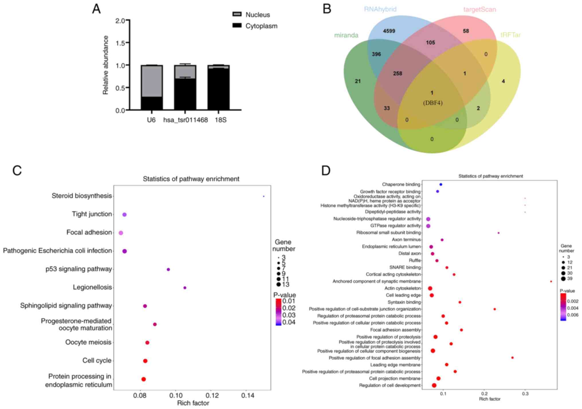
The present study also investigated its potential mechanism in LUAD
and observed via cellular localization experiments that
hsa_tsr011468, consistent with control 18S, was predominantly
located in the cytoplasm (Fig.
6A). Subsequently, TargetScan, miRanda, RNAhybrid and tRFTar
databases were used to predict the downstream target molecules of
hsa_tsr011468, and a Venn diagram was generated. The analysis
revealed an overlapping gene in these databases: DBF4 (Fig. 6B). In addition, KEGG bioinformatics
analysis revealed that it was associated with the ‘p53 signaling
pathway’, ‘Cell cycle’ and ‘Protein processing in endoplasmic
reticulum’ (Fig. 6C). GO
bioinformatics revealed that hsa_tsr011468 was associated with the
‘regulation of cell development’, ‘cell projection membrane’ and
‘ctin cytoskeleton’ (Fig. 6D).
Therefore, hsa_tsr011468 may regulate LUAD progression at the
cellular level via DBF4.
Discussion
Epidemiological data indicated that, in 2022, lung
cancer was the most common type of cancer in China and accounted
for the largest number of cancer-related deaths (21). LUAD is the most prevalent subtype
of lung cancer, and researchers are increasingly investigating
various functions of non-coding RNAs in LUAD. Specifically, long
non-coding RNAs and circular RNAs have demonstrated potential as
biomarkers for the detection, progression and drug resistance
mechanisms of LUAD, thereby presenting new opportunities for
scientific investigation (22–25).
However, there are few studies on tsRNAs in LUAD. Therefore, the
present study evaluated the effect of hsa_tsr011468 on LUAD
pathogenesis and its potential as a LUAD marker.
tsRNAs are RNA molecules cut from mature tRNA or
pre-RNA that have a specific sequence structure and biological
function (26). A growing number
of studies have shown that aberrant expression of tsRNAs affects
tumor development, and is indicative of early diagnosis and
prognosis. For example, tRF-19-3L7L73JD expression in preoperative
patients with gastric cancer was observed to be reduced compared
with that in postoperative and healthy individuals, indicating its
potential as a clinical diagnostic indicator for gastric cancer
(27). Furthermore, the expression
of 5′-tRF-GlyGCC in the plasma of patients with colorectal cancer
was significantly higher than that in healthy individuals, and the
AUC value of the working characteristics of the subjects verified
that 5′-tRF-GlyGCC had a better diagnostic value than CEA or CA19-9
(28). Additionally, hsa_tsr016141
has been reported to have more diagnostic value than CA724 in
gastric cancer (29). The
aforementioned studies showed that tsRNAs have potential for cancer
diagnosis, and provide new directions for cancer prognosis and
targeted therapies.
The present study screened the OncotRF database and
identified the highly expressed tsRNA, hsa_tsr011468, in LUAD.
Statistical data on clinicopathological features showed that
hsa_tsr011468 was associated with tumor size. ROC curve analysis
was conducted to assess the diagnostic value of hsa_tsr011468
compared with other lung cancer indicators in LUAD, and a survival
curve was generated to evaluate the prognostic value of
hsa_tsr011468. Furthermore, GO and KEGG analyses revealed that the
downstream target molecule of hsa_tsr011468 may be DBF4. DBF4 acts
as a crucial cell cycle regulator by binding to cell division cycle
7 to form a DBF4-dependent kinase, which serves a role in
positively regulating DNA replication in the nuclear cell cycle and
in regulating cell cycle phase transitions, and is significant in
tumor development (30,31). In addition, the ‘p53 signaling
pathway’ was identified as an enriched KEGG pathway. Notably, p53
is a classical signaling pathway that regulates cell survival and
death, which has also been shown to be significant in a number of
studies on the mechanism of RNA molecule-regulated cancer (32,33).
A previous analysis of 201 microarray samples from various
genetically engineered mouse breast cancer models indicated that
p53-altered mammary tumors had elevated DBF4 mRNA expression
(34). In addition, p53 as a
transcription factor can regulate the expression of various genes
including those associated with the cell cycle (35–37).
However, further experiments are needed to verify the specific
mechanisms regarding whether DBF4 can act as a target gene to
affect the p53 pathway and hsa_tsr011468.
The present study has some limitations. Primarily,
insufficient clinicopathological features related to LUAD were
assessed, leading to an incomplete analysis of the results.
Recently, EGFR mutation status, EML4-ALK and PD-L1 expression have
been introduced as new targets and research directions for clinical
lung cancer treatment (38–40);
however, the present study lacks a discussion of these factors.
Therefore, these factors will be explored in subsequent studies on
the specific mechanism and targeted therapy of hsa_tsr011468 in
LUAD to further determine the role of hsa_tsr011468 in LUAD.
In conclusion, the present study analyzed
significant tsRNAs in LUAD. Firstly, RT-qPCR analysis of LUAD serum
and tissue samples was performed, which indicated that patients
with LUAD had increased expression of hsa_tsr011468. Subsequently,
the ROC curve and survival analyses showed that hsa_tsr011468 had a
good diagnostic and prognostic value. By comparing the relative
expression levels of hsa_tsr011468 in the preoperative and
postoperative serum of patients, it was revealed that its
expression was decreased in the postoperative serum of patients.
This finding suggested that hsa_tsr011468 may possess strong
dynamic detection capabilities. Therefore, it could be proposed
that hsa_tsr011468 may serve as a potential novel serological
biomarker for LUAD. Finally, the target genes and signaling
pathways of hsa_tsr011468 in LUAD were predicted, establishing a
foundation for further research into its specific mechanism of
action.
Acknowledgements
Not applicable.
Funding
This work was financially supported by the Nantong Science and
Technology Project (grant nos. MS22021001 and 20231044312).
Availability of data and materials
The data generated in the present study may be
requested from the corresponding author.
Authors' contributions
PZ and KZ performed the experiments, analyzed and
interpreted the data, and wrote the paper. CX contributed reagents,
materials, analysis tools and collected data. SL and CX conceived
and designed the experiments, and wrote the paper. PZ and KZ
confirm the authenticity of all the raw data. All authors read and
approved the final version of the manuscript.
Ethics approval and consent for
participation
The present study was approved by the Nantong First
People's Hospital and Affiliated Hospital of Nantong University
(approval nos. 2023KT197 and 2019-L071) and all participants
provided written informed consent for this study.
Patient consent for publication
Not applicable.
Competing interests
The authors declare that they have no competing
interests.
References
|
1
|
Bray F, Laversanne M, Sung H, Ferlay J,
Siegel RL, Soerjomataram I and Jemal A: Global cancer statistics
2022: GLOBOCAN estimates of incidence and mortality worldwide for
36 cancers in 185 countries. CA Cancer J Clin. 74:229–263. 2024.
View Article : Google Scholar : PubMed/NCBI
|
|
2
|
Luo G, Zhang Y, Etxeberria J, Arnold M,
Cai X, Hao Y and Zou H: Projections of lung cancer incidence by
2035 in 40 countries worldwide: Population-based study. JMIR Public
Health Surveill. 9:e436512023. View
Article : Google Scholar : PubMed/NCBI
|
|
3
|
Lu T, Yang X, Huang Y, Zhao M, Li M, Ma K,
Yin J, Zhan C and Wang Q: Trends in the incidence, treatment, and
survival of patients with lung cancer in the last four decades.
Cancer Manag Res. 11:943–953. 2019. View Article : Google Scholar : PubMed/NCBI
|
|
4
|
Chen Q, Zhang X, Shi J, Yan M and Zhou T:
Origins and evolving functionalities of tRNA-derived small RNAs.
Trends Biochem Sci. 46:790–804. 2021. View Article : Google Scholar : PubMed/NCBI
|
|
5
|
Kumar P, Anaya J, Mudunuri SB and Dutta A:
Meta-analysis of tRNA derived RNA fragments reveals that they are
evolutionarily conserved and associate with AGO proteins to
recognize specific RNA targets. BMC Biol. 12:782014. View Article : Google Scholar : PubMed/NCBI
|
|
6
|
Lee YS, Shibata Y, Malhotra A and Dutta A:
A novel class of small RNAs: tRNA-derived RNA fragments (tRFs).
Genes Dev. 23:2639–2649. 2009. View Article : Google Scholar : PubMed/NCBI
|
|
7
|
Zhang Y, Gu X, Li Y, Huang Y and Ju S:
Multiple regulatory roles of the transfer RNA-derived small RNAs in
cancers. Genes Dis. 11:597–613. 2023. View Article : Google Scholar : PubMed/NCBI
|
|
8
|
Wen JT, Huang ZH, Li QH, Chen X, Qin HL
and Zhao Y: Research progress on the tsRNA classification,
function, and application in gynecological malignant tumors. Cell
Death Discov. 7:3882021. View Article : Google Scholar : PubMed/NCBI
|
|
9
|
Veneziano D, Tomasello L, Balatti V,
Palamarchuk A, Rassenti LZ, Kipps TJ, Pekarsky Y and Croce CM:
Dysregulation of different classes of tRNA fragments in chronic
lymphocytic leukemia. Proc Natl Acad Sci USA. 116:24252–24258.
2019. View Article : Google Scholar : PubMed/NCBI
|
|
10
|
Wang S, Wang R, Hu D, Zhang C, Cao P,
Huang J and Wang B: Epigallocatechin gallate modulates ferroptosis
through downregulation of tsRNA-13502 in non-small cell lung
cancer. Cancer Cell Int. 24:2002024. View Article : Google Scholar : PubMed/NCBI
|
|
11
|
Tan L, Wu X, Tang Z, Chen H, Cao W, Wen C,
Zou G and Zou H: The tsRNAs (tRFdb-3013a/b) serve as novel
biomarkers for colon adenocarcinomas. Aging (Albany NY).
16:4299–4326. 2024.PubMed/NCBI
|
|
12
|
Balatti V, Nigita G, Veneziano D, Drusco
A, Stein GS, Messier TL, Farina NH, Lian JB, Tomasello L, Liu CG,
et al: tsRNA signatures in cancer. Proc Natl Acad Sci USA.
114:8071–8076. 2017. View Article : Google Scholar : PubMed/NCBI
|
|
13
|
Li J, Zhu L, Cheng J and Peng Y: Transfer
RNA-derived small RNA: A rising star in oncology. Semin Cancer
Biol. 75:29–37. 2021. View Article : Google Scholar : PubMed/NCBI
|
|
14
|
Jin F, Yang L, Wang W, Yuan N, Zhan S,
Yang P, Chen X, Ma T and Wang Y: A novel class of tsRNA signatures
as biomarkers for diagnosis and prognosis of pancreatic cancer. Mol
Cancer. 20:952021. View Article : Google Scholar : PubMed/NCBI
|
|
15
|
Zhou J, Wan F, Wang Y, Long J and Zhu X:
Small RNA sequencing reveals a novel tsRNA-26576 mediating
tumorigenesis of breast cancer. Cancer Manag Res. 11:3945–3956.
2019. View Article : Google Scholar : PubMed/NCBI
|
|
16
|
Jiang J, Shi S, Zhang W, Li C, Sun L, Ge Q
and Li X: Circ_RPPH1 facilitates progression of breast cancer via
miR-1296-5p/TRIM14 axis. Cancer Biol Ther. 25:23607682024.
View Article : Google Scholar : PubMed/NCBI
|
|
17
|
Xia M, Chen J, Hu Y, Qu B, Bu Q and Shen
H: miR-10b-5p promotes tumor growth by regulating cell metabolism
in liver cancer via targeting SLC38A2. Cancer Biol Ther.
25:23156512024. View Article : Google Scholar : PubMed/NCBI
|
|
18
|
Zong T, Yang Y, Zhao H, Li L, Liu M, Fu X,
Tang G, Zhou H, Aung LHH, Li P, et al: tsRNAs: Novel small
molecules from cell function and regulatory mechanism to
therapeutic targets. Cell Prolif. 54:e129772021. View Article : Google Scholar : PubMed/NCBI
|
|
19
|
Livak KJ and Schmittgen TD: Analysis of
relative gene expression data using real-time quantitative PCR and
the 2(−Delta Delta C(T)) Method. Methods. 25:402–408. 2001.
View Article : Google Scholar : PubMed/NCBI
|
|
20
|
Goldstraw P, Chansky K, Crowley J,
Rami-Porta R, Asamura H, Eberhardt WE, Nicholson AG, Groome P,
Mitchell A, Bolejack V, et al: The IASLC lung cancer staging
project: Proposals for Revision of the TNM Stage Groupings in the
Forthcoming (Eighth) Edition of the TNM classification for lung
cancer. J Thorac Oncol. 11:39–51. 2016. View Article : Google Scholar : PubMed/NCBI
|
|
21
|
Xia C, Dong X, Li H, Cao M, Sun D, He S,
Yang F, Yan X, Zhang S, Li N and Chen W: Cancer statistics in China
and United States, 2022: Profiles, trends, and determinants. Chin
Med J (Engl). 135:584–590. 2022. View Article : Google Scholar : PubMed/NCBI
|
|
22
|
Chen Z, Hu Z, Sui Q, Huang Y, Zhao M, Li
M, Liang J, Lu T, Zhan C, Lin Z, et al: LncRNA FAM83A-AS1
facilitates tumor proliferation and the migration via the
HIF-1α/glycolysis axis in lung adenocarcinoma. Int J Biol Sci.
18:522–535. 2022. View Article : Google Scholar : PubMed/NCBI
|
|
23
|
Liu X, Feng Y, Wang L, Shi L, Ji K, Hu N,
Du Y, Liu M and Wang M: Silencing of circ_0088036 inhibits growth
and invasion of lung adenocarcinoma through miR-203/SP1 axis. J
Biochem Mol Toxicol. 38:e236842024. View Article : Google Scholar : PubMed/NCBI
|
|
24
|
Zhang LX, Gao J, Long X, Zhang PF, Yang X,
Zhu SQ, Pei X, Qiu BQ, Chen SW, Lu F, et al: The circular RNA
circHMGB2 drives immunosuppression and anti-PD-1 resistance in lung
adenocarcinomas and squamous cell carcinomas via the
miR-181a-5p/CARM1 axis. Mol Cancer. 21:1102022. View Article : Google Scholar : PubMed/NCBI
|
|
25
|
Zhang H, Wang SQ, Wang L, Lin H, Zhu JB,
Chen R, Li LF, Cheng YD, Duan CJ and Zhang CF: m6A
methyltransferase METTL3-induced lncRNA SNHG17 promotes lung
adenocarcinoma gefitinib resistance by epigenetically repressing
LATS2 expression. Cell Death Dis. 13:6572022. View Article : Google Scholar : PubMed/NCBI
|
|
26
|
Wang Y, Weng Q, Ge J, Zhang X, Guo J and
Ye G: tRNA-derived small RNAs: Mechanisms and potential roles in
cancers. Genes Dis. 9:1431–1442. 2022. View Article : Google Scholar : PubMed/NCBI
|
|
27
|
Shen Y, Xie Y, Yu X, Zhang S, Wen Q, Ye G
and Guo J: Clinical diagnostic values of transfer RNA-derived
fragment tRF-19-3L7L73JD and its effects on the growth of gastric
cancer cells. J Cancer. 12:3230–3238. 2021. View Article : Google Scholar : PubMed/NCBI
|
|
28
|
Wu Y, Yang X, Jiang G, Zhang H, Ge L, Chen
F, Li J, Liu H and Wang H: 5′-tRF-GlyGCC: A tRNA-derived small RNA
as a novel biomarker for colorectal cancer diagnosis. Genome Med.
13:202021. View Article : Google Scholar : PubMed/NCBI
|
|
29
|
Gu X, Ma S, Liang B and Ju S: Serum
hsa_tsr016141 as a Kind of tRNA-Derived fragments is a novel
biomarker in gastric cancer. Front Oncol. 11:6793662021. View Article : Google Scholar : PubMed/NCBI
|
|
30
|
Zhang L, Hong J, Chen W, Zhang W, Liu X,
Lu J, Tang H, Yang Z, Zhou K, Xie H, et al: DBF4 dependent kinase
inhibition suppresses hepatocellular carcinoma progression and
potentiates anti-programmed cell death-1 therapy. Int J Biol Sci.
19:3412–3427. 2023. View Article : Google Scholar : PubMed/NCBI
|
|
31
|
Qi Y, Hou Y and Qi L: miR-30d-5p represses
the proliferation, migration, and invasion of lung squamous cell
carcinoma via targeting DBF4. J Environ Sci Health C Toxicol
Carcinog. 39:251–268. 2021.PubMed/NCBI
|
|
32
|
Yuan K, Lan J, Xu L, Feng X, Liao H, Xie
K, Wu H and Zeng Y: Long noncoding RNA TLNC1 promotes the growth
and metastasis of liver cancer via inhibition of p53 signaling. Mol
Cancer. 21:1052022. View Article : Google Scholar : PubMed/NCBI
|
|
33
|
Zhang L, Liao Y and Tang L: MicroRNA-34
family: a potential tumor suppressor and therapeutic candidate in
cancer. J Exp Clin Cancer Res. 38:532019. View Article : Google Scholar : PubMed/NCBI
|
|
34
|
Herschkowitz JI, Simin K, Weigman VJ,
Mikaelian I, Usary J, Hu Z, Rasmussen KE, Jones LP, Assefnia S,
Chandrasekharan S, et al: Identification of conserved gene
expression features between murine mammary carcinoma models and
human breast tumors. Genome Biol. 8:R762007. View Article : Google Scholar : PubMed/NCBI
|
|
35
|
Feng J, Xie L, Lu W, Yu X, Dong H, Ma Y
and Kong R: Hyperactivation of p53 contributes to mitotic
catastrophe in podocytes through regulation of the Wee1/CDK1/cyclin
B1 axis. Ren Fail. 46:23654082024. View Article : Google Scholar : PubMed/NCBI
|
|
36
|
Hernández Borrero LJ and El-Deiry WS:
Tumor suppressor p53: Biology, signaling pathways, and therapeutic
targeting. Biochim Biophys Acta Rev Cancer. 1876:1885562021.
View Article : Google Scholar : PubMed/NCBI
|
|
37
|
Pritchard A, Tousif S, Wang Y, Hough K,
Khan S, Strenkowski J, Chacko BK, Darley-Usmar VM and Deshane JS:
Lung tumor cell-derived exosomes promote M2 macrophage
polarization. Cells. 9:13032020. View Article : Google Scholar : PubMed/NCBI
|
|
38
|
Qin Z, Yue M, Tang S, Wu F, Sun H, Li Y,
Zhang Y, Izumi H, Huang H, Wang W, et al: EML4-ALK fusions drive
lung adeno-to-squamous transition through JAK-STAT activation. J
Exp Med. 221:e202320282024. View Article : Google Scholar : PubMed/NCBI
|
|
39
|
Li N, Zuo R, He Y, Gong W, Wang Y, Chen L,
Luo Y, Zhang C, Liu Z, Chen P and Guo H: PD-L1 induces autophagy
and primary resistance to EGFR-TKIs in EGFR-mutant lung
adenocarcinoma via the MAPK signaling pathway. Cell Death Dis.
15:5552024. View Article : Google Scholar : PubMed/NCBI
|
|
40
|
Gu W, Liu P, Tang J, Lai J, Wang S, Zhang
J, Xu J, Deng J, Yu F, Shi C and Qiu F: The prognosis of TP53 and
EGFR co-mutation in patients with advanced lung adenocarcinoma and
intracranial metastasis treated with EGFR-TKIs. Front Oncol.
13:12884682024. View Article : Google Scholar : PubMed/NCBI
|


