Introduction
Alcohol consumption is linked to cancer development
and is considered to be the cause of certain types of cancers,
including oropharynx, larynx, esophageal, liver, breast, colon and
rectal cancers (1,2). Although many factors contribute to
cancer in women, approximately 60% of alcohol-induced cancers in
women are breast cancer (3).
Various mechanisms, such as mutagenic effects, changes in retinoic
acid, folate or estrogen metabolism and oxidative stress induced by
alcohol consumption have been reported to be involved in breast
cancer. However, the effects of alcohol on the carcinogenesis and
metastasis of breast cancer have yet to be determined (2).
Alcohol affects both the upstream and downstream
elements within the mitogen-activated protein kinase (MAPK) pathway
in many tissue types (4). MAPK
plays a role in relaying cellular responses to the nucleus; such
responses include proliferation, differentiation, survival and
apoptosis. MAPK can activate parallel cascades with such downstream
targets as extracellular signal-regulated kinases 1/2 (ERK1/2), p38
MAPK and stress-activated protein kinase/c-Jun NH2-terminal kinase
(SAPK/JNK). ERK1/2 is activated by receptor tyrosine kinases (RTKs)
in response to alcoholic stress; it relays signals to the nucleus.
In previous studies, the activity of ERK1/2 was found to be higher
in breast cancer tissue in comparison to normal tissue (4–6).
Increases in cell proliferation induced by activated ERK1/2 in
response to alcohol have been observed in the human breast cancer
cell line MCF-7 (7).
Epigenetic histone modification was recently
reported as a carcinogenic mechanism induced by alcohol consumption
(8,9). Histone modification affects gene
expression by remodeling the chromatin structure. Histones are
exposed to post-translational modifications, such as
phosphorylation, acetylation, methylation, ubiquitination and
SUMOylation, which regulate the activity of transcription factors,
nucleosome remodelers, histone chaperones and other histone
modifiers (10–12). The phosphorylation of histones
regulates cellular processes, including transcription, DNA repair,
chromosome condensation and apoptosis. For instance, the
phosphorylation of histone 3 serine 10 (H3S10p) regulates the
transcriptional activities of various genes during the interphase
stage of the cell cycle, and the crosstalk with other modifications
on the H3 tail residues (methylation, phosphorylation and
acetylation) aids in the initiation of transcription (13). The activation of the MAPK pathway by
UV irradiation increases the level of H3S10p and H3 serine 28
phosphorylation (H3S28p) (14,15).
Rat hepatocytes exposed to alcohol and acetaldehyde also showed
increases in the levels of H3S10p and H3S28p (16). Acute alcohol exposure also resulted
in the same pattern of H3S10p and H3S28p levels, which, in turn,
regulates the expression of c-fos, c-jun and
MKP-1(9).
The following are known kinases that phosphorylate
histones: Aurora B, VRK1, mitogen- and stress-activated kinase-1/2
(MSK1/2), PIM1, Rsk2, PKB/Akt and IKKα (13). MSK1/2, an H3 kinase, is activated by
stress-activated ERK1/2 (17,18),
and the resulting phosphorylation of H3 leads to the regulation of
the expression of immediate-early (IE) genes, such as c-fos
and c-jun(19–22). Activated by extracellular signals,
the MAPK pathway was found to regulate alcohol-induced stress in
cells, influencing the chromatin structure and gene expression
(4). The present study analyzed the
effect of alcohol on breast cancer cell proliferation through
proteome profiling and observed the changes in histone modification
patterns induced by signals relayed through the MAPK pathway in
alcohol-treated breast cancer cells.
Materials and methods
Cell culture
Human ductal breast carcinoma T47D (KCLB No. 30133)
cells were obtained from the Korean Cell Line Bank (Seoul, Korea).
The T47D cells were grown in RPMI-1640 medium (Invitrogen,
Carlsbad, CA, USA) supplemented with 10% fetal bovine serum
(Invitrogen) and penicillin (100 U/ml)/streptomycin (100 μg/ml)
(Invitrogen) at 37°C in a 5% CO2 atmosphere. The T47D
cells were exposed for 48 h to 100 mM alcohol and 10 μM U0126
(MEK1/2 inhibitor), and the medium with alcohol was changed every
24 h. To prevent the evaporation of the alcohol from the culture
dishes, the alcohol-treated cells were cultured in a separate
CO2 incubator at the same alcohol concentration, as
previously described (23).
Proteomic analysis
To analyze the protein expression profile in T47D
cells in the absence or presence of alcohol, we used the following
proteomic techniques: two-dimensional gel electrophoresis (2-DE),
matrix-assisted laser-desorption ionization time-of-flight mass
spectrometry (MALDI-TOF MS) and database searches, as previously
described by Jung et al(24). The protein samples for the 2-DE were
extracted in lysis buffer [9.5 M urea, 2.5% 3-[(3-cholamidopropyl)
dimethylammonio]-1-propanesulfonate (CHAPS), 40 mM dithiothreitol
(DTT), 0.12% carrier ampholytes and 0.0012% bromophenol blue] and
were normalized using the Bradford assay. Each sample (100 μg) was
analyzed using immobilized pH gradient (IPG) DryStrips (pH 4.0–7.0)
(Bio-Rad Laboratories, Hercules, CA, USA) for isoelectric focusing
(IEF) and SDS-polyacrylamide gels. The IEF was performed at 20°C
using a Protean® IEF Cell (Bio-Rad Laboratories)
following the manufacturer’s instructions. After electrophoresis,
the 2-DE gels were stained with Coomassie brilliant blue and were
analyzed to quantify the spot densities using PDQuest software
(version 7.3, Bio-Rad Laboratories). Following the quantitative
analysis, the differentially expressed protein spots were extracted
from each gel. In-gel digestion was performed on selected protein
spots, and the peptides were analyzed using an Ultraflex MALDI-TOF
MS (Bruker Daltonics, Bremen, Germany) using a procedure similar to
that previously described (24,25).
The search program ProFound developed by Rockefeller University
(http://prowl.rockefeller.edu/prowl-cgi/profound.exe)
was used for the protein identification.
Western blotting
The extraction of proteins from the cells was
performed using RIPA buffer [1% Triton X-100 in 50 mM phosphate
buffer (pH 7.4)] containing both a complete EDTA-free protease
inhibitor cocktail (Roche Diagnostics, Mannheim, Germany) and a
phosphatase inhibitor (GenDEPOT, Barker, TX, USA). The extracted
proteins were separated on SDS polyacrylamide gels and transferred
to polyvinylidene difluoride (PVDF) membranes (Schleicher &
Schuell BioScience, Inc., Keene, NH, USA). The western blot
analysis was performed using an anti-ROS1 antibody (Cell Signaling,
Danvers, MA, USA), anti-phospho-ROS1 (Tyr2274) antibody (Cell
Signaling), the phospho-ERK1/2 Pathway Sampler kit (Cell
Signaling), anti-β-actin antibody (Sigma-Aldrich, St. Louis, MO,
USA), anti-histone H3 (phospho-S10) antibody (Abcam, Cambridge, UK)
and anti-histone H3 antibody (Abcam).
Real-time reverse transcriptase
(RT)-PCR
Total RNA was isolated from the T47D cells exposed
or unexposed to alcohol and/or U0126 using TRIzol (Invitrogen). The
total RNA was reverse-transcribed into cDNA using PrimeScript™
Reverse Transcriptase (Takara, Shinga, Japan), and the real-time
PCR was performed using the 7500 Real-Time PCR system (Applied
Biosystems, Foster City, CA, USA) and 2X SYBR-Green PCR Master mix
(Takara). The sequences of the primers used in this study were as
follows: c-fos forward, 5′-GTCTCCAGTG CCAACTTCATT-3′, and
reverse, 5′-CCTCCTGTCATGG TCTTCACA-3′; and β-actin forward,
5′-TGGAGAAAATCT GGCACCACACC-3′, and reverse, 5′-GATGGGCACAGT
GTGGGTGACCC-3′. β-actin was used as an internal control. The
gene expression levels were analyzed using the 2-ΔΔCT
method (26).
Determination of cell proliferation
The proliferation of the cells was evaluated using
WST-1 (Takara) after exposure to 100 mM alcohol, 10 μM U0126 (Cell
Signaling) for 12 and 24 h. The WST-1 reagent was added to each
well, and the cells were incubated at 37°C in a 5% CO2
atmosphere for 4 h. The results of the WST-1 assay were measured
using a Model 680 microplate reader (Bio-Rad Laboratories) at 440
nm.
Chromatin immunoprecipitation (ChIP)
assay
ChIP assay was performed as previously described by
Choe et al with minor modifications (27). T47D cells were treated with 1%
formaldehyde for 10 min at 37°C. After harvesting, 2×10⁷
cells were suspended with Tris-EDTA buffer (10 mM Tris-HCl, pH 7.6,
1 mM EDTA) including 5 mM butyrate, 1X proteinase inhibitor
cocktail (Roche Diagnostics) and 0.5 mM fresh PMSF. After
sonication, the cells were dialyzed with RIPA buffer (10 mM Tris,
pH 7.4, 1 mM EDTA, 0.1% SDS, 0.1% sodium deoxycholate, 1% Triton
X-100) and subject to immunoprecipitation with antibodies against
anti-14-3-3ɛ, anti-14-3-3ζ and IgG (Santa Cruz Biotechnology, Santa
Cruz, CA, USA). Isolated chipped DNA was validated by PCR. The
sequences of primers for the 14-3-3 protein recruitment assay are
listed in Table I.
 | Table IList of primer sequences used to
amplify chipped DNA. |
Table I
List of primer sequences used to
amplify chipped DNA.
| Primer sequence 5′
to 3′ |
|---|
|
|
|---|
| Forward | Reverse |
|---|
| c-fos
(-999) |
CGTGGTTGAGCCCGTGACGTT |
TGCGGTTGGAGTACGAGGCG |
| c-fos
(-480) |
GGGCGGGACGCTCCAGTAGAT |
TCAGAGCAAGTCCCGAGCCC |
| GAPDH |
GCAAGGAGAGCTCAAGGTCA |
AGCGCGAAAGGAAAGAAAG |
Statistical analysis
All of the values were analyzed using OriginPro 8
(OriginLab Corp., Northampton, MA, USA). All of the values are
expressed as the mean ± standard error of the mean (SEM). All of
the statistical analyses were performed using SPSS 17.0 (SPSS Inc.,
Chicago, IL, USA). P-values <0.05 were considered to indicate a
statistically significant difference.
Results
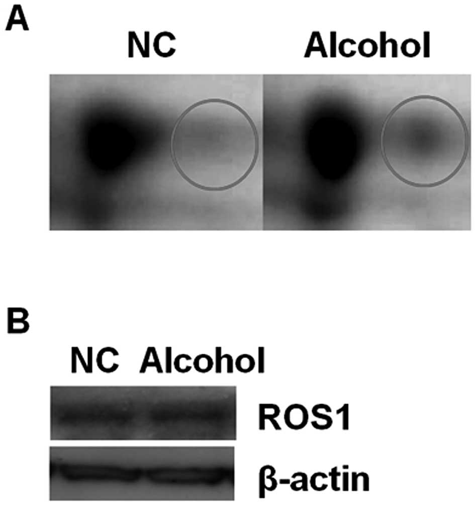
Identification of upregulated ROS1
protein in alcohol-treated T47D cells by proteomic analysis
To assay the influence of alcohol on breast cancer,
proteins extracted from untreated and alcohol-exposed T47D cells
(100 mM) for 48 h were isolated with 2-DE and identified using
MALDI-TOF MS. A total of 20 proteins displaying more than a 2-fold
difference in expression level were identified: 7 proteins
increased in response to alcohol exposure, whereas 13 proteins
displayed reductions in their expression levels (Tables II and III). ROS1, a RTK differentially induced
by alcohol, demonstrated an increased expression ~2.3 times more
than that under normal conditions in both 2-DE imaging (Fig. 1A) and western blotting (Fig. 1B).
| Table IIUpregulated proteins in T47D cells
exposed to alcohol in comparison to the unexposed cells. |
Table II
Upregulated proteins in T47D cells
exposed to alcohol in comparison to the unexposed cells.
| Intensity | | |
|---|
|
| | |
|---|
| Protein | NC | Alcohol | Est’d Z | Accession
number |
|---|
| Unnamed protein
product | 2,126.3 | 6,055.9 | 2.4 | AAA73055.1 |
| Nestin, isoform
CRA_b | 7,729.2 | 13,638.2 | 0.3 | EAW52924.1 |
| 26S protease
regulatory subunit 6B isoform 1 | 5,411.6 | 6,413.1 | 2.4 | NP_006494.1 |
| ATPase,
H+ transporting, lysosomal 50/57 kDa, V1 subunit H,
isoform CRA_c | 5,748.3 | 10,912.2 | 2.43 | EAW86730.1 |
| RNA binding motif
protein 4, isoform CRA_b | 2,086.9 | 4,221.9 | 2.43 | EAW74555.1 |
| RUN and FYVE
domain-containing protein 4 | 3,851.3 | 6,874.7 | 0.6 | NP_940885.2 |
| ROS1 | 4,316.6 | 10,284.5 | 1.43 | AAA60277.1 |
| Table IIIDownregulated proteins in the T47D
cells treated with alcohol in comparison to the untreated
cells. |
Table III
Downregulated proteins in the T47D
cells treated with alcohol in comparison to the untreated
cells.
| Intensity | | |
|---|
|
| | |
|---|
| Protein | NC | Alcohol | Est'd Z | Accession
number |
|---|
| SRB7 suppressor of
RNA polymerase B homolog (yeast), isoform CRA_a | 7,694.7 | 3,585.2 | 1.4 | EAW96544.1 |
| CD44 molecule
(Indian blood group) | 2,125.9 | 1,339.9 | 1.4 | CAC10349.1 |
| G α-q | 8,732 | 3,843.1 | 1.3 | AAB06875.1 |
| β-tubulin | 4,769.7 | 1,569 | 2.4 | AAB59507.1 |
| Keratin, type I
cytoskeletal 19 | 21,769.7 | 12,874.6 | 2.4 | P08727.3 |
| Keratin, type I
cytoskeletal 19 | 20,520.4 | 2,340.1 | 2.4 | P08727.3 |
| RIN2 protein | 10,761.2 | 3,308.4 | 0.9 | AAI28066.1 |
| Keratin, type I
cytoskeletal 19 | 10,293.7 | 2,111.6 | 2.4 | NP_002267.2 |
| β-tubulin | 11,069 | 4,955.7 | 2.4 | AAB59507.1 |
| HMMR protein | 6,474 | 5,410.5 | 1.4 | AAH06984.1 |
| α-amylase | 13,589.9 | 9,157.8 | 2.4 | NP_004029.2 |
|
3-hydroxyisobutyrate dehydrogenase | 1,523.5 | 279.9 | 1.9 | NP_689953.1 |
| Keratin, type II
cytoskeletal 79 | 17,336.5 | 12,267.3 | 2.4 | Q5XKE5.1 |
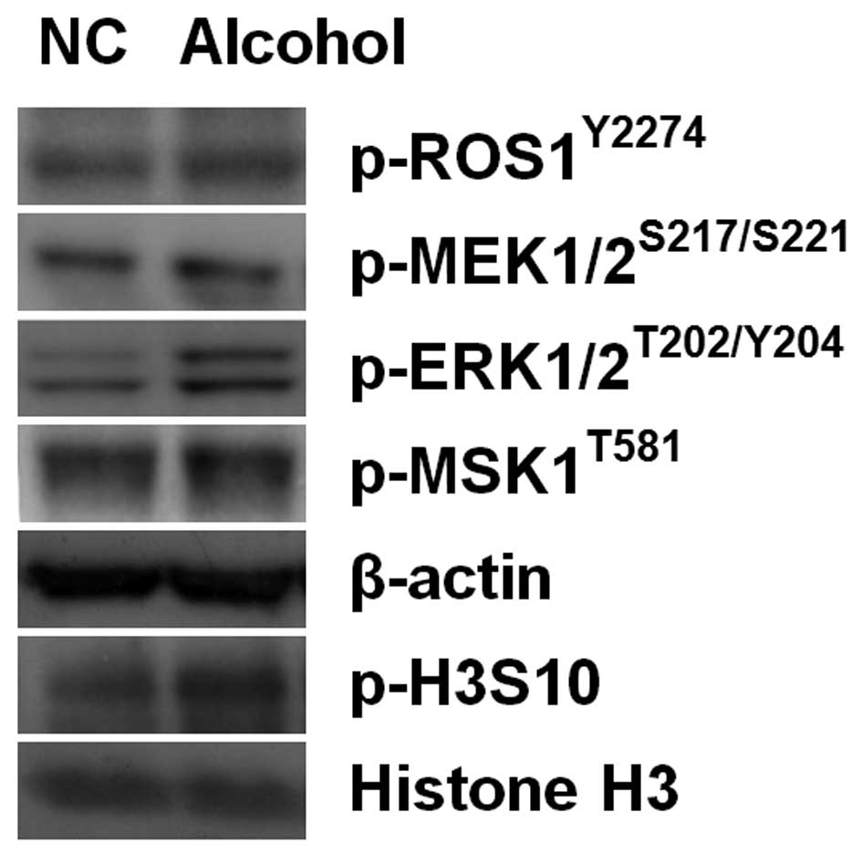
Effect of alcohol on the phosphorylation
of ROS1 and the MAPK pathway
Western blotting was performed to determine the
activation of the ROS1 protein and MAPK pathway proteins by
alcohol-induced phosphorylation (Fig.
2). The phosphorylation of the Y2274 residue on the ROS1
protein increased at 24 h after the exposure of T47D cells to 100
mM alcohol; the phosphorylation of MEK1/2, ERK1/2, MSK1 and H3S10
also showed similar patterns after alcohol treatment. These results
suggest that the level of H3S10p was increased via activated MAPK
pathways in alcohol-exposed T47D cells.
To further investigate the role of alcohol in the
activation of the MAPK pathway, cell proliferation was examined in
the alcohol- and/or MEK1/2 inhibitor U0126-treated T47D cells.
First of all, we assessed the optimal inhibition time of MAPK
pathway by U0126 treatment in T47D cells. A gradual decrease in the
level of the MEK1/2 phosphorylation was observed in the
U0126-treated T47D cells at 1, 3 and 6 h, while it increased after
12 h. The phosphorylation of ERK1/2, on the other hand, was
inhibited during the entire observation period and was not affected
by activated MEK1/2. The phosphorylation level of MSK1, a kinase
activated downstream of the MAPK cascade, was also reduced at 12
and 24 h (Fig. 3A).
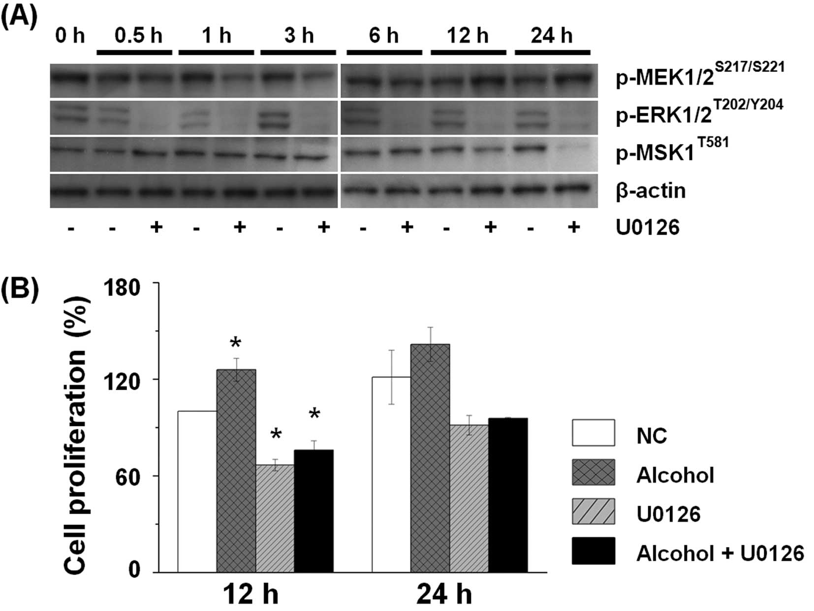 | Figure 3Exposure to alcohol resulted in the
activation of the MAPK pathway and increased cell proliferation.
(A) T47D cells were incubated in the presence or absence of 10 μM
U0126, a MEK1/2 inhibitor, for 0.5, 1, 3, 6, 12 and 24 h. Equal
amounts of proteins were obtained from the cells and were analyzed
using western blotting to detect p-MEK1/2 (S217/S2221), p-ERK1/2
(T202/Y204) and p-MSK1 (T581). Phosphorylated MEK was decreased at
0.5, 1 and 3 h in the T47D cells exposed to alcohol in the presence
of U0126. Phosphorylated ERK1/2 was decreased at 0.5, 1, 3, 6, 12
and 24 h in the T47D cells treated with alcohol in the presence of
U0126. Phosphorylated MSK was decreased at 12 and 24 h in the T47D
cells treated with alcohol in the presence of U0126. (B) The T47D
cells were exposed to 100 mM alcohol and/or 10 μM U0126 for 12 and
24 h; the cell proliferation was evaluated, and the proliferation
of the cells was measured using WST-1. The T47D cells treated with
alcohol demonstrated an increase in cell proliferation and the
U0126-treated cells showed a decrease compared with the untreated
cells. However, exposure to both alcohol and U0126 induced an
increase in cell proliferation when compared to the U0126-treated
cells. The values are presented as means ± SEM (n=3).
*Significantly different from the NC group. A one-way
ANOVA followed by Tukey’s HSD post-hoc test was used to evaluate
the statistical significance of the differences. NC, untreated
control. |
Cell proliferation was evaluated after exposure to
alcohol and/or U0126 at 12 and 24 h (Fig. 3B). When T47D cells were exposed to
alcohol, the cell numbers exhibited a greater increase than that of
the unexposed cells after 12 and 24 h of incubation. In contrast,
when U0126 was added for 12 h, the number of cells was reduced to
~66%, and a slight increase in the number of cells was observed
after 24 h. When T47D cells were exposed to both alcohol and U0126
for 12 and 24 h, the number of cells increased to a greater extent
than that of the U0126-treated cells. It can be inferred that
alcohol leads to an increase in cell proliferation through
activation of the MAPK pathway.
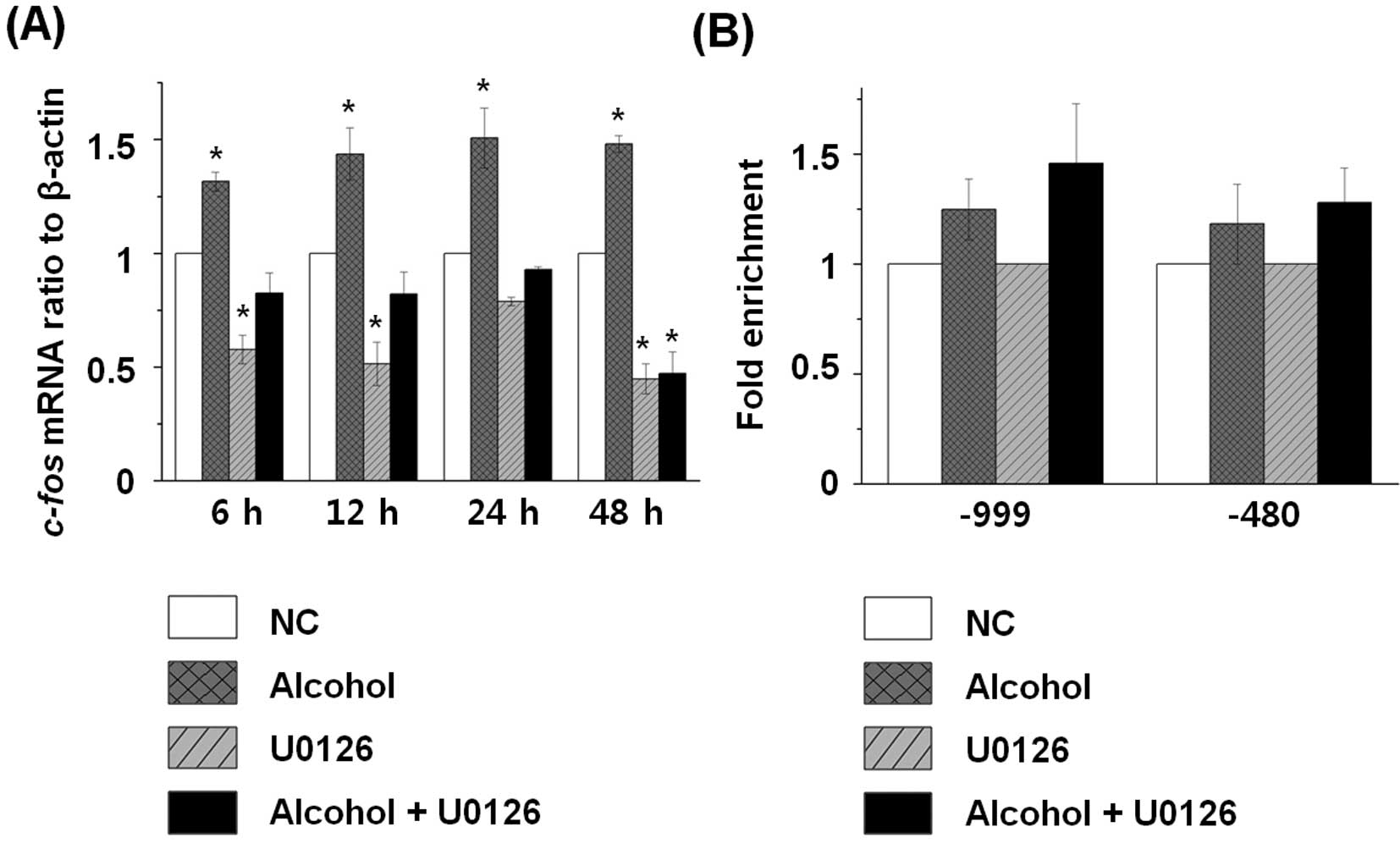
Regulation of immediate-early gene
expression following exposure to alcohol and the MEK1/2 inhibitor
treatment
As cell proliferation was increased due to the
activation of the alcohol-induced MAPK cascade, we analyzed the
effect of alcohol and U0126 on relevant IE gene expression patterns
(Fig. 4A). The expression of
c-fos, one of the IE genes, was controlled by increases in
H3S10p, as confirmed using real-time RT-PCR. The expression of
c-fos was increased after exposure to alcohol during the
entire exposure, while the expression level was reduced to half
when U0126 was added. However, the expression level was slightly
increased when both alcohol and U0126 were administered to T47D
cells. These results indicate that the expression level of
c-fos is increased according to elevated H3S10p through
activation of the MAPK pathway in alcohol-exposed cells.
Regulation of recruitment of the 14-3-3
proteins in response to alcohol exposure
In previous research, 14-3-3 proteins were reported
to act as adaptors between phosphorylated histone H3 and another
phosphoprotein (28). Additionally,
recruitment of 14-3-3 proteins such as 14-3-3ɛ and 14-3-3ζ were
found to be increased by ERK1/2 MAPK pathway activation for
inducible genes (29). To determine
the composition of 14-3-3 proteins of the c-fos gene after
alcohol exposure in T47D cells, we performed a ChIP assay (Fig. 4B). Upon alcohol exposure,
recruitment of 14-3-3 proteins was increased in both upstream
regions (−999, −480) of the c-fos gene, indicating that
recruitment of 14-3-3 proteins is induced after alcohol exposure at
the upstream regions of c-fos.
Discussion
In this study, we observed that the expression of
ROS1 and its phosphorylation level were enhanced in alcohol-exposed
T47D cells. ROS1, a proto-oncogene expressed in various tumor cell
lines, is a cellular signaling transduction pathway regulator that
mediates cell proliferation, migration and cell-to-cell
communication (30,31). ROS1 is activated via the
autophosphorylation of Y2274 and Y2334 residues and regulates
signaling transduction pathways, e.g., the MAPK, insulin receptor
substrate 1 (IRS-1), phosphatidylinositol 3-kinase (PI3K), protein
kinase B (AKT), STAT3 and VAV3 signaling pathways (30).
Among these pathways, the MAPK pathway influences
the chromatin structure and gene expression, thereby regulating
cellular processes such as cell proliferation, apoptosis,
inflammation and cell cycle progression (5,32,33).
Cell proliferation is enhanced through the MAPK pathway in various
tissues due to the chronic intake of alcohol (34–36).
When 10–40 mM alcohol was added to human hepatocellular carcinoma
cell lines HepG2 and SKHep, ERK1/2 was activated, and cell
proliferation was enhanced, while the alcohol-induced proliferation
was inhibited when a MEK1/2 inhibitor was added (37). ERK1/2, p38 MAPK and JNK were found
to be activated in alcohol-exposed mouse hippocampal HT22 cells,
and p38 MAPK activation was proposed to be related to the
production of reactive oxygen species (38).
In many case studies, the activation of ERK1/2 in
breast cancer cells is usually higher than that in normal cells.
This observation is consistent with the results from comparative
studies of primary breast tumors and nearby normal tissues in
breast cancer patients in which the severity of symptoms is
dependent on the level of ERK1/2 activation (4–6).
ERK1/2 was activated by a pathophysiologically relevant
concentration (65 mM) of alcohol in the human breast cancer cell
line MCF-7, and activated ERK1/2-induced increases in cell
proliferation up to 400%, but when an MEK1 inhibitor (PD98059) was
administered with alcohol, the cell proliferation was reduced to
200% (7). Similar to MCF-7 and
various tissue cells such as hepatocytes and hippocampal cells, the
breast cancer cell line T47D exhibited an activation of ERK1/2 and
MSK1 via phosphorylation when 100 mM alcohol was added.
Furthermore, we demonstrated that the proliferation of T47D cells
showed a greater increase when 100 mM alcohol was added when
compared with untreated cells. In addition, cell proliferation was
reduced in the MEK1/2 inhibitor U0126-treated T47D cells. However,
we found that exposure to both alcohol and U0126 restored cell
proliferation when compared with U0126-treated T47D cells. For
these reasons, we suggest that alcohol-induced ROS1 activity
influences MAPK pathway activation and cell proliferation.
Our results showed that the MSK1 phosphorylation
level increased via the MAPK pathway in alcohol-exposed breast
cancer cells and that phosphorylated MSK1 acts as an H3 kinase,
increasing the level of H3S10p. A previous study showed that the
level of H3S10p and H3S28p increased at 1–4 h after the
intraperitoneal injection of 5 g/kg alcohol in rats, leading to the
regulation of c-fos, c-jun and MKP-1
expression (9). When 100 mM alcohol
and 5 mM acetaldehyde were administered separately to rat
hepatocytes, the phosphorylation level of p38 MAPK reached its peak
at 24 h and 30 min, respectively, and the induced level of p38 MAPK
increased the levels of H3S10p and H3S28p (16). Similar to previous results, we
showed that histone phosphorylation is increased by alcohol
exposure in breast cancer cells. Therefore, we confirmed that
alcohol is closely related to histone phosphorylation.
In this study, we established that alcohol-induced
H3S10p regulates the recruitment of 14-3-3 proteins at the upstream
regions of c-fos. The 14-3-3 proteins including the isoforms
ɛ and ζ, an abundant family of phospho-specific binding proteins,
were reported to bind to phosphorylated histone residues such as
serine 10 and 28 (28,39). In addition, it functions as a link
between phosphorylated histone residues and 14-3-3 binding proteins
(29). A previous study reported
that phosphorylated H3S10 by TPA-stimulated MSK1 is recruited to
14-3-3 proteins mediating the recruitment of SWI/SNF, FOS/JUN and
RNA polymerase II at the target gene promoter region finally
inducing the expression of target IE genes (29). After alcohol exposure, we also
observed that binding of 14-3-3 proteins to the upstream regions of
the c-fos gene was increased and induced gene expression.
Notably, all the events such as H3S10 phosphorylation, the
recruitment of 14-3-3 proteins and the expression of the IE genes
were reduced by MSK1 knockdown (29). Thus, we suggest that activated MSK1
by external stimuli including alcohol plays an important role in
histone remodeling and IE gene expression.
When T47D human breast cancer cells are exposed to
alcohol, the expression of ROS1 is upregulated, and
autophosphorylated ROS1, in turn, activates MSK1 via ERK1/2 in the
MAPK pathway. The MSK1-induced increase in H3S10p induces the
expression of c-fos through 14-3-3 protein recruitment,
influencing cell growth under alcohol-exposed conditions. This is
the first study to show that activation of the ROS1 protein by
alcohol exposure in breast cancer cells is associated with the MAPK
pathway. Furthermore, this study demonstrated the relationship
between alcohol and cell proliferation associated with activation
of the MAPK pathway by ROS1 protein. Based on our results and
previous studies, it is believed that breast cancer cells exposed
to alcohol show increased cancer cell growth by activating the MAPK
pathway through ROS1 protein, therefore confirming that alcohol
consumption is detrimental to breast cancer patients.
Acknowledgements
We thank Professor Adam Turner for her kind
assistance in the language editing of our manuscript. This research
was supported by the Basic Science Research Program through the
National Research Foundation of Korea (NRF) funded by the Ministry
of Education, Science and Technology (no. 2010-0023808 to M.R.C.),
and the National Research Foundation of Korea Grant funded by the
Korean Government (no. 2011-0030768 to Y.G.C. and no. 2011-0006001
to K.H.J.).
References
|
1
|
Boffetta P and Hashibe M: Alcohol and
cancer. Lancet Oncol. 7:149–156. 2006. View Article : Google Scholar
|
|
2
|
Seitz HK and Stickel F: Molecular
mechanisms of alcohol-mediated carcinogenesis. Nat Rev Cancer.
7:599–612. 2007. View
Article : Google Scholar
|
|
3
|
Boffetta P, Hashibe M, La Vecchia C,
Zatonski W and Rehm J: The burden of cancer attributable to alcohol
drinking. Int J Cancer. 119:884–887. 2006. View Article : Google Scholar : PubMed/NCBI
|
|
4
|
Aroor AR and Shukla SD: MAP kinase
signaling in diverse effects of ethanol. Life Sci. 74:2339–2364.
2004. View Article : Google Scholar : PubMed/NCBI
|
|
5
|
Santen RJ, Song RX, McPherson R, Kumar R,
Adam L, Jeng MH and Yue W: The role of mitogen-activated protein
(MAP) kinase in breast cancer. J Steroid Biochem Mol Biol.
80:239–256. 2002. View Article : Google Scholar : PubMed/NCBI
|
|
6
|
Mueller H, Flury N, Eppenberger-Castori S,
Kueng W, David F and Eppenberger U: Potential prognostic value of
mitogen-activated protein kinase activity for disease-free survival
of primary breast cancer patients. Int J Cancer. 89:384–388. 2000.
View Article : Google Scholar : PubMed/NCBI
|
|
7
|
Izevbigie EB, Ekunwe SI, Jordan J and
Howard CB: Ethanol modulates the growth of human breast cancer
cells in vitro. Exp Biol Med (Maywood). 227:260–265.
2002.PubMed/NCBI
|
|
8
|
Park PH, Lim RW and Shukla SD: Involvement
of histone acetyltransferase (HAT) in ethanol-induced acetylation
of histone H3 in hepatocytes: potential mechanism for gene
expression. Am J Physiol Gastrointest Liver Physiol.
289:G1124–G1136. 2005. View Article : Google Scholar : PubMed/NCBI
|
|
9
|
James TT, Aroor AR, Lim RW and Shukla SD:
Histone H3 phosphorylation (Ser10, Ser28) and phosphoacetylation
(K9S10) are differentially associated with gene expression in liver
of rats treated in vivo with acute ethanol. J Pharmacol Exp Ther.
340:237–247. 2012. View Article : Google Scholar : PubMed/NCBI
|
|
10
|
Chi P, Allis CD and Wang GG: Covalent
histone modifications - miswritten, misinterpreted and mis-erased
in human cancers. Nat Rev Cancer. 10:457–469. 2010. View Article : Google Scholar : PubMed/NCBI
|
|
11
|
Jenuwein T and Allis CD: Translating the
histone code. Science. 293:1074–1080. 2001. View Article : Google Scholar : PubMed/NCBI
|
|
12
|
Kouzarides T: Chromatin modifications and
their function. Cell. 128:693–705. 2007. View Article : Google Scholar : PubMed/NCBI
|
|
13
|
Baek SH: When signaling kinases meet
histones and histone modifiers in the nucleus. Mol Cell.
42:274–284. 2011. View Article : Google Scholar : PubMed/NCBI
|
|
14
|
Zhong S, Jansen C, She QB, Goto H, Inagaki
M, Bode AM, Ma WY and Dong Z: Ultraviolet B-induced phosphorylation
of histone H3 at serine 28 is mediated by MSK1. J Biol Chem.
276:33213–33219. 2001. View Article : Google Scholar : PubMed/NCBI
|
|
15
|
He Z, Cho YY, Ma WY, Choi HS, Bode AM and
Dong Z: Regulation of ultraviolet B-induced phosphorylation of
histone H3 at serine 10 by Fyn kinase. J Biol Chem. 280:2446–2454.
2005. View Article : Google Scholar : PubMed/NCBI
|
|
16
|
Lee YJ and Shukla SD: Histone H3
phosphorylation at serine 10 and serine 28 is mediated by p38 MAPK
in rat hepatocytes exposed to ethanol and acetaldehyde. Eur J
Pharmacol. 573:29–38. 2007. View Article : Google Scholar : PubMed/NCBI
|
|
17
|
Soloaga A, Thomson S, Wiggin GR,
Rampersaud N, Dyson MH, Hazzalin CA, Mahadevan LC and Arthur JS:
MSK2 and MSK1 mediate the mitogen- and stress-induced
phosphorylation of histone H3 and HMG-14. EMBO J. 22:2788–2797.
2003. View Article : Google Scholar : PubMed/NCBI
|
|
18
|
Reul JM and Chandramohan Y: Epigenetic
mechanisms in stress-related memory formation.
Psychoneuroendocrinology. 32(Suppl 1): S21–S25. 2007. View Article : Google Scholar : PubMed/NCBI
|
|
19
|
Clayton AL, Rose S, Barratt MJ and
Mahadevan LC: Phosphoacetylation of histone H3 on c-fos- and
c-jun-associated nucleosomes upon gene activation. EMBO J.
19:3714–3726. 2000. View Article : Google Scholar : PubMed/NCBI
|
|
20
|
Hazzalin CA and Mahadevan LC:
MAPK-regulated transcription: a continuously variable gene switch?
Nat Rev Mol Cell Biol. 3:30–40. 2002. View
Article : Google Scholar : PubMed/NCBI
|
|
21
|
Strelkov IS and Davie JR: Ser-10
phosphorylation of histone H3 and immediate early gene expression
in oncogene-transformed mouse fibroblasts. Cancer Res. 62:75–78.
2002.PubMed/NCBI
|
|
22
|
Chadee DN, Hendzel MJ, Tylipski CP, Allis
CD, Bazett-Jones DP, Wright JA and Davie JR: Increased Ser-10
phosphorylation of histone H3 in mitogen-stimulated and
oncogene-transformed mouse fibroblasts. J Biol Chem.
274:24914–24920. 1999. View Article : Google Scholar : PubMed/NCBI
|
|
23
|
Choi MR, Jung KH, Park JH, Das ND, Chung
MK, Choi IG, Lee BC, Park KS and Chai YG: Ethanol-induced small
heat shock protein genes in the differentiation of mouse embryonic
neural stem cells. Arch Toxicol. 85:293–304. 2011. View Article : Google Scholar : PubMed/NCBI
|
|
24
|
Jung KH, Das ND, Park JH, Lee HT, Choi MR,
Chung MK, Park KS, Jung MH, Lee BC, Choi IG and Chai YG: Effects of
acute ethanol treatment on NCCIT cells and NCCIT cell-derived
embryoid bodies (EBs). Toxicol In Vitro. 24:1696–1704. 2010.
View Article : Google Scholar : PubMed/NCBI
|
|
25
|
Shevchenko A, Wilm M, Vorm O and Mann M:
Mass spectrometric sequencing of proteins silver-stained
polyacrylamide gels. Anal Chem. 68:850–858. 1996. View Article : Google Scholar : PubMed/NCBI
|
|
26
|
Baik SY, Jung KH, Choi MR, Yang BH, Kim
SH, Lee JS, Oh DY, Choi IG, Chung H and Chai YG: Fluoxetine-induced
up-regulation of 14-3-3zeta and tryptophan hydroxylase levels in
RBL-2H3 cells. Neurosci Lett. 374:53–57. 2005. View Article : Google Scholar : PubMed/NCBI
|
|
27
|
Choe MK, Hong CP, Park J, Seo SH and Roh
TY: Functional elements demarcated by histone modifications in
breast cancer cells. Biochem Biophys Res Commun. 418:475–482. 2012.
View Article : Google Scholar : PubMed/NCBI
|
|
28
|
Macdonald N, Welburn JP, Noble ME, Nguyen
A, Yaffe MB, Clynes D, Moggs JG, Orphanides G, Thomson S, Edmunds
JW, Clayton AL, Endicott JA and Mahadevan LC: Molecular basis for
the recognition of phosphorylated and phosphoacetylated histone h3
by 14-3-3. Mol Cell. 20:199–211. 2005. View Article : Google Scholar : PubMed/NCBI
|
|
29
|
Drobic B, Perez-Cadahia B, Yu J, Kung SK
and Davie JR: Promoter chromatin remodeling of immediate-early
genes is mediated through H3 phosphorylation at either serine 28 or
10 by the MSK1 multi-protein complex. Nucleic Acids Res.
38:3196–3208. 2010. View Article : Google Scholar : PubMed/NCBI
|
|
30
|
Acquaviva J, Wong R and Charest A: The
multifaceted roles of the receptor tyrosine kinase ROS in
development and cancer. Biochim Biophys Acta. 1795:37–52.
2009.PubMed/NCBI
|
|
31
|
Hubbard SR and Miller WT: Receptor
tyrosine kinases: mechanisms of activation and signaling. Curr Opin
Cell Biol. 19:117–123. 2007. View Article : Google Scholar : PubMed/NCBI
|
|
32
|
Aroor AR, James TT, Jackson DE and Shukla
SD: Differential changes in MAP kinases, histone modifications, and
liver injury in rats acutely treated with ethanol. Alcohol Clin Exp
Res. 34:1543–1551. 2010. View Article : Google Scholar : PubMed/NCBI
|
|
33
|
Hong SK, Yoon S, Moelling C, Arthan D and
Park JI: Non-catalytic function of ERK1/2 can promote
Raf/MEK/ERK-mediated growth arrest signaling. J Biol Chem.
284:33006–33018. 2009. View Article : Google Scholar : PubMed/NCBI
|
|
34
|
Chung J, Liu C, Smith DE, Seitz HK,
Russell RM and Wang XD: Restoration of retinoic acid concentration
suppresses ethanol-enhanced c-Jun expression and hepatocyte
proliferation in rat liver. Carcinogenesis. 22:1213–1219. 2001.
View Article : Google Scholar : PubMed/NCBI
|
|
35
|
Wang XD: Alcohol, vitamin A, and cancer.
Alcohol. 35:251–258. 2005. View Article : Google Scholar
|
|
36
|
Simoni D and Tolomeo M: Retinoids,
apoptosis and cancer. Curr Pharm Des. 7:1823–1837. 2001. View Article : Google Scholar
|
|
37
|
Hennig M, Yip-Schneider MT, Klein P, Wentz
S, Matos JM, Doyle C, Choi J, Wu H, O’Mara A, Menze A, Noble S,
McKillop IH and Schmidt CM: Ethanol-TGFalpha-MEK signaling promotes
growth of human hepatocellular carcinoma. J Surg Res. 154:187–195.
2009. View Article : Google Scholar : PubMed/NCBI
|
|
38
|
Ku BM, Lee YK, Jeong JY, Mun J, Han JY,
Roh GS, Kim HJ, Cho GJ, Choi WS, Yi GS and Kang SS: Ethanol-induced
oxidative stress is mediated by p38 MAPK pathway in mouse
hippocampal cells. Neurosci Lett. 419:64–67. 2007. View Article : Google Scholar : PubMed/NCBI
|
|
39
|
Aitken A: 14-3-3 and its possible role in
co-ordinating multiple signalling pathways. Trends Cell Biol.
6:341–347. 1996. View Article : Google Scholar : PubMed/NCBI
|