Introduction
Lung cancer prognosis remains very poor, with a
5-year survival rate generally <20%, lower than several other
leading cancers, underlining the need for a better understanding of
lung cancer formation and, ultimately, for identification of better
therapeutic targets (1). Cigarette
smoking is acknowledged to be the most important risk factor for
human lung carcinogenesis and
4-(methylnitrosamino)-1-(3-pyridyl)-1-butanone (NNK) is a
tobacco-specific N-nitrosamine which is considered to play
important roles in tobacco-related human lung cancer (2,3). NNK
is also a strong lung carcinogen in rodents, inducing
bronchiolo-alveolar hyperplasia, adenoma and adenocarcinoma
(4). Previously, we demonstrated
that NNK-induced lung carcinogenesis in female A/J mice was
strongly inhibited by treatment with 8-methoxypsoralen, a potent
human cytochrome P450 2A6 (CYP2A6) inhibitor, before a single
intraperitoneal injection (i.p.) of NNK (5–9). We
also reported carcinogenic mechanisms and modifying effects of
other factors using NNK-induced lung cancer animal models (10–15).
In the mouse, it is well known that females are
generally more sensitive to chemical lung carcinogenesis, such as
that induced by NNK, than males (16). It is hypothesized that the
difference is due to difference in the amount of sex hormones such
as testosterone and estradiol. Also, in humans, the incidence of
adenocarcinoma type lung cancer is known to be generally higher in
women than in men (17–19). The available data suggest that
gender and sex hormones affect the character of lung cancer
(20,21). For the purpose of these experiments,
we hypothesized that the carcinogenesis of the lung may be
influenced by sex hormone levels.
The present study was conducted to investigate
effects of gonadectomy, ovariectomy and castration in female and
male mice, respectively, on lung carcinogenesis and to determine
associations with sex hormone levels, i.e. estradiol and
testosterone. The experiments were conducted for 16 and 54 weeks
after NNK treatment, since hyperplasia and adenoma lesions were
earlier detected after 16 weeks (9), and adenocarcinomas were detected after
54 weeks (6).
Materials and methods
Chemicals
NNK was purchased from Toronto Research Chemicals
(Toronto, Ontario, Canada).
Animals
A total of 72 female and 72 male A/J mice (5 weeks
of age), purchased from Shizuoka Laboratory Animal Center
(Shizuoka, Japan), were maintained in the Division of Animal
Experiments, Life Science Research Center, Kagawa University,
according to the Institutional Regulations for Animal Experiments.
The regulations included the best considerations on animal welfare
and good practice of animal handling contributing to the
replacement, refinement and reduction of animal testing (3Rs). The
protocols of the experiments were approved by the Animal Care and
Use Committee for Kagawa University. All animals were housed in
polycarbonate cages with white wood chips for bedding, and given
free access to drinking water and a basal diet, Oriental MF
(Oriental Yeast Co., Ltd., Tokyo, Japan), under controlled
conditions of humidity (60±10%), lighting (12 h light/dark cycle)
and temperature (24±2°C).
Experimental design
At 6 weeks of age, the mice were separated into 8
groups (Groups 1–8) of 15–21 animals each (Table I). Groups 1 and 5 were female groups
undergoing ovariectomy. Groups 2 and 6 were females without
ovariectomy. Groups 3 and 7 were male groups with castration, while
Groups 4 and 8 were males without surgery. At the operations, the
ovaries or the testes were removed under deep anesthesia (Groups 1,
3, 5 and 7) with i.p. of 0.025 ml pentobarbital sodium
(Somunopentyl; Kyoritsu Seiyaku Co., Tokyo, Japan) diluted with
0.225 ml saline (Otsuka isotonic sodium chloride solution; Otsuka
Pharmaceutical Factory, Inc., Tokushima, Japan). The ovaries of
female mice were resected from the retroperitoneum with a vertical
incision in the back skin. The testes of male mice were resected
from the center of the ventral scrota. The incision sites were
closed with metal clips. At 2 weeks after surgery, all mice were
treated with i.p. of NNK (2 mg/0.1 ml saline/mouse). After 3 weeks
from surgery, Groups 1–4 were treated with another i.p. of NNK (2
mg/0.1 ml saline/mouse). The experiment was terminated after 18
weeks for Groups 1–4 and after 56 weeks for Groups 5–8. All
surviving mice were sacrificed under deep anesthesia. Blood samples
were collected from the mice in Groups 1–4 to measure the serum
concentrations of estradiol and testosterone. These hematological
examinations were performed at SRL Inc. (Tokyo, Japan). The
detection limit of the estradiol was 10 pg/ml, and that of
testosterone was 0.03 ng/ml. Values lower than these limits were
considered as 0 and means for each group were calculated. At
autopsy, the lungs, livers and kidneys were removed. The lungs were
weighed including trachea and heart first, infused with 10% neutral
buffered formalin after separation from the trachea and heart, and
then immersed in 10% neutral buffered formalin. Lung weights were
finally calculated by subtraction of the weight of the remaining
trachea and heart. These procedures are appropriate for accurate
weighing and good tissue preservation. After fixation in formalin,
all lungs were carefully inspected grossly using a
stereomicroscope. All macroscopically detected lung nodules were
counted and trimmed for histopathological evaluation. The livers
and kidneys were weighed and immersed in 10% neutral buffered
formalin. Slices of lungs, livers, kidneys and macroscopic mass
lesions were routinely processed for embedding in paraffin for
histopathological examination of hematoxylin and eosin stained
sections.
 | Table IBody and relative organ weights. |
Table I
Body and relative organ weights.
| Group | Gendera | Treatmenta | Durationb | No.c | Body weightd (g) | Lungd Relative (%) | Liverd Relative (%) | Kidneysd Relative (%) |
|---|
| 1 | F | NNKx2 OVX | 18 | 21 | 24.81±2.80 | 0.70±0.08 | 4.50±0.26e | 1.10±0.10 |
| 2 | F | NNKx2 N | 18 | 15 | 23.76±2.44 | 0.70±0.08 | 4.28±0.23 | 1.13±0.07 |
| 3 | M | NNKx2 CAST | 18 | 16 | 26.50±2.67 | 0.66±0.08 | 4.22±0.18f | 0.98±0.05g |
| 4 | M | NNKx2 N | 18 | 15 | 27.96±2.20 | 0.65±0.09 | 4.61±0.22 | 1.26±0.11 |
| 5 | F | NNKx1 OVX | 56 | 18 | 31.04±4.07h | 1.23±0.53 | 4.20±0.52 | 1.57±2.08 |
| 6 | F | NNKx1 N | 56 | 12 | 25.80±3.09 | 1.27±0.23 | 4.18±0.76 | 1.19±0.27 |
| 7 | M | NNKx1 CAST | 56 | 21 | 33.19±4.17 | 0.86±0.21 | 3.67±0.23i | 0.80±0.09j |
| 8 | M | NNKx1 N | 56 | 14 | 30.33±4.41 | 0.86±0.18 | 4.28±0.40 | 1.34±0.13 |
Histopathological analysis
Each lung lobe of all mice was examined
histopathologically. Lung proliferative lesions were diagnosed as
bronchiolo-alveolar hyperplasia, adenoma or adenocarcinoma
according to the criteria of ‘International Classification of
Rodent Tumors: The Mouse’ (22).
Statistical analysis
The incidences of lung proliferative lesions
(macroscopically and histopathologically) were analyzed by the
Fischer’s exact probability test. Body and organ weights were
analyzed by the Student’s t-test or the Welch’s t-test between
groups. Multiplicities of lung proliferative lesions
(macroscopically and histopathologically) were analyzed by the
Student’s t-test or the Welch’s t-test between groups. The
Student’s t-test was applied when equal variance was obtained and
the Welch’s t-test with unequal variance.
Results
General conditions
One mouse from Group 3 died 3 weeks after
castration. In Group 5, one mouse was sacrificed after 25 weeks
from ovariectomy due to poor general condition and another died 54
weeks after ovariectomy. In Group 6, 2 mice were sacrificed 4 and
17 weeks after surgery due to poor general condition and another
mouse died 54 weeks after surgery. In addition, one mouse of Group
8 died 53 weeks after surgery. All other mice demonstrated no
marked change in their general condition.
Body and organ weights
Body and organ weights are shown in Table I. In the 18-week study (Groups 1–4),
body weights exhibited no significant variation across groups. In
the 56-week study (Groups 5–8), body weights in Group 5
(ovariectomy group) were significantly increased as compared with
Group 6. Lung weights showed no significant differences between
ovariectomy or castration groups and each unoperated group. Liver
weights of Group 1 (ovariectomy group) were significantly increased
as compared with Group 2, while liver and kidney weights of Group 3
and 7 (castration groups) were significantly decreased as compared
with Groups 4 and 8, respectively.
Sex hormones in the blood samples
Serum concentrations of estradiol and testosterone
in each group in the 18-week study (Groups 1–4) are shown in
Table II. Blood samples with
quantities sufficient for estradiol measurement were obtained from
12 mice of Group 4, and from 9 mice of Groups 1, 2 and 3. For
testosterone measurement, 5 mice from Groups 1 and 3, and 8 mice
from Groups 2 and 4 were used. In ovariectomized females (Group 1),
the estradiol concentration decreased to below the detection limit
while the concentration of testosterone was slightly increased
compared to Group 2, but without statistical significance. In
castrated male mice (Group 3), the concentration of testosterone
decreased to below the detection limit compared to Group 4.
| Table IISerum concentrations of estradiol and
testosterone. |
Table II
Serum concentrations of estradiol and
testosterone.
| | | | Estradiol | Testosterone |
|---|
| | | |
|
|
|---|
| Group | Gendera | Treatmenta | Durationb | No.c | (pg/ml)d | No. | (ng/ml)d |
|---|
| 1 | F | NNKx2 OVX | 18 | 9 | DLe | 5 | 0.14±0.1 |
| 2 | F | NNKx2 N | 18 | 9 | 19.7±6.9 | 8 | DL |
| 3 | M | NNKx2 CAST | 18 | 9 | DL | 5 | DL |
| 4 | M | NNKx2 N | 18 | 12 | DL | 8 | 1.06±2.4 |
Macroscopical analysis
Macroscopically, lung white nodules were detected in
all mice of all groups (Fig. 1). In
Group 7 (56-week study), 2 mice were excluded from macroscopical
and histopathological analysis due to the failure in fixation of
lungs. Nodules in the 56-week study (Groups 5–8) were clearly
larger than those observed after 18 weeks (Groups 1–4). Data for
incidences and multiplicities of macroscopical lung nodules are
summarized in Table III. The
incidences were 100% in all groups. In unoperated groups, the
multiplicities of lung nodules were significantly greater in
females than in males of both 18- and 56-week studies. In the
56-week study, the multiplicity of lung nodules in the male
castrated group (Group 7) was significantly increased as compared
to the male unoperated group (Group 8).
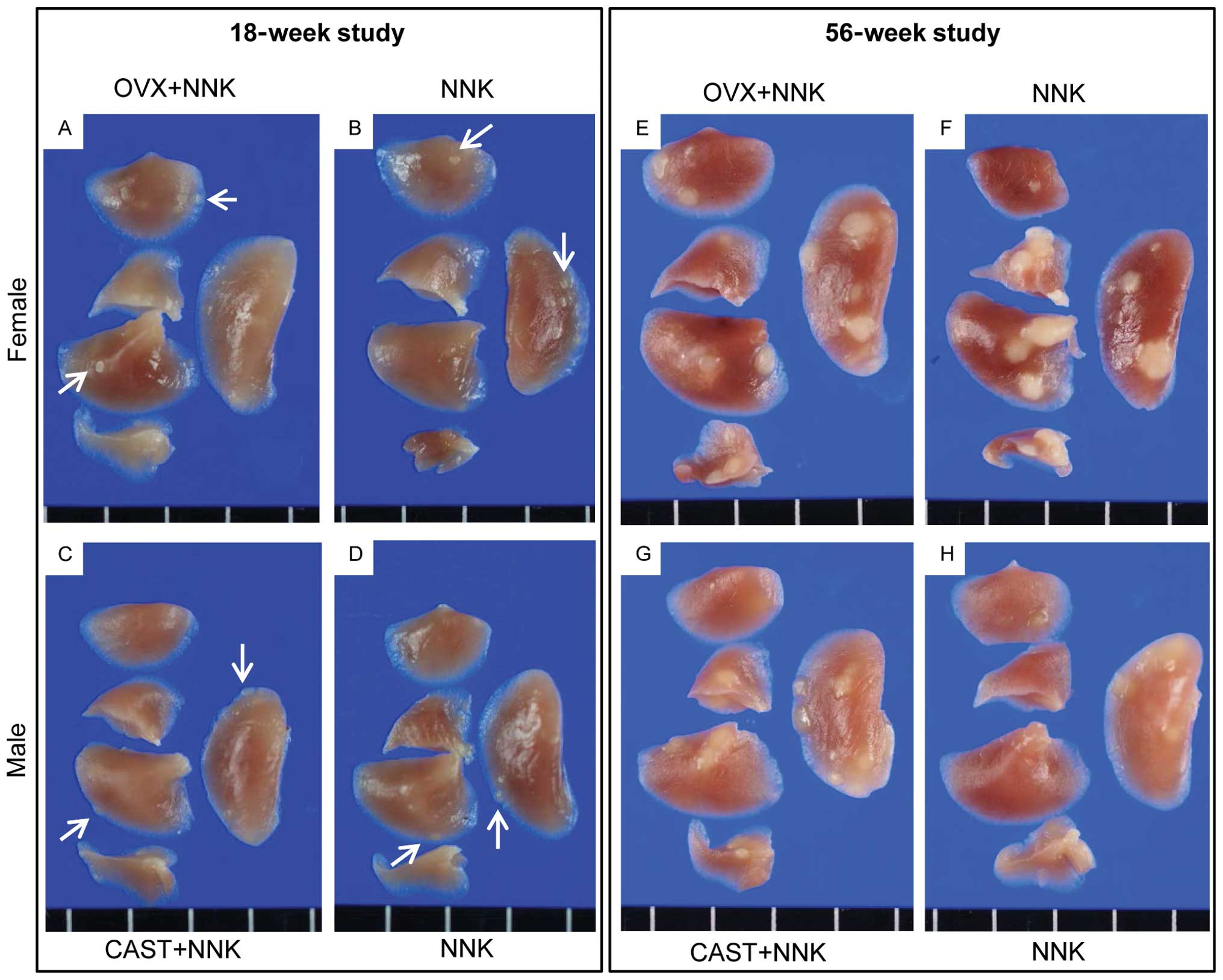 | Figure 1Macroscopical lung nodules. (A-D) In
the 18-week study, small white nodules were observed in all groups
(arrows indicate representative nodules in Groups 1–4). The sizes
of the nodules observed in the 56-week study (E-H) were clearly
larger than after 18 weeks (A-D). (A) Female ovariectomized group,
Group 1; (B) female unoperated group, Group 2; (C) male castrated
group, Group 3; (D) male unoperated group, Group 4; (E) female
ovariectomized group, Group 5; (F) female unoperated group, Group
6; (G) male castrated group, Group 7; (H) male unoperated group,
Group 8. OVX, ovariectomy; CAST, castration. |
| Table IIIIncidences and multiplicities of
macroscopical lung nodules. |
Table III
Incidences and multiplicities of
macroscopical lung nodules.
| | | | | Macroscopical
nodule |
|---|
| | | | |
|
|---|
| Group | Gendera | Treatmenta | Durationb | No.c | Incidence (%) |
Multiplicityd |
|---|
| 1 | F | NNKx2 OVX | 18 | 21 | 21/21 (100.0) | 17.2±8.4 |
| 2 | F | NNKx2 N | 18 | 15 | 15/15 (100.0) | 19.7±6.9e |
| 3 | M | NNKx2 CAST | 18 | 16 | 16/16 (100.0) | 14.3±6.2 |
| 4 | M | NNKx2 N | 18 | 15 | 15/15 (100.0) | 12.2±5.7 |
| 5 | F | NNKx1 OVX | 56 | 18 | 18/18 (100.0) | 18.2±7.6 |
| 6 | F | NNKx1 N | 56 | 12 | 12/12 (100.0) | 20.6±6.0f |
| 7 | M | NNKx1 CAST | 56 | 19 | 19/19 (100.0) | 12.1±4.6g |
| 8 | M | NNKx1 N | 56 | 14 | 14/14 (100.0) | 7.6±3.3 |
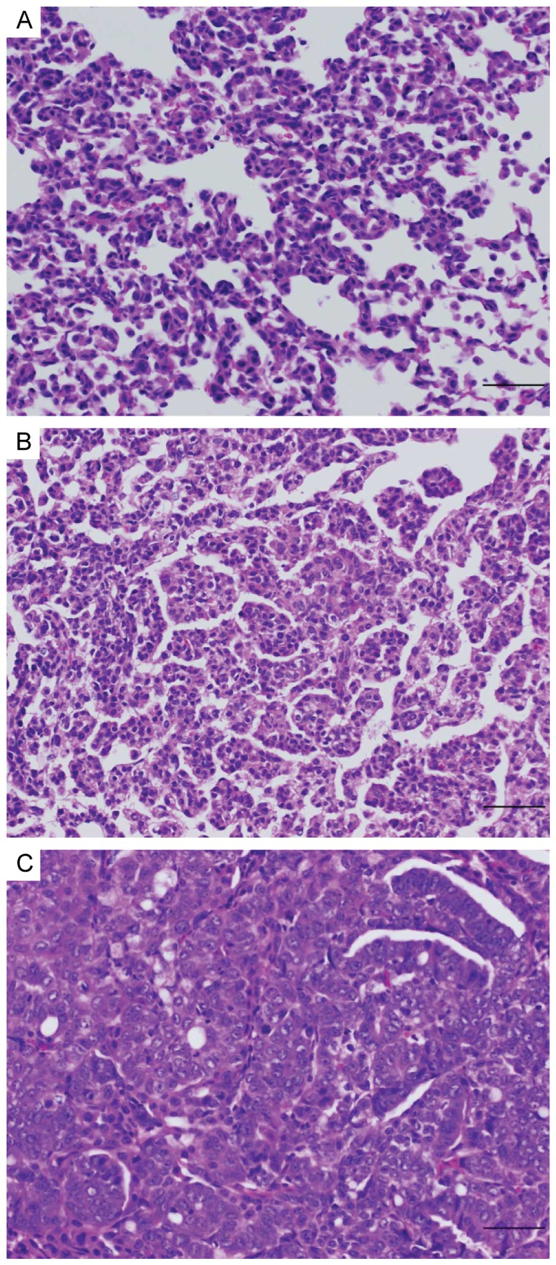
Histopathological analysis
In histopathological analysis of lung proliferative
lesions, bronchiolo-alveolar hyperplasias and adenomas were
observed in the 18-week study (Groups 1–4) and bronchiolo-alveolar
hyperplasias, adenomas and adenocarcinomas after 56 weeks (Groups
5–8) (Fig. 2). Incidences and
multiplicities of each proliferative lesion are summarized in
Table IV. The incidences showed no
significant intergroup variation in either 18- or 56-week studies.
In the 18-week study, the multiplicity of adenomas in the female
ovariectomized group (Group 1) was significantly lower than in the
female unoperated group (Group 2). In the 56-week study, the
multiplicities of hyperplasias, adenomas and tumors (adenomas and
adenocarcinomas) in the male castrated group (Group 7) were
significantly increased, and carcinomas showed a tendency to
increase, compared with the male unoperated group (Group 8). In
liver, a hepatocellular carcinoma was observed in only one case of
Group 5. No other lesions were observed in liver
histopathologically. There were no lesions detected in kidneys in
any of the groups. A lipoma surrounding the kidney and a thymoma
were observed in Group 5, each at incidences of 1/18 (6%). No other
tumors were observed in any of the groups.
| Table IVIncidences and multiplicities of lung
proliferative lesions. |
Table IV
Incidences and multiplicities of lung
proliferative lesions.
| | | | | Hyperplasia | Adenoma | Adenocarcinoma | Tumord |
|---|
| | | | |
|
|
|
|
|---|
| Group | Gendera | Treatmenta | Durationb | No.c | Incidence (%) |
Multiplicitye | Incidence | Multiplicity | Incidence | Multiplicity | Incidence | Multiplicity |
|---|
| 1 | F | NNKx2 OVX | 18 | 21 | 21/21 (100.0) | 4.8±3.6 | 18/21 (85.7) | 3.1±2.3f | 0/21 (0.0) | 0 | - | - |
| 2 | F | NNKx2 N | 18 | 15 | 14/15 (93.3) | 3.7±2.4 | 15/15 (100.0) | 5.8±3.7 | 0/15 (0.0) | 0 | - | - |
| 3 | M | NNKx2 CAST | 18 | 16 | 16/16 (100.0) | 5.3±2.9 | 14/16 (87.5) | 2.9±2.8 | 0/15 (0.0) | 0 | - | - |
| 4 | M | NNKx2 N | 18 | 15 | 13/15 (86.7) | 4.1±2.7 | 14/15 (93.3) | 3.3±2.0 | 0/15 (0.0) | 0 | - | - |
| 5 | F | NNKx1 OVX | 56 | 18 | 17/18 (94.4) | 5.3±3.5 | 18/18 (100.0) | 9.7±3.8 | 9/18 (50.0) | 0.9±1.0 | 18/18 (100.0) | 10.6±4.2 |
| 6 | F | NNKx1 N | 56 | 12 | 11/12 (91.7) | 4.8±2.7 | 12/12 (100.0) | 10.2±4.6 | 7/12 (58.3) | 0.8±0.8 | 12/12 (100.0) | 11.0±4.5 |
| 7 | M | NNKx1 CAST | 56 | 19 | 19/19 (100.0) | 4.5±2.2g | 19/19 (100.0) | 4.9±3.7g | 9/19 (47.4) | 0.7±1.1 | 19/19 (100.0) | 5.6±4.0g |
| 8 | M | NNKx1 N | 56 | 14 | 14/14 (100.0) | 3.1±1.1 | 12/14 (85.7) | 2.5±1.7 | 4/14 (28.6) | 0.3±0.5 | 12/14 (85.7) | 2.8±2.0 |
Discussion
In the present 18-week study, the serum
concentration of estradiol of female mice was significantly
decreased by ovariectomy, while the concentration of testosterone
was slightly increased, and the serum concentration of testosterone
of male mice was significantly decreased by castration. Estradiol
is one of the most important sex steroid hormones secreted by the
ovary and testosterone is a principal androgen secreted by the
testis (23,24). The decrease with gonadectomy was in
line with expectation and the increase in testosterone in
ovariectomized females may be due to the secretion by the adrenal
cortex. It is known that steroid sex hormones, particularly
androgen, are also secreted from adrenal cortex (25), and secretion may be augmented in
ovariectomized female mice as a reaction to ovariectomy (26). However, the concentration of
testosterone in ovariectomized female mice was much lower than that
in unoperated males.
In males, body weights showed no significant
difference with castration after both 18 and 56 weeks. In females
of the 56-week study, body weights in the operated group (Group 5)
were significantly increased and this seemed to be due to the
ovariectomy. In ovariectomized female mice, increase of body fat,
reduction of lipid metabolism and activation of lipid synthesis
have been reported (27–29). In males of the 18- and 56-week
studies, the liver and kidney weights of castrated groups (Groups 3
and 7) were significantly decreased compared with the unoperated
groups (Groups 4 and 8), and this may be due to the castration, as
similar weight loss has been reported in castrated CF-1 mice
(30). In females in the 18-week
study, the liver weight of the ovariectomized group (Group 1)
showed significant increase compared with the unoperated group
(Group 2), although no such difference was noted after 56 weeks. As
there is no report to support this, the liver weights may be
influenced by estradiol derived from the ovary.
In unoperated groups, the multiplicities of lung
nodules in females (Groups 2 and 6) were significantly greater than
in males (Groups 4 and 8), confirming female A/J mice to be more
susceptible to NNK-induced lung carcinogenesis than males. Our
results indicated that lung carcinogenesis was increased by
castration, suggesting the possibility that testosterone inhibits
NNK-induced lung carcinogenesis. In addition, the results of the
18-week study suggest that female sex hormones contribute to the
malignant transformation of lung proliferative lesions, such as
tumorous alteration from hyperplasia to adenoma. However, the
results of the 56-week study did not support this suggestion.
In humans, there have been several reports that sex
hormones affect incidences of lung adenocarcinoma. The
characteristics of gene mutations in lung cancer are reported to
differ between the sexes. In human lung cancer, tobacco-related p53
and EGFR mutations are more common in women than in men (20). Thus, there is a possibility that
lung carcinogenesis progresses through different pathways between
men and women. Estradiol influences the activity of various
metabolic enzymes, such as CYP2A6, CYP1A2, CYP3A4, CYP2C19,
UDP-glucuronyltransferase, which can activate NNK to ultimate
carcinogenic species (2,31,32).
In women, nicotine metabolism by CYP2A6 is reported to be
accelerated by estrogen (33).
Previously, we demonstrated that CYP2A6 plays important roles in
NNK-induced lung carcinogenesis (5–9). In
addition, oral contraceptives (estrogen) increase drug metabolism
by glucuronidation (34–36). However, there are also reports that
oral contraceptives decelerate drug metabolism by CYP1A2, CYP3A4
and CYP2C19 (37–39). Whatever the case, it appears clear
that metabolic enzymes of NNK are influenced by estradiol. There is
also the possibility that androgen influences lung carcinogenesis.
Androgen receptors are present in human lung adenocarcinomas and
normal lung tissue of humans and mice, and their expression in
normal lung tissue of mice may be affected by castration and
testosterone administration (40).
In male mice of the present study, tumors (adenomas
and adenocarcinomas) were significantly increased in the castrated
group only after 56 weeks, indicating that it is necessary to use
long-term experiments in order to determine modifying potential of
male sex hormones on lung carcinogenesis. The present study, in
fact, pointed to the possibility that NNK-induced lung
carcinogenesis may be inhibited by testosterone and accelerated by
estradiol, although the effect was only slight in females. One
explanation is that lung carcinogenic effects of NNK are so strong
in females, that modifying effects of estradiol are masked.
In conclusion, female A/J mice were confirmed to be
more susceptible to NNK-induced lung carcinogenesis than males. In
males, lung carcinogenesis was increased by castration, whereas in
females, malignant transformation of lung proliferative lesions
tended to be inhibited by ovariectomy. These results suggested that
NNK-induced lung carcinogenesis is inhibited by testosterone and
accelerated by estradiol. These findings indicate the possibility
that sex hormones play important roles in determining sex
differences in lung carcinogenesis in A/J mice initiated by NNK.
Additional experiments are ongoing to confirm the effects of the
sex hormones themselves.
Acknowledgements
We thank Dr Malcolm A. Moore for his help in the
critical reading of this manuscript.
References
|
1
|
Balbo S, Upadhyaya P, Villalta PW, Qian X
and Kassie F: DNA adducts in aldehyde dehydrogenase-positive lung
stem cells of A/J mice treated with the tobacco specific lung
carcinogen 4-(methylnitrosamino)-1-(3-pyridyl)-1-butanone (NNK).
Chem Res Toxicol. Mar 12–2013.(Epub ahead of print).
|
|
2
|
Akopyan G and Bonavida B: Understanding
tobacco smoke carcinogen NNK and lung tumorigenesis (Review). Int J
Oncol. 29:745–752. 2006.PubMed/NCBI
|
|
3
|
Chen RJ, Chang LW, Lin P and Wang YJ:
Epigenetic effects and molecular mechanisms of tumorigenesis
induced by cigarette smoke: an overview. J Oncol.
2011:6549312011.PubMed/NCBI
|
|
4
|
Belinsky SA, Devereux TR, Foley JF,
Maronpot RR and Anderson MW: Role of the alveolar type II cell in
the development and progression of pulmonary tumors induced by
4-(methylnitrosamino)-1-(3-pyridyl)-1-butanone in the A/J mouse.
Cancer Res. 52:3164–3173. 1992.PubMed/NCBI
|
|
5
|
Imaida K, Yokohira M and Kuno T: Detection
of carcinogenic and modifying potentials by test compounds using a
mouse lung carcinogenesis bioassay (Review). J Toxicol Pathol.
20:117–123. 2007. View Article : Google Scholar
|
|
6
|
Takeuchi H, Saoo K, Matsuda Y, et al:
8-Methoxypsoralen, a potent human CYP2A6 inhibitor, inhibits lung
adenocarcinoma development induced by
4-(methylnitrosamino)-1-(3-pyridyl)-1-butanone in female A/J mice.
Mol Med Rep. 2:585–588. 2009.
|
|
7
|
Takeuchi H, Saoo K, Matsuda Y, et al: Dose
dependent inhibitory effects of dietary 8-methoxypsoralen on
NNK-induced lung tumorigenesis in female A/J mice. Cancer Lett.
234:232–238. 2006. View Article : Google Scholar : PubMed/NCBI
|
|
8
|
Takeuchi H, Saoo K, Yokohira M, et al:
Pretreatment with 8-methoxypsoralen, a potent human CYP2A6
inhibitor, strongly inhibits lung tumorigenesis induced by
4-(methylnitrosamino)-1-(3-pyridyl)-1-butanone in female A/J mice.
Cancer Res. 63:7581–7583. 2003.
|
|
9
|
Yokohira M, Takeuchi H, Saoo K, et al:
Establishment of a bioassay model for lung cancer chemoprevention
initiated with 4-(methylnitrosamino)-1-(3-pyridyl)-1-butanone (NNK)
in female A/J mice. Exp Toxicol Pathol. 60:469–473. 2008.
View Article : Google Scholar : PubMed/NCBI
|
|
10
|
Kuno T, Yokohira M, Matsuda Y, et al: Lack
of modifying potential of 8-methoxypsoralen in the promotion or
progression stages of lung carcinogenesis in A/J female mice. Oncol
Rep. 20:767–772. 2008.PubMed/NCBI
|
|
11
|
Matsuda Y, Saoo K, Hosokawa K, et al:
Post-initiation chemopreventive effects of dietary bovine
lactoferrin on
4-(methylnitrosamino)-1-(3-pyridyl)-1-butanone-induced lung
tumorigenesis in female A/J mice. Cancer Lett. 246:41–46. 2007.
View Article : Google Scholar : PubMed/NCBI
|
|
12
|
Yamakawa K, Kuno T, Hashimoto N, et al:
Molecular analysis of carcinogen-induced rodent lung tumors:
Involvement of microRNA expression and Kras or Egfr mutations. Mol
Med Rep. 3:141–147. 2010.PubMed/NCBI
|
|
13
|
Yokohira M, Hashimoto N, Yamakawa K, Saoo
K, Kuno T and Imaida K: Lack of promoting effects from physical
pulmonary collapse in a female A/J mouse lung tumor initiated with
4-(methylnitrosamino)-1-(3-pyridyl)-1-butanone (NNK) with
remarkable mesothelial cell reactions in the thoracic cavity by the
polymer. Exp Toxicol Pathol. 63:181–185. 2011. View Article : Google Scholar
|
|
14
|
Yokohira M, Hashimoto N, Yamakawa K, et
al: Lack of modifying effects of intratracheal instillation of
quartz or dextran sulfate sodium (DSS) in drinking water on lung
tumor development initiated with
4-(Methylnitrosamino)-1-(3-pyridyl)-1-butanone (NNK) in female A/J
mice. J Toxicol Pathol. 22:179–185. 2009. View Article : Google Scholar : PubMed/NCBI
|
|
15
|
Miyazaki M, Yamazaki H, Takeuchi H, et al:
Mechanisms of chemopreventive effects of 8-methoxypsoralen against
4-(methylnitrosamino)-1-(3-pyridyl)-1-butanone-induced mouse lung
adenomas. Carcinogenesis. 26:1947–1955. 2005. View Article : Google Scholar : PubMed/NCBI
|
|
16
|
Igarashi M, Watanabe M, Yoshida M, et al:
Enhancement of lung carcinogenesis initiated with
4-(N-hydroxymethylnitrosamino)-1-(3-pyridyl)-1-butanone by
Ogg1 gene deficiency in female, but not male, mice. J
Toxicol Sci. 34:163–174. 2009.
|
|
17
|
Morita T: A statistical study of lung
cancer in the annual of pathological autopsy cases in Japan, from
1958 to 1997, with reference to time trends of lung cancer in the
world. Jpn J Cancer Res. 93:15–23. 2002. View Article : Google Scholar : PubMed/NCBI
|
|
18
|
Sobue T, Ajiki W, Tsukuma H, Oshima A,
Hanai A and Fujimoto I: Trends of lung cancer incidence by
histologic type: a population-based study in Osaka, Japan. Jpn J
Cancer Res. 90:6–15. 1999. View Article : Google Scholar : PubMed/NCBI
|
|
19
|
Zang EA and Wynder EL: Differences in lung
cancer risk between men and women: examination of the evidence. J
Natl Cancer Inst. 88:183–192. 1996. View Article : Google Scholar : PubMed/NCBI
|
|
20
|
Belani CP, Marts S, Schiller J and
Socinski MA: Women and lung cancer: epidemiology, tumor biology,
and emerging trends in clinical research. Lung Cancer. 55:15–23.
2007. View Article : Google Scholar : PubMed/NCBI
|
|
21
|
Nishio M, Ohyanagi F, Horiike A, et al:
Gefitinib treatment affects androgen levels in non-small-cell lung
cancer patients. Br J Cancer. 92:1877–1880. 2005. View Article : Google Scholar : PubMed/NCBI
|
|
22
|
Mohr U: International Classification of
Rodent Tumors: The Mouse. 1st edition. Springer-Verlag; Berlin:
2001, View Article : Google Scholar
|
|
23
|
de Kretser DM: The Testis. Reproduction in
Mammals: Hormonal Control of Reproduction. Austin CR and Short RV:
3. 2nd edition. Cambridge University Press; Cambridge: pp. 76–90.
1984
|
|
24
|
Baird DT: The Ovary. Reproduction in
Mammals: Hormonal Control of Reproduction. Austin CR and Short RV:
3. 2nd edition. Cambridge University Press; Cambridge: pp. 91–114.
1984, View Article : Google Scholar
|
|
25
|
Pelletier G, Luu-The V, Li S and Labrie F:
Localization of type 5 17β-hydroxysteroid dehydrogenase mRNA in
mouse tissues as studied by in situ hybridization. Cell Tissue Res.
320:393–398. 2005.
|
|
26
|
Matsuura S and Suzuki K: Morphological
changes in the submandibular glands and in the X zone of the
adrenal gland following ovariectomy in mice. Cell Tissue Res.
246:549–556. 1986. View Article : Google Scholar : PubMed/NCBI
|
|
27
|
Kamei Y, Suzuki M, Miyazaki H, et al:
Ovariectomy in mice decreases lipid metabolism-related gene
expression in adipose tissue and skeletal muscle with increased
body fat. J Nutr Sci Vitaminol. 51:110–117. 2005. View Article : Google Scholar : PubMed/NCBI
|
|
28
|
Wu J, Wang X, Chiba H, et al: Combined
intervention of soy isoflavone and moderate exercise prevents body
fat elevation and bone loss in ovariectomized mice. Metabolism.
53:942–948. 2004. View Article : Google Scholar : PubMed/NCBI
|
|
29
|
Yamaguchi M, Katoh S, Morimoto C, et al:
The hormonal responses of lipoprotein lipase activity and lipolysis
in adipose tissue differ depending on the stage of the estrous
cycle in female rats. Int J Obes Relat Metab Disord. 26:610–617.
2002. View Article : Google Scholar : PubMed/NCBI
|
|
30
|
Duffel MW, Graham JM and Ziegler DM:
Changes in dimethylaniline N-oxidase activity of mouse liver and
kidney induced by steroid sex hormones. Mol Pharmacol. 19:134–139.
1981.PubMed/NCBI
|
|
31
|
Kamataki T, Fujita KI, Nakayama K,
Yamazaki Y, Miyamoto M and Ariyoshi N: Role of human cytochrome
P450 (CYP) in the metabolic activation of nitrosamine derivatives:
application of genetically engineered Salmonella expressing
human CYP. Drug Metab Rev. 34:667–676. 2002. View Article : Google Scholar : PubMed/NCBI
|
|
32
|
Wiener D, Doerge DR, Fang JL, Upadhyaya P
and Lazarus P: Characterization of N-glucuronidation of the
lung carcinogen 4-(methylnitrosamino)-1-(3-pyridyl)-1-butanol
(NNAL) in human liver: importance of UDP-glucuronosyltransferase
1A4. Drug Metab Dispos. 32:72–79. 2004.
|
|
33
|
Benowitz NL, Lessov-Schlaggar CN, Swan GE
and Jacob P III: Female sex and oral contraceptive use accelerate
nicotine metabolism. Clinl Pharmacol Ther. 79:480–488. 2006.
View Article : Google Scholar : PubMed/NCBI
|
|
34
|
Miners JO, Grgurinovich N, Whitehead AG,
Robson RA and Birkett DJ: Influence of gender and oral
contraceptive steroids on the metabolism of salicytic acid and
acetylsalicylic acid. Br J Clin Pharmacol. 22:135–142. 1986.
View Article : Google Scholar : PubMed/NCBI
|
|
35
|
Mitchell MC, Hanew T, Meredith CG and
Schenker S: Effects of oral contraceptive steroids on acetaminophen
metabolism and elimination. Clin Pharmacol Ther. 34:48–53. 1983.
View Article : Google Scholar : PubMed/NCBI
|
|
36
|
Stoehr GP, Kroboth PD, Juhl RP, Wender DB,
Phillips JP and Smith RB: Effect of oral contraceptives on
triazolam, temazepam, alprazolam, and lorazepam kinetics. Clin
Pharmacol Ther. 36:683–690. 1984. View Article : Google Scholar : PubMed/NCBI
|
|
37
|
Balogh A, Klinger G, Henschel L, Börner A,
Vollanth R and Kuhnz W: Influence of ethinylestradiol-containing
combination oral contraceptives with gestodene or levonorgestrel on
caffeine elimination. Eur J Clin Pharmacol. 48:161–166. 1995.
View Article : Google Scholar : PubMed/NCBI
|
|
38
|
Laine K, Tybring G and Bertilsson L: No
sex-related differences but significant inhibition by oral
contraceptives of CYP2C19 activity as measured by the probe drugs
mephenytoin and omeprazole in healthy Swedish white subjects. Clin
Pharmacol Ther. 68:151–159. 2000. View Article : Google Scholar
|
|
39
|
Slayter KL, Ludwig EA, Lew KH, Middleton E
Jr, Ferry JJ and Jusko WJ: Oral contraceptive effects on
methylprednisolone pharmacokinetics and pharmacodynamics. Clin
Pharmacol Ther. 59:312–321. 1996. View Article : Google Scholar : PubMed/NCBI
|
|
40
|
Mikkonen L, Pihlajamaa P, Sahu B, Zhang FP
and Jänne OA: Androgen receptor and androgen-dependent gene
expression in lung. Mol Cell Endocrinol. 317:14–24. 2010.
View Article : Google Scholar : PubMed/NCBI
|