Introduction
Lung cancer is the leading cause of cancer-related
mortality worldwide (1). The
prognosis of lung cancer patients is poor despite the development
of several new therapeutic modalities, and this may be attributed
to the difficulties associated with the detection of lung cancer in
its early stages. Therefore, new approaches and alternative
diagnostic modalities are urgently needed.
Squamous cell carcinoma (SCC) of the lung, a major
type of non-small cell lung carcinoma (NSCLC), is more strongly
linked with smoking than other forms of NSCLC (2,3). The
genes responsible for lung SCC (LSCC) have been identified, but the
molecular origins of LSCC remain unclear. In our studies on the
mechanism of tumorigenesis in LSCC, we focused on protein tyrosine
kinase 7 (PTK7), which has been previously reported to play a key
role in carcinogenesis (4–8).
PTK7, also known as colon carcinoma kinase-4
(CCK-4), is a catalytically inactive receptor tyrosine kinase,
cloned from colon carcinoma tissue (4). It is composed of a transmembrane
region, seven immunoglobulin domains, and a C-terminal domain that
shows homology to tyrosine kinases.
PTK7 expression has been shown to be high in several
tumors, including gastric, colon, esophageal cancer, and
liposarcoma (4–7). PTK7 knockdown is known to inhibit
proliferation of liposarcoma cells, esophageal cancer cells, and
colon cancer cells, and silencing of PTK7 in colon cancer cells
induces caspase-10-dependent apoptosis via the mitochondrial
pathway (6–8). These studies suggested the probability
of an oncogenic role of PTK7 in gastric, colon and esophageal
cancer, as well as in liposarcoma. However, PTK7 has been reported
to be downregulated in clear cell renal cell carcinoma, metastatic
melanoma and breast cancer cell lines (9–11).
Taken together, these previous findings suggested
that PTK7 could exert dual effects (upregulation or
downregulation), depending on specific tissues or tumors, although
the exact mechanism is unknown. In this study, we attempted to
explore the role of PTK7 in lung cancer.
Materials and methods
Recruitment of patients and preparation
of tissue samples
This study was approved by the institutional review
board of the Korea Cancer Center Hospital. For RT-PCR and western
blot analysis, frozen samples of tumor and adjacent normal lung
tissue were obtained from 12 lung cancer patients who underwent
curative surgery for LSCC at the Korea Cancer Center Hospital.
Pathologic staging was performed based on the recommendations in
the 7th edition of the report of the American Joint
Committee on Cancer.
Cell lines
Human LSCC cell lines, HCC-1588 and HCC-95, were
purchased from the Korean Cell Line Bank (http://cellbank.snu.ac.kr); SW900, SK-MES-1, Calu-1
and the normal human lung fibroblast cell line WI-38 were purchased
from the American Type Culture Collection. HCC-1588 and HCC-95 were
maintained in RPMI-1640 medium supplemented with 10% fetal bovine
serum (FBS) and 100 U/ml penicillin and streptomycin. SK-MES-1 and
WI-38 cells were maintained in EMEM medium supplemented with 10%
FBS and 100 U/ml penicillin and streptomycin. SW900 and Calu-1
cells were maintained in L-15 and McCoy’s 5A media supplemented
with 10% FBS and 100 U/ml penicillin and streptomycin,
respectively. All cells were cultured at 37°C in the presence of 5%
CO2.
Transfection
Cells were transfected with 2 μg of pcDNA3
(Invitrogen, Carlsbad, CA, USA) and pcDNA3-PTK7-Flag (12) by using FuGENE HD reagents (Promega,
Madison, WI, USA) according to the manufacturer’s instructions.
RNA isolation and RT-PCR
Total RNA was isolated from frozen lung samples (SCC
and normal lung tissues) and cell lines using an RNeasy Mini Kit
(Qiagen, Cambridge, MA, USA) according to the manufacturer’s
protocol. Single-stranded cDNA was produced by
reverse-transcription of total RNA (2 μg) by using an oligo-dT
primer and Superscript II reverse transcriptase (Invitrogen).
RT-PCR was performed (13) with the
following sets of synthesized primers: PTK7 forward,
5′-AGAGATGCCCCATGGTGGGC-3′ and reverse, 5′-ACGG
CTTGCTGTCCACGGTG-3′. GAPDH-specific primers were used as internal
controls.
Cell proliferation assay
Cell growth was determined by the MTS assay using
the CellTiter 96®Aqueous One Solution Cell Proliferation
Assay kit (Promega). Cells (2×10³/well) were seeded in
96-well plates after transfection. After incubation for 24, 48, 72
and 96 h, 20 μl of MTS solution was added to each well. Plates were
incubated for an additional 1–2 h at 37°C in a 5% CO2
environment, and then the absorbance at 490 nm was recorded using a
microplate reader (Biotek, Winooski, VT, USA) to calculate the
percentage of surviving cells.
Migration and invasion assay
Cells (5×10⁵/well) were seeded in a
24-well plate and incubated with fresh medium. After overnight
incubation, a wound was introduced by scraping the monolayer with a
0.2-ml micropipette tip. The attached cells were washed twice with
PBS to remove debris, incubated for 48 h in RPMI-1640 supplemented
with 10% FBS, and observed by light microscopy. The invasion assay
was performed using BioCot™ Matrigel™ invasion chamber (8 μm,
24-well; BD Biosciences, Franklin Lakes, NJ, USA). Cells were
starved in serum-free medium overnight, trypsinized. Then, cells
were suspended in FBS-free RPMI-1640 (2×10³ cells) and
were seeded into the upper compartment of the chamber, and 600 μl
of RPMI-1640 containing 10% FBS was placed in the lower chamber.
After incubation for 24 h or 48 h at 37°C in a 5% CO2,
the cells remaining in the upper compartment of the chamber were
removed. Cells on the lower surface of the chamber were fixed using
4% paraformaldehyde and stained with 0.5% crystal violet.
Western blot analysis
Proteins from frozen tissues and cell lines were
extracted using RIPA buffer (Thermo Fisher Scientific, Waltham, MA,
USA) containing protease inhibitors (Roche, Basel, Switzerland).
Lysates containing the same amounts of protein were separated on
10% SDS-polyacrylamide gels and transferred onto nitrocellulose or
PVDF membranes. Western blotting was performed as previously
described (14) using the following
antibodies: anti-phospho-ERK, anti-ERK, and anti-β-actin (Santa
Cruz Biotechnology, Santa Cruz, CA, USA); anti-phospho-AKT
(Ser473), and anti-AKT (Cell Signaling Technology, Beverly, MA,
USA) primary antibodies and horseradish peroxidase-conjugated
secondary antibodies (Santa Cruz Biotechnology). Rabbit anti-PTK7
anti-serum, which recognizes both human and murine PTK7, was used
as previously described (13). The
immune reactions were visualized using Immobilon Western
Chemiluminescent HRP Substrate (Millipore, Bedford, MA, USA) and a
LAS-3000 imaging system (Fuji, Tokyo, Japan).
Statistical analysis
Each experiment was performed at least three times.
All values are reported as mean ± SD. Differences between
experimental groups and controls were assessed by Tukey’s test
using SigmaStat (SPSS, Inc., Chicago, IL, USA). The level of
statistical significance was set at P<0.05.
Results
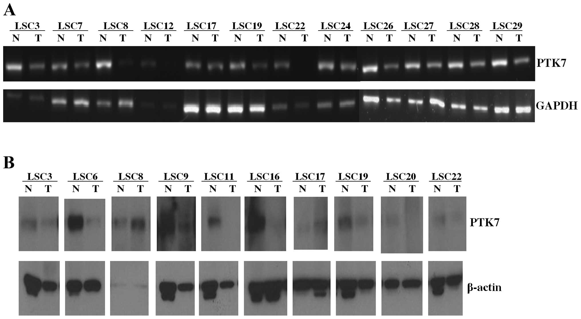
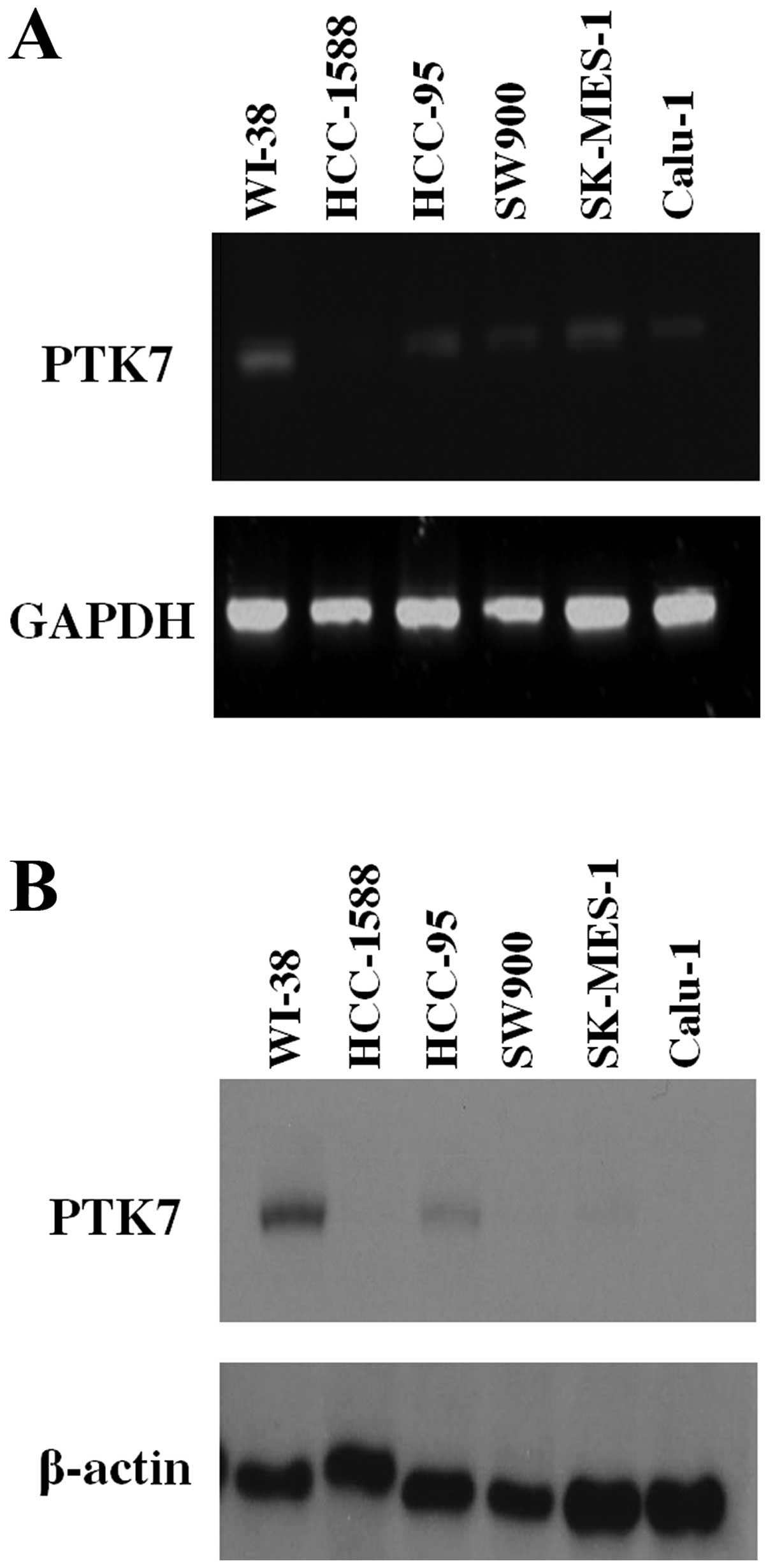
Low expression of PTK7 in LSCC patient
samples and LSCC cell lines
The expression of PTK7 in LSCC tissues was low at
the mRNA level in 11/12 samples and at the protein level in 8/10
samples (Fig. 1). The LSCC cell
lines also showed a significantly lower expression of PTK7 than the
normal lung cell line WI-38 (Fig.
2). These results showed that PTK7 is downregulated in both
LSCC tissues and LSCC cell lines.
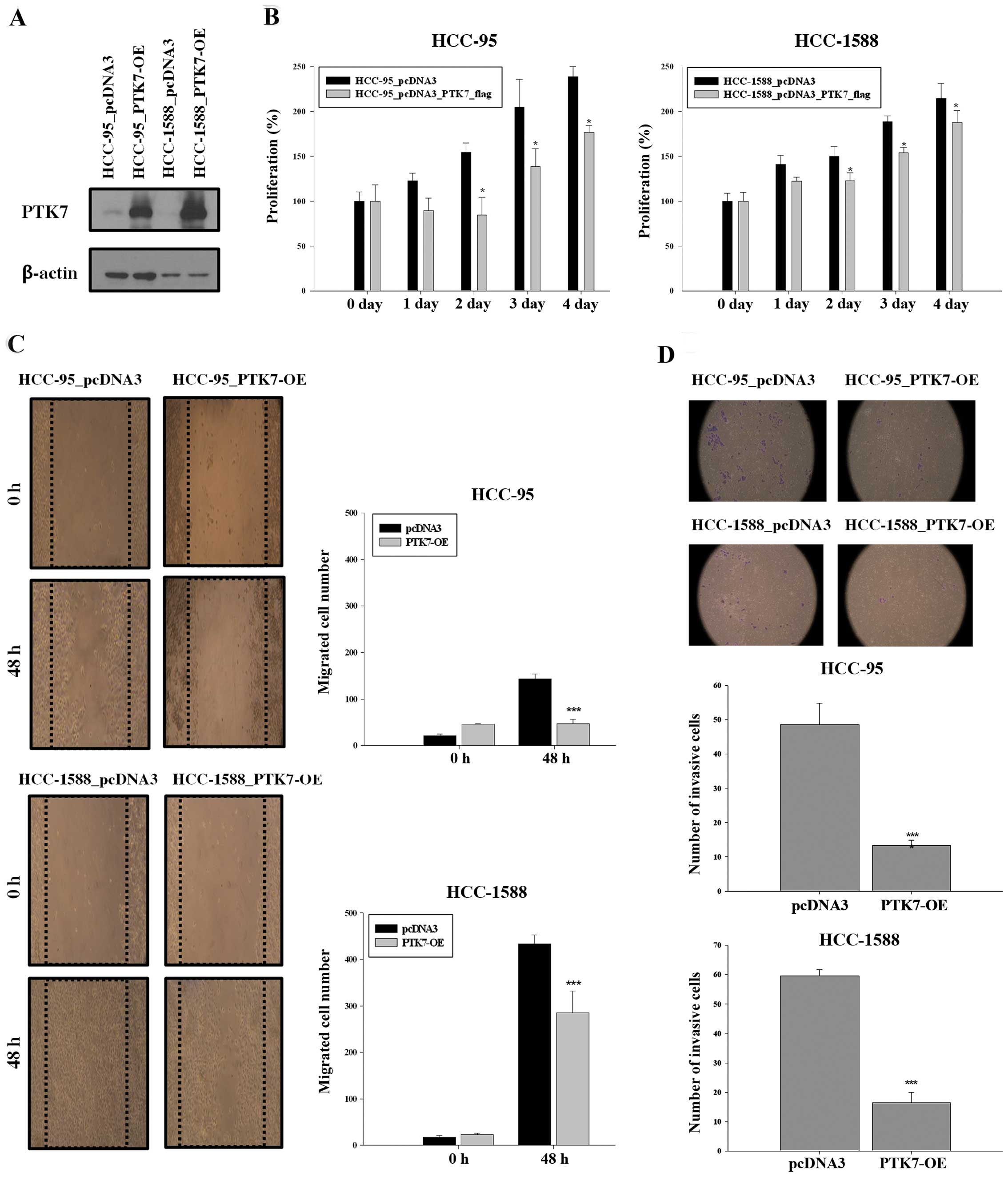
Overexpression of PTK7 in transfected
LSCC cells inhibits cell proliferation, wound healing and
invasion
To confirm the biological role of PTK7 in LSCC, we
transfected the LSCC cell lines HCC-95 and HCC-1588 with a
PTK7-expression vector pcDNA3-PTK7-flag and with an empty vector
pcDNA3. The results of the western blot analysis of the cell lines
transfected with the PTK7-expression vector showed overexpression
of PTK7 (Fig. 3A). The effect of
this PTK7 overexpression on the proliferation of LSCC cells was
evaluated by the MTS assay. The results of the MTS assay showed
that the proliferation of cells transfected with the
PTK7-expression vector was significantly less than that of cells
transfected with the empty vector (P<0.05; Fig. 3B). We then investigated the role of
PTK7 in wound healing and invasion of HCC-95 and HCC-1588 cells. In
the wound healing assay, control cells repaired the wound in 48 h,
whereas the PTK7-overexpressing cells showed significantly delayed
wound repair (Fig. 3C). The
invasive potential of the PTK7-overexpressing HCC-95 and HCC-1588
cells was 73% lower than that of control cells (Fig. 3D). The effect of PTK7 overexpression
on LSCC cell growth was also assessed by the colony formation
assay; however, both control and PTK7-overexpressing cells showed
similar growth rates (data not shown).
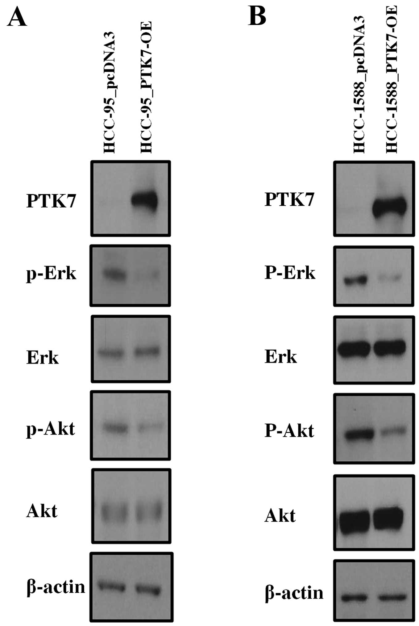
Overexpression of PTK7 inhibits the
activation of ERK and AKT in LSCC cells
We conducted a microarray analysis to investigate
genetic alterations in PTK7-overexpressed LSCC cell lines. Results
of this analysis revealed mutations in the genes coding for AKT and
ERK, p38, and JNK-enzymes involved in the mitogen activated protein
kinase (MAPK) pathways (data not shown). This finding led us to
consider that these pathways likely act together to regulate
PTK7-influenced cell proliferation, invasion and migration.
Therefore, we investigated the activation of AKT and MAPKs in
PTK7-overexpressed LSCC cells. In both HCC-95 and HCC-1588 cells,
PTK7 overexpression decreased the phosphorylation levels of AKT and
ERK (Fig. 4). However, activation
of JNK and p38 were found to be similar in control as well as in
PTK7 overexpressing cells (data not shown). These results suggest
that PTK7 effectively modulates signaling pathways involving AKT
and ERK, which are important determinants of cell proliferation,
migration and invasion in LSCC cells.
Discussion
Several studies have reported the increase in PTK7
expression in gastric cancer, colon cancer, liposarcoma and
esophageal cancer (4–7). We have also previously reported a
correlation between high expression of PTK7 and poor prognosis in
esophageal cancer and that knockdown of PTK7 in esophageal cancer
cells inhibits cell proliferation, survival, invasion and migration
through inhibition of AKT, ERK, FAK, JNK, and p38 MAPK (6). In addition, suppression of PTK7 has
been shown to inhibit cell proliferation and to induce apoptosis
via the mitochondria pathway in colon cancer cells (8). These results suggest that PTK could be
regarded as an oncogene (5–8).
On the other hand, PTK7 has been shown to be
downregulated in several types of cancer, including clear cell
renal cell carcinoma, metastatic melanoma and pulmonary
adenocarcinoma (9,10,15).
Easty et al (10) explored
the probable correlation between loss of PTK7 expression in
melanoma cells and tumorigenicity. In addition, Endoh et al
(15) and Garber et al
(16) reported that decreased
expression of PTK7 is correlated with poor clinical outcome. These
findings suggested that PTK7 expression may be upregulated or
downregulated, depending on the organ and tumor and that disease
prognosis would differ according to the rate of PTK7 expression.
However, expression of PTK7 and its role as a tumor suppressor in
LSCC have not been studied.
In the current study, we showed that PTK7 expression
is downregulated by approximately >80% in tumor cells from
tissue samples from LSCC patients in comparison to that observed in
normal tissues (control) at both mRNA and protein levels. PTK7
expression was also low at both levels in the LSCC cell lines.
Based on these results, we sought to link PTK7 expression to tumor
suppression in LSCC. In the present study, we found that PTK7
overexpression decreases the proliferation and inhibits wound
healing and invasion of HCC-95 and HCC-1588 LSCC cells, although no
significant difference in growth was observed between control and
PTK7-overexpressing cells. Moreover, we found that overexpression
of PTK7 decreased the phosphorylation of AKT and ERK in LSCC cells.
AKT and MAPK are important signaling cascades involved in cell
proliferation, tumor invasion and migration (17–20).
Therefore, we suggest that PTK7 may be involved in tumor and
metastasis repression in LSCC, particularly through inactivation of
AKT and MAPK, which promote proliferation, invasion and
migration.
In conclusion, our results show that PTK7 is
downregulated in clinical samples of LSCC and that it plays a tumor
suppressor role (via the AKT and MAPK pathways) in LSCC cells.
However, further studies are required to elucidate the tissue and
organ-specific differences in the oncogenic patterns of PTK7 and to
determine their clinical significance in LSCC.
Acknowledgements
This work was supported by grants from the National
R&D Program for Cancer Control (1120260 to J.H.P), Ministry of
Health and Welfare of the Republic of Korea, and from the
Radiological Translational Research Program (RTR), Korea Institute
of Radiological & Medical Sciences (KIRAMS 50455-2013).
References
|
1
|
Parkin DM, Bray F, Ferlay J and Pisani P:
Global cancer statistics, 2002. CA Cancer J Clin. 55:74–108. 2005.
View Article : Google Scholar
|
|
2
|
Koyi H, Hillerdal G and Brandėn E: A
prospective study of a total material of lung cancer from a county
in Sweden 1997–1999: gender, symptoms, type, stage, and smoking
habits. Lung Cancer. 36:9–14. 2002.PubMed/NCBI
|
|
3
|
Visbal AL, Williams BA, Nichols FC 3rd,
Marks RS, Jett JR, Aubry MC, Edell ES, Wampfler JA, Molina JR and
Yang P: Gender differences in non-small-cell lung cancer survival:
an analysis of 4,618 patients diagnosed between 1997 and 2002. Ann
Thorac Surg. 78:209–215. 2004. View Article : Google Scholar : PubMed/NCBI
|
|
4
|
Mossie K, Jallal B, Alves F, Sures I,
Plowman GD and Ullrich A: Colon carcinoma kinase-4 defines a new
subclass of the receptor tyrosine kinase family. Oncogene.
11:2179–2184. 1995.PubMed/NCBI
|
|
5
|
Lin Y, Zhang L, Wang X, Xing X, Cheng X,
Dong B, Hu Y, Du H, Li Y and Zhu Y: PTK7 as a novel marker for
favorable gastric cancer patient survival. J Surg Oncol.
106:880–886. 2012. View Article : Google Scholar : PubMed/NCBI
|
|
6
|
Shin WS, Kwon JH, Lee HW, Kang MC, Na HW,
Lee ST and Park JH: The oncogenic role of PTK7 in esophageal
squamous cell carcinoma. Cancer Sci. 104:1120–1126. 2013.
View Article : Google Scholar : PubMed/NCBI
|
|
7
|
Gobble RM, Qin L, Brill ER, Angeles CV,
Ugras S, O’Connor RB, Moraco NH, DeCarolis PL, Antonescu C and
Singer S: Expression profiling of liposarcoma yields a multigene
predictor of patient outcome and identifies genes that contribute
to liposarcomagenesis. Cancer Res. 71:2697–2705. 2011. View Article : Google Scholar : PubMed/NCBI
|
|
8
|
Meng L, Sefah K, O’Donoghue MB, Zhu G,
Shangguan D, Noorali A, Chen Y, Zhou L and Tan W: Silencing of PTK7
in colon cancer cells: caspase-10-dependent apoptosis via
mitochondrial pathway. PLoS One. 5:e140182010. View Article : Google Scholar : PubMed/NCBI
|
|
9
|
Behbahani TE, Thierse C, Baumann C, Holl
D, Bastian PJ, von Ruecker A, Müller SC, Ellinger J and Hauser S:
Tyrosine kinase expression profile in clear cell renal cell
carcinoma. World J Urol. 30:559–565. 2012. View Article : Google Scholar : PubMed/NCBI
|
|
10
|
Easty DJ, Mitchell PJ, Patel K, Florenes
VA, Spritz RA and Bennett DC: Loss of expression of receptor
tyrosine kinase family genes PTK7 and SEK in metastatic melanoma.
World J Urol. 71:1061–1065. 1997.PubMed/NCBI
|
|
11
|
Su YA, Yang J, Tao L, Nguyen H and He P:
Undetectable and decreased expression of KIAA1949 (Phostensin)
encoded on chromosome 6p21.33 in human breast cancers revealed by
transcriptome analysis. J Cancer. 38:38–50. 2010. View Article : Google Scholar : PubMed/NCBI
|
|
12
|
Na HW, Shin WS, Ludwig A and Lee ST: The
cytosolic domain of protein-tyrosine kinase 7 (PTK7), generated
from sequential cleavage by a disintegrin and metalloprotease 17
(ADAM17) and γ-secretase, enhances cell proliferation and migration
in colon cancer cells. J Biol Chem. 287:25001–25009.
2012.PubMed/NCBI
|
|
13
|
Shin WS, Maeng YS, Jung JW, Min JK, Kwon
YG and Lee ST: Soluble PTK7 inhibits tube formation, migration, and
invasion of endothelial cells and angiogenesis. Biochem Biophys Res
Commun. 371:793–798. 2008. View Article : Google Scholar : PubMed/NCBI
|
|
14
|
Lee SJ, Yoo HJ, Bae YS, Kim HJ and Lee ST:
TIMP-1 inhibits apoptosis in breast carcinoma cells via a pathway
involving pertussis toxin-sensitive G protein and c-Src. Biochem
Biophys Res Commun. 312:1196–1201. 2003. View Article : Google Scholar : PubMed/NCBI
|
|
15
|
Endoh H, Tomida S, Yatabe Y, Konishi H,
Osada H, Tajima K, Kuwano H, Takahashi T and Mitsudomi T:
Prognostic model of pulmonary adenocarcinoma by expression
profiling of eight genes as determined by quantitative real-time
reverse transcriptase polymerase chain reaction. J Clin Oncol.
22:811–819. 2004. View Article : Google Scholar
|
|
16
|
Garber ME, Troyanskaya OG, Schluens K,
Petersen S, Thaesler Z, Pacyna-Gengelbach M, van de Rijn M, Rosen
GD, Perou CM and Whyte RI: Diversity of gene expression in
adenocarcinoma of the lung. Proc Natl Acad Sci USA. 98:13784–13789.
2001. View Article : Google Scholar : PubMed/NCBI
|
|
17
|
Manning BD and Cantley LC: AKT/PKB
signaling: navigating downstream. Cell. 129:1261–1274. 2007.
View Article : Google Scholar : PubMed/NCBI
|
|
18
|
Johnson GL and Lapadat R:
Mitogen-activated protein kinase pathways mediated by ERK, JNK, and
p38 protein kinases. Science. 298:1911–1912. 2002. View Article : Google Scholar : PubMed/NCBI
|
|
19
|
Shin I, Kim S, Song H, Kim HR and Moon A:
H-Ras-specific activation of Rac-MKK3/6-p38 pathway: its critical
role in invasion and migration of breast epithelial cells. J Biol
Chem. 280:14675–14683. 2005. View Article : Google Scholar : PubMed/NCBI
|
|
20
|
Huang C, Jacobson K and Schaller MD: MAP
kinases and cell migration. J Cell Sci. 117:4619–4628. 2004.
View Article : Google Scholar : PubMed/NCBI
|