Introduction
On a global scale, gastric cancer (GC) remains the
fourth most common malignancy and the second leading cause of
cancer-associated mortality (1).
Surgical resection remains the mainstay of curative treatment.
However, the majority of patients are diagnosed at an advanced
stage when surgery is no longer feasible (2). Consequently, cytotoxic chemotherapy
has been proven to be an effective treatment. Nevertheless, drug
resistance and therapy-associated side-effects remain issues of
concern (3). Thus, more effective
antitumor drugs with fewer side-effects for the treatment of GC are
required.
A diverse range of phytochemicals have confirmed the
capacity to selectively inhibit the growth of tumor cells (4–8).
Flavonoids are polyphenolic compounds that have a wide range of
biological activities, including antioxidant ability,
anti-inflammatory capacity and ability to combat cancer (9,10).
Kaempferol is one of the general flavonoids that is often present
in tea, broccoli, apples, strawberries and beans (11). It has received much attention due to
its anticancer potential, preferable biocompatibility and few
side-effects. Kaempferol has been found to inhibit proliferation,
angiogenesis and metastasis, induce cell cycle arrest and promote
apoptosis in a variety of human cancer cell lines (12–19).
However, the potential role of these phytochemicals in GC treatment
has yet to be evaluated. Therefore, we investigated the therapeutic
potential and molecular mechanisms of kaempferol on GC cells in
vitro and in vivo.
Results of the present study demonstrated that
kaempferol potently inhibits the proliferation of GC cells and
induces apoptosis via the mitochondrial pathway in vitro and
in vivo. G2/M arrest of GC cells was also observed and the
expression levels of G2/M cell cycle regulating factors, cyclin B1,
Cdk1 and Cdc25C, were decreased following kaempferol treatment.
Additionally, we observed that kaempferol inhibited the ERK1/2 and
PI3K/AKT signaling pathway. In conclusion, the present study
results revealed the therapeutic potential of kaempferol in GC, and
we demonstrated several possible mechanisms that may be significant
in the activity of kaempferol, although this remains to be
determined in future studies.
Materials and methods
Cell lines, reagents and antibodies
MKN28, SGC7901 and GSE-1 cells were purchased from
the Cell Resource Center of Shanghai Institutes for Biological
Sciences, Chinese Academy of Sciences (Shanghai, China). All the
cells were cultured in RPMI-1640 medium (Gibco-BRL) supplemented
with 10% fetal bovine serum (FBS) at 37°C and 5% CO2.
Kaempferol was purchased from Winherb Medical S&T Development
Co., Ltd. (Shanghai, China). Primary antibodies against Bcl-2, Bax,
survivin, Bcl-xL, caspase-3, cleaved-caspase-3, caspase-9,
cleaved-caspase-9, PARP, cleaved-PARP, p-Akt, Akt, cyclin B1, Cdk1,
Cdc25C, GAPDH and secondary antibodies against mouse
IgG-horseradish peroxidase (HRP) and rabbit IgG-HRP were obtained
from Santa Cruz Biotechnology, Inc. (Santa Cruz, CA, USA).
Antibodies against ERK1/2, phospho-ERK1/2, Ki67 and COX-2 were
purchased from Cell Signaling Technology, Inc. (Trask Lane,
Danvers, MA, USA).
Cell viability assay
To investigate cell viability MKN28, SGC7901 and
GSE-1 cells were seeded in 96-well plates at a density of
3×10³/well and cultured for 24 h. After validation of
cell adherence, the cells were treated with different doses of
kaempferol for 24, 48 or 72 h. A Cell Counting Kit-8 (CCK-8;
Dojindo Molecular Technologies, Kumamoto, Japan) was used to assess
the cell viability. Cell viability was calculated as a percentage
of absorbance in treated wells relative to that of untreated wells.
Three independent experiments were performed.
Cell cycle analysis
Cells were seeded in 6-well plates at
4×10⁵/dish. After incubation with kaempferol (60 or 120
μM) for 48 h, the cells were trypsinized and washed twice with
phosphate-buffered saline (PBS). The cells were then cultured with
reagents A-C according to the manufacturer’s instructions and
subjected to flow cytometry.
Analysis of apoptosis
GC cells (4×10⁵ cells/well) were
incubated with kaempferol (60 or 120 μM) for 48 h. Cells
(1×10⁴) were collected and washed twice with cold PBS.
Apoptotic cells were evaluated by double staining with propidium
iodide (PI) and Annexin V labeled with FITC using an Annexin V-FITC
apoptosis detection kit (BD Biosciences, San Jose, CA, USA)
according to the manufacturer’s instructions.
Western blotting
MKN28 and SGC7901 cells were plated at a density of
4×10⁵/well in 6-well plates. After incubation with
kaempferol (60 or 120 μM) the cells were washed twice with ice-cold
PBS and treated with 120 μl sample buffer on ice for 30 min. The
cell lysate was centrifuged at 12,000 rpm for 10 min at 4°C.
Protein lysates (20 μl) were electrophoresed on a 12 or 10% SDS
gel. The proteins were then electrotransferred to a PVDF membrane
and the membrane was blocked for 30 min with blocking solution (5%
non-fat dry milk in PBS-0.5% Tween-20). The membrane was then
incubated overnight at 4°C with primary antibodies (1:1,000).
Subsequently, the membrane was washed in PBST for 30 min, exposed
to HRP-conjugated secondary antibody (diluted 1:2,000), and washed
again in PBST for 30 min. Final detection was performed using
enhanced chemiluminescence solution for 5 min.
Ki-67 immunohistochemistry
Formalin-fixed, paraffin-embedded sections (5 mm)
were rinsed with PBS, blocked with 10% bovine serum albumin for 30
min and then stained with an anti-Ki-67 antibody overnight. The
sections were subsequently incubated for 1 h with the appropriate
secondary antibody, and immunoreactivity was developed with
SigmaFAST DAB. Positively stained cells from three tumors/group
were counted in 10 randomly selected fields under ×400 high-power
magnification. A proliferative index (%) was calculated using the
formula: Number of Ki-67-positive cells/total cell count.
Tumor xenograft experiments
SGC7901 cells (3×10⁶) were injected into
the flanks of 4-week-old athymic mice to establish the tumor. When
the tumor volume reached 100 mm3, kaempferol (20 mg/kg)
was administered i.p. for 3 weeks daily. The mice in the treatment
and control groups (n=8 in each group) were sacrificed to obtain
the snap-frozen and paraffin-embedded tumor tissue for further
analysis. Body weight was recorded starting from the first day of
treatment, and tumor volumes were also calculated at the same time
points using the equation: Tumor volume = length ×
(width)2 × π/6. Operative procedures and care were
approved by the Institutional Ethics Committee at Harbin Medical
University. All the experiments were performed in accordance with
the guidelines of the Committee on the Use of Live Animals in
Teaching and Research of Harbin Medical University, Harbin,
China.
Statistical analysis
Data were presented as mean values ± standard
deviation (SD). Analysis of variance (ANOVA) and a Student’s t-test
were used to evaluate statistical significance. P<0.05 was
considered to indicate a statistically significant result.
Results
Kaempferol inhibits GC cell
proliferation
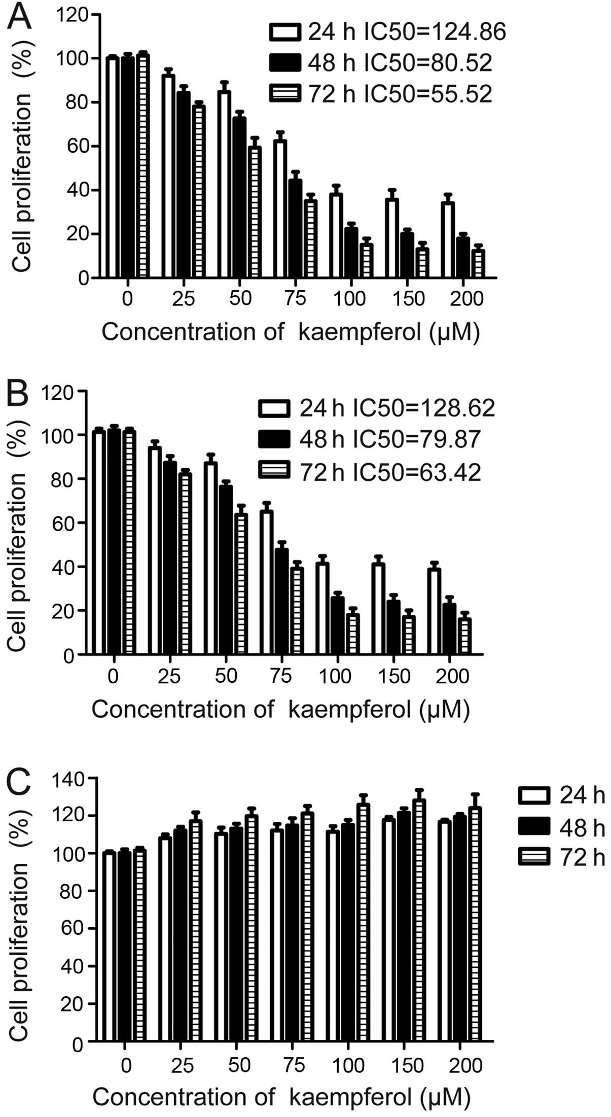
First, we assessed the effects of kaempferol on the
cell proliferation of GC cell lines. We used MKN28 and SGC7901 GC
cell lines, which are widely utilized in GC investigations to
examine the effects of kaempferol. We also used a GSE-1 cell line
to determine whether kaempferol had the same effects on normal
gastric epithelial cells. The results showed that kaempferol
significantly inhibited the proliferation of all the GC cell lines
within a period of 24–72 h (P<0.05), and these effects were more
apparent at a dose of 120 μM (Fig. 1A
and B). However, no marked inhibition was observed in the GSE-1
cell line with our experimental dose (Fig. 1C).
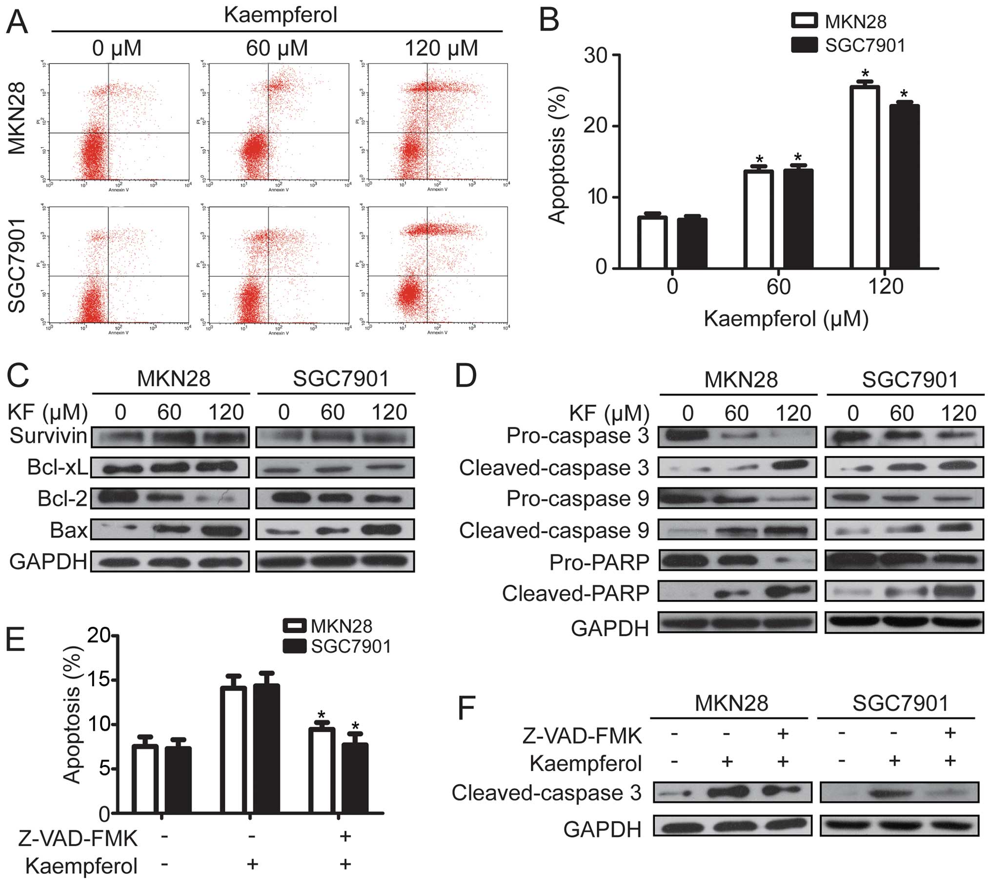
Kaempferol induces apoptosis in GC cells. To
determine whether kaempferol suppressed cell proliferation by
inducing apoptosis, we used an Annexin V/PI assay as described
above to investigate the apoptotic effect on GC cells. The results
showed that kaempferol induced the apoptosis of SGC-7901 and MKN28
cells in a dose-dependent manner (Fig.
2A and B). When the concentration of kaempferol reached 60 μM,
the apoptotic rate of the GC cell lines was markedly higher than
that of the untreated cells (P<0.05). Moreover, 120 μM
kaempferol resulted in a higly significant difference in the rate
of apoptosis compared to the kaempferol-treated (60 μM) and
untreated cells (P<0.05). As apoptosis is regulated by a variety
of pro- and anti-apoptotic proteins (20–22),
we investigated the expression levels of apoptosis-associated
proteins, including Bcl-2, Bax, Bcl-xL and survivin. The results
showed that kaempferol decreased the expression of Bcl-2 and
concomitantly increased the expression of Bax in a
concentration-dependent manner (Fig.
2C). However, no significant change of Bcl-xL and survivin was
observed in the GC cell line. Caspase family proteins are important
enzymes to execute apoptosis.
We also assessed the role of kaempferol in the
caspase cascade pathway. Western blotting showed that kaempferol
treatment lead to a dose-dependent elevation of cleaved caspase-3
and -9, and cleaved-PARP, and a dose-dependent decrease of
pro-caspase-3 and -9, and pro-PARP (Fig. 2D). To further investigate the role
of caspase activation in kaempferol-induced apoptosis, we treated
SGC-7901 and MKN28 cells with pan-caspase inhibitor Z-VAD-FMK (10
mmol/l) before kaempferol treatment. The pan-caspase inhibitor
Z-VAD-FMK pretreatment reduced the expression of cleaved caspase-3
and kaempferol-induced apoptosis (Fig.
2E and F). These data demonstrated that kaempferol induced the
apoptosis of GC cells by regulating the expression levels of
apoptosis-related proteins and partly activating caspase-dependent
cell death pathway.
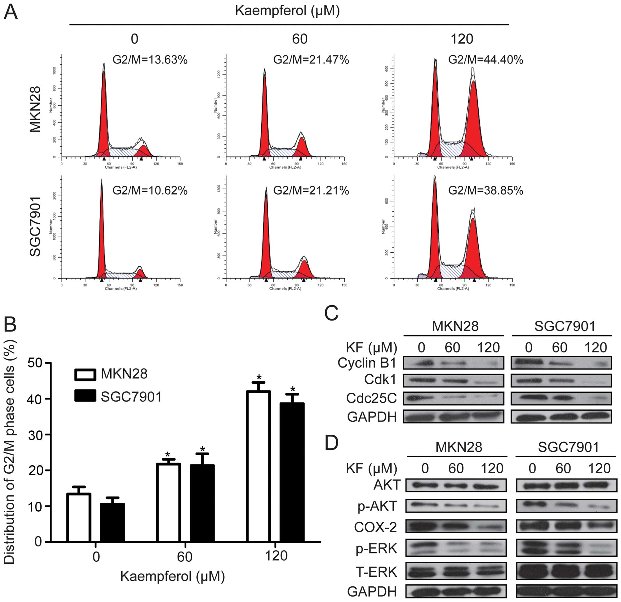
Kaempferol induces G2/M cell cycle arrest
and inhibits tumor cell survival signaling in GC cells
To examine the underlying mechanism of reduced cell
viability observed in the CCK-8 assay, we examined the cell
distribution by flow cytometry following treatment with 60 or 120
μM kaempferol for 48 h. Kaempferol treatment for 48 h arrested
cells at the G2/M stage (Fig. 3A and
B). To investigate the mechanism for G2/M arrest in
kaempferol-treated cells, we detected the expression of proteins
that are pivotal for G2/M transition, including cyclin B1, Cdk1 and
Cdc25C. The results showed that kaempferol led to a marked decrease
in the protein levels of cyclin B1, Cdk1 and Cdc25C in a
dose-dependent manner (Fig. 3C).
These data revealed that the inhibitory effect of kaempferol on GC
was associated with the induction of G2/M phase arrest. Our data
indicated that in vitro kaempferol treatment significantly
reduced the expression levels of COX-2, p-AKT and p-ERK, which were
involved in cell proliferation and cell cycle arrest. However,
kaempferol had no impact on the level of total Akt and ERK
(Fig. 3D). These findings revealed
that kaempferol suppresses tumor cell growth via the PI3K/AKT or
ERK-MAPK pathway.
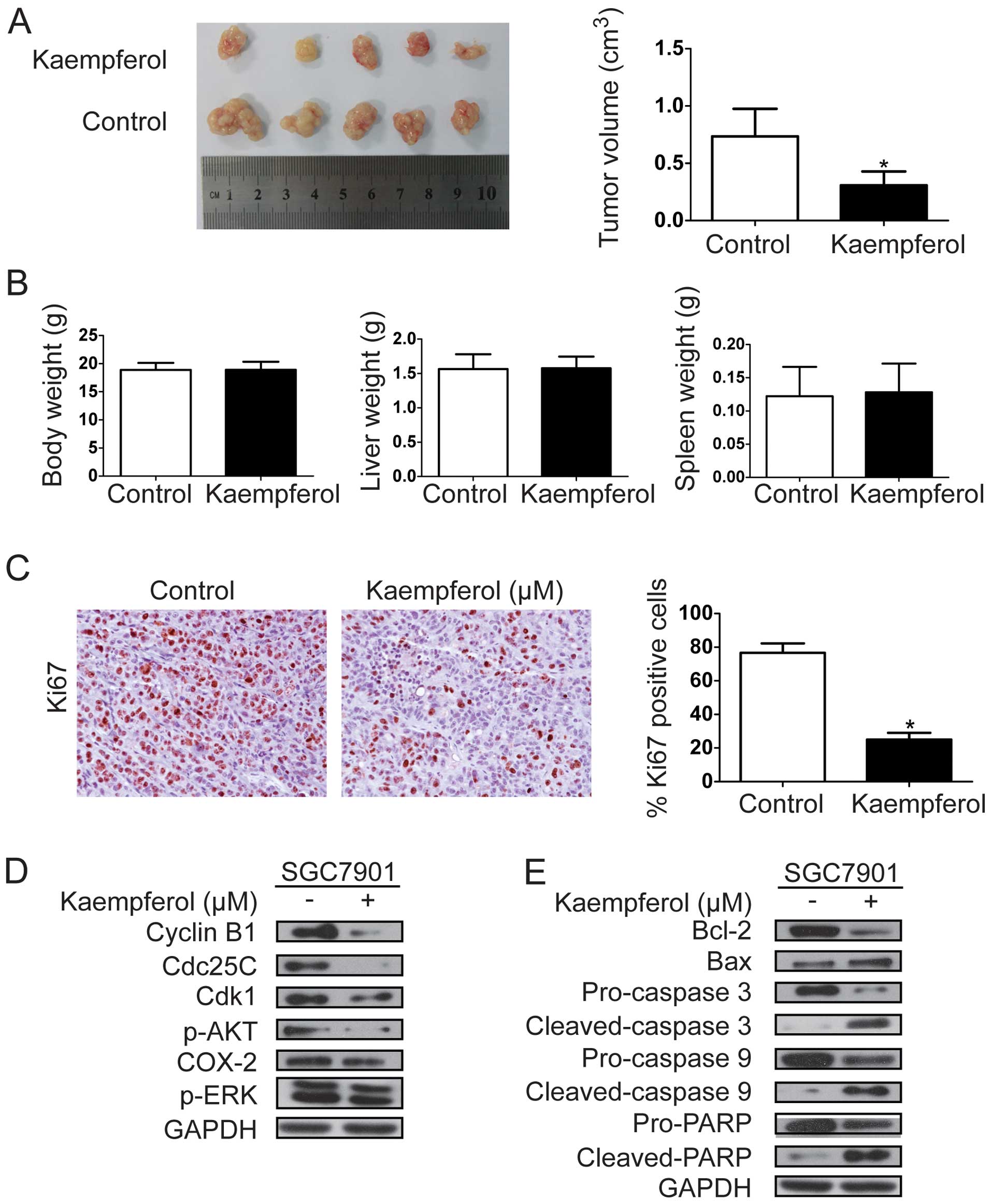
Kaempferol suppresses tumor growth in
vivo
We established a subcutaneous tumor model, which was
derived from the SGC7901 cell line, to examine the role of
kaempferol in tumor growth in vivo. SGC7901 cell-derived
xenograft tumors were allowed to develop and grow to a size of 100
mm3, and then kaempferol (20 mg/kg/day) was administered
i.p. for 3 weeks daily. The mice were sacrificed following 3 weeks
of kaempferol treatment and tumors were excised. Data showed that
kaempferol significantly suppressed the growth of the tumor
xenografts (Fig. 4A). However, no
marked change was observed in body, liver or spleen weight in the
animals (Fig. 4B). The results
revealed that kaempferol was a potential therapeutic drug for the
treatment of GC and it was relatively non-toxic to mice. Ki-67
staining for cell proliferation was performed in these xenografts,
and the number of Ki-67-positive tumor cells was lower in tumors
from kaempferol-treated mice than that from non-kaempferol-treated
mice (Fig. 4C). Western blot
analysis revealed that kaempferol treatment decreased the
expression of cyclin B1, Cdk1, Cdc25C, Bcl-2/Bax ratio, p-AKT,
P-ERK and increased the expression of cleaved caspase-3 and -9, and
cleaved PARP (Fig. 4D and E). These
findings demonstrated that kaempferol significantly suppressed GC
cell growth in vivo.
Discussion
Despite recent advancement in revealing the
tumorigenetic mechanism of gastric cancer (GC), the ever expanding
incidence and relatively low relief rate of chemotherapy have led
to identification of a more efficacious treatment method. The
present study shows that kaempferol significantly suppressed the
proliferation of GC cells. Moreover, it did not influence the
proliferation of normal gastric GSE-1 cells when treated with a
concentration of 120 μM.
Bcl-2 family proteins, including pro-apoptotic
proteins (Bid, Bak and Bax) and anti-apoptotic proteins (Bcl-2,
cIAP-2, XIAP, Bcl-xL and survivin), are critical in the control of
apoptosis (23,24). The ratio of anti- and pro-apoptotic
protein expression, such as Bcl-2/Bax, is crucial for the induction
of apoptosis, and it determines the susceptibility of cells to
undergo apoptosis (25). However,
Bcl-2 family proteins conduct the release of cytochrome c
from the mitochondria into cytosol, which lead to the activation of
caspase-9 and induces a subsequent caspase cascade (the intrinsic
cell death pathway). Luo et al showed that kaempferol
induced apoptosis in ovarian cancer cells through the intrinsic
apoptosis pathway (16). In the
present study, we observed that kaempferol increased the expression
of Bax and decreased the expression of Bcl-2 in SGC-7901 and MKN28
cells. Moreover, the result of kaempferol-induced activation of
caspase-3 and -9, and subsequent cleavage of PARP, as well as the
data that the pan-caspase inhibitor Z-VAD-FMK decreased
kaempferol-mediated apoptosis in SGC-7901 and MKN28 cells, indicate
that kaempferol induced the apoptosis of GC cells partly through a
mitochondrial cell death pathway.
Eukaryotic cell cycle progression is involved in the
successive activation of Cdks whose activation is dependent on
their conjunction with cyclins. A complex shaped by the conjunction
of Cdk1 and cyclin B1 plays an important role at entry into mitosis
(26). Choi and Ahn showed that
kaempferol induced G2/M phase cell cycle arrest in MDA-MB-453 human
breast cancer cells (13). The
results of the present study showed that the treatment of MKN28 and
SGC7901 cells with kaempferol led to the arrest of GC cells in G2/M
phase and that kaempferol-mediated G2/M arrest is connected with a
decrease in the protein levels of Cdk1, cyclin B1 and Cdc25C.
Therefore, kaempferol may induce cell cycle arrest by decreasing
activity of the Cdk1/cyclin B kinase complex through downregulation
of various G2/M-associated proteins.
The PI3K-Akt signaling pathway is a crucial
regulator of a number of cell processes including proliferation,
differentiation and metastasis in cancer development (27). Activated AKT phosphorylates numerous
proteins that have been involved in the control of the cell cycle
to ultimately lead to cell growth and suppress apoptosis (28). As kaempferol can block the PI3K/AKT
pathway in mouse epidermal JB6P+ cells by neutralizing
PI3K (29), we determined whether
the effect of kaempferol on GC cells was associated with the
inhibition of this pathway as well. Our data showed that the
expression level of p-Akt was reduced in a dose-dependent manner,
without any changes in the total Akt protein level following
kaempferol treatment. ERK is known to be involved in the promotion
of cell proliferation and is generally upregulated in many cancers,
including GC. Results of this study also showed a marked
dose-dependent reduced ERK phosphorylation in cells treated with
kaempferol. Therefore, we suggest that kaempferol suppresses GC
through the PI3K/AKT and ERK-MAPK pathway, highlighting a potential
mechanism for kaempferol activity, which may be used as a
therapeutic agent for GC. Nevertheless, the exact mechanism should
be further investigated.
The in vivo antitumor effect of kaempferol
was analyzed in a SGC7901 xenograft tumor model in nude mice. An
apparent decrease in the relative tumor volume was observed in
kaempferol-treated mice compared to non-kaempferol-treated
controls. However, a significant inhibition of proliferation was
observed on the results of immunohistochemistry for Ki-67 in
kaempferol-treated mice. Of note, kaempferol treatment did not
affect liver, spleen or total body weight. Although kaempferol
seemed to have potent antitumor activity with few side effects in
the present study, absorption and pharmacokinetic properties of
kaempferol need to be further investigated to confirm kaempferol as
an efficacious therapy for GC.
Kaempferol, which is a natural flavonoid present in
various fruits and vegetables, exerts antitumor activity in a
variety of cancer cells. The antitumor role of kaempferol and its
underlying mechanisms was assessed in the MKN28 and SGC7901 GC cell
lines. Kaempferol was found to suppress the proliferation of the
two GC cell lines, however, no significant inhibition effect was
observed in normal gastric epithelial cell line, GSE-1. G2/M phase
cell cycle arrest and apoptosis were observed by using flow
cytometry. Accordingly, kaempferol treatment downregulated the
expression of G2/M cell cycle-associated proteins cyclin B1, Cdk1
and Cdc25C. Kaempferol treatment also upregulated the expression of
Bax concomitant with a decrease in Bcl-2 and increased the
expression of cleaved caspase-3 and -9, and cleaved PARP. We also
observed that kaempferol decreased the protein level of p-Akt and
p-ERK in MKN28 and SGC7901 cells. In vivo kaempferol
significantly suppressed the growth of the tumor xenografts with no
marked change in liver, spleen or body weight and protein
expression data in vitro were further confirmed. In
conclusion, our data provide a basis for further inquiry of
kaempferol as a therapeutic agent for GC.
Acknowledgements
We would like to thank Dr Y.H. Gu for his support in
the pathobiology examination. This study was supported by
Foundation of Health and Family Planning Commission of Heilongjiang
Province.
Abbreviations:
|
GC
|
gastric cancer
|
|
KF
|
kaempferol
|
|
IC50
|
half-maximal inhibitory
concentration
|
References
|
1
|
Alberts SR, Cervantes A and van de Velde
CJ: Gastric cancer: epidemiology, pathology and treatment. Ann
Oncol. 14(Suppl 2): ii31–ii36. 2003. View Article : Google Scholar : PubMed/NCBI
|
|
2
|
Hundahl SA, Menck HR, Mansour EG and
Winchester DP: The National Cancer Data Base report on gastric
carcinoma. Cancer. 80:2333–2341. 1997. View Article : Google Scholar : PubMed/NCBI
|
|
3
|
Ajani JA: Evolving chemotherapy for
advanced gastric cancer. Oncologist. 10(Suppl 3): S49–S58. 2005.
View Article : Google Scholar
|
|
4
|
Aggarwal BB and Shishodia S: Molecular
targets of dietary agents for prevention and therapy of cancer.
Biochem Pharmacol. 71:1397–1421. 2006. View Article : Google Scholar : PubMed/NCBI
|
|
5
|
Naithani R, Huma LC, Moriarty RM,
McCormick DL and Mehta G: Comprehensive review of cancer
chemopreventive agents evaluated in experimental carcinogenesis
models and clinical trials. Curr Med Chem. 15:1044–1071. 2008.
View Article : Google Scholar : PubMed/NCBI
|
|
6
|
Kaefer CM and Milner JA: The role of herbs
and spices in cancer prevention. J Nutr Biochem. 19:347–361. 2008.
View Article : Google Scholar : PubMed/NCBI
|
|
7
|
Russo GL: Ins and outs of dietary
phytochemicals in cancer chemoprevention. Biochem Pharmacol.
74:533–544. 2007. View Article : Google Scholar : PubMed/NCBI
|
|
8
|
Moiseeva EP and Manson MM: Dietary
chemopreventive phytochemicals: too little or too much? Cancer Prev
Res. 2:611–616. 2009. View Article : Google Scholar
|
|
9
|
Seifried HE, Anderson DE, Fisher EI and
Milner JA: A review of the interaction among dietary antioxidants
and reactive oxygen species. J Nutr Biochem. 18:567–579. 2007.
View Article : Google Scholar : PubMed/NCBI
|
|
10
|
Luo H, Jiang BH, King SM and Chen YC:
Inhibition of cell growth and VEGF expression in ovarian cancer
cells by flavonoids. Nutr Cancer. 60:800–809. 2008. View Article : Google Scholar : PubMed/NCBI
|
|
11
|
Somerset SM and Johannot L: Dietary
flavonoid sources in Australian adults. Nutr Cancer. 60:442–449.
2008. View Article : Google Scholar : PubMed/NCBI
|
|
12
|
Zhang Y, Chen AY, Li M, Chen C and Yao Q:
Ginkgo biloba extract kaempferol inhibits cell proliferation and
induces apoptosis in pancreatic cancer cells. J Surg Res.
148:17–23. 2008. View Article : Google Scholar : PubMed/NCBI
|
|
13
|
Choi EJ and Ahn WS: Kaempferol induced the
apoptosis via cell cycle arrest in human breast cancer MDA-MB-453
cells. Nutr Res Pract. 2:322–325. 2008. View Article : Google Scholar : PubMed/NCBI
|
|
14
|
Luo H, Rankin GO, Liu L, Daddysman MK,
Jiang BH and Chen YC: Kaempferol inhibits angiogenesis and VEGF
expression through both HIF dependent and independent pathways in
human ovarian cancer cells. Nutr Cancer. 61:554–563. 2009.
View Article : Google Scholar : PubMed/NCBI
|
|
15
|
Luo H, Daddysman MK, Rankin GO, Jiang BH
and Chen YC: Kaempferol enhances cisplatin’s effect on ovarian
cancer cells through promoting apoptosis caused by down regulation
of cMyc. Cancer Cell Int. 10:162010. View Article : Google Scholar
|
|
16
|
Luo H, Rankin GO, Li Z, Depriest L and
Chen YC: Kaempferol induces apoptosis in ovarian cancer cells
through activating p53 in the intrinsic pathway. Food Chem.
128:513–519. 2011. View Article : Google Scholar : PubMed/NCBI
|
|
17
|
Nguyen TT, Tran E, Ong CK, et al:
Kaempferol-induced growth inhibition and apoptosis in A549 lung
cancer cells is mediated by activation of MEK-MAPK. J Cell Physiol.
197:110–121. 2003. View Article : Google Scholar : PubMed/NCBI
|
|
18
|
Chen HJ, Lin CM, Lee CY, et al: Kaempferol
suppresses cell metastasis via inhibition of the ERK-p38-JNK and
AP-1 signaling pathways in U-2 OS human osteosarcoma cells. Oncol
Rep. 30:925–932. 2013.PubMed/NCBI
|
|
19
|
Song W, Dang Q, Xu D, et al: Kaempferol
induces cell cycle arrest and apoptosis in renal cell carcinoma
through EGFR/p38 signaling. Oncol Rep. 31:1350–1356.
2014.PubMed/NCBI
|
|
20
|
Debatin KM: Apoptosis pathways in cancer
and cancer therapy. Cancer Immunol Immunother. 53:153–159. 2004.
View Article : Google Scholar : PubMed/NCBI
|
|
21
|
Fisher DE: Pathways of apoptosis and the
modulation of cell death in cancer. Hematol Oncol Clin North Am.
15:931–956. ix2001. View Article : Google Scholar
|
|
22
|
Fulda S and Debatin KM: Targeting
apoptosis pathways in cancer therapy. Curr Cancer Drug Targets.
4:569–576. 2004. View Article : Google Scholar : PubMed/NCBI
|
|
23
|
Susnow N, Zeng L, Margineantu D and
Hockenbery DM: Bcl-2 family proteins as regulators of oxidative
stress. Semin Cancer Biol. 19:42–49. 2009. View Article : Google Scholar : PubMed/NCBI
|
|
24
|
Daniel PT, Schulze-Osthoff K, Belka C and
Güner D: Guardians of cell death: the Bcl-2 family proteins. Essays
Biochem. 39:73–88. 2003.PubMed/NCBI
|
|
25
|
Cory S and Adams JM: The Bcl2 family:
regulators of the cellular life-or-death switch. Nat Rev Cancer.
2:647–656. 2002. View
Article : Google Scholar : PubMed/NCBI
|
|
26
|
Molinari M: Cell cycle checkpoints and
their inactivation in human cancer. Cell Prolif. 33:261–274. 2000.
View Article : Google Scholar : PubMed/NCBI
|
|
27
|
Cully M, You H, Levine AJ and Mak TW:
Beyond PTEN mutations: the PI3K pathway as an integrator of
multiple inputs during tumorigenesis. Nat Rev Cancer. 6:184–192.
2006. View
Article : Google Scholar : PubMed/NCBI
|
|
28
|
Chang F, Lee JT, Navolanic PM, et al:
Involvement of PI3K/Akt pathway in cell cycle progression,
apoptosis, and neoplastic transformation: a target for cancer
chemotherapy. Leukemia. 17:590–603. 2003. View Article : Google Scholar : PubMed/NCBI
|
|
29
|
Lee KM, Lee DE, Seo SK, et al:
Phosphatidylinositol 3-kinase, a novel target molecule for the
inhibitory effects of kaempferol on neoplastic cell transformation.
Carcinogenesis. 31:1338–1343. 2010. View Article : Google Scholar : PubMed/NCBI
|