Spandidos Publications Logo
  • About
    • About Spandidos
    • Aims and Scopes
    • Abstracting and Indexing
    • Editorial Policies
    • Reprints and Permissions
    • Job Opportunities
    • Terms and Conditions
    • Contact
  • Journals
    • All Journals
    • Oncology Letters
      • Oncology Letters
      • Information for Authors
      • Editorial Policies
      • Editorial Board
      • Aims and Scope
      • Abstracting and Indexing
      • Bibliographic Information
      • Archive
    • International Journal of Oncology
      • International Journal of Oncology
      • Information for Authors
      • Editorial Policies
      • Editorial Board
      • Aims and Scope
      • Abstracting and Indexing
      • Bibliographic Information
      • Archive
    • Molecular and Clinical Oncology
      • Molecular and Clinical Oncology
      • Information for Authors
      • Editorial Policies
      • Editorial Board
      • Aims and Scope
      • Abstracting and Indexing
      • Bibliographic Information
      • Archive
    • Experimental and Therapeutic Medicine
      • Experimental and Therapeutic Medicine
      • Information for Authors
      • Editorial Policies
      • Editorial Board
      • Aims and Scope
      • Abstracting and Indexing
      • Bibliographic Information
      • Archive
    • International Journal of Molecular Medicine
      • International Journal of Molecular Medicine
      • Information for Authors
      • Editorial Policies
      • Editorial Board
      • Aims and Scope
      • Abstracting and Indexing
      • Bibliographic Information
      • Archive
    • Biomedical Reports
      • Biomedical Reports
      • Information for Authors
      • Editorial Policies
      • Editorial Board
      • Aims and Scope
      • Abstracting and Indexing
      • Bibliographic Information
      • Archive
    • Oncology Reports
      • Oncology Reports
      • Information for Authors
      • Editorial Policies
      • Editorial Board
      • Aims and Scope
      • Abstracting and Indexing
      • Bibliographic Information
      • Archive
    • Molecular Medicine Reports
      • Molecular Medicine Reports
      • Information for Authors
      • Editorial Policies
      • Editorial Board
      • Aims and Scope
      • Abstracting and Indexing
      • Bibliographic Information
      • Archive
    • World Academy of Sciences Journal
      • World Academy of Sciences Journal
      • Information for Authors
      • Editorial Policies
      • Editorial Board
      • Aims and Scope
      • Abstracting and Indexing
      • Bibliographic Information
      • Archive
    • International Journal of Functional Nutrition
      • International Journal of Functional Nutrition
      • Information for Authors
      • Editorial Policies
      • Editorial Board
      • Aims and Scope
      • Abstracting and Indexing
      • Bibliographic Information
      • Archive
    • International Journal of Epigenetics
      • International Journal of Epigenetics
      • Information for Authors
      • Editorial Policies
      • Editorial Board
      • Aims and Scope
      • Abstracting and Indexing
      • Bibliographic Information
      • Archive
    • Medicine International
      • Medicine International
      • Information for Authors
      • Editorial Policies
      • Editorial Board
      • Aims and Scope
      • Abstracting and Indexing
      • Bibliographic Information
      • Archive
  • Articles
  • Information
    • Information for Authors
    • Information for Reviewers
    • Information for Librarians
    • Information for Advertisers
    • Conferences
  • Language Editing
Spandidos Publications Logo
  • About
    • About Spandidos
    • Aims and Scopes
    • Abstracting and Indexing
    • Editorial Policies
    • Reprints and Permissions
    • Job Opportunities
    • Terms and Conditions
    • Contact
  • Journals
    • All Journals
    • Biomedical Reports
      • Information for Authors
      • Editorial Policies
      • Editorial Board
      • Aims and Scope
      • Abstracting and Indexing
      • Bibliographic Information
      • Archive
    • Experimental and Therapeutic Medicine
      • Information for Authors
      • Editorial Policies
      • Editorial Board
      • Aims and Scope
      • Abstracting and Indexing
      • Bibliographic Information
      • Archive
    • International Journal of Epigenetics
      • Information for Authors
      • Editorial Policies
      • Editorial Board
      • Aims and Scope
      • Abstracting and Indexing
      • Bibliographic Information
      • Archive
    • International Journal of Functional Nutrition
      • Information for Authors
      • Editorial Policies
      • Editorial Board
      • Aims and Scope
      • Abstracting and Indexing
      • Bibliographic Information
      • Archive
    • International Journal of Molecular Medicine
      • Information for Authors
      • Editorial Policies
      • Editorial Board
      • Aims and Scope
      • Abstracting and Indexing
      • Bibliographic Information
      • Archive
    • International Journal of Oncology
      • Information for Authors
      • Editorial Policies
      • Editorial Board
      • Aims and Scope
      • Abstracting and Indexing
      • Bibliographic Information
      • Archive
    • Medicine International
      • Information for Authors
      • Editorial Policies
      • Editorial Board
      • Aims and Scope
      • Abstracting and Indexing
      • Bibliographic Information
      • Archive
    • Molecular and Clinical Oncology
      • Information for Authors
      • Editorial Policies
      • Editorial Board
      • Aims and Scope
      • Abstracting and Indexing
      • Bibliographic Information
      • Archive
    • Molecular Medicine Reports
      • Information for Authors
      • Editorial Policies
      • Editorial Board
      • Aims and Scope
      • Abstracting and Indexing
      • Bibliographic Information
      • Archive
    • Oncology Letters
      • Information for Authors
      • Editorial Policies
      • Editorial Board
      • Aims and Scope
      • Abstracting and Indexing
      • Bibliographic Information
      • Archive
    • Oncology Reports
      • Information for Authors
      • Editorial Policies
      • Editorial Board
      • Aims and Scope
      • Abstracting and Indexing
      • Bibliographic Information
      • Archive
    • World Academy of Sciences Journal
      • Information for Authors
      • Editorial Policies
      • Editorial Board
      • Aims and Scope
      • Abstracting and Indexing
      • Bibliographic Information
      • Archive
  • Articles
  • Information
    • For Authors
    • For Reviewers
    • For Librarians
    • For Advertisers
    • Conferences
  • Language Editing
Login Register Submit
  • This site uses cookies
  • You can change your cookie settings at any time by following the instructions in our Cookie Policy. To find out more, you may read our Privacy Policy.

    I agree
Search articles by DOI, keyword, author or affiliation
Search
Advanced Search
presentation
Oncology Reports
Join Editorial Board Propose a Special Issue
Print ISSN: 1021-335X Online ISSN: 1791-2431
Journal Cover
July-2016 Volume 36 Issue 1

Full Size Image

Sign up for eToc alerts
Recommend to Library

Journals

International Journal of Molecular Medicine

International Journal of Molecular Medicine

International Journal of Molecular Medicine is an international journal devoted to molecular mechanisms of human disease.

International Journal of Oncology

International Journal of Oncology

International Journal of Oncology is an international journal devoted to oncology research and cancer treatment.

Molecular Medicine Reports

Molecular Medicine Reports

Covers molecular medicine topics such as pharmacology, pathology, genetics, neuroscience, infectious diseases, molecular cardiology, and molecular surgery.

Oncology Reports

Oncology Reports

Oncology Reports is an international journal devoted to fundamental and applied research in Oncology.

Experimental and Therapeutic Medicine

Experimental and Therapeutic Medicine

Experimental and Therapeutic Medicine is an international journal devoted to laboratory and clinical medicine.

Oncology Letters

Oncology Letters

Oncology Letters is an international journal devoted to Experimental and Clinical Oncology.

Biomedical Reports

Biomedical Reports

Explores a wide range of biological and medical fields, including pharmacology, genetics, microbiology, neuroscience, and molecular cardiology.

Molecular and Clinical Oncology

Molecular and Clinical Oncology

International journal addressing all aspects of oncology research, from tumorigenesis and oncogenes to chemotherapy and metastasis.

World Academy of Sciences Journal

World Academy of Sciences Journal

Multidisciplinary open-access journal spanning biochemistry, genetics, neuroscience, environmental health, and synthetic biology.

International Journal of Functional Nutrition

International Journal of Functional Nutrition

Open-access journal combining biochemistry, pharmacology, immunology, and genetics to advance health through functional nutrition.

International Journal of Epigenetics

International Journal of Epigenetics

Publishes open-access research on using epigenetics to advance understanding and treatment of human disease.

Medicine International

Medicine International

An International Open Access Journal Devoted to General Medicine.

Journal Cover
July-2016 Volume 36 Issue 1

Full Size Image

Sign up for eToc alerts
Recommend to Library

  • Article
  • Citations
    • Cite This Article
    • Download Citation
    • Create Citation Alert
    • Remove Citation Alert
    • Cited By
  • Similar Articles
    • Related Articles (in Spandidos Publications)
    • Similar Articles (Google Scholar)
    • Similar Articles (PubMed)
  • Download PDF
  • Download XML
  • View XML
Article

Effects of TMEM9 gene on cell progression in hepatocellular carcinoma by RNA interference

  • Authors:
    • Yi Zhang
    • Yan Ran
    • Yan Xiong
    • Zi-Biao Zhong
    • Zheng‑Hua Wang
    • Xiao-Li Fan
    • Qi-Fa Ye
  • View Affiliations / Copyright

    Affiliations: Zhongnan Hospital of Wuhan University, Institute of Hepatobiliary Diseases of Wuhan University, Transplant Center of Wuhan University, Hubei Key Laboratory of Medical Technology on Transplantation, Wuhan University, Wuchang, Hubei 430071, P.R. China, Department of Anesthesiology, Zhongnan Hospital of Wuhan University, Department of Anesthesiology, Wuhan University, Wuchang, Hubei 430071, P.R. China
  • Pages: 299-305
    |
    Published online on: May 19, 2016
       https://doi.org/10.3892/or.2016.4821
  • Expand metrics +
Metrics: Total Views: 0 (Spandidos Publications: | PMC Statistics: )
Metrics: Total PDF Downloads: 0 (Spandidos Publications: | PMC Statistics: )
Cited By (CrossRef): 0 citations Loading Articles...

This article is mentioned in:


Abstract

Hepatocellular carcinoma (HCC) is a malignant tumor that has become a global health issue. The aim of the present study was to examine the role of transmembrane protein 9 (TMEM9) in cell progression, such as cell growth, cell cycle, cell metastasis of hepatoma cells, and to discuss the TMEM9 gene‑encoding protein as a potential therapy target of hepatoma. RT-qPCR was performed to examine TMEM9 expression in tumor tissues and adjacent tissues of patients with liver cancer. siRNAs were used to interfere TMEM9 in HepG2 and 7721 cells. A CCK-8 assay was performed to evaluate cell growth at 24, 48 and 72 h. Cell cycle and apoptosis were analyzed using flow cytometry. Transwell assays were used to determine cell invasion, migration and adhesion. The results showed that TMEM9 was expressed abnormally in liver cancers. TMEM9 expression increased significantly in the 34 examined patients. TMEM9 knockdown inhibited proliferation in the HepG2 and 7721 cells. The flow cytometric analysis revealed that TMEM9 knockdown by RNA interference resulted in G1 arrest and induced apoptosis. Cell invasion, migration and adhesion ability were also decreased. Western blotting indicated that expression of the cell cycle‑related proteins CDK1, EIF3H, RPL10L, S100A10, CCNB1 and CCNB2 was significantly decreased. In conclusion, TMEM9 plays an important role in the cell growth of hepatoma cells.

Introduction

The incidence of cancer is on the increase and one of the main causes of global mortality (1). Hepatocellular carcinoma (HCC) is a primary malignant tumor and the leading cause of cancer among cirrhotic patients, making it a global health issue (2,3). HCC usually develops in the context of inflammation and organ injury (4). The pathogenic factors are varied. Hepatitis B and C viruses, and autoimmune hepatitis cause progressive liver disease and are major risk factors for the development of HCC (5–9). Due to the lack of effective therapies, such as standard chemotherapeutic agents, and the challenges experienced in early diagnosis, HCC patients have a poor prognosis (10). Therefore, new therapeutic targets may be identified from investigations into the molecular mechanism involved in liver cancer (11).

The membrane protein is a unique structure of protein that plays an important role in cell contact, signal transduction and enzyme activity. It has various functions and becomes the ideal drug target. CD151, as a 4 transmembrane protein gene, is associated with the invasion and metastasis of HCC (12). The transmembrane protein (TMEM) family sequence functions remain unknown only individual protein function has been reported. TMEM9 was characterized as a novel human transmembrane protein, belonging to a new protein family (13). The gene is localized to chromosome 1q41. To the best of our knowledge, the role of TMEM9 in HCC studies remains to be investigated.

In the present study, we investigated the function of the TMEM9 gene in HCC. The results suggest that this gene is closely associated with liver cancer. Thus, this may be a candidate gene for further study of molecular or therapeutic targets.

Materials and methods

Patients and tissue samples

Between 2008 and 2013, 70 HCC patients presenting to the Zhongnan Hospital of Wuhan University (Hubei, China) were enrolled in the present study. All the patients had complete clinical and pathological follow-up data. Adjacent normal hepatocellular tissues were also collected as negative controls. These normal hepatocellular tissues were resected within at least 5 cm of the tumor margin when the patients underwent definitive surgery. Clinical fresh tissue samples were detected by qPCR. Approval for the study was provided by the independent Ethics Committee of the Zhongnan Hospital of Wuhan University. Informed and written consent was obtained from all the patients or their advisers according to the ethics committee guidelines.

Cell culture and transfection conditions

Human 97H, 97L, HepG2, 7721, 7404 and HuH7 HCC cell lines were obtained from the American Type Culture Collection (ATCC; Manassas, VA, USA). The cells were grown in Dulbecco's modified Eagle's medium (DMEM) supplemented with 10% fetal bovine serum (FBS) and 1% double antibiotics (penicillin/streptomycin) and maintained in a 37°C incubator with a 5% CO2 humidified atmosphere. Transfections were performed using the Lipofectamine™ 2000 reagent according to the manufacturer's instructions (Invitrogen Life Technologies, Carlsbad, CA, USA). After 48 h of transfection, the cells were used for cell proliferation assays, cell cycle analysis, and apoptosis, Matrigel invasion, migration and adhesion assays. Silencer negative control siRNA was used as a negative control.

RT-qPCR analysis

Cellular RNA was isolated using the TRIzol kit (Invitrogen Life Technologies). SYBR-Green RT-qPCR was performed to detect the mRNA expression. GADPH was used to normalize the RNA inputs. The primers used were: TMEM9 sense, 5′-GGGCACATTTACAACCAG-3′ and antisense, 5′-ATCAGGAAGGCCATGTAG-3′; GADPH sense, 5′-CACCCACTCCTCCACCTTTG-3′ and antisense, 5′-CCACCACCCTGTTGCTGTAG-3′.

Cell proliferation assay

Viability of cells 72 h after transfection was assessed using the Cell Counting Kit-8 (CCK-8) (Qihai, Shanghai, China). Briefly, cells were seeded at a density of 3×10⁴ in each 96-well plate and cultured for 0, 24, 48 and 72 h, respectively. CCK-8 reagent (100 µl/well) was added to each well and incubated for 1 h at 37°C. The optical density (OD) values were determined at 450 nm using a microplate reader. Three different experiments were performed for each experimental condition.

Flow cytomery

The cell cycle was assessed by flow cytometric analysis at different time points using a propidium iodide (PI) cell cycle detection kit (Beyotime, Shanghai, China). The cells were collected, treated and stained with PI according to the manufacturer's instructions. The cell cycle was detected using a flow cytometer (BD Biosciences, Heidelberg, Germany).

Apoptotic cells were visualized using an Annexin V-FITC/PI kit (BD Biosciences, San Jose, CA, USA). The apoptosis of HCC-transfected cells were determined by flow cytometric (FCM) analysis using a FACSCalibur.

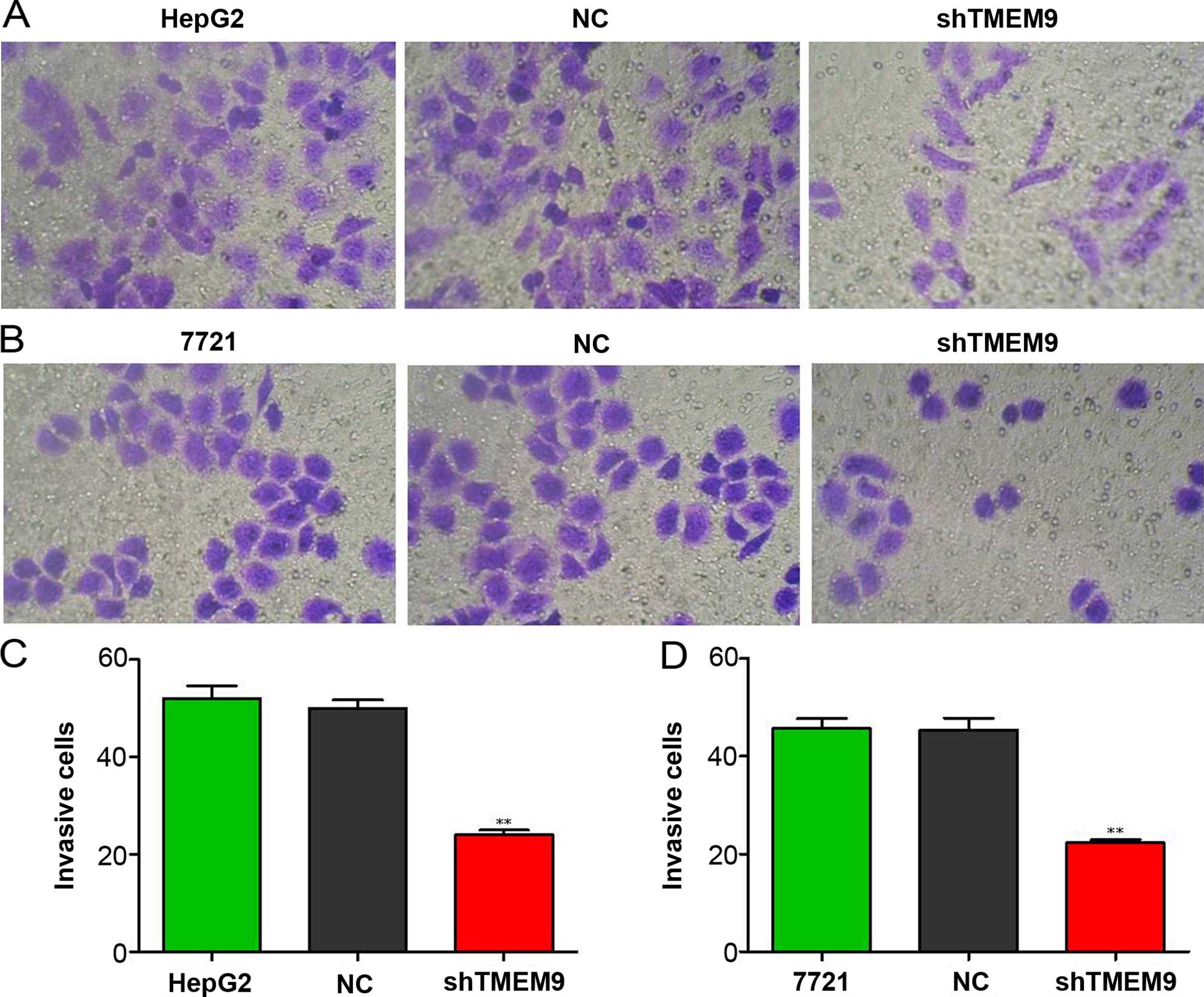
Cell invasion and migration

After transfection, the cells were detached and washed twice in PBS. Then, 1×10⁵ cells/ml were seeded in the upper chamber of a Transwell insert (8-µm pore size) coated (invasion) or not coated (migration) with 80 µl Matrigel (BD Biosciences). The lower chamber was filled with 0.75 ml of DMEM. After a 48-h (invasion) or 24-h (migration) incubation period, the non-migrated cells in the upper chamber were scraped away, and adherent cells were stained with formaldehyde solution. Any cells on the underside were counted and photographed under ×200 microscope fields.

Cell adhesion

To determine the adhesion cells, 12-well plates were used. Cell suspension (1×10⁵ cells/ml) was added to the well and incubated for 1 h at 37°C. Adherent cells were fixed with 4% methanol and stained with crystal violet for 20 min. The number of adherent cells were photographed and counted from three random selected ×200 fields of microscope.

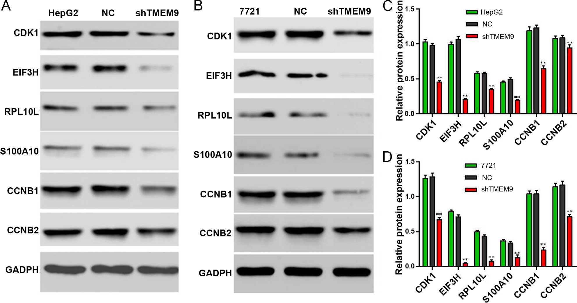
Channel protein expression detection

To detect the role of TMEM9 in liver cancer cells, we selected the proteins CCNB1, CCNB2, CDK1, PRL10A, S100A10 and EIF3H to detect the protein expression using western blotting. Protein lysates were prepared. Equal amounts of samples were resolved by SDS-PAGE and transferred to nitrocellulose membranes. The membranes were then blocked with 5% low-fat milk for 1 h or overnight at 4°C; incubated with CCNB1, CCNB2, CDK1, PRL10A, S100A10 and EIF3H with primary antibodies for 2 h, followed by secondary antibodies for 1 h at room temperature; and analyzed. GADPH protein levels were determined as a loading control.

Statistical analysis

Statistical significances were determined using the GraphPad Prism v5.0 software (GraphPad Software, La Jolla, CA, USA). Kaplan-Meier analysis was used to determine that the overall survival time between low and high expression of HCC. Data are presented as the mean ± SD of at least three independent replicates. Differences were considered significant when P<0.05 or P<0.01.

Results

TMEM9 is highly expressed in HCC with poor patient survival

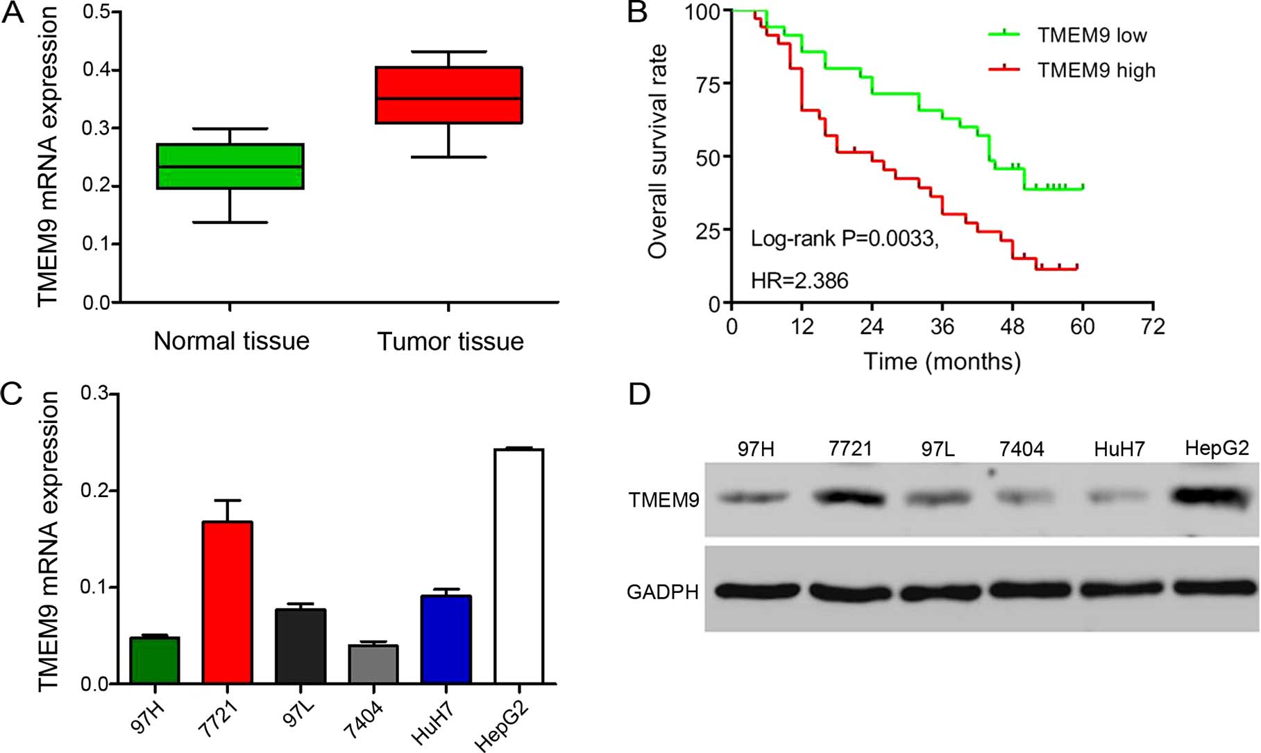
To investigate the expression of TMEM9, we used RT-qPCR to investigate in the HCC tissues of 30 patients. The results showed a higher level of TMEM9 expression (Fig. 1A). Then we investigated the correlation between TMEM9 expression and prognosis of the patients with HCC. As shown in Fig. 1B, Kaplan-Meier analysis showed that the overall survival time of lower-TMEM9-expressing patients was notably higher than that of higher-TMEM9-expressing patients. The expression levels of TMEM9 in the six HCC cells were also evaluated by qPCR and western blotting (Fig. 1C and D). The HepG2 and 7721 cell lines had a higher TMEM9 mRMA and protein expression.

Figure 1

High expression of TMEM9 is associated with the patient survival rate. (A) The mRNA expression level of TMEM9 was detected in tumor and adjacent tissues. (B) High expression of TMEM9 had a slightly higher survival rate. (C and D) The expression of TMEM9 was analyzed by RT-qPCR and western blotting in the six HCC cell lines. TMEM9, transmembrane protein 9; HCC, hepatocellular carcinoma.

Knockdown of TMEM9 inhibits cell proliferation and induces apoptosis

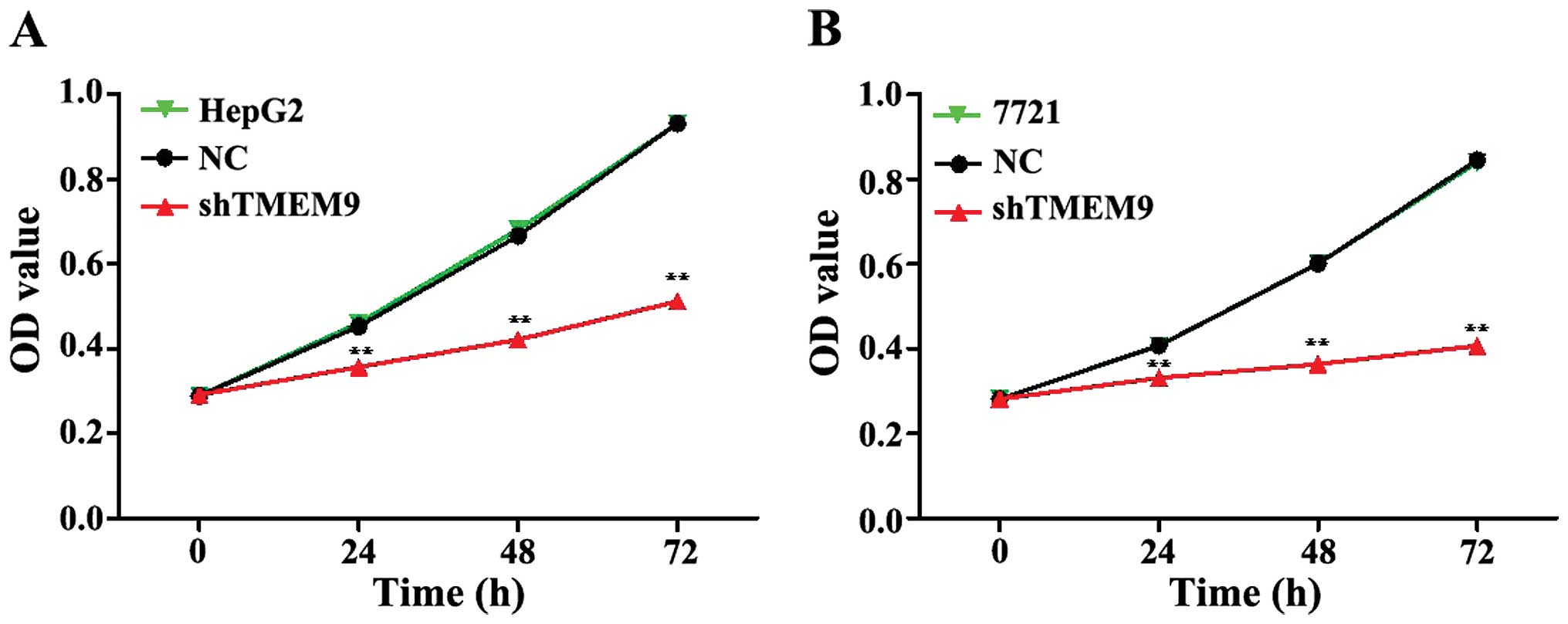
To assess the potential effects of RNAi silencing TMEM9 on proliferation, CCK-8 analysis was performed 72 h after transfection. The proliferative ability of HepG2 and 7721 cells was significantly inhibited at 24, 48 and 72 h (Fig. 2).

Figure 2

Effects of TMEM9 on cell proliferation of HepG2 and 7721 cells. (A) Cell proliferation was detected at 0, 24, 48 and 72 h in HepG2, NC and sh-TMEM9 cells. (B) Cell proliferation was detected in 7721, NC and sh-TMEM9 cells. NC, negative control; TMEM9, transmembrane protein 9.

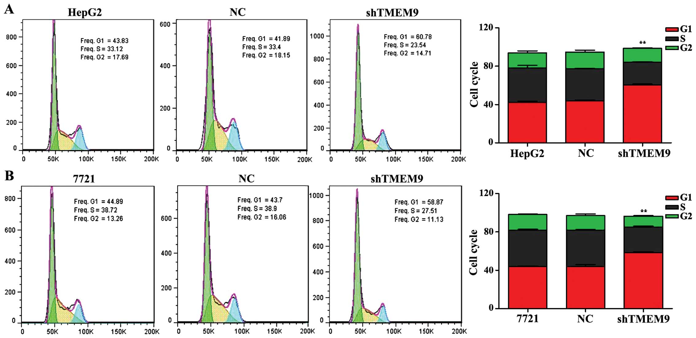
Moreover, we investigated the effects of TMEM9 on cell cycle and apoptosis in HCC. The flow cytometric analysis revealed that the population of G0/G1 phase was significantly increased but that of S and G2/M phase was decreased in HepG2 and 7721 cells, when compared with the negative control (NC) (P<0.01) (Fig. 3).

Figure 3

Effects of TMEM9 on cell cycle of HCC cells. (A) After TMEM9-siTNA transfection for 48 h, cell cycle distribution of HepG2 cells was identified by flow cytometry. (B) Cell cycle distribution of 7721, NC and shTMEM9 were detected. Data are based on at least 3 independent experiments and shown as mean ± SD. **P<0.01, compared with the negative control. NC, negative control; TMEM9, transmembrane protein 9.

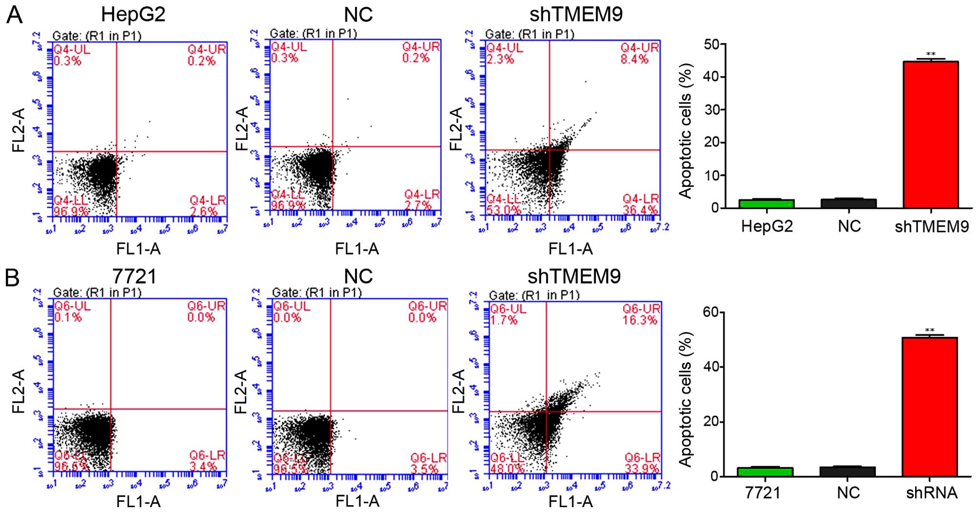
In addition, we assessed the apoptotic function of TMEM9 in HepG2 and 7721 cells using the Annexin V-FITC/PI staining assay. As shown in Fig. 4, the results showed that knockdown of TMEM9 in HCC cells markedly induced the cell apoptotic rate compared with NC (P<0.01).

Figure 4

Effects of TMEM9 on cell apoptosis of HCC cells. (A) After HepG2 cells were transfected for 48 h, the apoptotic rates of HepG2 cells were analyzed using flow cytometry. (B) The apoptotic rates of 7721, NC and shTMEM9 were detected. **P<0.01, compared with the negative control.TMEM9, transmembrane protein 9; HCC, hepatocellular carcinoma.

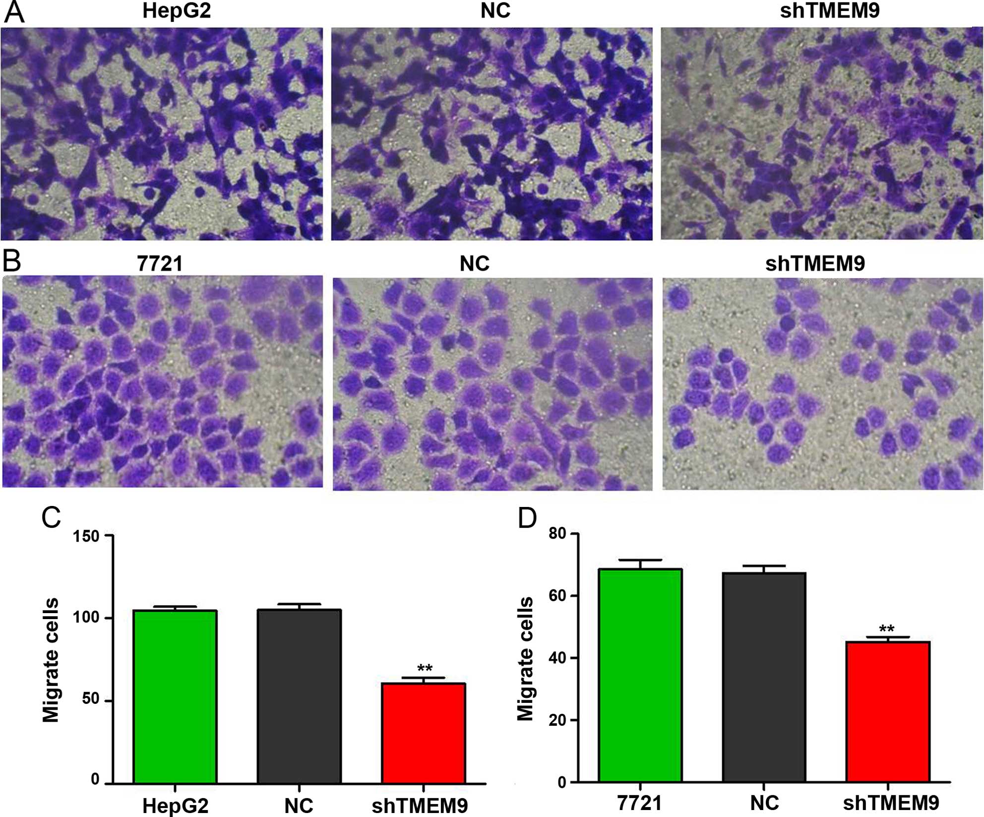
Knockdown of TMEM9 decreases metastasis of HCC cells

Cell metastasis plays an important role in cancer progression. We determined whether TMEM9 regulated metastasis of HCC cells. Cell invasion, migration and adhesion assays were then used to detect the metastatic capacity. As shown in Fig. 5, the cell invasion ability was reduced when compared with NC (P<0.01). The migration and adhesion cells were also decreased (P<0.01) (Figs. 6 and 7).

Figure 5

TMEM9 knockdown decreased invasion capacity of HCC cells in vivo. (A and B) Images of HepG2 and 7721 invasion cells were captured in 48 h after seeding. (C and D) Invasion assays showed that invasion cells were decreased than those in the negative group. **P<0.01, compared with the negative control. TMEM9, transmembrane protein 9; HCC, hepatocellular carcinoma.

Figure 6

Knockdown of TMEM9 decreases cell migration in HCC cells. (A and B) The images of HepG2 and 7721 migrated cells were captured in 48 h after seeding. (C and D) Migrations of HCC cells were assayed using the 24-Transwell system. Data are one of three similar experiments. **P<0.01, when compared with the negative controls. TMEM9, transmembrane protein 9; HCC, hepatocellular carcinoma.

Figure 7

Knockdown of TMEM9 decreased cell adhesion in HCC cells. (A and B) The images of HepG2 and 7721 migrate cells were captured in 48 h after seeding. (C and D) The adhesion of HCC cells were identified after transfection. **P<0.01, when compared with the negative controls. TMEM9, transmembrane protein 9; HCC, hepatocellular carcinoma.

Knockdown of TMEM9 decreases protein expression in HCC

Signaling pathways are often activated in tumor cells. We assessed the protein expression using western blotting. The results showed that the protein expression of CDK1, EIF3H, RPL10L, S100-A10, CCNB1 and CCNB2 was decreased compared to the control group (P<0.01) (Fig. 8).

Figure 8

Western blotting results of TMEM9 influenced proteins. (A and B) After 48 h of TMEM9 siRNA treatment, the protein expression of CDK1, EIF3H, RPL10L, S100A10, CCNB1 and CCNB2 in HepG2 and 7721 cells were detected by western blotting, GADPH was detected as the control of sample loading. (C and D) Western blotting data are analyzed to identify the protein levels. **P<0.01 compared with the NC cells.

Discussion

HCC is one of the most common types of cancer worldwide. Of an estimated 700,000 cancer-associated mortalities that arising in 2008, 50% occurred in China (2,14). The 5-year survival rate remains at <40% following surgery (15). HCC cause serious damage to human health; thus, investigation of its development mechanism and identification of effective measures of prevention, diagnosis and treatment of HCC is crucial. In the present study, we investigated the biological function of TMEM9 in HCC cells (16). The clinical data show that TMEM9 was highly expressed in HCC patients. Moreover, TMEM9 expression was associated with the patient survival rate. The in vitro experiments showed that knockdown of TMEM9 in HCC HepG2 and 7721 cancer cells inhibited cell growth and metastasis, and promoted cell apoptosis. Thus, TMEM9 serves as a potential target for the treatment of HCC.

In order to elucidate the possible mechanism involved, we identified the related protein expression. CDK1 is a highly conserved protein and a key player in the cell cycle regulation (17). Eukaryotic translation initiation factors (EIFs) are involved in the protein translation initiation process, and EIF2, EIF3, EIF4 and EIF5 have been previously investigated (18). EIF3H is an important subunit of the EIF3 family. The EIF3H expression level is closely associated with a variety of tumors, and is overexpressed in numerous malignant tumors (19,20). Ribosomal protein L 10-like (RPL10L), a protein-coding gene, has a structural constituent of ribosome. Furthermore, S100A10, CCNB1 and CCNB2 are involved in mitosis and the regulation of cell cycle progression (21–23). Our results show that the cell cycle-related protein expressions were significantly decreased when compared with NC.

In summary, to the best of our knowledge, the present study provides evidence for the first time that TMEM9 is crucial in the cell proliferation, apoptosis and metastasis of HCC cells. Additionally, TMEM9 regulates these biological processes by regulating cell cycle-related proteins. As TMEM9 expression level is associated with the patient survival rate, inhibition of TMEM9 in tumor tissues provides a therapeutic strategy. However, additional investigations should be conducted to validate its therapeutic function in the future.

Acknowledgments

The present study was supported by the Natural Science Fund of Hubei Province (no. 2012FFA044), the Health Department Found of Hubei Province (no. JX6B18), and the Public Service Platform Construction Projects of Wuhan Technology Bureau (no. 2013060705010326).

References

1 

Mathers C, Fat DM and Boerma JT: The Global Burden of Disease: 2004 Update. World Health Organization; 2008

2 

Jemal A, Bray F, Center MM, Ferlay J, Ward E and Forman D: Global cancer statistics. CA Cancer J Clin. 61:69–90. 2011. View Article : Google Scholar : PubMed/NCBI

3 

Bosch FX, Ribes J, Díaz M and Cléries R: Primary liver cancer: Worldwide incidence and trends. Gastroenterology. 127(Suppl 1): S5–S16. 2004. View Article : Google Scholar : PubMed/NCBI

4 

Villanueva A, Newell P, Chiang DY, Friedman SL and Llovet JM: Genomics and signaling pathways in hepatocellular carcinoma. Semin Liver Dis. 27:55–76. 2007. View Article : Google Scholar : PubMed/NCBI

5 

Yuen MF, Tanaka Y, Fong DY-T, Fung J, Wong DK, Yuen JC, But DY, Chan AO, Wong BC, Mizokami M, et al: Independent risk factors and predictive score for the development of hepatocellular carcinoma in chronic hepatitis B. J Hepatol. 50:80–88. 2009. View Article : Google Scholar

6 

Yang HI, Sherman M, Su J, Chen PJ, Liaw YF, Iloeje UH and Chen CJ: Nomograms for risk of hepatocellular carcinoma in patients with chronic hepatitis B virus infection. J Clin Oncol. 28:2437–2444. 2010. View Article : Google Scholar : PubMed/NCBI

7 

Lok AS, Seeff LB, Morgan TR, di Bisceglie AM, Sterling RK, Curto TM, Everson GT, Lindsay KL, Lee WM, Bonkovsky HL, et al HALT-C Trial Group: Incidence of hepatocellular carcinoma and associated risk factors in hepatitis C-related advanced liver disease. Gastroenterology. 136:138–148. 2009. View Article : Google Scholar

8 

Yeoman AD, Al-Chalabi T, Karani JB, Quaglia A, Devlin J, Mieli-Vergani G, Bomford A, O'Grady JG, Harrison PM and Heneghan MA: Evaluation of risk factors in the development of hepatocellular carcinoma in autoimmune hepatitis: Implications for follow-up and screening. Hepatology. 48:863–870. 2008. View Article : Google Scholar : PubMed/NCBI

9 

Wang X, Wang N, Cheung F, Lao L, Li C and Feng Y: Chinese medicines for prevention and treatment of human hepatocellular carcinoma: Current progress on pharmacological actions and mechanisms. J Integr Med. 13:142–164. 2015. View Article : Google Scholar : PubMed/NCBI

10 

Lopez PM, Villanueva A and Llovet JM: Systematic review: Evidence-based management of hepatocellular carcinoma - an updated analysis of randomized controlled trials. Aliment Pharmacol Ther. 23:1535–1547. 2006. View Article : Google Scholar : PubMed/NCBI

11 

Villanueva A, Toffanin S and Llovet JM: Linking molecular classification of hepatocellular carcinoma and personalized medicine: Preliminary steps. Curr Opin Oncol. 20:444–453. 2008. View Article : Google Scholar : PubMed/NCBI

12 

Ke AW, Shi GM, Zhou J, Huang XY, Shi YH, Ding ZB, Wang XY, Devbhandari RP and Fan J: CD151 amplifies signaling by integrin α6β1 to PI3K and induces the epithelial-mesenchymal transition in HCC Cells. Gastroenterology. 140:1629–1641.e15. 2011. View Article : Google Scholar

13 

Kveine M, Tenstad E, Døsen G, Funderud S and Rian E: Characterization of the novel human transmembrane protein 9 (TMEM9) that localizes to lysosomes and late endosomes. Biochem Biophys Res Commun. 297:912–917. 2002. View Article : Google Scholar : PubMed/NCBI

14 

Ferlay J, Shin HR, Bray F, Forman D, Mathers C and Parkin DM: Estimates of worldwide burden of cancer in 2008: GLOBOCAN 2008. Int J Cancer. 127:2893–2917. 2010. View Article : Google Scholar

15 

Fang F, Yang L, Tao Y and Qin W: FBI-1 promotes cell proliferation and enhances resistance to chemotherapy of hepatocellular carcinoma in vitro and in vivo. Cancer. 118:134–146. 2012. View Article : Google Scholar

16 

Zhai XF, Chen Z, Li B, Shen F, Fan J, Zhou WP, Yang YK, Xu J, Qin X, Li LQ and Ling CQ: Traditional herbal medicine in preventing recurrence after resection of small hepatocellular carcinoma: a multicenter randomized controlled trial. J Integr Med. 11:90–100. 2013. View Article : Google Scholar : PubMed/NCBI

17 

Morgan DO: The Cell Cycle: Principles of Control. New Science Press. 2007.

18 

Spilka R, Ernst C, Mehta AK and Haybaeck J: Eukaryotic translation initiation factors in cancer development and progression. Cancer Lett. 340:9–21. 2013. View Article : Google Scholar : PubMed/NCBI

19 

Okamoto H, Yasui K, Zhao C, Arii S and Inazawa J: PTK2 and EIF3S3 genes may be amplification targets at 8q23–q24 and are associated with large hepatocellular carcinomas. Hepatology. 38:1242–1249. 2003. View Article : Google Scholar : PubMed/NCBI

20 

Cappuzzo F, Varella-Garcia M, Rossi E, Gajapathy S, Valente M, Drabkin H and Gemmill R: MYC and EIF3H coamplification significantly improve response and survival of non-small cell lung cancer patients (NSCLC) treated with gefitinib. J Thorac Oncol. 4:472–478. 2009. View Article : Google Scholar : PubMed/NCBI

21 

Jackman M, Firth M and Pines J: Human cyclins B1 and B2 are localized to strikingly different structures: B1 to microtubules, B2 primarily to the Golgi apparatus. EMBO J. 14:1646–1654. 1995.PubMed/NCBI

22 

Porter LA, Singh G and Lee JM: Abundance of cyclin B1 regulates γ-radiation-induced apoptosis. Blood. 95:2645–2650. 2000.

23 

Rescher U and Gerke V: S100A10/p11: Family, friends and functions. Pflugers Arch. 455:575–582. 2008. View Article : Google Scholar

Related Articles

  • Abstract
  • View
  • Download
  • Twitter
Copy and paste a formatted citation
Spandidos Publications style
Zhang Y, Ran Y, Xiong Y, Zhong Z, Wang ZH, Fan X and Ye Q: Effects of TMEM9 gene on cell progression in hepatocellular carcinoma by RNA interference. Oncol Rep 36: 299-305, 2016.
APA
Zhang, Y., Ran, Y., Xiong, Y., Zhong, Z., Wang, Z., Fan, X., & Ye, Q. (2016). Effects of TMEM9 gene on cell progression in hepatocellular carcinoma by RNA interference. Oncology Reports, 36, 299-305. https://doi.org/10.3892/or.2016.4821
MLA
Zhang, Y., Ran, Y., Xiong, Y., Zhong, Z., Wang, Z., Fan, X., Ye, Q."Effects of TMEM9 gene on cell progression in hepatocellular carcinoma by RNA interference". Oncology Reports 36.1 (2016): 299-305.
Chicago
Zhang, Y., Ran, Y., Xiong, Y., Zhong, Z., Wang, Z., Fan, X., Ye, Q."Effects of TMEM9 gene on cell progression in hepatocellular carcinoma by RNA interference". Oncology Reports 36, no. 1 (2016): 299-305. https://doi.org/10.3892/or.2016.4821
Copy and paste a formatted citation
x
Spandidos Publications style
Zhang Y, Ran Y, Xiong Y, Zhong Z, Wang ZH, Fan X and Ye Q: Effects of TMEM9 gene on cell progression in hepatocellular carcinoma by RNA interference. Oncol Rep 36: 299-305, 2016.
APA
Zhang, Y., Ran, Y., Xiong, Y., Zhong, Z., Wang, Z., Fan, X., & Ye, Q. (2016). Effects of TMEM9 gene on cell progression in hepatocellular carcinoma by RNA interference. Oncology Reports, 36, 299-305. https://doi.org/10.3892/or.2016.4821
MLA
Zhang, Y., Ran, Y., Xiong, Y., Zhong, Z., Wang, Z., Fan, X., Ye, Q."Effects of TMEM9 gene on cell progression in hepatocellular carcinoma by RNA interference". Oncology Reports 36.1 (2016): 299-305.
Chicago
Zhang, Y., Ran, Y., Xiong, Y., Zhong, Z., Wang, Z., Fan, X., Ye, Q."Effects of TMEM9 gene on cell progression in hepatocellular carcinoma by RNA interference". Oncology Reports 36, no. 1 (2016): 299-305. https://doi.org/10.3892/or.2016.4821
Follow us
  • Twitter
  • LinkedIn
  • Facebook
About
  • Spandidos Publications
  • Careers
  • Cookie Policy
  • Privacy Policy
How can we help?
  • Help
  • Live Chat
  • Contact
  • Email to our Support Team