Introduction
Tumor necrosis factor α-induced protein 8 (TIPE,
TNFAIP8) is a member of the TNFAIP8 family (1). TIPE is overexpressed in many types of
malignancies, such as hepatocellular carcinoma, gastric
adenocarcinoma, non-small cell lung cancer (NSCLC), breast and
epithelial ovarian cancer (2–7).
Apoptosis is the process of programmed cell death which maintains a
healthy cell survival/cell death balance. Defects in the apoptotic
process may cause cancer or autoimmunity, while enhanced apoptosis
may cause degenerative diseases (8). In despite of the fact that TIPE is
involved in the carcinogenesis of many tumor types (2–7), the
exact role of TIPE in defective apoptosis-associated carcinogenesis
is still uncertain.
The family of mitogen-activated protein kinases
(MAPKs), which are specific to the amino acids serine and
threonine, is involved in directing cellular responses to mitogens,
osmotic stress and proinflammation (9). While the MAPK family regulates cell
functions including proliferation, differentiation, mitosis and
cell survival (1), it was
documented that MAPK signaling, such as extracellular
signal-regulated kinases (ERK), c-Jun N-terminal kinases (JNK) and
p38 mitogen-activated protein kinases (p38) regulate apoptosis and
play a prominent role in tumor development (10,11).
Blockade of p38 signaling was found to significantly inhibit the
proliferation of head and neck squamous cell carcinoma cells
(12), and p38 activation was also
revealed to have counteracting effects with JNK on myeloid cell
leukemia sequence (Mcl-1) expression and lead to pro-apoptotic
effects in NSCLC cells (13).
Nevertheless, JNK activation is essential for cell transformation
and proliferation in response to Ras signaling (14). Hence, the role of MAPK activation in
TIPE-associated carcinogenesis needs to be elucidated.
In the present study, we provide evidence that TIPE
overexpression suppressed the apoptotic effect of radiation and
cisplatin treatment in Raw264.7 and EL4 cells via JNK and p38
activation, which ultimately promoted tumor formation. This was
supported by the fact that TIPE overexpression promoted the
proliferation of Raw264.7 and EL4 cells. Secondly, TIPE
overexpression increased the anti-apoptotic effects and the levels
of phosphorylated JNK and p38. Moreover, inhibition of JNK and p38
efficiently abolished the pro-survival effects of TIPE. Most
importantly, in vivo tumor implantation test revealed that
TIPE overexpression obviously augmented the volume and weight of
implanted tumors in mice, indicating that TIPE facilitated tumor
formation. Hence, the present study revealed that activation of JNK
and p38 kinases contributed to the TIPE-mediated anti-apoptotic
effect, indicating that JNK and p38 may be potential therapeutic
molecules for TIPE overexpression-associated diseases.
Materials and methods
Reagents and antibodies
Reagents were purchased from the following
companies. Annexin V/PI apoptosis detection kit was obtained from
Promega (Madison, WI, USA). JNK inhibitor SP600125 and p38 MAPK
inhibitor SB203580 were obtained from Cayman Chemical (Ann Arbor,
MI, USA) and used as previously described (15–17).
Antibodies to β-actin (#8457), proliferating cell nuclear antigen
(PCNA) (#13110), phospho(p)-p38 (#4511), phosphorylated
mitogen-activated protein kinase kinase (p-MEK) (#9154), p-JNK
(#4668), cleaved caspase-3 (#9664), cleaved caspase-9 (#7237) and
cleaved poly(ADP-ribose) polymerase (PARP) (#5625) were purchased
from Cell Signaling Technology, Inc. (Beverly, MA, USA). Antibody
to TIPE (#SC-82054) was purchased from Santa Cruz Biotechnology,
Inc. (Dallas, TX, USA). The concentrations of the used antibodies
were determined according to the manufacturer's instructions. The
PrimeScript RT-PCR kit and SYBR Premix ExTaq™ kit were purchased
from Tarkara Bio (Dalian, Liaoning, China). JC-1 (Molecular Probes,
Eugene, OR, USA) was the dye used to detect early stage cell
apoptosis. RPMI-1640 medium, Dulbecco's modified Eagle's medium
(DMEM) and fetal bovine serum (FBS) were purchased from HyClone (GE
Healthcare Life Sciences, Logan, UT, USA). UltraSensitive™ SP
immunohistochemistry (IHC) kit was obtained from Maixin Biotech
(Fuzhou, Fujian, China).
Animals
Pathogen-free BALB/c nude mice (5–6 weeks old,
female) were obtained from the Shanghai Laboratory Animal Center of
the Chinese Academy of Sciences and were housed at the Animal
Center of Xiamen University under a 12 h light-dark cycle,
temperature (20–26°C) and humidity (40–70%) conditions. They had
free access to water and food. The animal study protocol was
approved by the Committee on the Ethics of Animal Experiments of
Xiamen University.
Cell lines
Abelson murine leukemia virus transformed Raw264.7
cells (18) and murine T lymphocyte
EL4 cells which were transfected with Migr1 control and TIPE vector
were kindly provided by Professor Y.H. Chen (University of
Pennsylvania, Philadelphia, PA, USA). Raw264.7 cells were cultured
in DMEM with 10% FBS at 37°C in 5% CO2. EL4 cells were
cultured in RPMI-1640 medium with 10% FBS at 37°C in 5%
CO2. Cells were synchronized by serum starvation for at
least 6 h before further treatment.
Flow cytometric measurements
Cell apoptosis was determined as previously
described (15). Briefly,
TIPE-overexpressing and mock cells (2×10⁵/ml) were
placed in 6-well plates containing 10% FBS (HyClone) in phenol
red-free RPMI-1640 (HyClone), were grown overnight, and washed once
in PBS. Cells were irradiated with 4 mJ/cm2 of UVC light
(HFsafe 1500; Class II, Type A2; Shanghai Lishen Scientific
Equipment Co., Ltd., Shanghai, China), for 30 min as previously
described (19). Then, the cells
were collected and stained with Annexin V-FITC and propidium iodide
for 20 min at room temperature. To determine the role of MAPK
activation in TIPE-mediated increased cell pro-survival, the cells
were pretreated with U0126, SP600125, SB203580 at a final
concentration of 10 µM prior to treatment with 2 µg/ml cisplatin,
as previously described (15). Flow
cytometry was performed using a FACSCalibur flow cytometer, and the
data were analyzed using CellQuest software (BD Biosciences, San
Jose, CA, USA).
Cell proliferation assay
Cell proliferation assay was performed as previously
described (16). Briefly,
5×10⁴ TIPE-overexpressing EL4 or Raw264.7 cells were
inoculated in a 96-well plate at 100 µl/well medium in a humidified
incubator (37°C, 5% CO2). Then, Cell Counting Kit-8
(CCK-8) solution (10 µl) was added to each well of the plate and
the OD450 value was determined at a wavelength of 450 nm.
Measurement of mitochondrial membrane
potential by flow cytometry
Mitochondrial membrane potential was assessed using
the fluorescent dye JC-1, which detects the early stage of cell
apoptosis (15). Briefly,
TIPE-expressing EL4 and control cells were treated with 2 µg/ml
cisplatin for 20 min. Then, the cells were rinsed with PBS and JC-1
was added at a final concentration of 1 µg/ml. After 15 min
co-incubation at 37°C, the cells were collected, washed twice with
PBS and then analyzed by flow cytometry.
Reverse transcription PCR
The expression of TIPE in Raw264.7 and EL4 cells was
investigated by RT-PCR analysis according to a previous description
(20). Briefly, total RNA was
isolated from the cells. Reverse transcription was performed using
PrimeScript Reverse Transcriptase kit (Takara Biotechnology Co.,
Ltd., Dalian, China) and cDNA was used for subsequent PCR reactions
using the Maxima SYBR-Green qPCR Master Mix (Takara Biotechnology
Co., Ltd.). The following primers were used (Santa Cruz
Biotechnology, Inc.): β-actin (sc-108070-PR), TNFα-IP 8 (m)-PR
(sc-76699-PR). However, primer sequences are not provided by Santa
Cruz Biotechnology, Inc., as stated in their datasheets:
‘Semi-quantitative RT-PCR may be performed to monitor TNFα-IP 8
gene expression using RT-PCR Primer: TNFα-IP 8 (m)-PR: sc-76699-PR,
β-Actin (m)-PR: sc-108070-PR’.
Western blot analysis
The whole cellular protein was extracted and western
blotting was performed as previously described (17,20).
Briefly, proteins were obtained in lysis buffer and loaded onto
sodiumdodecylsulfate-polyacrylamide gel electrophoresis (SDS-PAGE)
gels for electrophoresis and transferred onto polyvinylidene
difluoride (PVDF) membranes. After blocking in 5% fat-free milk in
Tris-buffered saline with Tween-20 (TBST) for 90 min, the membranes
were incubated with primary antibodies at 4°C overnight.
Subsequently, the membranes were incubated with corresponding
horseradish peroxidase (HRP)-conjugated secondary antibodies at
room temperature for 90 min. After washing 4 times with TBST (for
10 min each), the bound antibodies were visualized using enhanced
chemiluminescence (ECL). β-actin was used as a loading control.
In vivo implantation tumor models
A xenograft tumor model in BALB/c mice was
established as previously described (21) by subcutaneous neck injection of
Raw264.7-TIPE and control cells (2×10⁶/nude mouse in 100
µl volume). Tumor size was measured by vernier caliper (Shanghai
Hui Yi Electronic Technology Co., Ltd., Shanghai, China), the
length (L), width (W), and diameter were measured every two days
from day 4, tumor volume (cm3) was calculated using the
formula W2x(L/2). Following 12 days of growth, animals
were sacrificed by CO2 asphyxiation and tumors were
removed.
Immunohistochemical (IHC)
staining
The expression of TIPE in the mouse tumor tissues
was determined via IHC staining as previously described (20). Briefly, free-floating lung sections
(4 µM) were obtained using a slicing system (Leica RM2135).
Endogenous peroxidase activity was quenched in
H2O2, and 90% formic acid was used to expose
the epitope. The primary antibody was applied overnight at 4°C, and
then biotinylated secondary antibody at room temperature. After a
final wash, the slides were developed with diaminobenzidine
substrate using the avidin-biotin HRP system.
Statistical analysis
All experiments were repeated at least 3 times to
confirm the results. The data are presented as the mean ± SEM.
Student's t-test and one-way ANOVA with post Newman-Keuls test were
applied. Differences were considered significant at P<0.05.
Results
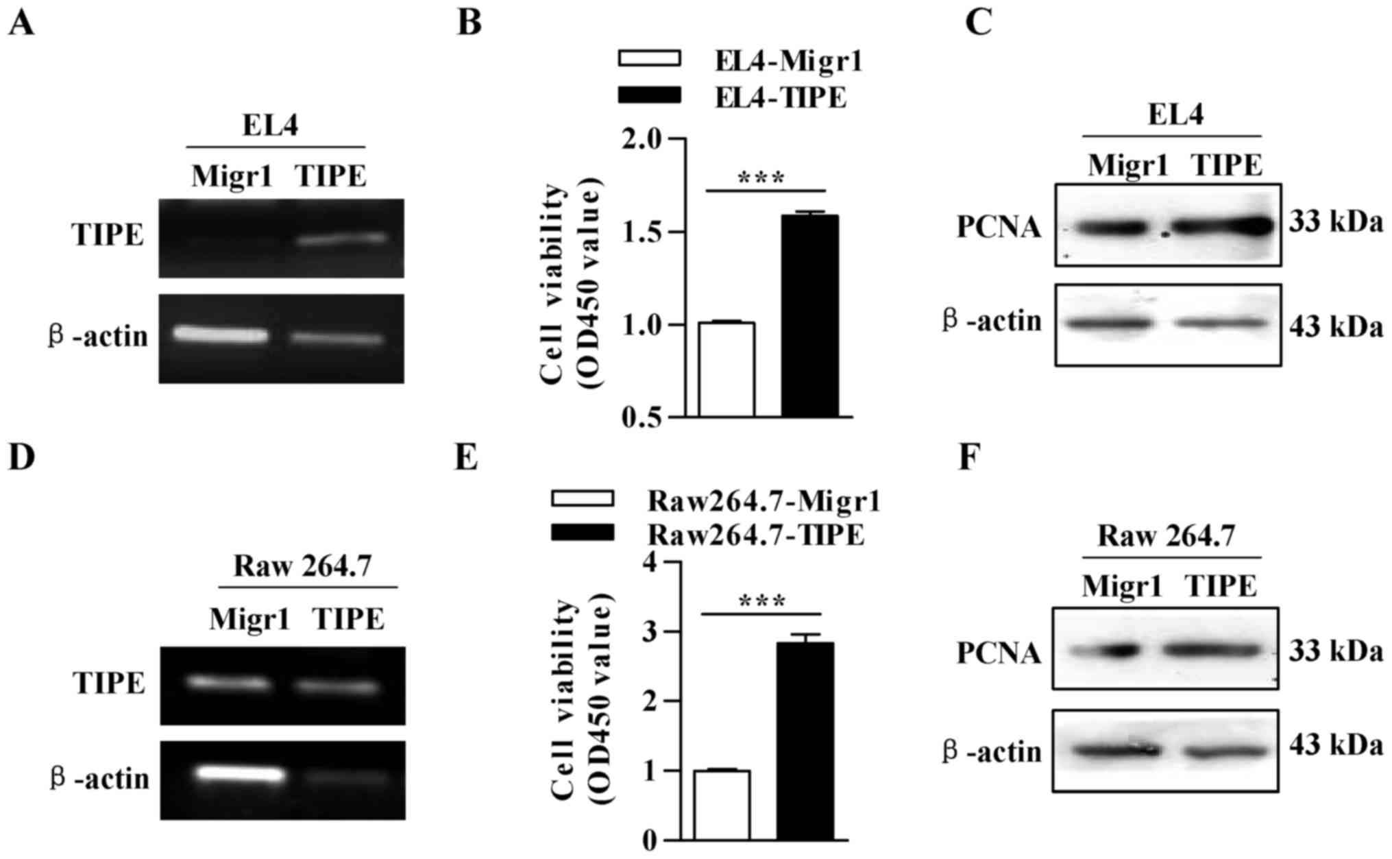
TIPE overexpression promotes the
proliferation of Raw264.7 and EL4 cells
The overexpression of TIPE in the Raw264.7 and EL4
cells transfected with the TIPE-overexpression vector was confirmed
by RT-PCR (Fig. 1A and D). Then,
the effect of TIPE overexpression on cell proliferation was
determined by CCK-8 assays and western blotting, respectively. The
results showed that TIPE overexpression increased the number of
viable cells in the Raw264.7 and EL4 cell lines (Fig. 1B and E). The expression of PCNA was
also upregulated in the TIPE-overexpressing cell lines (Fig. 1C and F). All these data indicate
that TIPE has the ability to promote cell proliferation.
TIPE overexpression inhibits
ultraviolet irradiation or cisplatin-induced apoptosis
To explore the effect of TIPE on cell apoptosis,
TIPE-overexpressing Raw264.7 and EL4 cells were exposed to
ultraviolet irradiation and the percentage of apoptotic cells was
determined by flow cytometry. Under ultraviolet irradiated
condition, TIPE overexpression exhibited a ~18.5% anti-apoptotic
ability in the EL4 cells, which was verified by the decreased
percentage of apoptotic cells from 53.24 to 43.39% (Fig. 2A and B). Flow cytometric
determination in Raw264.7 cells also revealed a similar trend
(Fig. 2C and D). When JC-1 was used
to measure mitochondrial membrane potential, TIPE overexpression
obviously decreased the cisplatin-augmented mean fluorescence
intensity (MFI) (Fig. 2E). Western
blot analyses also revealed that TIPE overexpression inhibited
cisplatin-induced activation of caspase-3, caspase-9 and PARP
(Fig. 2F). All of these
observations indicate that the cells acquired anti-apoptotic
abilities following induction of TIPE overexpression.
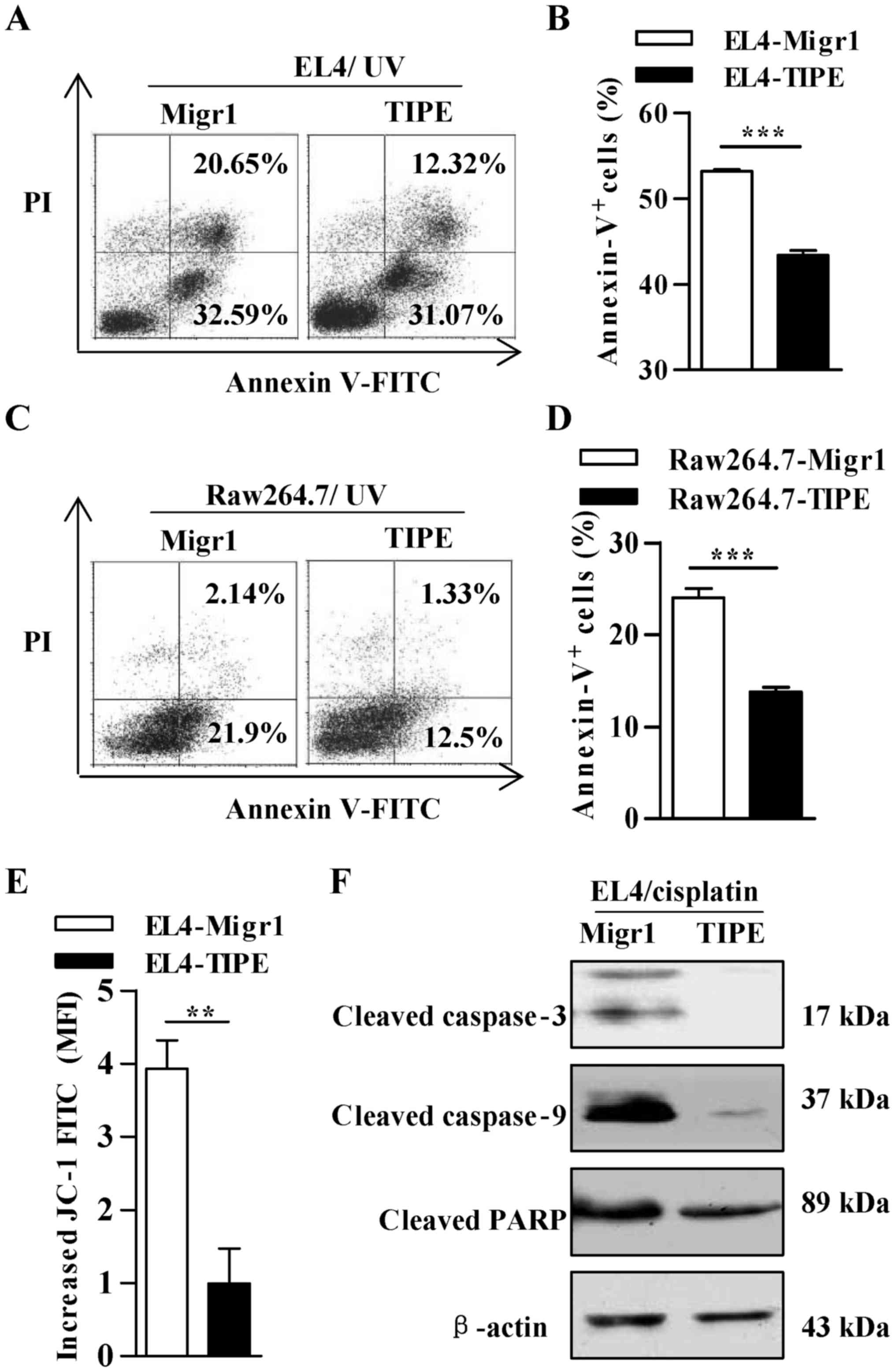 | Figure 2.TIPE inhibits ultraviolet irradiation-
or cisplatin-induced apoptosis in Raw264.7 and EL4 cells. (A-D) The
cells were exposed to 4 mJ/cm2 ultraviolet irradiation and cell
apoptosis was determined by Annexin V/PI staining and flow
cytometry. Data in the histograms represent the percentages of the
analyzed population. Data are presented as mean ± SEM, n=3,
***P<0.001, Student's t-test. (E and F) Next, the cells were
treated with 2 µg/ml cisplatin, and (E) mitochondrial membrane
potential was determined by JC-1 analyses, data are presented as
mean ± SEM, n=3, **P<0.01, Student's t-test and (F) levels of
cleaved caspase-3, caspase-9 and PARP were determined by western
blotting. For western blotting, β-actin was used as an internal
control. A representative of 3 independent experiments is shown.
TIPE, tumor necrosis factor α-induced protein 8. MFI, mean
fluorescence intensity. |
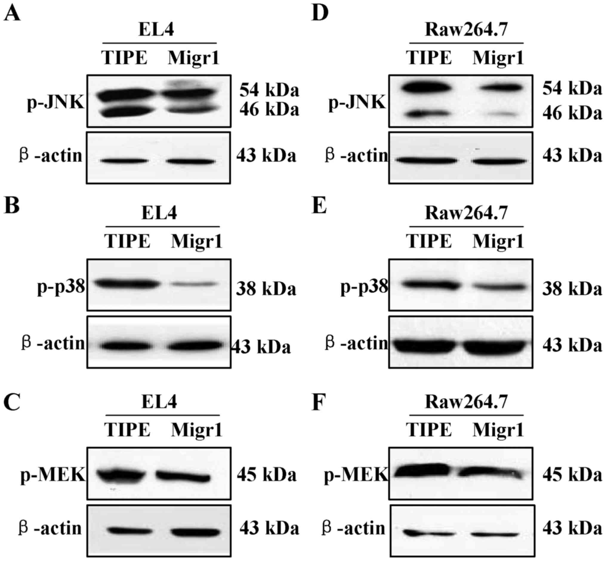
TIPE overexpression augments
phosphorylation of JNK, p38 and MEK
MAPK was reported to regulate cell proliferation,
differentiation and cell survival (1). The levels of phosphorylated MAPKs such
as MEK, JNK, and p38 were determined to evaluate the role of MAPK
in the TIPE-mediated increased cell pro-survival. An obvious
increase in phosphorylated JNK, MEK and p38 was observed in the
TIPE-overexpressing EL4 cells (Fig.
3A-C). A similar result was found in the TIPE-overexpressing
Raw264.7 cells (Fig. 3D-F). All
these data indicate that MAPK pathways may play an important role
in the TIPE-mediated cell proliferation and anti-apoptotic
effects.
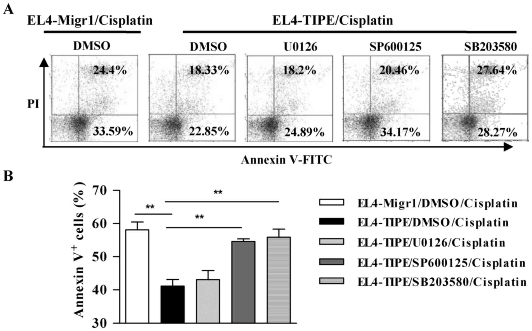
Inhibition of p38 and JNK activation
abolishes TIPE-induced anti-apoptotic effects
As TIPE overexpression efficiently increased cell
survival (Fig. 2) and obviously
augmented MAPK activation (Fig. 3),
we aimed to ascertain whether the TIPE-mediated anti-apoptotic
effects were due to the activation of these kinases. Toward this
end, TIPE-overexpressing and Migr1 control EL4 cells were incubated
with related kinase inhibitors prior to cisplatin treatment and
apoptotic cells were determined by flow cytometry. Compared with
the Migr1 control, TIPE overexpression efficiently inhibited
cisplatin-induced cell apoptosis, which was confirmed by reduction
in apoptotic cells from 58 to 41.18% (Fig. 4A and B). Compared with the
U0126-treated group, inhibition of JNK phosphorylation by JNK
inhibitor SP600125 efficiently abolished the promotive effect of
TIPE on cell survival, which was confirmed by the increase in the
percentages of apoptotic cells from 41.18 to 54.63% (Fig. 4). Treatment with SB203580 also
showed a similar trend (Fig. 4).
All these observations suggest that the activation of JNK, p38, but
not MEK contributes to the anti-apoptotic effect of TIPE
overexpression.
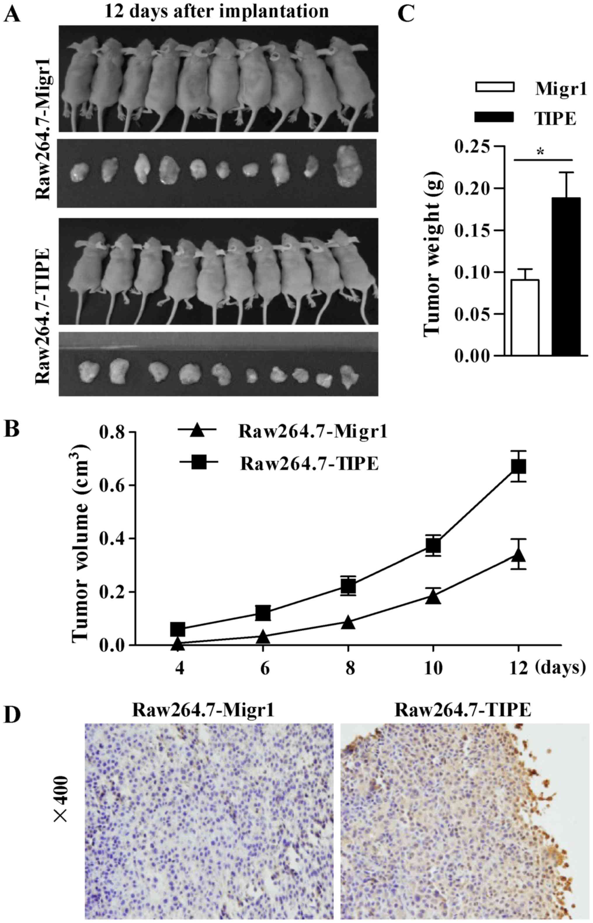
TIPE overexpression promotes tumor
formation in vivo
We next constructed a tumor implantation model to
observe the effect of TIPE overexpression on tumor formation.
TIPE-overexpressing Raw264.7 and mock cells were subcutaneously
implanted into BALB/c nude mice and the corresponding neoplasm
volumes were measured every two days. TIPE overexpression not only
increased tumor volume (Fig. 5A and
B) but also augmented tumor weight (Fig. 5C). Immunohistochemical staining for
TIPE in the implanted tumor tissues clearly showed that the
transfected tumor cells consistently expressed TIPE (Fig. 5D). The results demonstrated that
TIPE overexpression augmented tumor weight and volume indicating
that TIPE overexpression facilitates tumor formation in
vivo.
Discussion
In the present study, murine macrophage Raw264.7 and
T lymphocyte EL4 cells were transfected with Migr1 control and a
TIPE overexpression vector due to their abilities for suitable DNA
transfection (18). In despite of
the fact that TIPE is a newly described regulator of immunity and
tumorigenesis and is the only known transfer protein of the lipid
second messengers (22,23), the exact mechanism of TIPE-mediated
pro-survival effects remain uncertain. In the present study, we
observed that TIPE inhibited cell apoptosis and promoted tumor
formation in vivo. We found that TIPE overexpression
augmented the levels of phosphorylated JNK and p38. Moreover,
inhibition of JNK and p38 efficiently abolished the promotive
effect of TIPE on cell survival. Most importantly, an in
vivo tumor mouse model revealed that TIPE overexpression
obviously augmented the volume and weight of the tumors, indicating
that TIPE facilitates tumor formation.
Apoptosis can be initiated by two alternative
signaling pathways: The death receptor-mediated extrinsic apoptotic
and the mitochondrion-mediated intrinsic apoptotic pathway.
Mitochondrial membrane potential reflects the pumping of hydrogen
ions across the inner membrane (15). Cellular stress responses cause
translocation of the Bcl-2 family from the cytosol to the
mitochondria, resulting in decreased mitochondrial membrane
potential, the release of cytochrome c, and the initiation
of apoptosis (24). In the present
study, we demonstrated that TIPE exhibited anti-apoptotic
abilities, yet the exact effects of TIPE on mitochondrial membrane
potential and the release of cytochrome c need further
investigation.
MAPK signaling, which includes ERK, p38 and JNK
pathways, can be subverted to facilitate tumor proliferation,
survival and invasion (25),
indicating that MAPK signaling is involved in the process of
carcinogenesis and tumor development. For example, an increased
level of phosphorylated p38 was reported to be related to tumor
size and the formation of satellite tumors (26). A higher activation of ERK was also
found in many types of tumor (27).
Activation of JNK signaling by the silencing of dual-specificity
phosphatase 9 was documented to induce the proliferation of gastric
cancer (28). Hence, in the present
study, it was no surprise to find that the activation of MAPK
signaling was upregulated by TIPE overexpression and promoted tumor
formation. Notably, in spite of the fact that the activation of JNK
and p38 contributed to the TIPE-induced anti-apoptotic effects, ERK
phosphorylation had no effect on cell pro-survival. As prolonged
JNK phosphorylation uncouples MEK-mediated ERK activation in a
c-Jun-dependent manner (29),
opposing effects of ERK and JNK-p38 MAPKs on apoptosis have been
found in nerve growth factor withdrawal cells (30). Meanwhile, TIPE expression was found
to be positively correlated with ERK1 but not ERK2 as documented in
gastric cancer (31). Hence, the
exact function of MEK phosphorylation in TIPE-mediated promotion of
tumor formation remains unclear and needs further elucidation.
Mitochondrial integrity plays an important role in
the process of cytochrome c release and anti-apoptosis
activity. Mcl-1, a member of the anti-apoptotic Bcl-2 family, was
found to relocate into the mitochondrial membrane, antagonize
Bcl-2-like protein 4 (Bax) and Bcl-2 homologous antagonist/killer
(Bak) activation, and exert anti-apoptotics activity (32). The amelioration of mitochondrial
membrane potential by regulating MAPK signaling was also
demonstrated to exert the ability of pro-survival (33). Hence, the activation of JNK and p38
signaling by TIPE overexpression may have potential positive
effects on the mitochondrial relocation of the components of the
Bcl-2 family, maintain mitochondrial integrity, and exert
pro-survival ability.
Taken together, our studies revealed that the
activation of JNK and p38 kinases contributed to the TIPE-mediated
anti-apoptotic effects and tumor formation, indicating that JNK and
p38 may be potential therapeutic molecules for TIPE
overexpression-associated diseases.
Acknowledgements
TIPE-overexpressing RAW264.7 and EL4 cells were
supported by grant from National Institutes of Health, USA
(AI-077533, AI-050059, and GM-085112 to YHC) and kindly provided by
Professor YH Chen (University of Pennsylvania, Philadelphia, PA,
USA).
Funding
This study was supported by grants from the National
Natural Science Foundation of China (nos. 81273203 and 81771669), a
grant from the Natural Science Foundation of Fujian Province of
China (no. 2017J05140) and a grant from the Fujian Provincial
Department of Education (JA15014).
Availability of data and materials
The datasets used during the present study are
available from the corresponding author upon reasonable
request.
Author's contributions
FGG designed the research and edited the manuscript.
YL performed the western blotting and analyzed the data. XYN
contributed to the flow cytometric analyses and CCK-8 assays and
analyzed the data; RLC and JL contributed to manuscript
preparation. All authors read and approved the manuscript and agree
to be accountable for all aspects of the research in ensuring that
the accuracy or integrity of any part of the work are appropriately
investigated and resolved.
Ethics approval and consent to
participate
The animal study protocol was approved by the
Committee on the Ethics of Animal Experiments of Xiamen
University.
Consent for publication
Not applicable.
Competing interests
All authors declare no competing interests.
References
|
1
|
Lou Y and Liu S: The TIPE (TNFAIP8) family
in inflammation, immunity, and cancer. Mol Immunol. 49:4–7. 2011.
View Article : Google Scholar : PubMed/NCBI
|
|
2
|
Dong Q, Fu L, Zhao Y, Xie C, Li Q and Wang
E: TNFAIP8 interacts with LATS1 and promotes aggressiveness through
regulation of Hippo pathway in hepatocellular carcinoma.
Oncotarget. 8:15689–15703. 2017.PubMed/NCBI
|
|
3
|
Hadisaputri YE, Miyazaki T, Suzuki S,
Yokobori T, Kobayashi T, Tanaka N, Inose T, Sohda M and Kuwano H:
TNFAIP8 overexpression: Clinical relevance to esophageal squamous
cell carcinoma. Ann Surg Oncol. 19:S589–S596. 2012. View Article : Google Scholar : PubMed/NCBI
|
|
4
|
Hu R, Qiu X, Hong S, Meng L, Hong X, Qiu
J, Yang J, Zhuang G and Liu Z: Clinical significance of TIPE
expression in gastric carcinoma. Onco Targets Ther. 9:4473–4481.
2016. View Article : Google Scholar : PubMed/NCBI
|
|
5
|
Dong QZ, Zhao Y, Liu Y, Wang Y, Zhang PX,
Jiang GY, Dong XJ, Cui QZ and Wang EH: Overexpression of SCC-S2
correlates with lymph node metastasis and poor prognosis in
patients with non-small-cell lung cancer. Cancer Sci.
101:1562–1569. 2010. View Article : Google Scholar : PubMed/NCBI
|
|
6
|
Xiao M, Xu Q, Lou C, Qin Y, Ning X, Liu T,
Zhao X, Jia S and Huang Y: Overexpression of TNFAIP8 is associated
with tumor aggressiveness and poor prognosis in patients with
invasive ductal breast carcinoma. Hum Pathol. 62:40–49. 2017.
View Article : Google Scholar : PubMed/NCBI
|
|
7
|
Liu T, Gao H, Chen X, Lou G, Gu L, Yang M,
Xia B and Yin H: TNFAIP8 as a predictor of metastasis and a novel
prognostic biomarker in patients with epithelial ovarian cancer. Br
J Cancer. 109:1685–1692. 2013. View Article : Google Scholar : PubMed/NCBI
|
|
8
|
Hassan M, Watari H, AbuAlmaaty A, Ohba Y
and Sakuragi N: Apoptosis and molecular targeting therapy in
cancer. Biomed Res Int. 2014:1508452014. View Article : Google Scholar : PubMed/NCBI
|
|
9
|
Pearson G, Robinson F, Gibson Beers T, Xu
BE, Karandikar M, Berman K and Cobb MH: Mitogen-activated protein
(MAP) kinase pathways: Regulation and physiological functions.
Endocr Rev. 22:153–183. 2001. View Article : Google Scholar : PubMed/NCBI
|
|
10
|
Huang P, Han J and Hui L: MAPK signaling
in inflammation-associated cancer development. Protein Cell.
1:218–226. 2010. View Article : Google Scholar : PubMed/NCBI
|
|
11
|
Low HB and Zhang Y: Regulatory roles of
MAPK phosphatases in cancer. Immune Netw. 16:85–98. 2016.
View Article : Google Scholar : PubMed/NCBI
|
|
12
|
Leelahavanichkul K, Amornphimoltham P,
Molinolo AA, Basile JR, Koontongkaew S and Gutkind JS: A role for
p38 MAPK in head and neck cancer cell growth and tumor-induced
angiogenesis and lymphangiogenesis. Mol Oncol. 8:105–118. 2014.
View Article : Google Scholar : PubMed/NCBI
|
|
13
|
Azijli K, Yuvaraj S, van Roosmalen I,
Flach K, Giovannetti E, Peters GJ, de Jong S and Kruyt FA: MAPK p38
and JNK have opposing activities on TRAIL-induced apoptosis
activation in NSCLC H460 cells that involves RIP1 and caspase-8 and
is mediated by Mcl-1. Apoptosis. 18:851–860. 2013. View Article : Google Scholar : PubMed/NCBI
|
|
14
|
Dérijard B, Hibi M, Wu IH, Barrett T, Su
B, Deng T, Karin M and Davis RJ: JNK1: A protein kinase stimulated
by UV light and Ha-Ras that binds and phosphorylates the c-Jun
activation domain. Cell. 76:1025–1037. 1994. View Article : Google Scholar : PubMed/NCBI
|
|
15
|
Ke SZ, Ni XY, Zhang YH, Wang YN, Wu B and
Gao FG: Camptothecin and cisplatin upregulate ABCG2 and MRP2
expression by activating the ATM/NF-κB pathway in lung cancer
cells. Int J Oncol. 42:1289–1296. 2013. View Article : Google Scholar : PubMed/NCBI
|
|
16
|
Wang YY, Liu Y, Ni XY, Bai ZH, Chen QY,
Zhang Y and Gao FG: Nicotine promotes cell proliferation and
induces resistance to cisplatin by α7 nicotinic acetylcholine
receptor-mediated activation in Raw264.7 and El4 cells. Oncol Rep.
31:1480–1488. 2014. View Article : Google Scholar : PubMed/NCBI
|
|
17
|
Jin HJ, Li HT, Sui HX, Xue MQ, Wang YN,
Wang JX and Gao FG: Nicotine stimulated bone marrow-derived
dendritic cells could augment HBV specific CTL priming by
activating PI3K-Akt pathway. Immunol Lett. 146:40–49. 2012.
View Article : Google Scholar : PubMed/NCBI
|
|
18
|
Hartley JW, Evans LH, Green KY, Naghashfar
Z, Macias AR, Zerfas PM and Ward JM: Expression of infectious
murine leukemia viruses by RAW264.7 cells, a potential complication
for studies with a widely used mouse macrophage cell line.
Retrovirology. 5:12008. View Article : Google Scholar : PubMed/NCBI
|
|
19
|
Kimura H, Lee C, Hayashi K, Yamauchi K,
Yamamoto N, Tsuchiya H, Tomita K, Bouvet M and Hoffman RM: UV light
killing efficacy of fluorescent protein-expressing cancer cells in
vitro and in vivo. J Cell Biochem. 110:1439–1446. 2010. View Article : Google Scholar : PubMed/NCBI
|
|
20
|
Jiang YN, Yan HQ, Huang XB, Wang YN, Li Q
and Gao FG: Interleukin 6 trigged ataxia-telangiectasia mutated
activation facilitates lung cancer metastasis via MMP-3/MMP-13
up-regulation. Oncotarget. 6:40719–40733. 2015. View Article : Google Scholar : PubMed/NCBI
|
|
21
|
Williams ES, Rodriguez-Bravo V,
Chippada-Venkata U, De Ia Iglesia-Vicente J, Gong Y, Galsky M, Oh
W, Cordon-Cardo C and Domingo-Domenech J: Generation of prostate
cancer patient derived xenograft models from circulating tumor
cells. J Vis Exp. 20:531822015.
|
|
22
|
Kumar D, Gokhale P, Broustas C,
Chakravarty D, Ahmad I and Kasid U: Expression of SCC-S2, an
antiapoptotic molecule, correlates with enhanced proliferation and
tumorigenicity of MDA-MB 435 cells. Oncogene. 23:612–616. 2004.
View Article : Google Scholar : PubMed/NCBI
|
|
23
|
Goldsmith JR and Chen YH: Regulation of
inflammation and tumorigenesis by the TIPE family of phospholipid
transfer proteins. Cell Mol Immunol. 14:482–487. 2017. View Article : Google Scholar : PubMed/NCBI
|
|
24
|
Czabotar PE, Lessene G, Strasser A and
Adams JM: Control of apoptosis by the BCL-2 protein family:
Implications for physiology and therapy. Nat Rev Mol Cell Biol.
15:49–63. 2014. View
Article : Google Scholar : PubMed/NCBI
|
|
25
|
Wagner EF and Nebreda AR: Signal
integration by JNK and p38 MAPK pathways in cancer development. Nat
Rev Cancer. 9:537–549. 2009. View
Article : Google Scholar : PubMed/NCBI
|
|
26
|
Wang SN, Lee KT, Tsai CJ, Chen YJ and Yeh
YT: Phosphorylated p38 and JNK MAPK proteins in hepatocellular
carcinoma. Eur J Clin Invest. 42:1295–1301. 2012. View Article : Google Scholar : PubMed/NCBI
|
|
27
|
Samatar AA and Poulikakos PI: Targeting
RAS-ERK signalling in cancer: Promises and challenges. Nat Rev Drug
Discov. 13:928–942. 2014. View
Article : Google Scholar : PubMed/NCBI
|
|
28
|
Wu F, Lv T, Chen G, Ye H, Wu W, Li G and
Zhi FC: Epigenetic silencing of DUSP9 induces the proliferation of
human gastric cancer by activating JNK signaling. Oncol Rep.
34:121–128. 2015. View Article : Google Scholar : PubMed/NCBI
|
|
29
|
Shen YH, Godlewski J, Zhu J,
Sathyanarayana P, Leaner V, Birrer MJ, Rana A and Tzivion G:
Cross-talk between JNK/SAPK and ERK/MAPK pathways: Sustained
activation of JNK blocks ERK activation by mitogenic factors. J
Biol Chem. 278:26715–26721. 2003. View Article : Google Scholar : PubMed/NCBI
|
|
30
|
Xia Z, Dickens M, Raingeaud J, Davis RJ
and Greenberg ME: Opposing effects of ERK and JNK-p38 MAP kinases
on apoptosis. Science. 270:1326–1331. 1995. View Article : Google Scholar : PubMed/NCBI
|
|
31
|
Hu R, Liu W, Qiu X, Lin Z, Xie Y, Hong X,
Paerhati R, Qi Z, Zhuang G and Liu Z: Expression of tumor necrosis
factor-α-induced protein 8 in stage III gastric cancer and the
correlation with DcR3 and ERK1/2. Oncol Lett. 11:1835–1840. 2016.
View Article : Google Scholar : PubMed/NCBI
|
|
32
|
Perciavalle RM, Stewart DP, Koss B, Lynch
J, Milasta S, Bathina M, Temirov J, Cleland MM, Pelletier S,
Schuetz JD, et al: Anti-apoptotic MCL-1 localizes to the
mitochondrial matrix and couples mitochondrial fusion to
respiration. Nat Cell Biol. 14:575–583. 2012. View Article : Google Scholar : PubMed/NCBI
|
|
33
|
Yu D, Li M, Tian Y, Liu J and Shang J:
Luteolin inhibits ROS-activated MAPK pathway in myocardial
ischemia/reperfusion injury. Life Sci. 122:15–25. 2015. View Article : Google Scholar : PubMed/NCBI
|