Introduction
Gastric cancer (GC) has the fourth highest incidence
among all types of cancer and is the third leading cause of
cancer-associated mortalities worldwide (1). Despite a steady decline in the
incidence and rate of mortality associated with GC due to improved
nutrition and the eradication of H. pylori in recent years,
GC remains a major public health concern (1). Although various treatment options are
available, GC has a poor prognosis (2). Human GC tumourigenesis is a multistep
and multifactorial process that is associated with several genetic
and molecular alterations, including the activation of various
oncogenes, inactivation of tumour suppressor genes and abnormal
expression of cell cycle-associated proteins (3–6).
The abnormal expression and dysregulation of
Cyclin-dependent kinases (CDKs), including CDK1, CDK2, CKD3, CDK4
and CDK6, have recently emerged as important mechanisms underlying
the tumourigenesis of certain types of cancer (7–18).
However, the role of CDK5 in GC remains relatively unknown. CDK5 is
a proline-directed serine/threonine kinase that was first
discovered and reported by Hellmich et al in 1992 (19). Unlike the other CDKs, CDK5 has no
known cell cycle or mitotic function and is not activated by
cyclins (20). Recently, CDK5
activities beyond the nervous system have emerged, and an
increasing body of evidence has indicated that CDK5 may serve a
role in cancer tumourigenesis and progression (21–25).
Our previous study demonstrated that CDK5 levels decrease in GC and
that CDK5 nuclear accumulation suppresses gastric tumourigenesis
(26).
Serine/threonine-protein phosphatase 2A (PP2A) is a
serine/threonine phosphatase that is comprised of catalytic,
scaffold and regulatory subunits. The catalytic and scaffold
subunits have 2 isoforms, and the regulatory subunit is derived
from 4 different families of isoforms. The regulatory subunit is
the most diverse, with temporal and spatial specificity. PP2A
dephosphorylates a number of critical cellular molecules, including
protein kinase B, mitogen-activated protein kinase kinase (MEK),
extracellular signal-regulated kinase (ERK), p53, c-Myc, and
β-catenin; it also regulates a variety of cellular processes,
including cell proliferation, signal transduction and apoptosis
(27). Aberrant expression,
mutations and somatic alterations of PP2A have been associated with
the development of human lung (28), breast (29), skin (27) and colon cancers (30). Tsuchiya et al (31) reported that the phosphatidylinositol
derivative,
1,2-O-bis-[8-{2-(2-pentyl-cyclopropylmeth-yl)-cyclopropyl}-octanoyl]-sn-glycero-3-phosphatidyl-D-1-inositol,
serves as an enhancer of PP2A to dephosphorylate and inactivate
MEK, thereby inducing the caspase-independent apoptosis of MKN28
human GC cells with high MEK activity. However, the role of PP2A in
GC metastasis has not been reported. Based on our previous
research, it was hypothesized that a functional association between
CDK5 and PP2A may affect GC metastasis.
Materials and methods
Cell culture
The human GC cell line HGC-27 was obtained from the
Cell Line Bank, Chinese Academy of Sciences (Shanghai, China). The
cell line was verified by polymerase chain reaction (PCR) and
cultured without mycoplasma contamination; the species origin was
also confirmed by PCR. In addition, the identity of the cell line
was authenticated with short tandem repeat profiling. HGC-27 cells
were cultured in RPMI-1640 (Gibco; Thermo Fisher Scientific, Inc.,
Waltham, MA, USA) supplemented with 10% foetal bovine serum (FBS;
Gibco; Thermo Fisher Scientific, Inc.) and incubated at 37°C in a
humidified atmosphere containing 5% CO2.
Immunoprecipitation (IP)
Cells were washed with ice-cold PBS and lysed in
Tris-buffered saline (pH 7.4) containing 50 mmol/l Tris, 150 mmol/l
NaCl, 1% Nonidet P-40, 1 mmol/l EDTA, 1 mmol/l
Na3VO4, 10 mmol/l NaF, 2.5 mg/ml aprotinin
and leupeptin, 1 mmol/l glycerophosphate plus 4-(2-aminoethyl)
benzenesulfonyl fluoride hydrochloride and 10 mmol/l iodoacetate,
on ice for 15 min prior to removing cellular debris and nuclei by
centrifugation at 10,000 × g for 5 min at 4°C. Cell lysates were
incubated with the corresponding primary antibody CDK5 (cat. no.
2506; Cell Signaling Technology, Inc., Danvers, MA, USA) overnight
at 4°C. Protein A-Sepharose beads (Amersham; GE Healthcare,
Chicago, IL, USA) in a 50:50 mixture in 50 mmol/l Tris buffer, pH
7.0, were added and further incubated for 4 h at 4°C. The
immunoprecipitates were washed 4 times with Tris-buffered saline
and boiled for 5 min in 40 l of Laemmli buffer containing 0.02%
blue bromophenol and 2% mercaptoethanol.
Western blot analysis
The cells were seeded into 60-mm dishes at a
concentration of 5×10⁴ cells/well (20–30% confluence)
and cultured to 80% confluence. The cells were then scraped and
lysed in Radioimmunoprecipitation Assay buffer, and the lysates
were centrifuged at 10,000 × g (4°C for 10 min). Protein
concentrations were determined using the BCA Protein Assay kit
(Thermo Fisher Scientific, Inc.) according to the manufacturer's
instructions. A total of 40 µg of protein from each sample was
denatured, loaded into each well, separated by 10% SDS-PAGE, and
transferred to a polyvinylidene difluoride membrane (EMD Millipore,
Billerica, MA, USA). The membranes were blocked with 5% non-fat
milk at room temperature for 1 h and then incubated overnight with
primary antibodies in TBST (1L TBS buffer with 0.5 ml, 0.05%
Tween-20; 1:1,000). Following washing with TBST, the membranes were
incubated for 1 h at room temperature with the corresponding
horseradish peroxidase (HRP)-conjugated secondary antibodies at
appropriate dilutions and then washed three times with TBST. The
proteins on the membranes were visualized using enhanced
chemiluminescence (Amersham; GE Healthcare). Primary antibodies
against enhanced green fluorescent protein (EGFP; cat. no. 2956;
1:1,000 dilution), CDK5/EGFP-CDK5 (cat. no. 2506; 1:1,000),
PP2A/PP2Ac (cat. no. 2038; 1:1,000), Lamin A (cat. no. 86846S;
1:1,000 dilution), a-tubulin (cat. no. 2144; 1:1,000) and GAPDH
(cat. no. 5174S; 1:1,000) were purchased from Cell Signaling
Technology, Inc. HRP-conjugated goat anti-rabbit immunoglobulin
(Ig)-G (cat. no. A4914) and anti-mouse IgG (cat. no. A0168) were
purchased from Sigma-Aldrich; Merck KGaA (Darmstadt, Germany).
Liquid chromatography (LC)-mass
spectrometry (MS)/MS and data analysis
A nano-ultra performance (UP)-LC system (Waters
Corporation, Milford, MA, USA) was used to separate the peptides.
Samples were loaded on a trap column and flushed with mobile phase
A (0.1% formic acid in H2O) at 5 µl/min for 3 min prior to being
delivered onto a nanoUPLC column (C18, 150×0.075 mm, 1.7 µm). The
peptides were eluted using a 7–45% B gradient (0.1% formic acid in
acetonitrile) over 90 min into a nano-electrospray ionization LTQ
Orbitrap mass spectrometer (Thermo Fisher Scientific, Inc.). The
mass spectrometer was operated in data-dependent mode in which an
initial FT scan recorded the mass range of 350–2,000 m/z, and the
eight most abundant ions were automatically selected for
collisional activated dissociation. The spray voltage was set as
2.0 kV. The normalized collision energy was set at 35% for MS/MS.
Raw data were compared against the Uniprot human protein database
(www.uniprot.org/uniprot/) containing
98,778 sequence entries using the SEQUEST algorithm embedded in the
Protein Discoverer 1.3 Software (Thermo Fisher Scientific, Inc.).
The following parameters were applied during the database search:
10 ppm precursor mass error tolerance, 1 Da fragment mass error
tolerance, static modifications of carbamidomethylation for all
cysteine residues, flexible modification of oxidation modifications
for methionine residues, and one missed cleavage site of trypsin
was allowed. False discovery rate <0.01 was used as filtering
criteria for all identified peptides. Only proteins identified with
≥2 unique peptides were considered, and proteins identified with
the same set of peptides were grouped.
Plasmids and the construction of
sTable transfectants for immunofluorescence
EGFP-CDK5-nuclear localisation signal (NLS),
EGFP-CDK5-nuclear export signal (NES), EGFP-CDK5-wild-type (WT),
pTRE-EGFP, short hairpin (sh)-CDK5 (Lenti-shCKD5) and the
corresponding empty vectors Lenti-scramble (Lenti-scr) were
constructed, packed and purified by Shanghai GeneChem Co., Ltd.
(Shanghai, China) and were transfected into the HGC-27 cell line
(5×10⁴ cells/well in 6-well plates) with the Xfect
Transfection Reagent (cat. nos. 631317 and 631318; Clontech
Laboratories, Inc., Mountain View, CA, USA) according to the
manufacturer's protocol. EGFP-CDK5-WT plasmids were then introduced
using doxycycline (Sigma-Aldrich; Merck KGaA; cat. no. D9891).
Then, once selected using puromycin or G418 (Sigma-Aldrich; Merck
KGaA), sTable cells were harvested and stored in liquid nitrogen
until subsequent use. CKD5 knockdown using Lenti-shCDK5, HGC-27
cells were transfected with shPP2A (lenti-shPP2A) and control
plasmid (Shanghai GeneChem Co., Ltd.) according to the
manufacturer's protocol using Lipofectamine™ 3000 Transfection
Reagent (Thermo Fisher Scientific, Inc.). All of the sTable cells
were then stored in liquid nitrogen for subsequent experimentation
within 6 months post-transfection. CDK5-overexpressing GC cells
were treated with Okadaic Acid (Cell Signaling Technology, Inc.;
cat. no. 5934) at 100 nM for 30 min according to the manufacturer's
protocol prior to subsequent experiments.
Immunofluorescence
Cells were grown on glass coverslips, washed twice
with PBS, fixed with PBS containing 4% formaldehyde at 4°C for 10
min, and permeabilized with 0.2% Triton X-100 in PBS at 4°C for 10
min. Following washing with PBS, cells were blocked with 10% goat
serum (Abcam, Cambridge, MA, USA; cat. no. ab7481) at room
temperature for 2 h. Then cells were incubated overnight at 4°C
with primary antibody against CDK5 (Abcam; cat. no. ab151233;
1:1,000), washed with PBS and incubated with Alexa Fluor 488 IgG
(Invitrogen; Thermo Fisher Scientific, Inc; cat. no. A-11008;
1:100; according to the manufacturer's instructions, cells were
incubated with 4 µg/ml PBS containing 0.2% bovine serum albumin for
45 min at room temperature.). Alexa Fluor® 568
phalloidin (Invitrogen; Thermo Fisher Scientific, Inc.) was
employed to observe the lamellipodia, and nuclei were stained with
DAPI (Sigma-Aldrich; Merck KGaA) at 37°C for 5 min. The coverslips
were mounted with the SlowFade® Gold reagent
(Invitrogen; Thermo Fisher Scientific, Inc.) and observed under a
laser confocal scanning microscope.
In vitro cell function
experiments
Cell migration (using non-Matrigel coated pore
membrane) and invasion assays (using Matrigel coated pore membrane)
were performed in Transwell chambers (polycarbonate filters of 8-mm
porosity; BD Biosciences, Franklin Lakes, NJ, USA). Cells with
serum-free RPMI-1640 were seeded in the upper chamber
(5×10⁴ cells/well) for 24 h at 37°C, then medium in the
lower chamber was replaced with complete growth medium containing
5% FBS (Gibco; Thermo Fisher Scientific, Inc.) and chambers were
cultured for 45 h at 37°C. Non-invading cells were removed from the
upper surfaces of the invasion membranes and the cells on the lower
surface were stained with 0.1% crystal violet for 5 min at room
temperature. Cells at the subcellular level of the microporous
membrane were observed and photographed under an optical microscope
(magnification, ×200).
Colony formation assays were performed to assess the
ability of the cells to form colonies. Cell Counting Kit-8 (CCK-8;
Dojindo Molecular Technologies, Inc., Kumamoto, Japan) and
Sulforhodamine B (SRB; Sigma-Aldrich; Merck KGaA; cat. no. S9012)
cell proliferation assays were performed to examine cell viability
and cell proliferation. Briefly, cells were seeded into 96-well
tissue culture plates at 1,000 cells/well. Plates were incubated
for 24, 48, 72, 96 or 120 h at 37°C with 5% CO2 in a
humidified incubator. Once cells were treated and following
incubation, 20 µl of CCK-8 was added to each well and cells were
further incubated for an additional 3 h. Plates were read on a
Biorad ELISA plate reader (Biotek Instruments, Inc., Winooski, VT,
USA) using a 450-nm filter. Results of at least three independent
experiments were analysed in duplicate. The relative cell
proliferation ratios were plotted along with non-treated controls
to determine the 100% level of activity. For the SRB colorimetric
assay, cells were seeded into 96-well tissue culture plates at
1,000 cells/well and allowed to adhere for 5 days. Following 24,
48, 72, 96 or 120 h, cells were harvested and subjected to the SRB
protocol, as described previously (32).
Human gastric tumour tissues
GC tissues and the respective adjacent non-tumour
tissues were obtained following R0 gastrectomy procedures from 80
patients admitted to Fujian Medical University Union Hospital
(Fujian, China) between February 2013 to February 2014 as well as
detailed information regarding the clinicopathological parameters.
All of the patients underwent R0 gastrectomy. None of the patients
underwent preoperative chemotherapy or radiotherapy. Cases with the
following characteristics were also excluded: Remnant GC, treated
with palliative surgery and concurrent malignancy in other organs.
Postoperative adjuvant chemotherapy was performed using
5-fluorouracil-based drugs plus oxaliplatin in advanced cases. The
pathological stage of the tumour was reassessed according to the
2010 International Union Against Cancer Tumour-Node-Metastasis
(TNM) classification on GC (7th edition) (33). The present study was approved by the
Ethics Committee of Fujian Medical University Union Hospital (no.
UH-2013009), and written informed consent was obtained from all
patients involved.
RNA isolation and reverse
transcription-quantitative PCR (RT-qPCR)
RNA isolation was performed using TRIzol reagent
(Thermo Fisher Scientific, Inc.) according to the manufacturer's
manual. RT-PCR was performed with the ReverTra Ace qPCR RT kit
(Toyobo Life Science, Osaka, Japan) using oligo-dT-primers and 1 µg
of mRNA/reaction as a template. The temperature protocol for RT-PCR
was as follows: 25°C for 5 min, 42°C for 60 min and 70°C for 5 min.
RT-qPCR analysis was performed in triplicate on the ABI PRISM 7500
Sequence Detection System (Thermo Fisher Scientific, Inc.) using
the Thunderbird SYBR qPCR Mix (Toyobo Life Science) according to
the manufacturer's recommendations. The thermocycling conditions
for qPCR were as follows: 40 cycles at 95°C for 3 min, 95°C for 15
sec and 60°C for 30 sec. The relative expression of PP2A was
normalized to GADPH expression levels. The primer sequences were as
follows: PP2A, 5′-CGAGAGTCATGACACGGAGG-3′ (forward) and
5′-GAGAACGCTTCCATTTGCCC-3′ (reverse); and GAPDH:
5′-GGTCGGAGTCAACGGATTTG-3′ (forward) and
5′-ATGAGCCCCAGCCTTCTCCAT-3′ (reverse). RNA expression levels were
obtained using the comparative 2−∆∆Cq method (34).
Follow-up
All patients were systematically followed up by
trained doctors based on the institutional follow-up protocol using
several approaches, including outpatient service, written letters,
telephone calls, mail or visitation. Follow-up was conducted every
3 months during the first year and every 6 months from the second
year onwards, and all surviving patients were followed for >3
years. The survival time was defined as the time from the date of
surgery until the date of last contact or mortality. All 80
patients involved in the immunohistochemical analysis were followed
up, and none were lost.
Statistical analysis
All statistical analyses were performed using SPSS
18.0 for Windows (SPSS, Inc., Chicago, IL, USA) and GraphPad Prism
5.0 software (GraphPad Software, Inc., La Jolla, CA, USA).
Chi-squared tests were used to evaluate differences in proportions.
Differences among groups were assessed by one-way analysis of
variance with a Student-Newman-Keuls post hoc test, as previously
described (35). Univariate
survival analysis was performed using the Kaplan-Meier method, and
the curves were compared using the log-rank test. Multivariate
analysis was performed using the Cox proportional hazards model to
further evaluate all significant prognostic factors that were
identified in the univariate analysis. P<0.05 was considered to
indicate a statistically significant different; all P-values were
two-sided.
Results
CDK5 overexpression in GC cells
inhibits the development of GC
To examine the function of CDK5 in different
subcellular locations, the present study first overexpressed
EGFP-CDK5-WT, EGFP-CDK5-NLS, EGFP-CDK5-NES or p-TRE-EGFP in the
HGC-27 GC cell line (Fig. 1A-C).
Then, CCK-8 and SRB assays were performed, which revealed that cell
proliferation and colony formation were significantly inhibited by
nuclear CDK5 expression (Fig. 1D and
E). Similarly, nuclear CDK5 expression markedly reduced the
colony formation of HGC-27 GC cells and the metastasis of HGC-27
cells in Transwell assays. (Fig. 1F and
G). Therefore, in vitro, CDK5 overexpression inhibited
HGC-27 cell proliferation, colony formation and metastasis, which
was consistent with our previous data obtained with the SGC-7901
and MGC-803 GC cell lines (26).
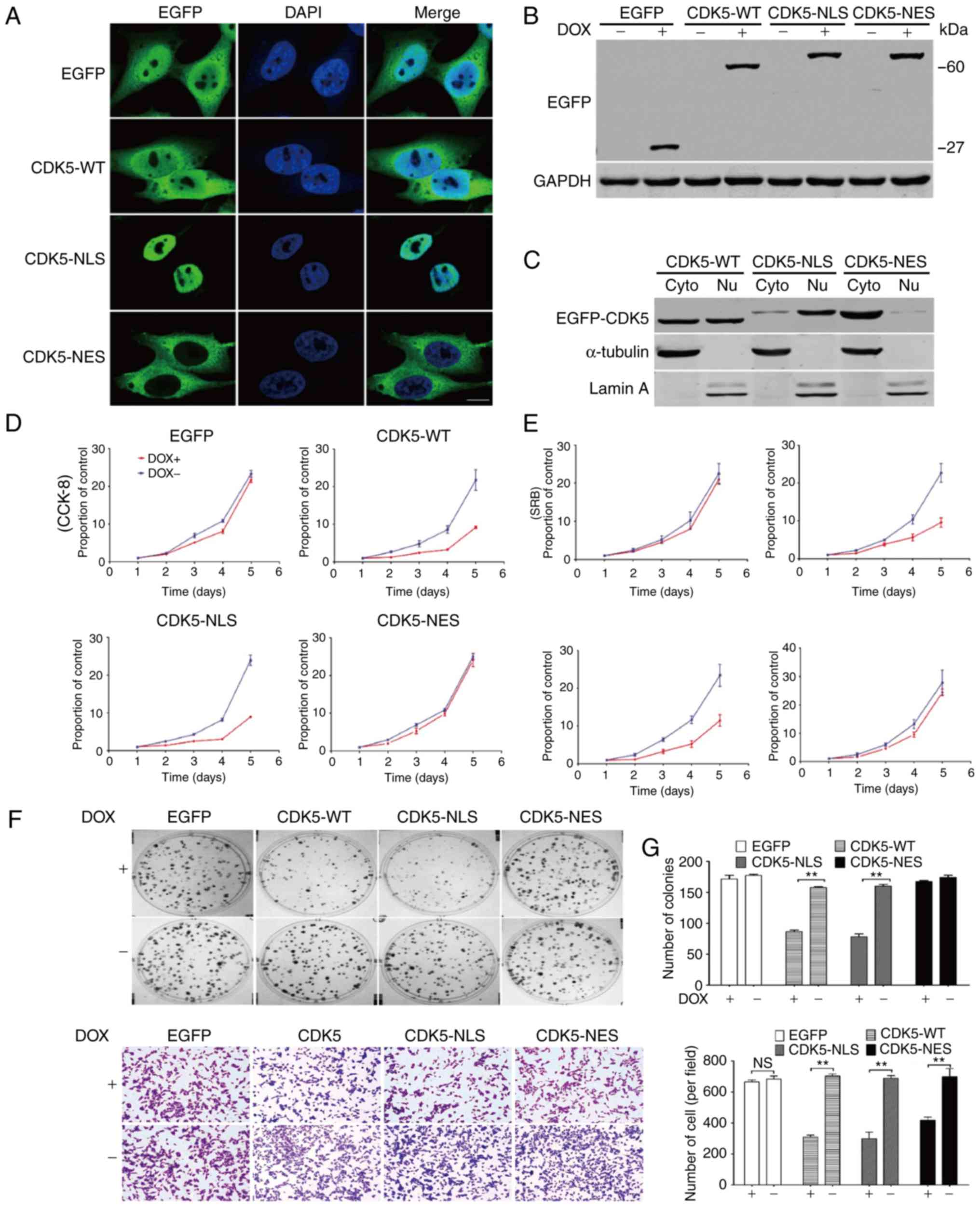 | Figure 1.Function of CDK5 in different
subcellular locations. (A) HGC-27 cells with stably overexpressed
CDK5 in different subcellular locations were generated and examined
by immunofluorescence. (B and C) The changes in CDK5 expression
(overexpression and knockdown) were confirmed using western
blotting. (D) CCK-8 and (E) SRB assays were performed to verify the
function of CDK5 in regard to cell proliferation. (F) CDK5
overexpression reduced the colony formation of HGC-27 GC cells. (G)
CDK5 overexpression reduced the metastasis of HGC-27 GC cells.
Magnification, ×200. **P<0.01, as indicated. CDK5, cyclin
dependent kinase 5; CCK-8, Cell Counting Kit-8; GC, gastric cancer;
EGFP, enhanced green fluorescent protein; WT, wild-type; SRB,
Sulforhodamine B; NLS, nuclear localisation signal; NES, nuclear
export signal; Nu, nuclear; Cyto, cytoplasm; DOX, doxycycline. |
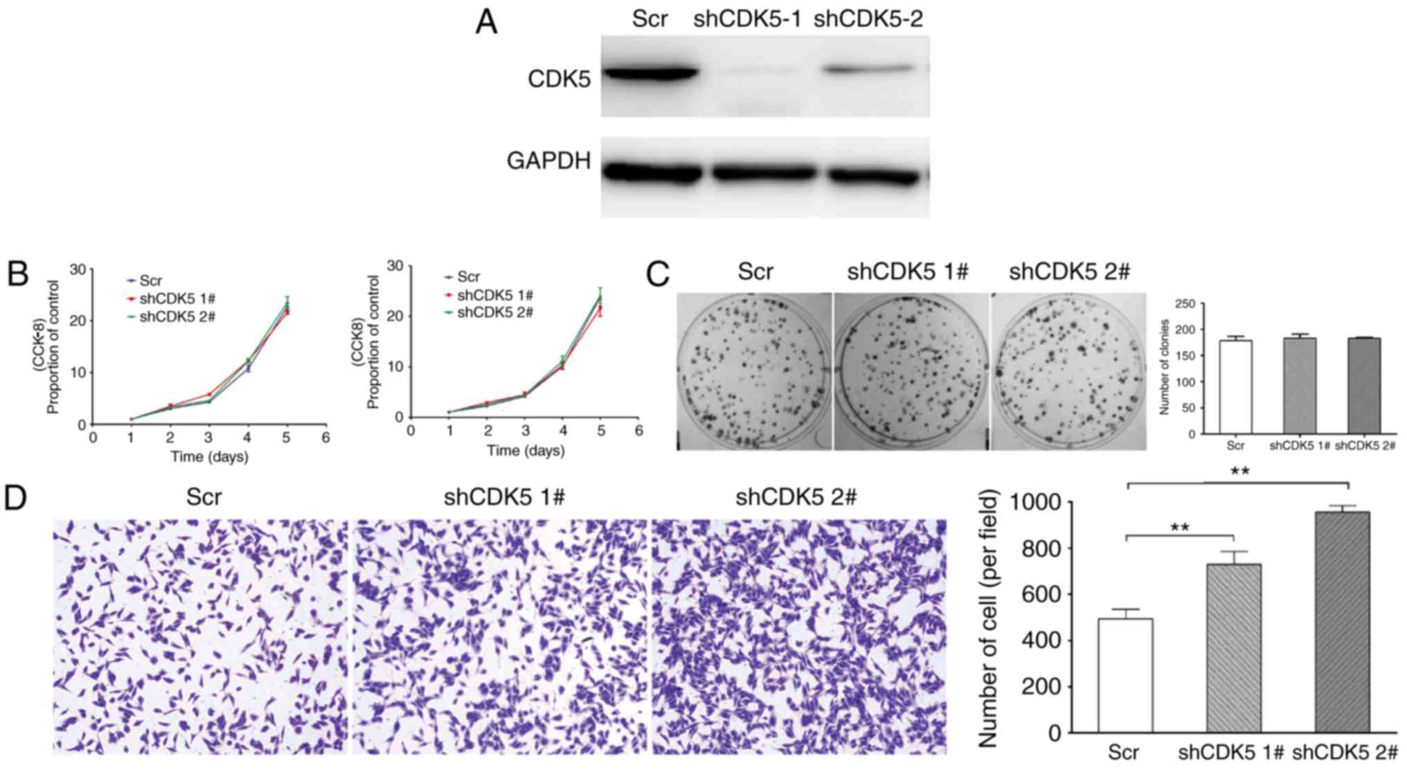
Downregulation of CDK5 in GC cells
promote metastasis
Furthermore, to examine the effect of CDK5 on GC
tumourigenicity, HGC-27 cells with CDK5 stably knocked down and
control cells were generated (Fig.
2A). In vitro, it was observed that no significant
differences were evident in terms of proliferation and colony
formation between the shCDK5 cells and control cells (Fig. 2B and C). However, the knockdown of
CDK5 in HGC-27 GC cells promoted metastasis (Fig. 2D).
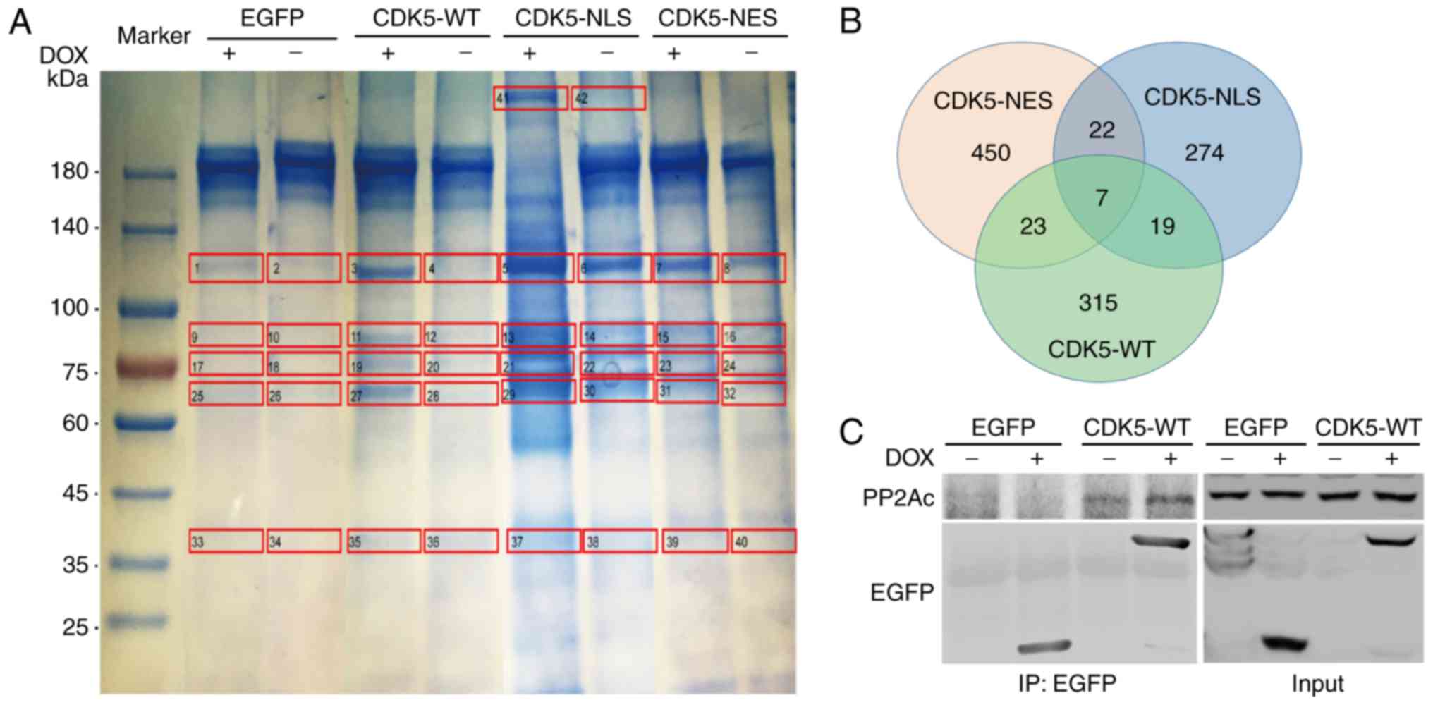
Screening and verification of novel
protein target of CDK5
To investigate the functional mechanism of CDK5 in
GC, the present study performed IP and LC-MS to identify potential
targets of CDK5 in HGC-27 GC cells overexpressing EGFP-tagged
CDK5-NLS, EGFP-tagged CDK5-NES or p-TRE-EGFP (Fig. 3A). Seven proteins were expressed in
all of the groups (Fig. 3B).
Further examination by co-IP and western blot analysis revealed
that CDK5 interacted with PP2A (Fig.
3C). Therefore, it was hypothesized that CDK5 in combination
with PP2A may serve a crucial role in GC.
Inhibition of PP2A antagonizes the
metastasis of CDK5-overexpressing GC cells
Next, cells were treated with okadaic acid, an
inhibitor of PP2A, and alterations in the proliferation, colony
formation and metastasis of HGC-27 GC cells overexpressing CDK5 and
in the control group were observed. No significant differences were
identified between the two groups in terms of proliferation
(Fig. 4A), though a reduction in
the metastasis of GC cells overexpressing CDK5 was revealed
(Fig. 4B). Furthermore, knockdown
of PP2A reduced metastasis (Fig. 4C and
D) in CKD5 knockdown (shCDK5 #1) HGC-27 cells, but it did not
affect proliferation (Fig. 4E).
Consequently, it was concluded that CDK5 may influence the
metastasis of GC by interacting with PP2A.
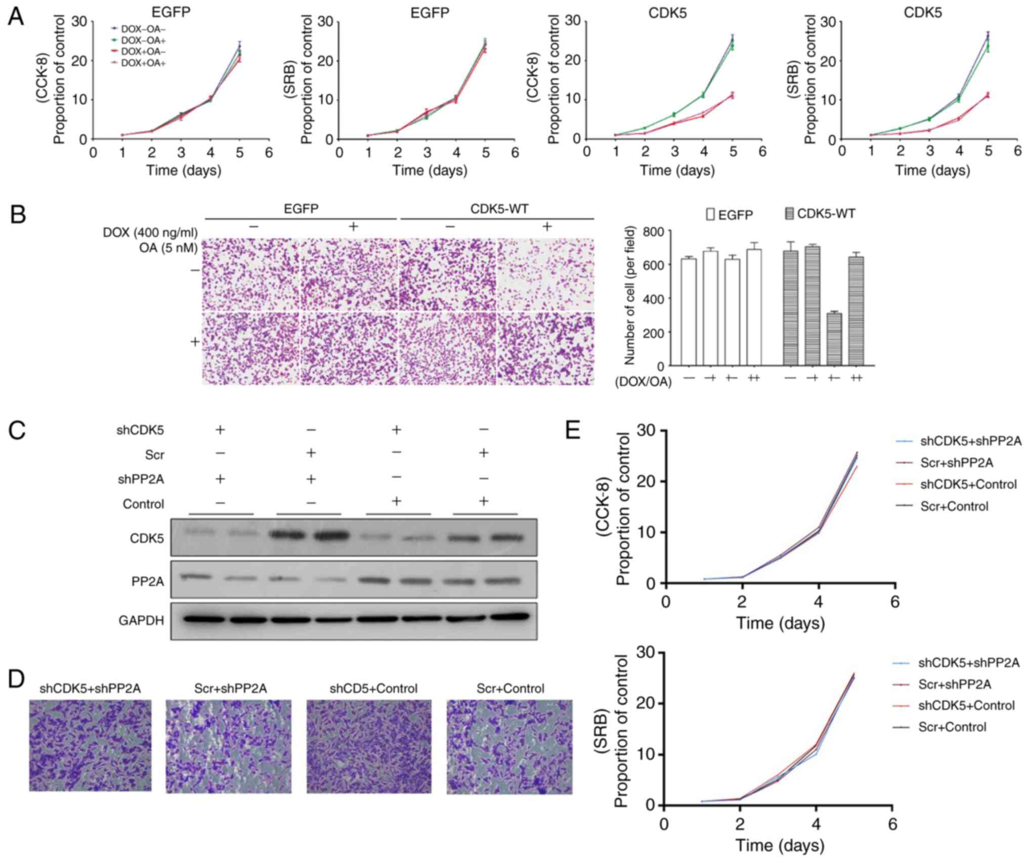 | Figure 4.Inhibition of PP2A antagonizes the
metastasis of CDK5-overexpressing GC cells. (A) CCK-8 and SRB
assays demonstrated that inhibition of PP2A did not significantly
affect proliferation. (B) Inhibition of PP2A antagonized the
metastasis of CDK5-overexpressing GC cells (magnification, ×200).
(C) Western blotting of PP2A knockdown in CDK5 knockdown (shCDK5
#1) HGC-27 cells. (D) Knockdown of PP2A in CDK5 knockdown HGC-27
cells reduced metastasis (magnification, ×200). (E) CCK-8 and SRB
assays demonstrated that knockdown of PP2A in CDK5 knockdown HGC-27
cells did not significantly affect proliferation. CDK5, cyclin
dependent kinase 5; PP2A, serine/threonine-protein phosphatase 2A;
CCK-8, Cell Counting Kit-8; GC, gastric cancer; sh-, short hairpin
RNA; Scr, scramble; EGFP, enhanced green fluorescent protein; WT,
wild-type; SRB, Sulforhodamine B; DOX, doxycycline; OA, Okadaic
Acid. |
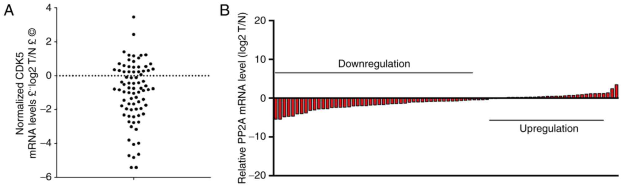
PP2A expression is downregulated in
GC
In the present study, it was demonstrated that PP2A
interacted with CDK5 and subsequently influenced the metastasis of
GC. For further analysis, RT-qPCR was performed to examine the
relative expression levels of PP2A in 80 pairs of GC tissues
matched with normal tissues (Fig. 5A
and B). PP2A expression in the GC tissues was lower than in the
corresponding adjacent non-tumour tissues (Fig. 5).
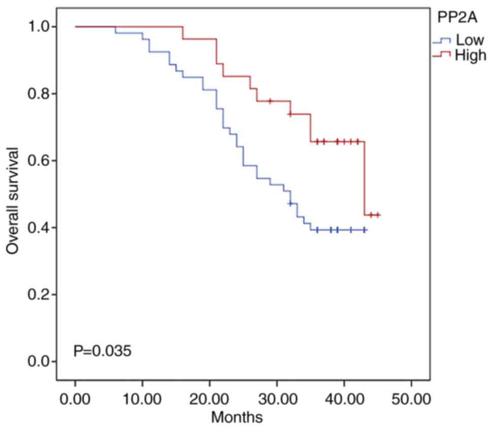
Associations between PP2A expression
and clinicopathological features and prognosis
To analyse the association between PP2A expression
and clinicopathological features, specimens were collected from 80
patients with GC. The cohort was divided into groups: Low PP2A
expression group and a high PP2A expression group, according to the
median PP2A value. No significant differences were observed in
terms of the clinical and pathological characteristics between the
high- and low-PP2A groups (Table
I). To investigate whether PP2A could serve as a prognostic
marker, overall survival (OS) curves were plotted using the
Kaplan-Meier method according to the PP2A expression level.
Patients with low levels of PP2A had a poorer OS rate when compared
with those with high levels of expression (P=0.035; Fig. 6). In addition, to assess whether the
use of PP2A to predict OS was independent of the other clinical or
pathological factors of the patients with GC, univariate and
multivariate Cox proportional hazards analyses were performed. The
results revealed that the PP2A levels and TNM stage were
independent prognostic factors for OS in patients with GC (Table II). Therefore, low levels of PP2A
may be indicative of a poor prognosis in patients with GC.
 | Table I.Association analyses between
expression levels of serine/threonine-protein phosphatase 2A and
clinicopathological features in 80 patients with gastric
cancer. |
Table I.
Association analyses between
expression levels of serine/threonine-protein phosphatase 2A and
clinicopathological features in 80 patients with gastric
cancer.
|
| Low expression
(n=53) | High expression
(n=27) |
|
|---|
|
|
|
|
|
|---|
| Characteristic | n | % | n | % | P-value |
|---|
| Age, years |
|
|
|
| 0.055 |
|
<60 | 27 | 50.9 | 7 | 25.9 |
|
|
≥60 | 26 | 49.1 | 20 | 74.1 |
|
| Sex |
|
|
|
| 0.604 |
|
Female | 15 | 28.3 | 6 | 22.2 |
|
|
Male | 38 | 71.7 | 21 | 77.8 |
|
| Tumour
location |
|
|
|
| 0.339 |
|
Upper | 13 | 24.5 | 5 | 18.5 |
|
|
Middle | 12 | 22.6 | 6 | 22.2 |
|
|
Lower | 27 | 50.9 | 13 | 48.1 |
|
|
Mixed | 1 | 1.9 | 3 | 11.1 |
|
| Tumour size,
mm |
|
|
|
| 0.235 |
|
<50 | 27 | 50.9 | 18 | 66.7 |
|
|
≥50 | 26 | 49.1 | 9 | 33.3 |
|
| Vascular
invasion |
|
|
|
| 0.267 |
|
Negative | 38 | 71.7 | 23 | 85.2 |
|
|
Positive | 15 | 28.3 | 4 | 14.8 |
|
| Lymphatic
invasion |
|
|
|
| 0.581 |
|
Negative | 39 | 73.6 | 22 | 81.5 |
|
|
Positive | 14 | 26.4 | 5 | 18.5 |
|
| Nervous
invasion |
|
|
|
| 0.999 |
|
Negative | 42 | 79.2 | 22 | 81.5 |
|
|
Positive | 11 | 20.8 | 5 | 18.5 |
|
| Tumour
differentiation |
|
|
|
| 0.154 |
|
Differentiated | 25 | 47.2 | 18 | 66.7 |
|
|
Undifferentiated | 28 | 52.8 | 9 | 33.3 |
|
| CEA, ng/ml |
|
|
|
| 0.999 |
| ≤5 | 40 | 75.5 | 20 | 74.1 |
|
|
>5 | 13 | 24.5 | 7 | 25.9 |
|
| CA 19-9, IU/ml |
|
|
|
| 0.999 |
|
≤37 | 44 | 83.0 | 23 | 85.2 |
|
|
>37 | 9 | 17.0 | 4 | 14.8 |
|
| AJCC TNM stage |
|
|
|
| 0.429 |
| I | 8 | 15.1 | 2 | 7.4 |
|
| II | 19 | 35.8 | 8 | 29.6 |
|
|
III | 26 | 49.1 | 17 | 63.0 |
|
| Table II.Univariate and multivariate Cox
proportional analysis for survival. |
Table II.
Univariate and multivariate Cox
proportional analysis for survival.
|
|
| Univariate
analysis | Multivariate
analysis |
|---|
|
|
|
|
|
|---|
| Parameter | Categories | HR (95% CI) | P-value | HR (95% CI) | P-value |
|---|
| Age (years) | (≥60 vs.
<60) | 1.616
(0.8484–3.081) | 0.136 |
|
|
| Sex | (male vs.
female) | 1.165
(0.596–2.279) | 0.658 |
|
|
| Tumour
location | (upper vs.
other) | 1.342
(0.545–3.308) | 0.522 |
|
|
| Tumour size | (≥50 mm vs. <50
mm) | 1.517
(0.826–2.786) | 0.179 |
|
|
| Vascular
invasion | (yes vs. no) | 1.998
(1.033–3.865) | 0.040 | 1.720
(0.882–3.352) | 0.111 |
| Lymphatic
invasion | (yes vs. no) | 1.718
(0.876–3.370) | 0.115 |
|
|
| Nervous
invasion | (yes vs. no) | 1.585
(0.795–3.158) | 0.190 |
|
|
| Tumour
differentiation | (undifferentiated
vs. differentiated) | 1.447
(0.789–2.652) | 0.232 |
|
|
| AJCC TNM stage | (III vs. I/II) | 3.398
(1.873–6.163) | <0.001 | 4.271
(2.289–7.969) | <0.001 |
| CEA level | (above normal vs.
normal) | 1.272
(0.651–2.485) | 0.481 |
|
|
| CA 19-9 level | (above normal vs.
normal) | 1.060
(0.470–2.388) | 0.889 |
|
|
| PP2A | (high vs. low) | 0.478
(0.234–0.973) | 0.042 | 0.317
(0.152–0.662) | 0.002 |
Discussion
Recently, many therapeutic strategies have been
developed to improve the functional outcomes of GC (36,37).
CDK5, a typical CDK that was previously considered to function in
processes unrelated to cell cycle regulation, has been shown to
serve a fundamental role in several types of carcinomas, including
multiple myeloma, and breast, lung, prostate and pancreatic cancers
(22,24,25,38,39).
In the present study, the results demonstrated that
nuclear CDK5 overexpression markedly reduced the proliferation,
colony formation and metastasis of HGC-27 GC cells, which was
consistent with our previous data obtained with the SGC-7901 and
MGC-803 GC cell lines (26).
However, CDK5 downregulation was observed to induce metastasis but
not proliferation. Zhang et al (40) revealed that CDK5 could function as a
cell cycle suppressor when localized to the nucleus of the neuron.
To date, the mechanism through which CDK5 regulates the development
of GC is unknown. Based on the present IP and liquid
chromatography/MS analysis, it is possible that PP2A may be a
potential target of CDK5.
PP2A, a serine/threonine phosphatase, comprises
catalytic, scaffold and regulatory subunits and is associated with
the development of numerous human cancers (27–30).
Therefore, the aim of the present study was to understand whether
CDK5 functioned with PP2A during the development of GC. When cells
were treated with an inhibitor of PP2A, it was demonstrated that
the metastasis of CDK5-overexpressing GC cells was restored without
affecting proliferation. When PP2A was knocked down in CKD5
knockdown HGC-27 cells, the results were consistent those
aforementioned. Therefore, it was proposed that CDK5 may influence
the metastasis of GC by interacting with PP2A. In addition,
Tsuchiya et al (31)
identified a novel PP2A enhancer that induced the
caspase-independent apoptosis of MKN28 GC cells, which is
consistent with the present results. Further analysis revealed that
relative PP2A expression was downregulated in 80 pairs of matched
GC and normal tissues. Based on the median PP2A value, the cohort
was divided into groups with low and high PP2A expression, and
notably, the patients with high levels of PP2A had better survival
outcomes than those with low PP2A expression (P=0.035).
Furthermore, univariate and multivariate Cox proportional hazards
analyses demonstrated that the PP2A level was an independent
prognostic factor for OS in patients with GC.
In conclusion, to the best of our knowledge, the
present study is the first to report that PP2A may affect
metastasis by interacting with CDK5 in GC and that low levels of
PP2A may indicate a tendency for poor prognosis in patients with
GC.
Acknowledgements
Not applicable.
Funding
The present study was supported by the Scientific
and Technological Innovation Joint Capital Projects of Fujian
Province (grant no. 2016Y9031), Construction Project of Fujian
Province Minimally Invasive Medical Center [grant no. (2017)171],
The Second Batch of Special Support Funds for Fujian Province
Innovation and Entrepreneurship Talents (grant no. 2016B013),
QIHANG funds of Fujian Medical University (grant no. 2016QH025) and
the Science Foundation of the Fujian Province, China (grant no.
2018J01307).
Availability of data and materials
The datasets analysed during the present study are
available from the corresponding author upon reasonable
request.
Authors' contributions
JL conceived and designed the present study. YL and
CMH contributed to the design of the present study. JL, YQS and PYZ
performed the in vitro experiments. PL, JWX, JBW, and QYC
obtained the tumor samples and the clinical data. JXL and LLC
analysed the data. JL, PYZ and CHZ wrote and revised the
manuscript. CHZ and CMH were also involved in the conception of the
study. All authors read and approved the manuscript and agree to be
accountable for all aspects of the research in ensuring that the
accuracy or integrity of any part of the work are appropriately
investigated and resolved.
Ethics approval and consent to
participate
The present study was approved by the Ethics
Committee of Fujian Medical University Union Hospital (Fujian,
China; no. UH-2013009), and written informed consent was obtained
from all patients involved.
Patient consent for publication
Not applicable.
Competing interests
The authors declare that they have no competing
interests.
References
|
1
|
Torre LA, Bray F, Siegel RL, Ferlay J,
Lortet-Tieulent J and Jemal A: Global cancer statistics, 2012. CA
Cancer J Clin. 65:87–108. 2015. View Article : Google Scholar : PubMed/NCBI
|
|
2
|
Chen H, Guan R, Lei Y, Chen J, Ge Q, Zhang
X, Dou R, Chen H, Liu H, Qi X, et al: Lymphangiogenesis in gastric
cancer regulated through Akt/mTOR-VEGF-C/VEGF-D axis. BMC Cancer.
15:1032015. View Article : Google Scholar : PubMed/NCBI
|
|
3
|
Johnson SM and Evers BM: Translational
research in gastric malignancy. Surg Oncol Clin N Am. 17:323–340.
2008. View Article : Google Scholar : PubMed/NCBI
|
|
4
|
Kim K, Chun KH, Suh PG and Kim IH:
Alterations in cell proliferation related gene expressions in
gastric cancer. Crit Rev Eukaryot Gene Expr. 21:237–254. 2011.
View Article : Google Scholar : PubMed/NCBI
|
|
5
|
Kim DH: Prognostic implications of cyclin
B1, p34cdc2, p27Kip1 and p53 expression in gastric cancer. Yonsei
Med J. 48:694–700. 2007. View Article : Google Scholar : PubMed/NCBI
|
|
6
|
Liang B, Wang S, Yang X, Ye Y, Yu Y and
Cui Z: Expressions of cyclin E, cyclin dependent kinase 2 and
p57KIP2 in human gastric cancer. Chin Med J. 116:20–23.
2003.PubMed/NCBI
|
|
7
|
Malumbres M and Barbacid M: Cell cycle,
CDKs and cancer: A changing paradigm. Nat Rev Cancer. 9:153–166.
2009. View
Article : Google Scholar : PubMed/NCBI
|
|
8
|
Lapenna S and Giordano A: Cell cycle
kinases as therapeutic targets for cancer. Nat Rev Drug Discov.
8:547–566. 2009. View
Article : Google Scholar : PubMed/NCBI
|
|
9
|
Wright RH, Castellano G, Bonet J, Le Dily
F, Font-Mateu J, Ballaré C, Nacht AS, Soronellas D, Oliva B and
Beato M: CDK2-dependent activation of PARP-1 is required for
hormonal gene regulation in breast cancer cells. Genes Dev.
26:1972–1983. 2012. View Article : Google Scholar : PubMed/NCBI
|
|
10
|
Yang S, Zhang L, Liu M, Chong R, Ding SJ,
Chen Y and Dong J: CDK1 phosphorylation of YAP promotes mitotic
defects and cell motility and is essential for neoplastic
transformation. Cancer Res. 73:6722–6733. 2013. View Article : Google Scholar : PubMed/NCBI
|
|
11
|
Cepeda D, Ng HF, Sharifi HR, Mahmoudi S,
Cerrato VS, Fredlund E, Magnusson K, Nilsson H, Malyukova A,
Rantala J, et al: CDK-mediated activation of the SCFFBXO (28)
ubiquitin ligase promotes MYC-driven transcription and
tumourigenesis and predicts poor survival in breast cancer. EMBO
Mol Med. 5:1067–1086. 2013. View Article : Google Scholar : PubMed/NCBI
|
|
12
|
Lu M, Breyssens H, Salter V, Zhong S, Hu
Y, Baer C, Ratnayaka I, Sullivan A, Brown NR, Endicott J, et al:
Restoring p53 function in human melanoma cells by inhibiting MDM2
and cyclin B1/CDK1-phosphorylated nuclear iASPP. Cancer Cell.
30:822–823. 2016. View Article : Google Scholar : PubMed/NCBI
|
|
13
|
Radomska HS, Alberich-Jordà M, Will B,
Gonzalez D, Delwel R and Tenen DG: Targeting CDK1 promotes
FLT3-activated acute myeloid leukemia differentiation through
C/EBPα. J Clin Invest. 122:2955–2966. 2012. View Article : Google Scholar : PubMed/NCBI
|
|
14
|
Sheppard KE and McArthur GA: The
cell-cycle regulator CDK4: An emerging therapeutic target in
melanoma. Clin Cancer Res. 19:5320–5328. 2013. View Article : Google Scholar : PubMed/NCBI
|
|
15
|
Rader J, Russell MR, Hart LS, Nakazawa MS,
Belcastro LT, Martinez D, Li Y, Carpenter EL, Attiyeh EF, Diskin
SJ, et al: Dual CDK4/CDK6 inhibition induces cell-cycle arrest and
senescence in neuroblastoma. Clin Cancer Res. 19:6173–6182. 2013.
View Article : Google Scholar : PubMed/NCBI
|
|
16
|
Sakaue-Sawano A, Kurokawa H, Morimura T,
Hanyu A, Hama H, Osawa H, Kashiwagi S, Fukami K, Miyata T, Miyoshi
H, et al: Visualizing spatiotemporal dynamics of multicellular
cell-cycle progression. Cell. 132:487–498. 2008. View Article : Google Scholar : PubMed/NCBI
|
|
17
|
Tetsu O and McCormick F: Proliferation of
cancer cells despite CDK2 inhibition. Cancer Cell. 3:233–245. 2003.
View Article : Google Scholar : PubMed/NCBI
|
|
18
|
van den Heuvel S and Harlow E: Distinct
roles for cyclin-dependent kinases in cell cycle control. Science.
262:2050–2054. 1993. View Article : Google Scholar : PubMed/NCBI
|
|
19
|
Hellmich MR, Pant HC, Wada E and Battey
JF: Neuronal cdc2-like kinase: A cdc2-related protein kinase with
predominantly neuronal expression. Proc Natl Acad Sci USA.
89:10867–10871. 1992. View Article : Google Scholar : PubMed/NCBI
|
|
20
|
Dhavan R and Tsai LH: A decade of CDK5.
Nat Rev Mol Cell Biol. 2:749–759. 2001. View Article : Google Scholar : PubMed/NCBI
|
|
21
|
Strock CJ, Park JI, Nakakura EK, Bova GS,
Isaacs JT, Ball DW and Nelkin BD: Cyclin-dependent kinase 5
activity controls cell motility and metastatic potential of
prostate cancer cells. Cancer Res. 66:7509–7515. 2006. View Article : Google Scholar : PubMed/NCBI
|
|
22
|
Pozo K, Castro-Rivera E, Tan C, Plattner
F, Schwach G, Siegl V, Meyer D, Guo A, Gundara J, Mettlach G, et
al: The role of Cdk5 in neuroendocrine thyroid cancer. Cancer Cell.
24:499–511. 2013. View Article : Google Scholar : PubMed/NCBI
|
|
23
|
Ehrlich SM, Liebl J, Ardelt MA, Lehr T, De
Toni EN, Mayr D, Brandl L, Kirchner T, Zahler S, Gerbes AL and
Vollmar AM: Targeting cyclin dependent kinase 5 in hepatocellular
carcinoma-A novel therapeutic approach. J Hepatol. 63:102–113.
2015. View Article : Google Scholar : PubMed/NCBI
|
|
24
|
Feldmann G, Mishra A, Hong SM, Bisht S,
Strock CJ, Ball DW, Goggins M, Maitra A and Nelkin BD: Inhibiting
the cyclin-dependent kinase CDK5 blocks pancreatic cancer formation
and progression through the suppression of Ras-Ral signaling.
Cancer Res. 70:4460–4469. 2010. View Article : Google Scholar : PubMed/NCBI
|
|
25
|
Demelash A, Rudrabhatla P, Pant HC, Wang
X, Amin ND, McWhite CD, Naizhen X and Linnoila RI: Achaete-scute
homologue-1 (ASH1) stimulates migration of lung cancer cells
through Cdk5/p35 pathway. Mol Biol Cell. 23:2856–2866. 2012.
View Article : Google Scholar : PubMed/NCBI
|
|
26
|
Cao L, Zhou J, Zhang J, Wu S, Yang X, Zhao
X, Li H, Luo M, Yu Q, Lin G, et al: Cyclin-dependent kinase 5
decreases in gastric cancer and its nuclear accumulation suppresses
gastric tumorigenesis. Clinical Cancer Res. 21:1419–1428. 2015.
View Article : Google Scholar
|
|
27
|
Seshacharyulu P, Pandey P, Datta K and
Batra SK: Phosphatase: PP2A structural importance, regulation and
its aberrant expression in cancer. Cancer Lett. 335:9–18. 2013.
View Article : Google Scholar : PubMed/NCBI
|
|
28
|
Zhou X, Updegraff BL, Guo Y, Peyton M,
Girard L, Larsen JE, Xie XJ, Zhou Y, Hwang TH, Xie Y, et al:
PROTOCADHERIN 7 acts through SET and PP2A to potentiate MAPK
signaling by EGFR and KRAS during lung tumorigenesis. Cancer Res.
77:187–197. 2017. View Article : Google Scholar : PubMed/NCBI
|
|
29
|
Baldacchino S, Saliba C, Petroni V, Fenech
AG, Borg N and Grech G: Deregulation of the phosphatase, PP2A is a
common event in breast cancer, predicting sensitivity to FTY720.
EPMA J. 5:32014. View Article : Google Scholar : PubMed/NCBI
|
|
30
|
Cristóbal I, Manso R, Rincón R, Caramés C,
Senin C, Borrero A, Martínez-Useros J, Rodriguez M, Zazo S,
Aguilera O, et al: PP2A inhibition is a common event in colorectal
cancer and its restoration using FTY720 shows promising therapeutic
potential. Mol Cancer Ther. 13:938–947. 2014. View Article : Google Scholar : PubMed/NCBI
|
|
31
|
Tsuchiya A, Kanno T, Shimizu T, Nakao S,
Tanaka A, Tabata C, Nakano T and Nishizaki T: A novel PP2A enhancer
induces caspase-independent apoptosis of MKN28 gastric cancer cells
with high MEK activity. Cancer Lett. 347:123–128. 2014. View Article : Google Scholar : PubMed/NCBI
|
|
32
|
Lombardo Y, Filipović A, Molyneux G,
Periyasamy M, Giamas G, Hu Y, Trivedi PS, Wang J, Yagüe E, Michel L
and Coombes RC: Nicastrin regulates breast cancer stem cell
properties and tumor growth in vitro and in vivo. Proc Natl Acad
Sci USA. 109:16558–16563. 2012. View Article : Google Scholar : PubMed/NCBI
|
|
33
|
Edge S, Byrd DR, Compton CC, Fritz AG,
Greene F and Trotti A: AJCC Cancer Staging Handbook: From the AJCC
Cancer Staging Manual. 7th. Springer-Verlag; New York: 2010
|
|
34
|
Livak KJ and Schmittgen TD: Analysis of
relative gene expression data using real-time quantitative PCR and
the 2-ΔΔCT method. Methods. 25:402–408. 2001. View Article : Google Scholar : PubMed/NCBI
|
|
35
|
Lei W, Wang ZL, Feng HJ, Lin XD, Li CZ and
Fan D: Long non-coding RNA SNHG12 promotes the proliferation and
migration of glioma cells by binding to HuR. Int J Oncol.
53:1374–1384. 2018.PubMed/NCBI
|
|
36
|
Li Y, Gong J, Zhang Q, Lu Z, Gao J, Li Y,
Cao Y and Shen L: Dynamic monitoring of circulating tumour cells to
evaluate therapeutic efficacy in advanced gastric cancer. Br J
Cancer. 114:138–145. 2016. View Article : Google Scholar : PubMed/NCBI
|
|
37
|
Fujiwara Y, Okada K, Omori T, Sugimura K,
Miyata H, Ohue M, Kobayashi S, Takahashi H, Nakano H, Mochizuki C,
et al: Multiple therapeutic peptide vaccines for patients with
advanced gastric cancer. Int J Oncol. 50:1655–1662. 2017.
View Article : Google Scholar : PubMed/NCBI
|
|
38
|
Liang Q, Li L, Zhang J, Lei Y, Wang L, Liu
DX, Feng J, Hou P, Yao R, Zhang Y, et al: CDK5 is essential for
TGF-β1-induced epithelial-mesenchymal transition and breast cancer
progression. Sci Rep. 3:29322013. View Article : Google Scholar : PubMed/NCBI
|
|
39
|
Hsu FN, Chen MC, Lin KC, Peng YT, Li PC,
Lin E, Chiang MC, Hsieh JT and Lin H: Cyclin-dependent kinase 5
modulates STAT3 and androgen receptor activation through
phosphorylation of Ser727 on STAT3 in prostate cancer
cells. Am J Physiol Endocrinol Metab. 305:E975–E986. 2013.
View Article : Google Scholar : PubMed/NCBI
|
|
40
|
Zhang J, Li H, Yabut O, Fitzpatrick H,
D'Arcangelo G and Herrup K: Cdk5 suppresses the neuronal cell cycle
by disrupting the E2F1-DP1 complex. J Neurosci. 30:5219–5228. 2010.
View Article : Google Scholar : PubMed/NCBI
|